Histologic Studies on Normal and Persistent Ductus Arteriosus in the Dog
ADRIANA C. GITTENBERGER-DE GROOT, MO, JAN L. M. STRENGERS, MD, MONICA MENTINK, ROBERT E. POELMANN, PHD, DONALD F. PATTERSON, DVM, DSc, FACC
Leiden, The Netherlands
The process of anatomic closure of the ductus arteriosus was studied at the ultrastructural level in 15 normal beagles (age 0 hour to 13 days) and in 18 specimens from a strain of dogs with hereditary persistent ductus arteriosus (age 4 hours to 27 days). Normal ductal closure takes place from the pulmonary artery to the aortic end. It is accompanied by a series of histologic changes: I) separation of the endothelial cells from the internal elastic lamina resulting in a wide region of subendothelial edema; 2) ingrowth and infolding of endothelial cells and migration of undifferentiated smooth muscle cells from the inner media into the subendothelial region; 3) apposition of endothelial cells bordering the lumen; and 4)
Studies of the functional and anatomic closure of the ductus arteriosus have been pursued with renewed interest since discovery of the possibility of medical manipulation of this process with prostaglandins and prostaglandin inhibitors (I). In this study we examined the relevant histologic changes accompanying ductal closure to gain a better understanding of the closing process of the ductus arteriosus and the extent to which it is prone to medical manipulation. In recent years we have concentrated on the morphology of the normal and persistent human ductus arteriosus (2-4). However, because human material is not easily available for study, detailed ultrastructural information is difficult to obtain and reported data are scarce (5).
In the present investigation, the ductus arteriosus in dogs was used as a model in which to study the structural and
From the Department of Anatomy and Embryology, Rijksuniversiteit, Leiden, The Netherlands. Dr. Strengers is a Pediatric Cardiology fellow at the Wilhelmina Children's Hospital, Utrecht, The Netherlands. Dr. Patterson is a professor of Veterinary Medicine and Human Genetics, University of Pennsylvania, Philadelphia, Pennsylvania. This study was supported in part by Grant HHL 18848 from the National Heart, Lung, and Blood Institute, National Institutes of Health, Bethesda, Maryland. Manuscript received June 12, 1984; revised manuscript received November 29, 1984, accepted March 29, 1985.
Address for reprints: A. C. Gittenberger-de Groot, MD, Department of Anatomy and Embryology, Postbus 9602, 2300 RC Leiden, The Netherlands.
degenerative changes. In persistent ductus arteriosus, these changes do not occur. The endothelial cells remain closely adhered to the internal elastic lamina and the underlying media is abnormal in structure. In the case of partial persistent ductus arteriosus (ductus diverticulum), both the normal and the abnormal type of wall are found in a single ductus arteriosus. The histologic features of the normal and the persistent ductus arteriosus in the dog resemble those of the norinal and the persistent ductus arteriosus in humans, suggesting a similar pathogenesis.
(J Am Coil CardioI1985;6:394-404)
ultrastructural characteristics of normal anatomic closure. The characteristics of closure of the ductus arteriosus in normal beagle dogs were compared with those of the ductus arteriosus from offspring of a strain of dogs that is genetically predisposed to persistent patency of the ductus (6-10).
Methods Study material. The study material comprised the duc
tus arteriosus from 15 normal beagles (Table l) and 18 specimens from a strain of dogs with hereditary persistent ductus arteriosus (Table 2). The beagles were purchased from a commercial breeder whose breeding stock had been examined previously for congenital defects. The strain with hereditary persistent ductus arteriosus was derived from dogs with naturally occurring persistent ductus arteriosus; this strain has been maintained as a colony at the University of Pennsylvania since 1971 (6).
Clinical and anatomic data. The postnatal age of the study animals varied from 0 hour to 13 days in the beagle and from 4 hours to 27 days in the strain of dogs with persistent ductus arteriosus. Clinical information was available concerning the presence of cardiovascular murmurs and failure to thrive. Holosystolic or continuous murmurs characteristic of persistent ductus arteriosus were detected in
0735-1097/85/$3.30
JACC Vol. 6, No.2 August 1985:394-404
Table 1. Normal Beagle Ductus Arteriosus Group
Age Anatomically Dog (days) Patent Ductus
I Oh + 2 3 h + 3 12 h ±
4 I day 5 3 days 6 3 days 7 5 days 8 5 days 9 7 days
10 7 days II 8 days 12 9 days 13 II days 14 II days 15 13 days
+ = patent; ± = almost closed at pulmonary end; - = closed; a = development of edema; b = infolding of the endothelial cells, more marked edema and protrusion of inner media cells in the subendothelial region; c = infiltration of the subendothelial region with various cell types; d = start of degenerative changes with accumulation of lipid droplets.
some dogs from the persistent ductus arteriosus strain but not in the normal beagles.
Previous studies have shown that abnormal closure of the ductus arteriosus occurs in 80% of the offspring of the persistent ductus arteriosus strain when both parents have this lesion. The anatomic abnormality varies in degree (6). In the mildest form recognizable by gross examination, the
Table 2. Normal and Abnormal Ductus Arteriosus From the Persistent Ductus Arteriosus Strain
Clinical Anatomic Dog Age Findings Features Intima
4h ? + a-b 2 24 h + e.f; a 3 2 days :j: a-c 4 2 days :j: ± a-b 5 4 days :j: a-c 6 4 days + e; a-b 7 4 days t + e.f; a-c 8 5 days t a-c 9 5 days t a-c
10 7 days * + c.f II 9 days * + e; a-b 12 9 days + e,f; a 13 II days + e.f 14 II days * Formalin-fixed 15 II days :j: a-c 16 13 days * + eJ 17 27 days * + e,f 18 27 days * + e,f; a
*Marked murmur; tsoft murmur; :j:no murmur. + = patent ductus; ± = ductus almost closed at pulmonary end; - = ductus closed; e = no development of edema; f = slight development of dark-staining edema without infolding of endothelial cells; other abbreviations as in Table I.
GITTENBERGER'DE GROOT ET AL. 395 NORMAL AND PERSISTENT DUCTUS ARTERIOSUS
ductus arteriosus closes at the pulmonary artery end, but remains open over the rest of its length, producing a funnelshaped diverticulum that is in communication with the aorta (ductus diverticulum). In most affected dogs, the ductus arteriosus remains open over its entire length but is smallest at the end adjoining the pulmonary artery. In the most severe form, the ductus arteriosus is cylindrical and its diameter equals that of the aorta. Dogs with the latter form of persistent ductus arteriosus have severe pulmonary hypertension.
Ultrastructural study. The dogs were killed immediately before the collection of tissues by intraperitoneal injection of pentobarbital sodium. All specimens were prepared for ultrastructural study by perfusion with half-strength Kamovsky's fixative (11) and immersed in the same fixative for 24 hours. Thereafter, the specimens were transferred to cacodylate buffer. For comparison with earlier light microscopic studies (8-10), two specimens were fixed in formalin (4%) for complete serial sectioning. One near-term dog (Table I) was delivered by cesarean section and not allowed to breathe at birth.
Blocks of tissue were taken at similar sites in each ductus (Fig. I) to allow better comparison of the results. Blocks A and C were sectioned sagittally and block B was sectioned transversely (Fig. I). Each block was sectioned in 1 /-Lm thin sections that were routinely stained with toluidine blue. Selected sections were stained with alcian blue (pH 2.2). Some sections were pretreated with hyaluronidase to digest hyaluronic acid. Ultrathin sections were made for electron microscopic survey. In four specimens, the inner surface of the ductus arteriosus and the aorta were studied with the scanning electron microscope.
Results
Normal Closing Process of the Ductus Arteriosus in the Beagle
The histologic changes of the ductal wall occurring se-quentially during anatomic closure are observed in a single
Figure 1. Schematic drawing of ductus arteriosus and aortic arch. A to C = blocks of tissue studied for ductus arteriosus; * = crista reuniens; AAo = ascending aorta; DAo = descending aorta; P = pulmonary artery.
396 GITTENBERGER-DE GROOT ET AL. NORMAL AND PERSISTENT DUCTUS ARTERIOSUS
A B
c D
E F
Figure 2. Schematic drawing of histologic changes in the wall of the normal (A to D) and abnormal (E, F) ductus arteriosus. A, Development of edema; B, infolding of the endothelial cells, more marked edema and protrusion of inner media cells in the subendothelial region; C, infiltration of the subendothelial region with various cell types; D, start of degenerative changes with accumulation of lipid droplets; E, no development of edema; F, slight development of dark-staining edema without infolding of endothelial cells.
ductus arteriosus when the entire length of the vessel is studied. The aortic end of the ductus is the least advanced toward closure, and the pulmonary end the most advanced. Parts A, Band C (Fig. 1) should be studied to ensure that all essential changes in the ductal wall are observed. If only part B is studied, the ductus should range in age from 0 hour to approximately 5 days. Apparently, ductal closure proceeds from the pUlmonary to the aortic end.
Intimal changes. Endothelium and subendothelial region. In the intimal region of the youngest animal at term (0 hour) (Table 1) and in part A (Fig. 1) of most other specimens, the endothelial cells are separated from the underlying internal elastic lamina by a clear space containing only a few cells (Fig. 2A and 3A). This subendothelial edema was present in all specimens from the normal beagle, including that from the beagle delivered by cesarean section and killed without breathing (Table 1, Case 1). Several hours
JACC Vol. 6, No.2 August 1985:394--404
after birth, the lumen of the ductus arteriosus is much smaller and the edema more marked. The endothelial cells form clumps and strands that project into the subendothelial region (Fig. 3B and 4). They have an ill defined basement membrane. In the strands, there is close apposition of the endothelial cells. Occasionally an erythrocyte is trapped. All endothelial cells that project into the subendothelial region have a swollen appearance (Fig. 2B and 3B). The endothelial cells remain connected with the inner media cells (Fig. 3A and B) by thin cytoplasmic protrusions and a fine fibrillar extracellular material. The fine, thready material resembles the microfibrillar part of the elastic fiber.
Medial cells and the subendothelial region. In the earliest phase (0 hour) (Fig. 2A and 3), there are a variable number of small spindle-shaped dark cells (1JLm toluidine blue-stained sections) in the inner media. They seem to pass the internal elastic lamina and protrude into the subendothelial region. These dark cells, which we provisionally consider to be undifferentiated smooth muscle cells, can be distinguished from the larger and paler-staining smooth muscle cells of the main part of the inner media. The change in structure of the inner media and the outer subendothelial region causes the internal elastic lamina to become fragmented and less recognizable.
Advancing closure, first noted at the pulmonary end of the ductus arteriosus in the 1 day old beagle, causes the dark, spindle-shaped cells to lose contact with the inner media and become distributed, seemingly at random, in the subendothelial region. The swollen endothelial cells that project into the subendothelial region lose their mutual contacts (Fig. 2C and 5). Leukocytes and erythrocytes may be found in the subendothelial region. The endothelial cells bordering the lumen are now in close apical apposition (Fig_ 6A). The gap between them can be distinguished only at the ultrastructural level (15 to 30 JLm). An occasional erythrocyte is wedged between these endothelial cells, which have a healthy appearance with many membrane-bound pinocytotic vesicles both at the lumen and the side bordering the subendothelial region (Fig. 6B).
Media. The structure of the media is different in various parts of the ductus arteriosus. Of particular interest is the portion of the ductal wall that is adjacent to the aortic wall and merges completely with it at the crista reuniens (Fig. 1). We refer to this portion as the roof of the ductus arteriosus. It is continuous by way of the lateral walls with the base of the ductus, which is a free wall.
Aortic, mixed and ductal type of media. In normal beagles, starting at the rim of the crista reuniens, the media has the appearance of an elastic artery (aortic type of media) with regular elastic lamellae alternating with smooth muscle cell layers. In the direction of the pulmonary artery, the roof of the ductus arteriosus changes its structure. The inner media develops an increasingly more disorganized muscular and elastic tissue pattern that is best seen in part B2 (Fig.
JACC Vol. 6, No.2 August 1985:394-404
A
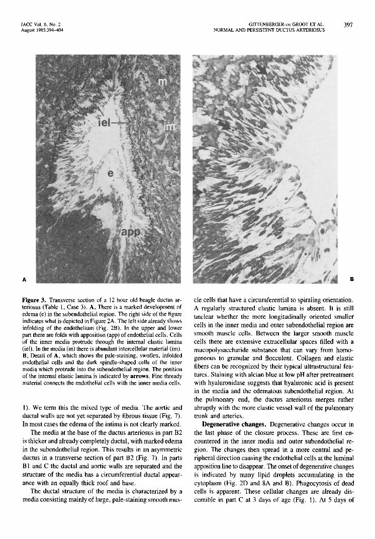
Figure 3. Transverse section of a 12 hour old beagle ductus arteriosus (Table 1, Case 3). A, There is a marked development of edema (e) in the subendothelial region. The right side of the figure indicates what is depicted in Figure 2A. The left side already shows infolding of the endothelium (Fig. 2B). In the upper and lower part there are folds with apposition (app) of endothelial cells. Cells of the inner media protrude through the internal elastic lamina (iel). In the media (m) there is abundant intercellular material (im). B, Detail of A, which shows the pale-staining, swollen, infolded endothelial cells and the dark spindle-shaped cells of the inner media which protrude into the subendothelial region. The position of the internal elastic lamina is indicated by arrows. Fine thready material connects the endothelial cells with the inner media cells.
I). We tenn this the mixed type of media. The aortic and ductal walls are not yet separated by fibrous tissue (Fig. 7). In most cases the edema of the intima is not clearly marked.
The media at the base of the ductus arteriosus in part B2 is thicker and already completely ductal, with marked edema in the subendothelial region. This results in an asymmetric ductus in a transverse section of part B2 (Fig. 7). In parts Bland C the ductal and aortic walls are separated and the structure of the media has a circumferential ductal appearance with an equally thick roof and base.
The ductal structure of the media is characterized by a media consisting mainly of large, pale-staining smooth mus-
GITTENBERGER'DE GROOT ET AL. 397 NORMAL AND PERSISTENT DUCTUS ARTERIOSUS
B
cle cells that have a circumferential to spiraling orientation. A regularly structured elastic lamina is absent. It is still unclear whether the more longitudinally oriented smaller cells in the inner media and outer subendothelial region are smooth muscle cells. Between the larger smooth muscle cells there are extensive extracellular spaces filled with a mucopolysaccharide substance that can vary from homogeneous to granular and flocculent. Collagen and elastic fibers can be recognized by their typical ultrastructural features. Staining with alcian blue at low pH after pretreatment with hyaluronidase suggests that hyaluronic acid is present in the media and the edematous subendothelial region. At the pulmonary end, the ductus arteriosus merges rather abruptly with the more elastic vessel wall of the pulmonary trunk and arteries.
Degenerative changes. Degenerative changes occur in the last phase of the closure process. These are first encountered in the inner media and outer subendothelial region. The changes then spread in a more central and peripheral direction causing the endothelial cells at the lumi~al apposition line to disappear. The onset of degenerative changes is indicated by many lipid droplets accumulating in the cytoplasm (Fig. 2D and 8A and B). Phagocytosis of dead cells is apparent. These cellular changes are already discernible in part C at 3 days of age (Fig. 1). At 5 days of
398 GITIENBERGER-DE GROOT ET AL. NORMAL AND PERSISTENT DUCTUS ARTERIOSUS
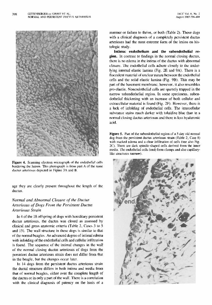
Figure 4. Scanning electron micrograph of the endothelial cells bordering the lumen. This photograph is from part A of the same ductus arteriosus depicted in Figure 3A and B.
age they are clearly present throughout the length of the ductus.
Normal and Abnormal Closure of the Ductus Arteriosus of Dogs From the Persistent Ductus Arteriosus Strain
In 4 of the 18 offspring of dogs with hereditary persistent ductus arteriosus, the ductus was closed as assessed by clinical and gross anatomic criteria (Table 2, Cases 3 to 5 and 15). The wall structure in these dogs is similar to that of the normal beagles. An advanced degree of intimal edema with infolding of the endothelial cells and cellular infiltration is found. The sequence of the intimal changes in the wall of the normal closing ductus arteriosus of dogs from the persistent ductus arteriosus strain does not differ from that in the beagle, but the changes occur later.
In 14 dogs from the persistent ductus arteriosus strain the ductal structure differs in both intima and media from that of normal beagles, either over the complete length of the ductus or in only a part of the wall. There is a correlation with the clinical diagnosis of patency on the basis of a
JACC Vol. 6, No.2 August 1985:394-404
murmur or failure to thrive, or both (Table 2). Those dogs with a clinical diagnosis of a completely persistent ductus arteriosus had the most extreme form of the lesion on histologic study.
Intima: endothelium and the subendothelial region. In contrast to findings in the normal closing ductus, there is no edema in the intima of the ductus with abnormal closure. The endothelial cells adhere closely to the underlying internal elastic lamina (Fig. 2E and 9A). There is a flocculent material of unclear nature between the endothelial cells and the solid elastic lamina (Fig. 9B). This may be part of the basement membrane; however, it also resembles pro-elastin. Nonendothelial cells are sparsely trapped in the narrow subendothelial region. In some specimens, subendothelial thickening with an increase of both cellular and extracellular material is found (Fig. 2F). However, there is a lack of infolding of endothelial cells. The intercellular substance stains much darker with toluidine blue than in a normal closing ductus arteriosus and there is less hyaluronic acid.
Figure 5. Part of the subendothelial region of a 5 day old normal dog from the persistent ductus arteriosus strain (Table 2, Case 8) with marked edema and a clear infiltration of cells (see also Fig. 2C). There are dark spindle-shaped cells derived from the inner media. The endothelial cells (end) form clumps and also capillarylike structures (arrow).
/
lACC Vol. 6, No.2 August 1985:394-404
A
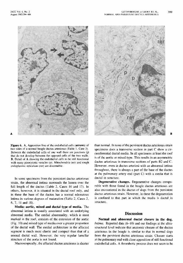
Figure 6. A, Apposition line of the endothelial cells (arrows) of two sides of a normal beagle ductus arteriosus (Table 1, Case 3). Between the endothelial cells of one wall there are junctions (j) that do not develop between the opposed cells of the two walls. B, Detail of A showing the endothelial cells to be still functional with many pinocytotic vesicles (p). Mitochondria (mi) and rough endoplasmic reticulum (rer) are discernible.
In some specimens from the persistent ductus arteriosus strain, the abnormal intima surrounds the lumen over the full length of the ductus (Table 2, Cases lO and 17). In others, however, it is situated in the ductal roof only, and in these the base of the ductus has a normal edematous intima in various degrees of maturation (Table 2, Cases 2, 6,7,11 and 18),
Media: aortic, mixed and ductal type of media. The abnormal intima is usually associated with an underlying abnormal media, The medial abnormality, which is most marked in the roof, consists of the extension of the aortic (Fig. lO) and mixed type of media over a greater proportion of the ductal wall. The medial architecture in the affected segment is much more elastic and compact than that of a normal ductal wall. However, the very regular lamellar structure of the aorta is not found.
Macroscopically, the affected ductus arteriosus is shorter
GITTENBERGER-\)~. GROOT ET AL. 399 NORMAL AND PERSISTENT DUCTUS ARTERIOSUS
B
than normal. In none of the persistent ductus arteriosus strain specimens does a transverse section in part C show a circumferential ductal media. In all specimens at least the roof is of the aortic or mixed type. This results in an asymmetric ductus arteriosus in transverse sections of parts B2 and C. However, even in ductus arteriosi with an abnormal intima throughout, there is always a part of the base of the ductus at the pulmonary artery end (part C) with a media that is ductal in structure.
Degenerative changes. Degenerative changes comparable with those found in the beagle ductus arteriosus are also encountered in the ductus of dogs from the persistent ductus arteriosus strain. However, in these the degeneration is confined to that part in which the media is ductal in structure.
Discussion Normal and abnormal ductal closure in the dog.
Intima. Reported data (6-lO) and our findings at the ultrastructural level indicate that anatomic closure of the ductus arteriosus in the beagle is similar to that in normal dogs from the persistent ductus arteriosus strain. Closure starts at the pulmonary end with close apposition of still functional endothelial cells. A thrombotic process does not seem to be
400 GIITENBERGER-DE GROOT ET AL. NORMAL AND PERSISTENT DUCTUS ARTERIOSUS
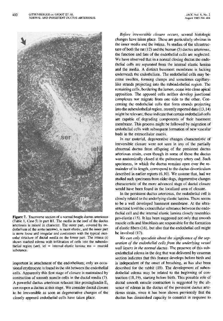
Figure 7. Transverse section of a normal beagle ductus arteriosus (Table 1, Case 5) in part B 1. The media in the roof of the ductus arteriosus is mixed in character. The outer part, covered by endothelium of the aorta (arrow), is more elastic, and the inner part is mote loose and irregular and continuous with the typical muscular structure of ductal media on the lower part. The intima (i) shows marked edema with infiltration of cells into the subendothelial region (ser). iel = internal elastic lamina; ms = mucoid substance.
important in attachment of the endothelium; only an occasional erythrocyte is found in the slit between the endothelial cells. Apparently this first stage of closure is maintained by contraction of smooth muscle cells in the ductal media (7). A powerful ductus arteriosus relaxant like prostaglandin EI can reopen a ductus at this stage. We consider ductal closure to be irreversible as soon as degenerative changes of the closely apposed endothelial cells have taken place.
JACC Vol. 6. No.2 August 1985:394-404
Before irreversible closure occurs, several histologic changes have taken place. These are particularly obvious in the inner media and the intima. In studies of the ultrastructure of both the rat (12) and the human (5) ductus arteriosus, the ftinction and fate of the endothelial cells are neglected. We have observed that in a normal closing ductus the endothelial cells are separated from the internal elastic lamina and the media. A distinct basement membrane is lacking underneath the endothelium. The endothelial cells may become swollen, forming clumps and sometimes capillarylike strands projecting into the subendothelial region. The remaining cells, bordering the lumen, come into close apical apposition. The apposed cells neither develop junctional complexes nor migrate from one side to the other. Concerning the endothelial cells that form strands projecting il1to the subendothelial region, recently reported data (13,14) might be relevant; these indicate that certain endothelial cells are capable of degrading components of their basement membrane. This process might be followed by migration of endothelial cells With subsequent formation of new vascular buds in the extracellular matrix.
In our material, degenerative changes characteristic of irreversible closure were not seen in any of the partially abnormal ductus from offspring of the persistent ductus arteriosus strain, even though in some of these the ductus was anatomically closed at the pulmonary artery end. Such specimens, in which the ductus remains open over the remainder of its length, correspond to the ductus diverticulum described in earlier reports (6,10). We assume that, had we studied such specimens from older dogs, degenerative changes characteristic of the more advanced stage of ductal closure would have been found in the localized area of closure.
In the persistent ductus arteriosus, the endothelial cell is closely related to the underlying elastic lamina. There seems to be a well developed basement membrane. At the ultrastructlirallevel the extracellular substance between the endothelial cell and the internal elastic lamina closely resembles pro-elastin (15). It has been suggested not only that smooth muscle cells and fibroblasts are responsible for the formation of elastic fibers (16), but also that the endothelial cell might be involved (17).
We can only speculate about the significance of the separation of the endothelial cells from the underlying vessel wall layers in the normal ductus. The presence of this subendothelial edema in the dog that was delivered by cesarean section indicates that this feature develops before birth and is independent of the onset of breathing, as has also been described for the rabbit (18). The development of subendothelial edema may be related to the beginning of contraction (18,19), starting before birth. This possible role of ductal smooth muscle contraction is suggested by the absence of edema in the ductus of the persistent ductus arteriosus strain, since it has been shown previously that the ductus has diminished capacity to constrict in response to
JACC Vol. 6, No.2 August 1985:394-404
A
Figure 8. Part of the intima and media of the ductus arteriosus of a 7 day old beagle (Table I, Case 10). A, In the center the apposition line (arrows) is still discernible. In the subendothelial region there is accumulation of lipid droplets in the cells indicated by the black dots (see also Fig. 20). B, Electron micrograph of cells showing a marked accumulation of lipid droplets, most probably indicative of a degenerative change. el = elastin; m = media.
oxygen and vasoconstrictor drugs (7). However, the occasional finding of an abrupt transition from an edematous intima to an intima without edema, independent of the amount of smooth muscle cells in the underlying media, does not support this view. Usually there is edema in the region of the crista reuniens at the aortic end of the normal ductus. Here the media is still rather elastic and poor in smooth muscle cells, suggesting that smooth muscle contraction need not to be a major factor in the formation of edema.
It has been proposed (20) that an intact endothelium may be important in maintaining relaxation of a muscular artery. This implies an interaction of the endothelium and the medial smooth muscle cells. It might be postulated that ductal patency in the fetal period is dependent on the presence of an intact normal endothelium. Ductal constriction might be enhanced by the loss of normal cellular relations as is seen
GllTENBERGER-DE GROOT ET AL. 401 NORMAL AND PERSISTENT DUCTUS ARTERIOSUS
B
in the intima of the normal closing ductus arteriosus. In the persistent ductus this cellular relation is maintained.
Media. The function and nature of the small dark cells in the inner media must be further evaluated. These are clearly discernible in the normal ductus arteriosus and seem to migrate into the subendothelial region. In various animal species these cells have been referred to as smooth or modified smooth muscle cells (5,12). The possibility that they are undifferentiated smooth muscle cells, as described in coronary artery intimal proliferation (21), cannot be ruled out.
In the persistent ductus, the small dark cells in the inner media are either far fewer in number or more difficult to detect. It can be hypothesized that the lack of endothelial detachment in the persistent ductus inhibits transformation and migration of smooth muscle cells from the medial to the intimal region. A similar mechanism has been postulated to be important in the pathogenesis of atherosclerosis (22).
It is apparent from these and previous studies of the persistent ductus arteriosus strain (7,9,10) that the roof of the ductus arteriosus in persistent ductus is more elastic than usual and that this phenomenon is accompanied by a decreased capacity of the ductus to constrict (7,10). In the present study, we found a clear relation between the abnormal presence of an aortic or a mixed type of ductal media
402 GITTENBERGER-DE GROOT ET AL. NORMAL AND PERSISTENT DUCTUS ARTERIOSUS
A
and the abnonnal intimal structure of persistent ductus: areas of the ductus with an abnonnal media tended to lack subendothelial edema. However, this relation is not exclusive. An abnonnal intima in some cases can accompany a muscular media in a persistent ductus, whereas a mixed type of media in a nonnal ductus can be accompanied by subendothelial edema.
Comparability of human and canine ductus arteriosus. Comparison of the nonnal human with the nonnal canine ductus at light microscopic level shows the main difference to be in the time of development of the intimal thickening. In the human, intimal cushions develop in the fetal ductus arteriosus during the last 3 months of pregnancy (4). In the dog, except for the edema, the intimal changes take place in the first hours to days after birth. When characteristics of intimal cushion fonnation in the human ductus (5) are compared with our findings in the dog, the timing rather than the character or the sequence of the histologic changes appears to be different.
As has been described (2,23), the only typical finding in the persistent ductus arteriosus in humans is the close adherence between the endothelium and a subendothelial elastic lamina. This can be either the nonnal internal elastic lamina, as found in the premature ductus arteriosus, or an additional elastic subendothelial elastic lamina lying above
JACC Vol. 6, No.2 August 1985:394-404
B
Figure 9. A, Intima and media of the ductus arteriosus of a 7 day old dog from the strain with persistent ductus arteriosus with an abnormal wall (Table 2, Case 10). There is no edema and the endothelial (end) cells adhere closely to the internal elastic lamina (iel). The media (m) is ductal in character. B, Scanning electron micrograph of the endothelium of a 27 day old dog from the strain with persistent ductus arteriosus (Table 2, Case 17) with an abnormal wall. The whitish substance is elastin of the internal elastic lamina (iel). Also collagen (coli) fibers are visible in the intercellular substance.
an intimal cushion. In the dog with hereditary persistent ductus arteriosus, this close relation of the endothelium with an underlying elastic lamina is also found.
Future research. It appears that in a normal closing ductus arteriosus the separation of the endothelial cells from the underlying media cells is an initial step in the progress of definitive anatomic sealing. The histologic changes in intima and media of the ductus arteriosus that occur during what can be considered a nonnal physiologic process, have a striking resemblance to the development of pathologiC intimal thickening in other arteries. This process is in some way disrupted by the genetic defect underlying persistence of the ductus in the persistent ductus arteriosus strain of dogs. Further study of the nonnal anatomic closing process of the ductus compared with the defect in the persistent
JACC Vol. 6, No.2 August 1985:394-404
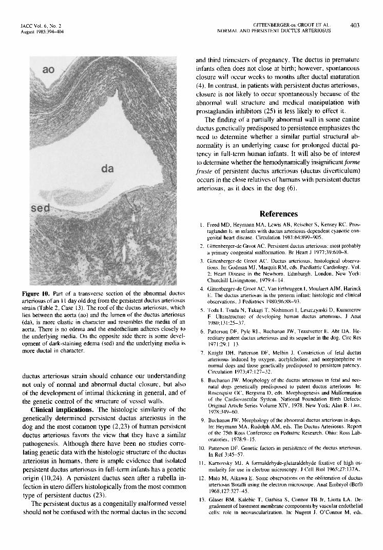
Figure 10. Part of a transverse section of the abnormal ductus arteriosus of an II day old dog from the persistent ductus arteriosus strain (Table 2, Case 13). The roof of the ductus arteriosus, which lies between the aorta (ao) and the lumen of the ductus arteriosus (da), is more elastic in character and resembles the media of an aorta. There is no edema and the endothelium adheres closely to the underlying media. On the opposite side there is some development of dark-staining edema (sed) and the underlying media is more ductal in character.
ductus arteriosus strain should enhance our understanding not only of normal and abnormal ductal closure, but also of the development of intimal thickening in general, and of the genetic control of the structure of vessel walls.
Clinical implications. The histologic similarity of the genetically determined persistent ductus arteriosus in the dog and the most common type (2,23) of human persistent ductus arteriosus favors the view that they have a similar pathogenesis. Although there have been no studies correlating genetic data with the histologic structure of the ductus arteriosus in humans, there is ample evidence that isolated persistent ductus arteriosus in full-term infants has a genetic origin 00,24). A persistent ductus seen after a rubella infection in utero differs histologically from the most common type of persistent ductus (23).
The persistent ductus as a congenitally malformed vessel should not be confused with the normal ductus in the second
GITIENBERGER-DE GROOT ET AL. 403 NORMAL AND PERSISTENT DUCTUS ARTERIOSUS
and third trimesters of pregnancy. The ductus in premature infants often does not close at birth; however, spontaneous closure will occur weeks to months after ductal maturation (4). In contrast, in patients with persistent ductus arteriosus, closure is not likely to occur spontaneously because of the abnormal wall structure and medical manipulation with prostaglandin inhibitors (25) is less likely to effect it.
The finding of a partially abnormal wall in some canine ductus genetically predisposed to persistence emphasizes the need to determine whether a similar partial structural abnormality is an underlying cause for prolonged ductal patency in full-term human infants. It will also be of interest to determine whether the hemodynamically insignificant forme fruste of persistent ductus arteriosus (ductus diverticulum) occurs in the close relatives of humans with persistent ductus arteriosus, as it does in the dog (6).
References l. Freed MD, Heymann MA, Lewis AB, Reischer S, KeIisey RC. Pros
taglandin E\ in infants with ductus arteriosus-dependent cyanotic congenital heart disease. Circulation 1981 ;64:899-905.
2. Gittenberger-de Groot AC. Persistent ductus arteriosus: most probably a primary congenital malformation. Br Heart J 1977;39:610-8.
3. Gittenberger-de Groot AC. Ductus arteriosus, histological observations. In: Godman MJ, Marquis RM, eds. Paediatric Cardiology, Vol. 2; Heart Disease in the Newborn. Edinburgh, London, New York: Churchill Livingstone, 1979:4-14.
4. Gittenberger-de Groot AC, Van Ertbruggen I, Moulaert AJM, Harinck E. The ductus arteriosus in the preterm infant: histologic and clinical observations. J Pediatrics 1980;96:88-93.
5. Toda I, Tsuda N. Takagi T, Nishimori I, Leszczynski D. Kummerov F. Ultrastructure of developing human ductus arteriosus. J Anat 1980; 131 :25-37.
6. Patterson OF. Pyle RL, Buchanan JW, Trautvetter E. Abt DA. Hereditary patent ductus arteriosus and its sequelae in the dog. Circ Res 1971;29:1-13.
7. Knight DH. Patterson OF. Melbin J. Constriction of fetal ductus arteriosus induced by oxygen. acetylcholine, and norepinephrine in normal dogs and those genetically predisposed to persistent patency. Circulation 1973;47: 127-32.
8. Buchanan JW. Morphology of the ductus arteriosus in fetal and neonatal dogs genetically predisposed to patent ductus arteriosus. In: Rosenquist GC, Bergsma D. eds. Morphogenesis and Malformation of the Cardiovascular System. National Foundation Birth Defects: Original Article Series Volume XIV. 1978. New York: Alan R. Liss, 1978:349-60.
9. Buchanan JW. Morphology of the abnormal ductus arteriosus in dogs. In: Heymann MA. Rudolph AM, eds. The Ductus Arteriosus. Report of the 75th Ross Conference on Pediatric Research. Ohio: Ross Laboratories. 1978:9-15.
10. Patterson OF. Genetic factors in persistence of the ductus arteriosus. In Ref 3:45-57.
II. Kamovsky MJ. A formaldehyde-glutaraldehyde fixative of high osmolarity for use in electron microscopy. J Cell BioI 1965;27:137A.
12. Mato M, Aikawa E. Some observations on the obliteration of ductus arteriosus Botalli using the electron microscope. Anat Embryol (Berl) 1968;127:327-45.
13. Glaser BM, Kalebic T, Garbisa S. Connor TB Jr, Liotta LA. Degradement of basement membrane components by vascular endothelial cells: role in neovascularization. In: Nugent J. O'Connor M, eds.
404 GITIENBERGER-DE GROOT ET AL. NORMAL AND PERSISTENT DUCTUS ARTERIOSUS
Development of the Vascular System. London: Pitman (Ciba Foundation Symposium 1(0), 1983:150-62.
14. Macaig T, Kadish J, Wilkins L, Steniennan MB, Weinstein R. Organizational behavior of human umbilical vein endothelial cells. J Cell Bioi 1982;94:511-20.
15. Dingemans KP, Jansen N, Becker AE. Ultrastructure of the nonnal human aortic media. Virchows Arch (Path Anat) 1981;392:199-216.
16. Roach MR. the pattern of elastin in the aorta and large arteries of maminals. In Ref 13:37-55.
17. Cantor JO, Keller S, Parshley MS, et al. Synthesis of cross linked elastin by an endothelial cell culture. Biochem Biophys Res Commun 1980;95:1381-6.
18. Yoder MJ, Baumann FG, Goodyear JI, Imparato AM. Endothelial alterations in the constricting rabbit ductus arteriosus: relationship to smooth muscle cell bleb fonnation. Chicago: Scanning Electron Microscopy/SEM Inc., I 980:III:27 1-6.
19. Kohori K, Suzuki K, Yoshida Y, Ooneda G. Light and electron mi-
JACC Vol. 6, No.2 August 1985:394-404
crdscopic studies on rat arterial lesions induced by experimental arterial contraction. Virchows Arch (AJ 1979;385:29-39.
20. Furchgott RF, Zawadski JV. The obligatory role of endothelial cells in relaxation of arterial smooth muscle by acetylcholine. Nature 1980;288:373-6.
21. Jaffe D, Hartroft WS, Manning M, Eleta G. Coronary arteries in newborn children (suppl). Acta Paediatr Scand 1971;219:3-28.
22. Ross R. A hypothesis of atherogenesis. In Ref 13:198-206.
23. Gittenberger-de Groot AC, Moulaert AJM, Hitchcock JF. Histology of the persistent ductus arteriosus in cases of congenital rubella. Circulation 1980;62: 183-6.
24. Wilkins JL. Risks to offspring with patent ductus arteriosus. J Med Genet 1969;6: 1-4.
25. Gittenberger-de Groot AC, Sutherland K, Sauer U, Kellner M, Schober JG, Biihlmeijer K. Nonnal and persistent ductus arteriosus influenced by prostaglandin E!. Herz 1980;5:361-8.