Histological alterations of a combination of Chlorpyrifos and Cypermethrin (Nurocombi) insecticide in the fresh water crab, Paratelphusa jacquemontii (Rathbun) A. Maharajan * , Y. Narayanasamy, V. Ganapiriya, K. Shanmugavel PG & Research Department of Zoology, Khadir Mohideen College, Adirampattinam 614701, Thanjavur Dist, Tamil Nadu, India Received 31 May 2015; revised 8 August 2015; accepted 8 August 2015 Available online 16 September 2015 KEYWORDS Paratelphusa jacquemontii; Histology; Nurocombi Abstract Chlorpyrifos (CPF) and Cypermethrin (CPM) are toxic and subject to long-term in vivo accumulation in different aquatic species throughout the world. The purpose of the present study was to examine the combined CPF/CPM (Nurocombi) exposure on histology in various tissues of Paratelphusa jacquemontii. The crabs were exposed to combined CPF/CPM concentrations of 0.0187 ppm and 0.0374 ppm (sub lethal) for 28 days with parallel untreated control. The experimen- tal gill tissue exhibited epithelial lifting, edema, necrosis, fusion of secondary lamellae and hemor- rhage. The deceased hepatopancreas revealed infiltration, formation of large lumen and disappearance of haemocytes. The pathologic symptoms like atrophy, necrosis, wavy appearance, accumulation of granular material in between muscle fibers, fragmentation, loss of muscle structure, appearance of basophilic deposits were displayed in the muscle tissue. The vas deferens showed remarkable epithelial vacuolar degeneration, irregular appearance of spermatophore matrix associ- ated with reduction in the number of spermatophores and dehiscence of most of the sper- matophores. It is concluded that histological biomarkers provide reliable and discriminatory data to augment pesticide pollution and therefore, long-term monitoring is necessary to assess the eco-health of the mangrove system. Ó 2015 The Egyptian German Society for Zoology. Production and hosting by Elsevier B.V. This is an open access article under the CC BY-NC-ND license (http://creativecommons.org/licenses/by-nc-nd/4.0/). Introduction Crabs play an important role in the maintenance, modification and regulation of the environment by influencing both abiotic and biotic components. They are abundant and serve both as the predator and the prey and hence are located at different trophic levels in the ecosystem (Siddon and Witman, 2004). * Corresponding author. Mobile: +91 9443286900. E-mail address: [email protected](A. Maharajan). Peer review under responsibility of The Egyptian German Society for Zoology. The Journal of Basic & Applied Zoology (2015) 72, 104–112 HOSTED BY The Egyptian German Society for Zoology The Journal of Basic & Applied Zoology www.egsz.org www.sciencedirect.com http://dx.doi.org/10.1016/j.jobaz.2015.08.002 2090-9896 Ó 2015 The Egyptian German Society for Zoology. Production and hosting by Elsevier B.V. This is an open access article under the CC BY-NC-ND license (http://creativecommons.org/licenses/by-nc-nd/4.0/).
Transcript
The Journal of Basic & Applied Zoology (2015) 72, 104–112
HO ST E D BYThe Egyptian German Society for Zoology
The Journal of Basic & Applied Zoology
www.egsz.orgwww.sciencedirect.com
Histological alterations of a combination of
Chlorpyrifos and Cypermethrin (Nurocombi)
insecticide in the fresh water crab, Paratelphusajacquemontii (Rathbun)
Peer review under responsibility of The Egyptian German Society for
Zoology.
http://dx.doi.org/10.1016/j.jobaz.2015.08.0022090-9896 � 2015 The Egyptian German Society for Zoology. Production and hosting by Elsevier B.V.This is an open access article under the CC BY-NC-ND license (http://creativecommons.org/licenses/by-nc-nd/4.0/).
A. Maharajan *, Y. Narayanasamy, V. Ganapiriya, K. Shanmugavel
PG & Research Department of Zoology, Khadir Mohideen College, Adirampattinam 614701, Thanjavur Dist, Tamil Nadu, India
Received 31 May 2015; revised 8 August 2015; accepted 8 August 2015Available online 16 September 2015
KEYWORDS
Paratelphusa jacquemontii;
Histology;
Nurocombi
Abstract Chlorpyrifos (CPF) and Cypermethrin (CPM) are toxic and subject to long-term in vivo
accumulation in different aquatic species throughout the world. The purpose of the present study
was to examine the combined CPF/CPM (Nurocombi) exposure on histology in various tissues
of Paratelphusa jacquemontii. The crabs were exposed to combined CPF/CPM concentrations of
0.0187 ppm and 0.0374 ppm (sub lethal) for 28 days with parallel untreated control. The experimen-
tal gill tissue exhibited epithelial lifting, edema, necrosis, fusion of secondary lamellae and hemor-
rhage. The deceased hepatopancreas revealed infiltration, formation of large lumen and
disappearance of haemocytes. The pathologic symptoms like atrophy, necrosis, wavy appearance,
accumulation of granular material in between muscle fibers, fragmentation, loss of muscle structure,
appearance of basophilic deposits were displayed in the muscle tissue. The vas deferens showed
remarkable epithelial vacuolar degeneration, irregular appearance of spermatophore matrix associ-
ated with reduction in the number of spermatophores and dehiscence of most of the sper-
matophores. It is concluded that histological biomarkers provide reliable and discriminatory data
to augment pesticide pollution and therefore, long-term monitoring is necessary to assess the
eco-health of the mangrove system.� 2015 The Egyptian German Society for Zoology. Production and hosting by Elsevier B.V. This is an
open access article under the CC BY-NC-ND license (http://creativecommons.org/licenses/by-nc-nd/4.0/).
Introduction
Crabs play an important role in the maintenance, modification
and regulation of the environment by influencing both abioticand biotic components. They are abundant and serve both asthe predator and the prey and hence are located at different
trophic levels in the ecosystem (Siddon and Witman, 2004).
Chlorpyrifos and Cypermethrin exposure on histology in fresh water crab 105
Many crab species are burrowing in nature and frequentlyalter the surface characteristics and drive the nutrient cycling(Pandya, 2011). A wide range of studies are available on macro
invertebrates as an indicator species of aquatic habitat butamong them specifically, brachyuran crabs are an effectiveindicator of different changes in both abiotic and biotic
factors.The marine pollution is mainly contributed by industrial,
domestic and agricultural wastes (Kasmin, 2010). Pesticides,
commonly used in agriculture and public health, adverselyaffect the natural environment and non target aquatic organ-isms through surface runoff from the treated area (Singhet al., 2008; Stueckle et al., 2008). When compared to single
pesticides, mixed pesticides, cause significant synergistic effectson target species, and also effective to beneficial species in mostcases. Cypermethrin is applied 2–3 times per growing season
and repeated pesticide applications represent a risk to adjacentsurface waters. However, the environmental impact of suchagricultural intensification remains largely unreported.
Currently many pesticides are extensively used in agricul-tural operations. These pesticides have various physiologicaleffects on the pests, such as inhibitory effects on growth, food
intake, metabolism, enzyme activity and general development(Tungare and Sawant, 2000) and also adverse effects on aqua-tic animals. Chlorpyrifos an organophosphate (OP) is the sec-ond largest used pesticide in India in controlling cutworms,
corn rootworms, flea beetles, flies, termites, fire ants and lice(Mathur and Tannan, 1999). There are some reports on thetoxicity of OP pesticides to crabs (Radhakrishnaiah and
Renukadevi, 1990; Senthil Kumar et al., 2007; Ghediraet al., 2009). The study of the impact of pesticides on aquaticanimals is an important aspect of chemical contamination of
the aquatic environment (Narra, 2014).Histopathological examination has been increasingly recog-
nized as a valuable tool for the assessment of the impact of
environmental pollutants on aquatic animals (Saravanabhavan and Geraldine, 2009; Maharajan et al., 2012a;Manosathiyadevan et al., 2012; Chourpagar and Kulkarani,2014; Paruruckumani et al., 2015). Gills apart from being the
primary respiratory organ in crabs, are also responsible forother vital physiological functions like excretion, acid basebalance and ion regulation. So when crabs are exposed to envi-
ronmental pollutants, these vital functions are deleteriouslyaffected and the functional impairment of gills can significantlydamage their health (Kumar and Tembhre, 2010). The gills are
efficient tools for biomonitoring potential impacts (OliveiraRibeiro et al., 2005) because of their large area in contact withwater and high permeability (Arellano et al., 2004; Viglianoet al., 2006).
The crustacean hepatopancreas or digestive gland involvesin food absorption, synthesis and secretion of digestiveenzymes, storage of lipids, glycogen and minerals during inter-
moult period. It is the main organ of reserve and detoxificationof xenobiotics and is highly sensitive to physiological and envi-ronmental changes (Johnson et al., 1998). Reddy et al. (1983)
observed that the sumithion affects the ovarian growth of crab,Oziotelphusa sensex sensex. Victor (1984) observed structuralchanges in ovary of freshwater prawn, Caridina rajadhari
exposed to malathion and DDT.Crab, Paratelphusa jacquemontii commonly inhabits the
rice field, ponds, lakes and rivers, and forms food for localpeople. The combined toxicity of Chlorpyrifos (CPF) and
Cypermethrin (CPM) to P. jacquemontii has not yet beenreported. Therefore, the major objective of the present paperis to study histopathological alterations in the gill, hepatopan-
creas, muscle and vas deference of P. jacquemontii.
Materials and methods
Animal collection and acclimatization
The experiments were performed in accordance with local/na-tional guidelines for experimentation in animals and all carewas taken to prevent cruelty of any kind. Fresh water crabs,
P. jacquemontii of carapace size ranging from 5.6 to 6.1 andweight 45–55 g were collected from the paddy field of Muthu-pettai, Thiruvarur Dist, Tamil Nadu. They were transported
and kept in 100 L tank containing well aerated filtered freshwater maintained at ambient temperature (27 ± 2 �C) for aperiod of one week. Before stocking, the tank was washed with0.1% KMnO4 for disinfection.
Chemicals
For preparation of stock solution 1 ml of insecticide NURO-
COMBI (Chlorpyriphos (CPF) 50% and Cypermethrin(CPM) 5% EC), Cheminova, FMC Corporation, Mumbai,diluted with 1 L of Milli-Q deionised water was purchased.
Test concentration
Crabs were exposed to 0.0187 and 0.0374 ppm sublethal con-
centration of combined insecticide doses at 10% and 20%respectively of the Maximum Acceptable Toxicant Concentra-tion (MATC), which was 0.187 ppm.
Test procedure
After 2 weeks of acclimatization in a holding tank, ten healthycrabs with carapace size ranging from 5.9 to 6.2 and weight
50–60 g were transferred to each aquarium. Three replicateswere performed for test concentration and control. Crabs werefed twice daily with commercially prepared pellet feed at 10:00
and 16:00 h. Uneaten food was quickly removed from the sys-tem. The media were renewed every alternate day. Mortalityand behavior were observed everyday in each concentration.
Two crabs from each aquarium were sampled at 0, 7and28 days post-exposure.
Histological study
The tissues gill, hepatopancreas, muscle and vas deferens werecollected from the treated as well as control crab after 0, 7 and28 days post-exposure and preserved in Davidson’s fixatives.
The method of Bernet et al. (1999) was adopted for processingof tissues for histological studies. In brief, after 24 h of tissuefixation in Davidson’s solution, tissues were dehydrated with
alcohol upgrades (30–100%). After dehydration, tissue blockswere prepared in paraffin wax. Sagittal sections (5 lm thick) ofthe paraffin blocks were cut and stained with hematoxylin-
alcoholic eosin for histological evaluation. Photomicrographsof stained sections were made using phase contrast microscopy
Plate 1 Histological changes of Gills in P. jacquemontii. Light microscope of a paraffin section stained with Haematoxylin and Eosin
(40X). (A & B) Control. (C) After 7 days of exposure to 0.0187 ppm concentration of Nurocombi. (D) After 28 days of exposure to
0.0187 ppm concentration of Nurocombi. (E) After 7 days of exposure to 0.0384 ppm concentration of Nurocombi. (F) After 28 days of
exposure to 0.0384 ppm concentration of Nurocombi. Abbreviations used: SL – secondary lamellae, PC – pillar cells, IC – Ionocytes,
DESGL – degeneration of epithelium in secondary gill lamellae, NEC – necrosis, IH – infiltration of haemocytes, RSLT – rupture of
secondary lamellar tip, RC – rupture of capillaries, DC – detached cuticle, ILS – inter lamellar space, PN – pyknotic nuclei, HYP –
hypertrophy, HP – hyperplasia, EREC – edema and rupture of epithelial cells, SSL – swelling of secondary lamellae, DSGL –
disarrangement of secondary gill lamellae, ESGL – enlargement of secondary gill lamellae, DPC – disruption of pillar cells.
106 A. Maharajan et al.
(Nikon, Eclipse Ci fitted with a Spot inside DS-Fi 2 digitalcamera).
Results
Histology of gill
The gills of P. jacquemontii are formed of a number of lamellaeor broad flattened plates arranged serially in pairs along a con-
trol gill stem. The central axis of gill tissue is the primary gilllamellae and it further divides into secondary gill lamellae orfilaments. The control gill exhibits a thin layer of cuticle which
covers the entire outer surface. Underlying the cuticle is a
continuous layer of epithelial cells. At irregular intervals pillarcells join the lamellae. (Plate 1a and b).
Histopathology of gill
In lower concentration of 0.0187 ppm the changes were per-ceptible enlargement of intralamellar space densely packed
with granular material, and loss of gill structure (Plate 1c).The gill lamellae get collapsed in exposed crab due to the dis-ruption of the pillar cells. In the case of higher concentration
0.0374 ppm after 7 days of exposure the changes are hemocoelfilled with coarse amorphous to fibrous materials, thickenedgill lamellae, and massive hemocytic infiltration (Plate 1d).
Chlorpyrifos and Cypermethrin exposure on histology in fresh water crab 107
Detached cuticle (DC) and rupture of capillaries (RC) at tip ofthe secondary lamellae releasing haemocytes and gills devel-oped bulbular swelling at the tip are evident in later stages
(Plate 1e). Epithelial necrosis and hyperplasia were alsoobserved. Enlargement and disarrangement of secondary gilllamellae (ESGL) (DSGL) and lamellar fusion in some regions
are seen in the treated crabs at higher concentrations after28 days of exposure (Plate 1f).
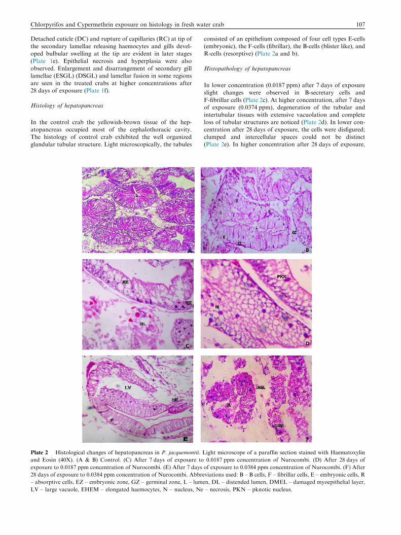
Histology of hepatopancreas
In the control crab the yellowish-brown tissue of the hep-atopancreas occupied most of the cephalothoracic cavity.
The histology of control crab exhibited the well organizedglandular tubular structure. Light microscopically, the tubules
Plate 2 Histological changes of hepatopancreas in P. jacquemontii.
and Eosin (40X). (A & B) Control. (C) After 7 days of exposure to
exposure to 0.0187 ppm concentration of Nurocombi. (E) After 7 days
28 days of exposure to 0.0384 ppm concentration of Nurocombi. Abbre
– absorptive cells, EZ – embryonic zone, GZ – germinal zone, L – lum
LV – large vacuole, EHEM – elongated haemocytes, N – nucleus, Ne
consisted of an epithelium composed of four cell types E-cells(embryonic), the F-cells (fibrillar), the B-cells (blister like), andR-cells (resorptive) (Plate 2a and b).
Histopathology of hepatopancreas
In lower concentration (0.0187 ppm) after 7 days of exposure
slight changes were observed in B-secretary cells andF-fibrillar cells (Plate 2c). At higher concentration, after 7 daysof exposure (0.0374 ppm), degeneration of the tubular and
intertubular tissues with extensive vacuolation and completeloss of tubular structures are noticed (Plate 2d). In lower con-centration after 28 days of exposure, the cells were disfigured;
clumped and intercellular spaces could not be distinct(Plate 2e). In higher concentration after 28 days of exposure,
Light microscope of a paraffin section stained with Haematoxylin
0.0187 ppm concentration of Nurocombi. (D) After 28 days of
of exposure to 0.0384 ppm concentration of Nurocombi. (F) After
viations used: B – B cells, F – fibrillar cells, E – embryonic cells, R
large numbers of vacuoles appeared and thickening of thebasal lamina was noticed. Necrosis, reduction in cell height,bulging and damaged myoepithelial layer with elongated
haemocytes were also conspicuous. (Plate 2f).
Histology of muscle
The muscle tissue of the control crab was made up of musclecells containing contractile filaments that move each otherand change the size of the cell. Muscle tissue derived from
mesoderm contains protein, and myosin filament (thread-like) forms multi nucleate cells that assemble into fibers calledmyofibrils (Plate 3a). The photomicrograph of the muscle
depicted the presence of normal myotomes (MT) with equallyspaced muscle bundles the fascicular arrangement of myofila-ments (MF) with emarginated epimysium, binding to
Plate 3 Histological changes of Muscle in P. jacquemontii. Light mic
(40X). (A & B) Control. (C) After 7 days of exposure to 0.0187 ppm
0.0187 ppm concentration of Nurocombi. (E) After 7 days of exposure
exposure to 0.0384 ppm concentration of Nurocombi. Abbreviations u
bundle, LV – large vacuole, FMB – fusion of muscle bundle, RMB – r
nuclei, CMB – congestion of muscle bundle, LMB – loosen of muscle
connective tissue and tendon at the extremities of the smoothmuscles. The striated muscle fibers (SM) were tightly packed.The nuclei were arranged along the margins of the muscle
bundles (Plate 3b).
Histopathology of muscle
After 7 days of exposure (0.0187 ppm) the muscle tissueshowed disintegrated epidermis (DE) with vacuolation, gapformation (GF) in between the muscle bundles, necrosis
(NE), marked thickening and separation of muscle bundleand pronounced intramuscular edema with minor dystrophicchanges (Plate 3c). In the higher concentration (0.0374 ppm),
the muscle bundles are completely disrupted with discontinuityof striations and complete disappearance of nuclei. In someregions muscle tissue shows the sloughing of epidermal layer
roscope of a paraffin section stained with Haematoxylin and Eosin
concentration of Nurocombi. (D) After 28 days of exposure to
to 0.0384 ppm concentration of Nurocombi. (F) After 28 days of
sed: SM – striated muscle, N – nuclei, DMB – disruption of muscle
upture of muscle bundle, GF – gap formation, CN – congestion of
bundle.
Chlorpyrifos and Cypermethrin exposure on histology in fresh water crab 109
(SEL) (Plate 3d), lesions (LN) and mild haemocyte infiltrations(HI) followed by fusion of muscle bundles (FMB) after 28 dayslow concentration (Plate 3e). In higher concentration the
muscle tissue expressed significant changes like broken myofib-rils (BMF), coagulative necrosis (CNE) congestion of musclebundles and rupture of muscle bundles. Severe haemocyte
infiltration (HI) and accumulation of granular materials inbetween the muscle fibers (GMF) are also noted (Plate 3f).
Histology of vas deferens
The light microscopy examination of the vas deferens of thecontrol crabs showed normal structure as evidenced by the well
organized structure of epithelium and spermatophores(Plate 4a). The vas deferens is morphologically coiled andinternally lined with squamous epithelium with definite long
Plate 4 Histological changes of Vas deferens in P. jacquemontii. Ligh
Eosin (40X). A & B Control. (C) After 7 days of exposure to 0.0187 pp
0.0187 ppm concentration of Nurocombi. (E) After 7 days of exposure
exposure to 0.0384 ppm concentration of Nurocombi. Abbreviations us
spermatophore, DEP – degenerated epithelium, DSF – disintegratio
collapsed spermatophore matrix, EN – extensive necrosis of spermato
nucleus and nucleolus. The lumen is filled with eosinophilichomogenous secretions in which spermatozoa are seensurrounded by wall called as spermatophore (Plate 4b). Under
light microscope spermatophore wall appears double layered.Within the spermatophores, the sperm cells are embedded inhomogenous material referred to as spermatophore matrix.
The size of the spermatophores ranging from 10 to 70 lmand enclose 2 to 80 sperm cells.
Histopathology of vas deferens
In the present study, Nurocombi treatment caused vacuolardegeneration on the epithelium and highly irregular
appearance of the spermatophore matrix after 7 days in lowerconcentration (0.0187 ppm) (Plate 4c). The day progresses andin 28 days the destruction was highly evident in the seminal
t microscope of a paraffin section stained with Haematoxylin and
m concentration of Nurocombi. (D) After 28 days of exposure to
to 0.0384 ppm concentration of Nurocombi. (F) After 28 days of
ed: SPH – spermatophore, SF – seminal fluid, BSPH – breakage of
n of seminal fluid, Ne – necrosis, GF – gap formation, CSM –
phore, AT – atrophy.
110 A. Maharajan et al.
plasma also. There was reduction in the number of sper-matophores and also dehiscence of most of the sper-matophores. The vas deferens exhibited severe necrosis and
atrophy when compared to control (Plate 4d). In higher con-centration (0.0374 ppm) after 28 days of exposure the sper-matophore matrix collapsed and spermatozoa exposed out in
most of the spermatophores. The double layered wall gotdehisced in some regions and spermatozoa are aggregated inseminal plasma (Plate 4e and f).
Discussion
The histopathological changes of gill can result in hypoxia, res-piratory failure problems with ionic and acid-base balance(Alazemi et al., 1996). Changes in the gill surfaces and
increased mucus production are consistent with observed his-tological effects such as hyperplasia, necrosis and lamellaraneurysms in the exposed crab with response to sub lethal con-
centrations of Nurocombi. Changes in the architecture of gillunder the Nurocombi, pesticide, stress would alter the diffus-ing capacity of gill with consequent hypoxic conditions andthus respiration becomes a problematic task for the crab in
fresh water habitat. Our results suggest that the lethal effectof Nurocombi is a result of damage to gas exchange mecha-nisms as consequence of the gill pathologies observed.
In the present study, epithelial lifting, edema, necrosis,fusion of adjacent secondary lamellae and hemorrhage at pri-mary lamellae were observed in the gills of the crab examined
after 28 days of exposure at higher concentration. Epithelialnecrosis and rupture of gill epithelium are direct deleteriouseffect of the irritants. The animal’s defense responses are exces-
sive mucus secretion due to the stress caused by the environ-mental change and pathologic agents which induce theproliferation of mucus cells (Richmonds and Dutta, 1989;Cardoso et al., 1996). Lifting of the epithelium, lamellar fusion
and club shaped lamellae could be protective in that it dimin-ishes the amount of vulnerable gill surface area (Richmondsand Dutta, 1989; Maharajan et al., 2012b; Maharajan et al.,
2013).Hepatopancreas is not only a digestive organ that possesses
abilities of absorption, digestion, storage and secretion but
also a major site where biotransformations and detoxificationundergo in crustaceans. In the present study, the hepatopan-creas showed changes in the F and B cells in low concentrationof Nurocombi, and cells were found clumped, and intercellular
spaces invisible in the medium concentration, and a generaldegeneration, loss of tubules structures, vacuolation, starshape of lumen and necrosis of cells in the high concentrations
of Nurocombi exposed P. jacquemontii. The star shape of thelumen was partially lost due to morphological changes of thetubular epithelial cells, because some cells decreased in height
from a normal columnar height to a low cuboidal form. In thepresent study, one of the most evident changes is a prolifera-tion of B-cells in the dosed crabs, indicating a high rate of
excretion from the hepatopancreas. The accumulation andelimination of the xenobiotic entering the hepatopancreatictubules are perhaps effected with a large number of F-cellsconverting into B-cells.
The hepatopancreas plays important roles in several meta-bolic processes in crustaceans (Caceci et al., 1988; Saravanabhavan and Geraldine, 2000). Nurocombi induced structural
changes were decrease in the cellular height of the tubularepithelium, reduction of secretory and lipid vacuoles, infiltra-tion of hemocytes, atrophy, pyknotic nuclei, cytolysis and
the melanised encapsulation of necrotic tissues.Krishnamoorthy and Subramanian (1996) also reportedchanges such as elongation of hepatopancreatic cells, and
shrunken cells in Macrobrachium lamerrei lamerrei exposedto low (0.0065 ppm), and high (0.0215 ppm) concentrationsof copper. Destructive and deteriorative changes in the hep-
atopancreas and gills were observed in Penaeus indicus exposedto Zn at a low concentration of 100 ppb (Viswanathan andManisseri, 1995). The noted histopathological changes in thehepatopancreas may be due to accumulation of the pesticide
since this organ is the center of storage, metabolism and detox-ification. The rupture of basal laminae observed in the hep-atopancreatic tubules suggest that tissue integrity was
affected in crab due to exposure to Nurocombi. Abnormalinfiltration of hemocytes in the interstitial sinuses noted inthe hepatopancreas of test animals suggest that the mechanism
of cellular/host defense was in operation to neutralize the tis-sue damage caused by Nurocombi and since hemocytes arethe most important form of cellular defense in crustaceans
(Bodhipaksha and Weeks-Perkins, 1994). The formation ofnecrotic hepatopancreatic tubules recorded in test crabs indi-cates the fact that the distortion, disintegration and death ofcells occurred in the hepatopancreas of P. jacquemontii
exposed to the highest sub-lethal concentration of Nurocombi.Therefore Nurocombi toxicity affects the normal integrity andcaused tissue damage in the hepatopancreas of P. jacquemontii.
In the present study, several histopathological alterationswere also noticed in the muscles of P. jacquemontii whenexposed to sub lethal concentration of Nurocombi. The patho-
logical findings include degeneration of muscles, necroses ofmuscle fibers with haemorrhages and RBC like pigmentedcells. The structural changes noticed in the muscle tissue as
atrophy, necrosis, wavy appearance and granular material inbetween the muscle fibers, fragmentation, loss of muscle struc-ture, appearance of basophilic deposits of the muscle fiberswere caused as a result of exposure of crabs to the sub lethal
concentrations.During pesticide exposure, pollutants affected the muscle
epidermis abruptly. Pigmented cells are a prominent feature
of chronic inflammatory response. The present investigationclosely agreed with a similar report by Tehrani et al. (2011)in the muscle tissues of Artemia urmiana in response to carba-
mates pesticide resulting in degeneration, Zenkers necrosis ofmuscle fiber with haemorrhages and RBC like cells. Exposureof Labeo rohita to hexachlorocyclohexane was found to induceseparation of muscle bundles and intracellular edema in the
muscle tissues (Das and Mukherjee, 2000). Such observationswere also made in muscle tissues of Oreochromis mossambicuson exposure to dimethoate (Parikh et al., 2010). Histopatho-
logical alterations in the muscle tissues of Heteropneustesfossilis exposed to polluted river water were also recorded byRakhi et al. (2013).
Interestingly, in the vas deferens, disruption in the tubulararchitecture, disarrangement of epithelial cells, reduction innumber of spermatophores, and aggregation of the granular
substance were exhibited by Nurocombi treated crabs at lowconcentration. At high concentration of Nurocombi exposure,severe disruption in the arrangement of spermatophores andepithelial cells and the reduction in tubule membrane thickness
Chlorpyrifos and Cypermethrin exposure on histology in fresh water crab 111
were reported. Our results clearly indicate that the abnormalcellular architecture of the vas deferens was noticed in P.jacquemontii on treatment with Nurocombi.
Bodkhe (1983) observed irregular arrangement of sperma-tozoa in the testicular tubules of the crab, Barytelphusa cunic-ularis following exposure to sevimol. Deshpande (1985)
reported thickening of lobular wall, impairment of lobules,reduced spermatogenic mass affected interstitial cells,vacuolization and degeneration of testis of Macrobrachium
kistnensis after pesticide treatment. Ovaries in M. kistnensisexhibited epithelial layer destruction, degeneration of oocytes,increased phagocytic cells, vacuolization appearance in cyto-plasm and nucleoplasm. TBTCL induced significant alteration
in the ovary of the prawn� M. kistnensis as increase in expo-sure leads to increase in damage to the ovary. This damageobserved in the ovary might be due to the direct effects of
TBTCL on developing oocytes intervening the enzyme systemin metabolism (Puccia et al., 2005).
Jegou (1992) found that dysfunction may lead to a reduc-
tion in sperm quality and possible infertility. Likewise, Fentand Hunn (1995) suggested that TBT can also affect spermcount and male reproductive systems in aquatic organisms.
Similarly, TBT at environmentally realistic concentrationshad an adverse effect on gametogenesis in cuvier Sebastiscusmarmoratus (Zhang et al., 2007). Kinnberg et al. (2000) alsodocumented concentration-dependent effects of nonylphenol
on the testicular structure of the fish Xiphophorus maculates.Zutshi and Murthy (2001) observed appreciable reduction insize, with spermatids and sperms in degenerating condition,
and necrosis of interstitial cells after fenthion treatment inthe fish Glossogobius giuris. They also reported extensive cyto-toxic damage in the testes of G. giuris after fenthion exposure.
Damage of vas deferens as a reproductive organ resulted in thedisturbance of overall metabolism and several physiologicalprocesses of crab. Hence, we can conclude that utilization of
pesticides should be minimized in the paddy field area ofMuthupettai mangrove ecosystem.
Acknowledgements
Authors would like to acknowledge their gratitude to Science
and Engineering Research Board, Department of Scienceand Technology, New Delhi, India (SB/YS/LS/254/2013) forthe financial assistance and Head of the Institution, Khadir
Mohideen College, Adirampattinam for the facilities provided.
References
Alazemi, B.M., Lewis, J.W., Andrews, E.B., 1996. Gill damage in the
freshwater fish Gnathonemus ptersii (Family: Mormyridae) exposed
to selected pollutants: an ultra structural study. Environ. Technol.
17, 225–238.
Arellano, J.M., Storch, V., Sarasquete, C., 2004. Ultrastructural and
histochemical study on gills and skin of the Senegal sole, Solea
senegalensis. J. Appl. Ichthyol. 20, 452–460.
Bernet, D., Schmidt, H., Meier, W., Burkhardtholm, P., Wahli, T.,
1999. Histopathology in fish: proposal for a protocol to assess
aquatic pollution. J. Fish Dis. 22, 25–34.
Bodhipaksha, N., Weeks Perkins, B.A., 1994. The effects of methyl
parathion on phagocytosis and respiratory burst activity of tiger