Histological assessments for toxicity and functionalization-dependent biodistribution of carbon nanohorns This content has been downloaded from IOPscience. Please scroll down to see the full text. Download details: IP Address: 134.117.10.200 This content was downloaded on 20/09/2013 at 10:46 Please note that terms and conditions apply. 2011 Nanotechnology 22 265106 (http://iopscience.iop.org/0957-4484/22/26/265106) View the table of contents for this issue, or go to the journal homepage for more Home Search Collections Journals About Contact us My IOPscience
Transcript
Histological assessments for toxicity and functionalization-dependent biodistribution of carbon
nanohorns
This content has been downloaded from IOPscience. Please scroll down to see the full text.
Download details:
IP Address: 134.117.10.200
This content was downloaded on 20/09/2013 at 10:46
Histological assessments for toxicity andfunctionalization-dependentbiodistribution of carbon nanohornsYoshio Tahara1, Jin Miyawaki2,7, Minfang Zhang1,2, Mei Yang1,5,Iwao Waga3, Sumio Iijima1,2,4,5, Hiroshi Irie6 andMasako Yudasaka1,2,7
1 Nanotube Research Center, National Institute of Advanced Industrial Science andTechnology, 5-2, 1-1-1 Higashi, Tsukuba, Ibaraki 305-8565, Japan2 JST/SORST, c/o NEC, 34 Miyukigaoka, Tsukuba, Ibaraki 305-8501, Japan3 VALWAY Technology Center, NEC Soft, Ltd, 1-18-7, Shinkiba, Koto-Ku, Tokyo 136-8627,Japan4 NEC, 34 Miyukigaoka, Tsukuba, Ibaraki 305-8501, Japan5 Department of Material Science and Engineering, Meijo University, 1-501 Shiogamaguchi,Tenpaku, Nagoya 468-8502, Japan6 Teikyo University School of Medicine, 2-11-1 Kaga, Itabashi-ku, Tokyo 173-8605, Japan
Received 14 January 2011, in final form 7 April 2011Published 18 May 2011Online at stacks.iop.org/Nano/22/265106
AbstractSingle-walled carbon nanohorns (SWNHs) intravenously administered to mice did not showsevere toxicity during a 26-week test period, which was confirmed by normal gross appearance,normal weight gain and the lack of abnormality in the tissues on histological observations of themice. SWNH biodistribution was influenced by chemical functionalization. Accumulation ofSWNH in the lungs reduced as SWNH hydrophilicity increased; however, the most hydrophilicSWNHs modified with bovine serum albumin (BSA) were most likely to be trapped in thelungs, suggesting that the BSA moiety enhanced macrophage phagocytosis in the lungs.Clearance of some of the hydrophobic SWNHs from the lungs was observed, the mechanism ofwhich is briefly discussed.
S Online supplementary data available from stacks.iop.org/Nano/22/265106/mmedia
(Some figures in this article are in colour only in the electronic version)
1. Introduction
Carbon nanotubes [1, 2] are attractive candidates forvarious biomedical applications such as drug delivery [3–5].Their toxicity and biodistribution depends on the size,shape, agglomeration and chemical functionalization of themolecules [6–9]. Recent reports suggest that long, rigid rod-like multi-walled carbon nanotubes (MWNTs) cause toxiclesions and inflammation because they are not completelyengulfed by macrophages due to their length, which results inphagocytotic frustration and leads to the generation of toxicmaterials such as reactive oxygen species [7, 10]. On the
7 Authors to whom any correspondence should be addressed.
other hand, short MWNTs engulfed by macrophages do notinduce phagocytotic frustration [11]. Similar to MWCNTs, thetoxicity of single-walled carbon nanotubes (SWNTs) dependson their length and aggregation states [8, 9].
Biodistribution studies have revealed that MWNTs andSWNTs are usually captured by macrophages and accumulatedin the lungs, liver, spleen and other reticuloendothelialsystems [12], suggesting the need for long-term toxicitystudies. On the other hand, depending on their length, diameterand chemical functionalization, they are excreted out of theliving body [13, 14].
To establish the relationship of toxicity and biodistri-bution of nanocarbons with size, shape, agglomeration andfunctionalization, toxicity and biodistribution with regard to
various types of nanocarbons should be studied. Therefore, westudied the toxicity and biodistribution of single-walled carbonnanohorns (SWNHs). SWNHs are single graphene tubuleswith diameters of 2–5 nm and lengths of 40–50 nm. Approx-imately 2000 SWNHs assembled to form a robust sphericalaggregate with a diameter of approximately 100 nm [15].Since SWNHs do not contain metals, their toxicity andbiodistribution can be discussed without considering the effectsof metals. We previously confirmed that in vivo acutetoxicity (dermatoxicity, mutagenicity test, peroral toxicity andothers) for pristine SWNHs was low [16]. In this study,we discuss how chemical functionalization influences SWNHtoxicity and biodistribution when administered intravenously.We used three types of functionalized nanohorns (figure 1):(1) hydrophobic oxSWNH obtained by slow combustion inair [17], (2) hydrophilic light-assisted H2O2 oxidation (LAOx)-SWNH obtained by LAOx [18] and (3) BSA–LAOx-SWNHprepared from LAOx-SWNH by chemical functionalizationwith bovine serum albumin (BSA). LAOx-SWNHs have highernumbers of oxygen-containing functional groups, such ascarboxyl groups, at the hole edges than oxSWNHs [18]. BSA–LAOx-SWNHs disperse well in phosphate buffered saline(PBS) and can be taken up by mammalian cells [18].
2. Experimental section
2.1. Sample preparation
SWNHs were prepared by CO2 laser ablation of a pure graphitetarget containing no metal catalyst in an Ne atmosphere(760 Torr) at room temperature [15]. SWNHs were then heatedat 1200 ◦C for 3 h under an H2 flow (300 ml min−1, 760 Torr)to remove amorphous carbon. The holes of the H2-treatedSWNHs were opened by slow combustion or LAOx methods.In the slow combustion method [17], the H2-treated SWNHswere heated up to 550 ◦C at a ramping rate of 1 ◦C min−1
under a dry air flow followed by natural cooling to roomtemperature (oxSWNHs). In the LAOx method [18], 50 mgof the H2-treated SWNHs were dispersed in 50 ml of 30%H2O2 and stirred on a hot plate at 100 ◦C under light irradiation(λ = 250–2000 nm, 3 W) for 2 h. After filtration with a 0.2 μmmembrane filter and rinsing with ultrapure water twice, half ofthe black material on the filter was dispersed in ultrapure waterand dried in air at 70 ◦C (LAOx-SWNHs). The other half wasdispersed in 25 ml of PBS (pH 7.4) and used for reaction withBSA. For chemical modification with BSA [17], carboxylicgroups of LAOx-SWNHs were activated by sonication with220 mg of 1-ethyl-3-[3-(dimethylamino)propyl]carbodiimide(EDAC, Sigma-Aldrich) for 2 h. Then 500 mg of BSA (Sigma-Aldrich, 66 kDa) was added to the slurry and sonication wascontinued for another 20 min, followed by stirring for 24 h atroom temperature. After repeated filtration/rinsing three timesto remove unreacted EDAC and BSA, the black material onthe filter was dispersed in ultrapure water and lyophilized. Thefinal product was designated as BSA–LAOx-SWNHs.
2.2. Sample characterization
The structure of nanohorn samples was observed under atransmission electron microscope (TEM; Topcon Co., EM-
002B) at an acceleration voltage of 120 kV. The quantityof covalently bonded BSA was roughly estimated bythermogravimetric analysis (TGA; TA instruments, TGA2950)in O2 as the weight-loss difference at 400 ◦C between samplesbefore and after BSA attachment. Total pore volume wasestimated from the thermal desorption quantity of m-xylene(Wxylene) measured by TGA in He [16, 17]. Briefly, m-xylene was pre-adsorbed on SWNHs by exposure to saturatedvapors of m-xylene in a closed container for 1 h at roomtemperature. TGA measurements were performed afterspecimens were placed in the TGA apparatus under an He gasflow (100 ml min−1) for 30 min. Wxylene was estimated fromthe weight-loss difference at 300 ◦C between samples withand without m-xylene adsorption. To estimate pore volume,a calibration curve between Wxylene and the pore volumeobtained from N2 adsorption isotherm measurements at77 K [17] (supporting information I available at stacks.iop.org/Nano/22/265106/mmedia) was used.
Particle size distributions in the nanohorn-PBS dispersionswere estimated by the dynamic light scattering method(DLS; Otsuka Electronics Co. Ltd, FPAR-1000). Their ζ
potentials were measured using a laser ζ -electrometer (OtsukaElectronics Co. Ltd, ELS-6000). In ζ -potential measurements,an aliquot (0.1 ml) of the administered solution was dilutedwith 15 ml of ultrapure water.
2.3. Animal tests
The animal experiments were performed at Toray ResearchCenter, Inc. Under the specific-pathogen-free condition, 20male mice (Slc:ICR, 8 weeks old upon arrival) obtained fromJapan SLC Inc.were housed individually in plastic cages undercontrolled laboratory conditions (19–25 ◦C, 40–60% relativehumidity, 12 h/12 h light/dark cycle). They were allowedfree access to standard pellets (CRF-1, Oriental Yeast Co.,Ltd) and ultraviolet-disinfected drinking water. The animalswere acclimated to this environment for 1 week before SWNHadministration. At the time of administration (day 0), theywere 9 weeks old and weighed 35.0–44.0 g. The animalswere treated according to the Institutional Animal Care andUse Guidelines.
We independently performed single-dose intravenousadministration tests for oxSWNHs (test 1) and for LAOx-SWNHs and BSA–LAOx-SWNHs (test 2). Each test containedrespective control groups in which animals were given asingle injection of Dulbecco’s PBS (Wako Pure ChemicalIndustries, Ltd) at a dosage of 8 ml kg−1 of body weight.Three observation periods of 2, 4 and 26 weeks (2 w,4 w and 26 w) were set. Each group contained fivemice. To prepare test samples, nanohorn samples weresuspended in PBS and sonicated using an ultrasonic bath forabout 10 min for homogeneous dispersion. Concentrationswere set at 0.75 mg ml−1 for oxSWNHs and LAOx-SWNHsand 1.0 m ml−1 for BSA–LAOx-SWNHs (LAOx-SWNH:0.75 mg ml−1). The suspensions were further vortexed andimmediately injected in the tail vein using a 1 ml disposablesyringe barrel with a 26 or 27 G needle at a dosage of 8 ml kg−1
of body weight (6 mg kg−1 of body weight for oxSWNHs and
Figure 1. Schematic illustration of oxSWNH, LAOx-SWNH andBSA–LAOx-SWNH agglomerates.
LAOx-SWNHs or 8 mg kg−1 of body weight for BSA–LAOx-SWNHs).
After the test periods, animals were euthanized byexsanguination under pentobarbital anesthesia, and theirorgans and tissues were autopsied. Organs (brain, heart, lungs,liver, kidneys and spleen) were fixed in 10% buffered formalin,embedded in paraffin, sectioned, stained with hematoxylinand eosin (HE), and then histopathologically observed underan optical microscope. Body weight was measured on days0, 1, 3, 4, 7, 11 and 14, and once a week thereafter.The Mitsui Toxicological Data Processing System (protocolno. 05T053) was used for animal grouping, which groupedanimals according to body weight after acclimation, and datacollection of clinical signs, mortality, gross autopsy findings,histopathological findings and body weights.
2.4. Immunohistochemistry
Immunohistochemical staining of macrophages was performedat Hist Science Laboratory Co., Ltd. In brief, 4 μmsections were prepared from the paraffin blocks. Thesections were deparaffinized in xylene and ethanol. Afterrinsing, endogenous peroxidase was blocked by incubationwith 3% H2O2 for 5 min. The sections were thenincubated overnight with rat anti-mouse LAMP-2 (CD107b)antibody (M3/84) (1:25) (Santa Cruz Biotechnology) at4 ◦C. Following rinsing, they were incubated with thesecondary antibody, Histofine Simple Stain Mouse MAX PO(Rat) (Nichirei), for 30 min at room temperature. Afterwashing, CD107b-specific immunolabeling was examinedusing diaminobenzidine (Nichirei). In addition, nuclei werestained with hematoxylin.
3. Results
3.1. Sample characterization
TEM images show that oxSWNHs (figure 2(a)), LAOx-SWNHs (figure 2(c)) and BSA–LAOx-SWNHs (figure 2(e))maintained the spherical aggregate forms of as-grown SWNHs.The holes opened at the tips, and sidewalls of oxSWNHsand LAOx-SWNHs were visible in their TEM images (arrowsin figures 2(b) and (d)). BSA molecules in BSA–LAOx-SWNHs, most likely damaged by electron beams, appearedto be attached to agglomerate surfaces (figure 2(f)) [18].
oxSWNH
LAOx-SWNH
a
c
e
b
d
fBSA-LAOx-SWNH
10 nm
10 nm
10 nm20 nm
20 nm
20 nm
Figure 2. TEM images of oxSWNH ((a) and (b)), LAOx-SWNH ((c)and (d)), and BSA–LAOx-SWNH ((e) and (f)) agglomerates. Thearrows indicate holes ((b) and (d)) and BSA moieties (f).
The pore volumes of oxSWNH (0.92 ml g−1) and LAOx-SWNH (0.68 ml g−1) were larger than those of as-grownSWNHs (0.42 ml g−1; table 1, supporting information I andIII (available at stacks.iop.org/Nano/22/265106/mmedia). Seealso section 2), indicating that their holes were open. The porevolume of BSA–LAOx-SWNH was similar to that of as-grownSWNHs, indicating that BSA molecules were attached to thehole edges [17] and blocked the holes (table 1).
Dispersion of the specimens in PBS was first examinedby sight (figures 3(a), (c) and (e)). The sonically dispersedoxSWNHs and LAOx-SWNHs settled down considerablyafter 5 min and 60 min, respectively (figures 3(a) and (c)),but BSA–LAOx-SWNHs maintained the almost-homogeneousdispersion, even after a day or more (figure 3(e)) [18].Corresponding to these dispersion states, the particle sizemeasured by DLS was large for oxSWNHs (sub-micrometersto micrometers) and LAOx-SWNHs (sub-micrometers) butsmall for BSA–LAOx-SWNHs (approximately 120 nm;figures 3(b), (d) and (f), table 1). These results suggestthat oxSWNH aggregates and LAOx-SWNH aggregates wereagglomerated in PBS, while BSA–LAOx-SWNH aggregatesalmost individually dispersed. These dispersion differences
a This value coincides with the pore volume of as-grown SWNHs, 0.42 ml g−1 [19].
Figure 3. Images of dispersions of oxSWNHs (a), LAOx-SWNHs (c)and BSA–LAOx-SWNHs (e) in PBS. From left to right: immediatelyafter sonication, 5 min, 60 min and 1 day. Particle size distributionsfor oxSWNHs (b), LAOx-SWNHs (d) and BSA–LAOx-SWNHs (f).
were correlated with the number of hydrophilic groups.The quantity of oxygenated groups estimated from TGAin O2 was small in oxSWNH (1–2%) and large in LAOx-SWNH and BSA–LAOx-SWNH (∼20%). Furthermore, BSA–LAOx-SWNH had additional BSA moieties (∼14%; table 1,supporting information II available at stacks.iop.org/Nano/22/265106/mmedia). The degree of dispersion of the specimensdid not have any correlation with ζ potential or the pore volume(table 1).
3.2. Animal tests
3.2.1. Toxicological assessments. All animals survived allthe test periods after single-dose intravenous administrationof nanohorn dispersion in PBS. No statistically significantdifferences were observed in body weight between the groups
treated with the vehicle, oxSWNHs, LAOx-SWNHs andBSA–LAOx-SWNHs (supporting information IV availableat stacks.iop.org/Nano/22/265106/mmedia). No clinicalsymptoms or signs of abnormalities were observed for anyanimals on gross observation (supporting information Vavailable at stacks.iop.org/Nano/22/265106/mmedia).
Histopathological observation revealed that black particlesof oxSWNHs, LAOx-SWNHs or BSA–LAOx-SWNHs werepresent in the lungs, liver and spleen of all SWNH-treatedanimals. Vessel wall thickening (yellow paste in supportinginformation VI and VII available at stacks.iop.org/Nano/22/265106/mmedia) caused by large agglomerates of oxSWNHsoccupying the vessel lumens were found at 2 w in thelungs. Vessel wall thickening was alleviated when theagglomerate size reduced during the later periods (supportinginformation VI and VII available at stacks.iop.org/Nano/22/265106/mmedia). Abnormal cellular degeneration/necrosiswas not observed (supporting information VI available atstacks.iop.org/Nano/22/265106/mmedia), which may suggestthat oxSWNH, LAOx-SWNH and BSA–LAOx-SWNH donot potentially cause severe tissue damage. The blackagglomerates of oxSWNHs, LAOx-SWNHs and BSA–LAOx-SWNHs were not found in the kidneys, heart and brain,since no toxicological signs were obvious on histologicalobservation.
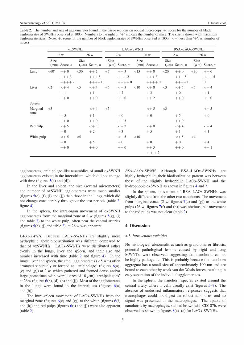
3.2.2. Biodistribution. The number and size of black particlesof oxSWNHs, LAOx-SWNHs and BSA–LAOx-SWNHs foundin the lungs, liver and spleen at 2 w and 26 w are presented intable 2. The details of the number of black particles are shownin the shadowed lines in supporting information VI (availableat stacks.iop.org/Nano/22/265106/mmedia). In table 2, thenumber of black agglomerates of nanohorns observed under anoptical microscope (100× magnification, visible agglomeratesizes >5 μm) are indicated with a ‘+’. The largest sizeof black agglomerates is also indicated in table 2. Figure 4shows the ‘maximum size’ and ‘average number of + symbols’indicated in table 2. The ‘maximum size’ in the spleen is shownonly for the zones (red pulp, white pulp or marginal zones),where the nanohorn number was the highest.
OxSWNH. The highly hydrophobic oxSWNHs mainlyaccumulated in the lungs and hardly in the liver and spleen(table 2, figure 4). The hydrophobic property induced manyoxSWNHs to form large agglomerates, reaching 50 μm in thelungs at 2 w, but their number and size decreased at 26 w(table 2, figures 4, 5(a) and (b)). In addition to the large
Table 2. The number and size of agglomerates found in the tissue sections on optical microscopy. +: score for the number of blackagglomerates of SWNHs observed at 100×. Numbers to the right of ‘+’ indicate the number of mice. The size is shown with maximumagglomerate sizes. (Note: +: score for the number of black agglomerates of SWNHs observed at 100×. <+: less than ‘+’. n: number ofmice.)
agglomerates, archipelago-like assemblies of small oxSWNHagglomerates existed in the interstitium, which did not changewith time (figures 5(c) and (d)).
In the liver and spleen, the size (several micrometers)and number of oxSWNH agglomerates were much smaller(figures 5(e), (f), (i) and (j)) than those in the lungs, which didnot change considerably throughout the test periods (table 2,figure 4).
In the spleen, the intra-organ movement of oxSWNHagglomerates from the marginal zone at 2 w (figures 5(g), (i)and table 2) to the white pulp, often near the central arteries(figures 5(h), (j) and table 2), at 26 w was apparent.
LAOx-SWNH. Because LAOx-SWNHs are slightly morehydrophilic, their biodistribution was different compared tothat of oxSWNHs. LAOx-SWNHs were distributed ratherevenly in the lungs, liver and spleen, and their size andnumber increased with time (table 2 and figure 4). In thelungs, liver and spleen, the small agglomerates (<5 μm) oftenarranged separately or formed an ‘archipelago’ (figures 6(a),(c) and (g)) at 2 w, which gathered and formed dense and/orlarge (sometimes with overall sizes of 10 μm) ‘archipelagoes’at 26 w (figures 6(b), (d), (h) and (j)). Most of the agglomeratesin the lungs were found in the interstitium (figures 6(a)and (b)).
The intra-spleen movement of LAOx-SWNHs from themarginal zone (figures 6(e) and (g)) to the white (figures 6(f)and (h)) and red pulps (figures 6(i) and (j)) were also apparent(table 2).
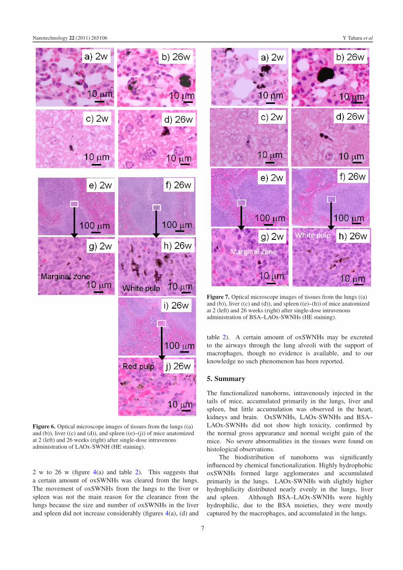
BSA–LAOx-SWNH. Although BSA–LAOx-SWNHs arehighly hydrophilic, their biodistribution pattern was betweenthose of the slightly hydrophilic LAOx-SWNH and thehydrophobic oxSWNH as shown in figures 4 and 7.
In the spleen, movement of BSA–LAOx-SWNHs wasslightly different from the other two nanohorns. The movementfrom marginal zones (2 w; figures 7(e) and (g)) to the whitepulps (26 w; figures 7(f) and (h)) was obvious, but movementto the red pulps was not clear (table 2).
4. Discussion
4.1. Intravenous toxicities
No histological abnormalities such as granuloma or fibrosis,potential pathological lesions caused by rigid and longMWNTs, were observed, suggesting that nanohorns cannotbe highly pathogenic. This is probably because the nanohornaggregate has a small size of approximately 100 nm and arebound to each other by weak van der Waals forces, resulting ineasy separation of the individual agglomerates.
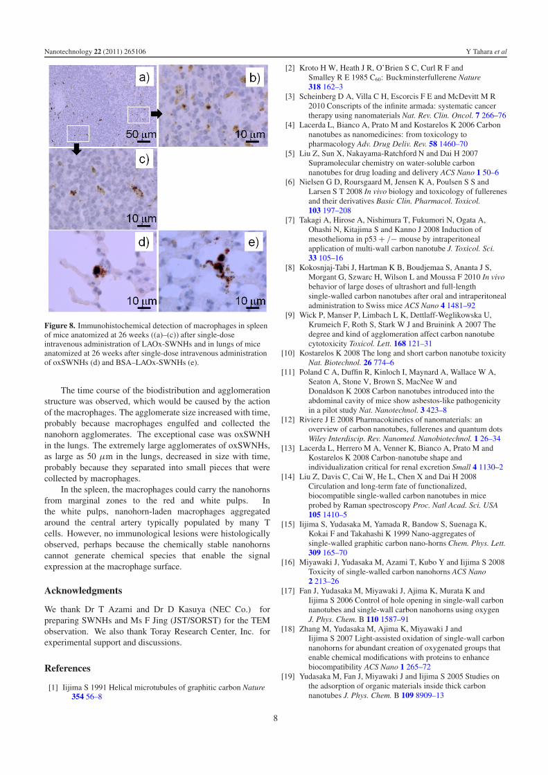
In the spleen, the nanohorn species existed around thecentral artery where T cells usually exist (figures 5–7). Theabsence of undesired inflammatory responses suggests thatmacrophages could not digest the robust nanohorns, and nosignal was presented at the macrophages. The uptake ofnanohorns by macrophages, stained brown with CD107b, wereobserved as shown in figures 8(a)–(c) for LAOx-SWNHs.
5
Nanotechnology 22 (2011) 265106 Y Tahara et al
Figure 4. Average number of ‘+’ symbols in table 2 foroxSWNHs (a), LAOx-SWNHs (b) and BSA–LAOx-SWNHs (c),which correspond to agglomerate number. Maximum size ofagglomerates in table 2 for oxSWNHs (d), LAOx-SWNH (e) andBSA–LAOx-SWNH (f). Color codes: blue (lung), magenta (liver)and red (spleen).
4.2. Biodistribution of nanohorns was influenced byfunctionalization
The nanohorn biodistribution and its changes with time dependon the functionalizations of nanohorns, perhaps because theinteraction of macrophages with nanohorns depends on thefunctionalized moieties. Both LAOx-SWNH and BSA–LAOx-SWNH tended to increase the agglomerate number, size anddensity with time in the lungs, liver and spleen (table 2,figures 4, 6 and 7). These changes may be attributedto the macrophages’ engulfing/collecting small agglomeratesas inferred from the CD107b brown-stained macrophagesinteracting with nanohorn agglomerates (figure 8(e)). Theeffect of BSA was apparent in the agglomeration of BSA–LAOx-SWNHs in the lungs, which was larger than thatof LAOx-SWNHs (figure 4(f)). This tendency contradictsthe results in PBS, in which BSA–LAOx-SWNH has asmaller agglomerate size than LAOx-SWNH (figure 3). Webelieve that the BSA moieties stimulated the macrophages andenhanced their uptake of nanohorns in the lungs, resulting inhigher accumulation. The macrophage uptake enhancement
Figure 5. Optical microscope images of tissues from the lungs ((a)and (d)), liver ((e) and (f)), and spleen ((g) and (j)) of miceanatomized at 2 (left) and 26 weeks (right) after single-doseintravenous administration of oxSWNHs. (HE staining).
of LAOx-SWNH by the BSA attachment has been confirmedin vitro (data not shown). The BSA moieties also influencedthe movement of BSA–LAOx-SWNH in the spleen, though itsmechanism is unclear.
OxSWNHs, which are different from LAOx-SWNHs andBSA–LAOx-SWNHs, form extremely large agglomerates dueto their highly hydrophobic properties, thus occupying the lungvessel lumens (figure 5). The agglomerate size of oxSWNHsin the lungs decreased from 2 w to 26 w (table 2 and figure 4),perhaps because the large oxSWNH agglomerates separatedinto small pieces in the bloodstream. These small pieces maybe captured by macrophages (figure 8(d)); however, the capturewas not particularly effective and, as a result, both the numberand size of oxSWNH agglomerates in the lungs decreased from
6
Nanotechnology 22 (2011) 265106 Y Tahara et al
Figure 6. Optical microscope images of tissues from the lungs ((a)and (b)), liver ((c) and (d)), and spleen ((e)–(j)) of mice anatomizedat 2 (left) and 26 weeks (right) after single-dose intravenousadministration of LAOx-SWNH (HE staining).
2 w to 26 w (figure 4(a) and table 2). This suggests thata certain amount of oxSWNHs was cleared from the lungs.The movement of oxSWNHs from the lungs to the liver orspleen was not the main reason for the clearance from thelungs because the size and number of oxSWNHs in the liverand spleen did not increase considerably (figures 4(a), (d) and
Figure 7. Optical microscope images of tissues from the lungs ((a)and (b)), liver ((c) and (d)), and spleen ((e)–(h)) of mice anatomizedat 2 (left) and 26 weeks (right) after single-dose intravenousadministration of BSA–LAOx-SWNHs (HE staining).
table 2). A certain amount of oxSWNHs may be excretedto the airways through the lung alveoli with the support ofmacrophages, though no evidence is available, and to ourknowledge no such phenomenon has been reported.
5. Summary
The functionalized nanohorns, intravenously injected in thetails of mice, accumulated primarily in the lungs, liver andspleen, but little accumulation was observed in the heart,kidneys and brain. OxSWNHs, LAOx-SWNHs and BSA–LAOx-SWNHs did not show high toxicity, confirmed bythe normal gross appearance and normal weight gain of themice. No severe abnormalities in the tissues were found onhistological observations.
The biodistribution of nanohorns was significantlyinfluenced by chemical functionalization. Highly hydrophobicoxSWNHs formed large agglomerates and accumulatedprimarily in the lungs. LAOx-SWNHs with slightly higherhydrophilicity distributed nearly evenly in the lungs, liverand spleen. Although BSA–LAOx-SWNHs were highlyhydrophilic, due to the BSA moieties, they were mostlycaptured by the macrophages, and accumulated in the lungs.
7
Nanotechnology 22 (2011) 265106 Y Tahara et al
Figure 8. Immunohistochemical detection of macrophages in spleenof mice anatomized at 26 weeks ((a)–(c)) after single-doseintravenous administration of LAOx-SWNHs and in lungs of miceanatomized at 26 weeks after single-dose intravenous administrationof oxSWNHs (d) and BSA–LAOx-SWNHs (e).
The time course of the biodistribution and agglomerationstructure was observed, which would be caused by the actionof the macrophages. The agglomerate size increased with time,probably because macrophages engulfed and collected thenanohorn agglomerates. The exceptional case was oxSWNHin the lungs. The extremely large agglomerates of oxSWNHs,as large as 50 μm in the lungs, decreased in size with time,probably because they separated into small pieces that werecollected by macrophages.
In the spleen, the macrophages could carry the nanohornsfrom marginal zones to the red and white pulps. Inthe white pulps, nanohorn-laden macrophages aggregatedaround the central artery typically populated by many Tcells. However, no immunological lesions were histologicallyobserved, perhaps because the chemically stable nanohornscannot generate chemical species that enable the signalexpression at the macrophage surface.
Acknowledgments
We thank Dr T Azami and Dr D Kasuya (NEC Co.) forpreparing SWNHs and Ms F Jing (JST/SORST) for the TEMobservation. We also thank Toray Research Center, Inc. forexperimental support and discussions.
References
[1] Iijima S 1991 Helical microtubules of graphitic carbon Nature354 56–8
[2] Kroto H W, Heath J R, O’Brien S C, Curl R F andSmalley R E 1985 C60: Buckminsterfullerene Nature318 162–3
[3] Scheinberg D A, Villa C H, Escorcis F E and McDevitt M R2010 Conscripts of the infinite armada: systematic cancertherapy using nanomaterials Nat. Rev. Clin. Oncol. 7 266–76
[4] Lacerda L, Bianco A, Prato M and Kostarelos K 2006 Carbonnanotubes as nanomedicines: from toxicology topharmacology Adv. Drug Deliv. Rev. 58 1460–70
[5] Liu Z, Sun X, Nakayama-Ratchford N and Dai H 2007Supramolecular chemistry on water-soluble carbonnanotubes for drug loading and delivery ACS Nano 1 50–6
[6] Nielsen G D, Roursgaard M, Jensen K A, Poulsen S S andLarsen S T 2008 In vivo biology and toxicology of fullerenesand their derivatives Basic Clin. Pharmacol. Toxicol.103 197–208
[7] Takagi A, Hirose A, Nishimura T, Fukumori N, Ogata A,Ohashi N, Kitajima S and Kanno J 2008 Induction ofmesothelioma in p53 + /− mouse by intraperitonealapplication of multi-wall carbon nanotube J. Toxicol. Sci.33 105–16
[8] Kokosnjaj-Tabi J, Hartman K B, Boudjemaa S, Ananta J S,Morgant G, Szwarc H, Wilson L and Moussa F 2010 In vivobehavior of large doses of ultrashort and full-lengthsingle-walled carbon nanotubes after oral and intraperitonealadministration to Swiss mice ACS Nano 4 1481–92
[9] Wick P, Manser P, Limbach L K, Dettlaff-Weglikowska U,Krumeich F, Roth S, Stark W J and Bruinink A 2007 Thedegree and kind of agglomeration affect carbon nanotubecytotoxicity Toxicol. Lett. 168 121–31
[10] Kostarelos K 2008 The long and short carbon nanotube toxicityNat. Biotechnol. 26 774–6
[11] Poland C A, Duffin R, Kinloch I, Maynard A, Wallace W A,Seaton A, Stone V, Brown S, MacNee W andDonaldson K 2008 Carbon nanotubes introduced into theabdominal cavity of mice show asbestos-like pathogenicityin a pilot study Nat. Nanotechnol. 3 423–8
[12] Riviere J E 2008 Pharmacokinetics of nanomaterials: anoverview of carbon nanotubes, fullerenes and quantum dotsWiley Interdiscip. Rev. Nanomed. Nanobiotechnol. 1 26–34
[13] Lacerda L, Herrero M A, Venner K, Bianco A, Prato M andKostarelos K 2008 Carbon-nanotube shape andindividualization critical for renal excretion Small 4 1130–2
[14] Liu Z, Davis C, Cai W, He L, Chen X and Dai H 2008Circulation and long-term fate of functionalized,biocompatible single-walled carbon nanotubes in miceprobed by Raman spectroscopy Proc. Natl Acad. Sci. USA105 1410–5
[15] Iijima S, Yudasaka M, Yamada R, Bandow S, Suenaga K,Kokai F and Takahashi K 1999 Nano-aggregates ofsingle-walled graphitic carbon nano-horns Chem. Phys. Lett.309 165–70
[16] Miyawaki J, Yudasaka M, Azami T, Kubo Y and Iijima S 2008Toxicity of single-walled carbon nanohorns ACS Nano2 213–26
[17] Fan J, Yudasaka M, Miyawaki J, Ajima K, Murata K andIijima S 2006 Control of hole opening in single-wall carbonnanotubes and single-wall carbon nanohorns using oxygenJ. Phys. Chem. B 110 1587–91
[18] Zhang M, Yudasaka M, Ajima K, Miyawaki J andIijima S 2007 Light-assisted oxidation of single-wall carbonnanohorns for abundant creation of oxygenated groups thatenable chemical modifications with proteins to enhancebiocompatibility ACS Nano 1 265–72
[19] Yudasaka M, Fan J, Miyawaki J and Iijima S 2005 Studies onthe adsorption of organic materials inside thick carbonnanotubes J. Phys. Chem. B 109 8909–13