Histological Structure of Skeletal Muscle in Disuse Muscle Atrophy and Role of Exercise
Sara Gamal Abd El-kawy Tayel, Fatma El-Nabawia Abdel-Hady El-Safty, Neveen Mohamed El-Sherif and Manar Ali Faried
Department of Anatomy and Embryology, Faculty of Medicine, Menoufia University, Menoufia, Egypt
ABSTRACTDisuse muscle atrophy is one of the most important research topics in fields of rehabilitation and clinical medicine. It refers to morphological and functional alteration of skeletal muscle under the states of hypokinesia, immobilization and weightlessness. This review analyzes a number of structural and functional changes in the skeletal muscle tissue in case of muscle disuse which appeared in loss in the muscle mass, reduction in the transverse diameter of most of muscle fibers, wide spaces between muscle fibers, increase collagen fiber deposition and appearance of apoptotic nuclei in muscle fibers besides sensorial dysfunction such as hypo mechanical responsiveness and diminished contractile function. In addition to the ultrastructural alteration, which include loss of sarcomere organization, indistinguishable A and I bands, irregular distorted Z-line with disruption of myofilaments and fused giant accumulated mitochondria. The review summarizes also the molecular mechanisms of skeletal muscle disuse atrophy which could be resulted from an alteration in the activity of oxidative stress enzymes and lead to increase in protein degradation and/or a reduction in protein synthesis. Moreover, the review discusses the efficiency of potential countermeasures of stretching exercise as a very powerful stimulant of muscle growth and muscle protein synthesis to counteract the structural and functional alterations in muscle tissue caused by disuse. The review showed that stretching exercise can increase the transverse sectional area of muscle fibers, produce a rise in the number of sarcomeres in series and prevent muscle atrophy. Passive stretching also completely prevented the decrease in myonuclear number induced by inactivity. Moreover, stretching accelerated the amelioration of the range of motion limitation and prevent the deposition of connective tissue resulted from disuse muscle atrophy.
Received: 23 February 2020, Accepted: 18 March 2020
Key Words: Disuse atrophy, skeletal muscle, stretching exercise. Corresponding Author: Sara Gamal Abd El-kawy Tayel, MSc, Department of Anatomy and Embryology, Faculty of Medicine, Menoufia University, Menoufia, Egypt, Tel.: +20 1023135739, E-mail: [email protected]: 1110-0559, Vol. 44, No.1
INTRODUCTION
Skeletal muscles constitute around 40 percent of human's body mass and participate in locomotion, production of body heat, metabolic regulation and protein storage[1]. Therefore, maintenance of the muscle mass and function is essential for health and survival[2]. The structure and function of the muscular tissue are greatly affected by several factors throughout life including nutritional status, neural activation, hormones/growth factors and cytokines production[3]. Striated muscle inactivity such as bed rest, spaceflight, limb immobilization and reduced steps lead to a deleterious changes in muscle extensibility, decrease in muscle mass, intramuscular fibrosis and limitation of joint motion[4]. This loss of musculoskeletal mass, size and strength ultimately lead to skeletal muscle disuse atrophy[5]. This disuse atrophy interferes with the routine daily activities of people as climbing stairs and even walking, resulting in loss of independence, decreased quality of life and poor health[6].
Exercise is recommended for the prevention and treatment of muscle atrophy and the most commonly
prescribed exercise in disuse atrophy is muscle stretching[7,8]. Muscle stretching is considered a very powerful stimulant of muscle protein synthesis and muscle growth[9].
The aims of this review were to show morphological changes in skeletal muscle tissue during disuse atrophy through its effects on muscle mass, cross sectional area of muscle fibers, presence of apoptotic nuclei and collagen fiber deposition in addition to ultrastructural changes during disuse atrophy. This review also summarized the role of stretching exercise in disuse muscle atrophy and discussed the underlying mechanisms responsible for the protective effects of stretching exercise against muscle atrophy.
Light microscopic structure of the normal skeletal muscle
In transverse section, the skeletal muscle is formed of small bundles and each bundle is separated by a connective tissue layer called perimysium. Another thin layer of reticular connective tissue fibers called endomysium invests the individual muscle fiber. Skeletal muscle fibers appeared
2
DISUSE MUSCLE ATROPHY AND EXERCISE
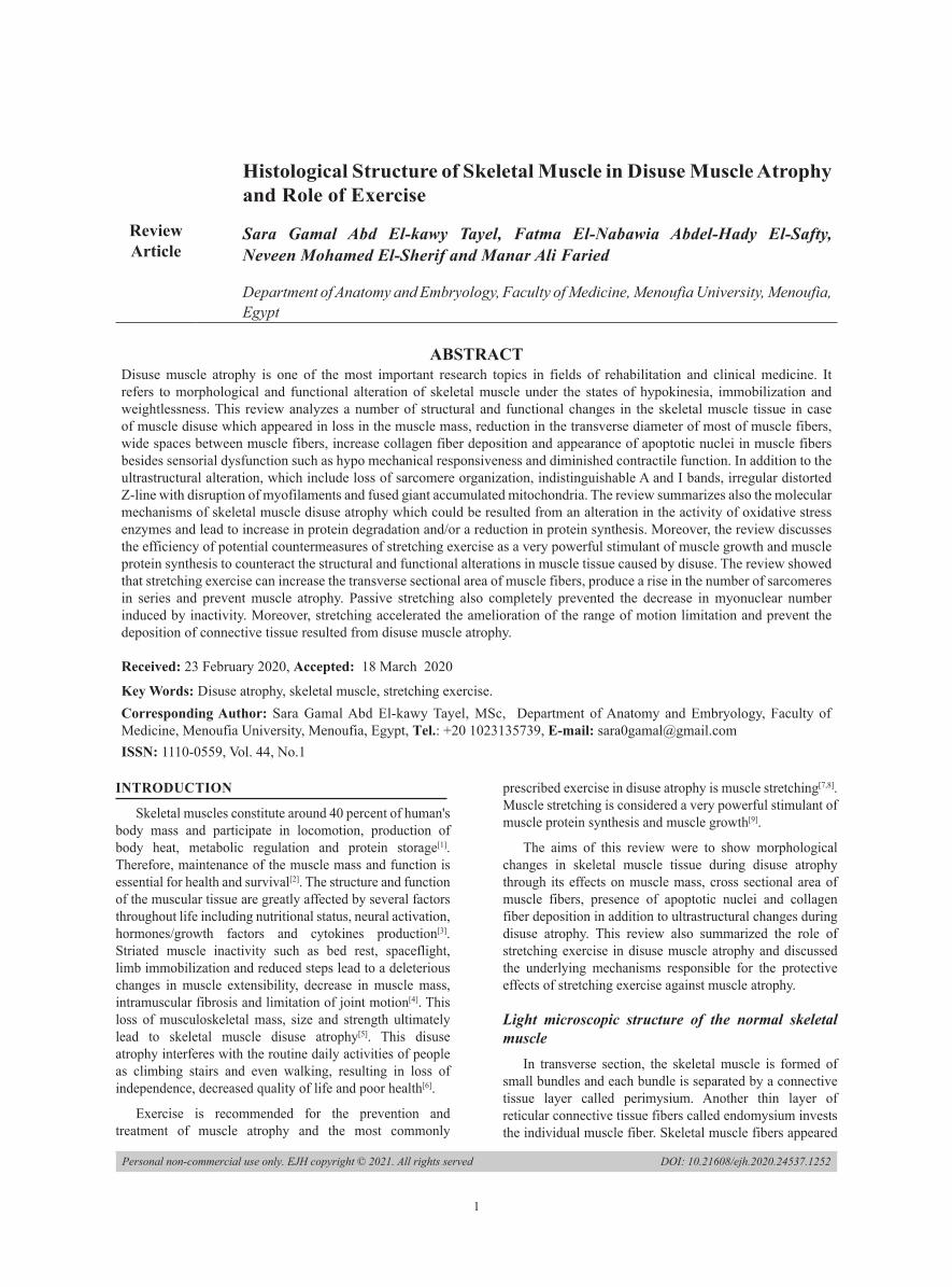
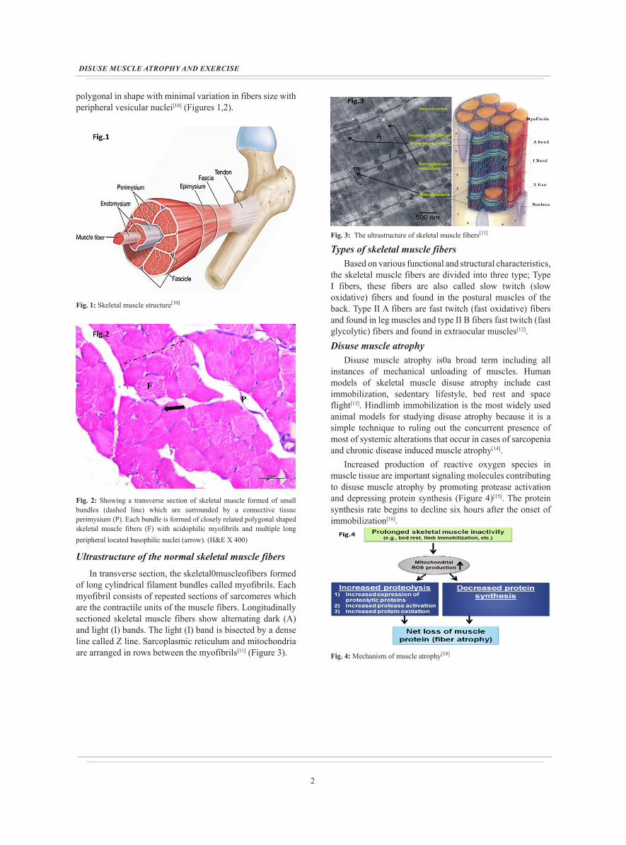
polygonal in shape with minimal variation in fibers size with peripheral vesicular nuclei[10] (Figures 1,2).
Fig. 1: Skeletal muscle structure[10]
Fig. 2: Showing a transverse section of skeletal muscle formed of small bundles (dashed line) which are surrounded by a connective tissue perimysium (P). Each bundle is formed of closely related polygonal shaped skeletal muscle fibers (F) with acidophilic myofibrils and multiple long peripheral located basophilic nuclei (arrow). (H&E X 400)
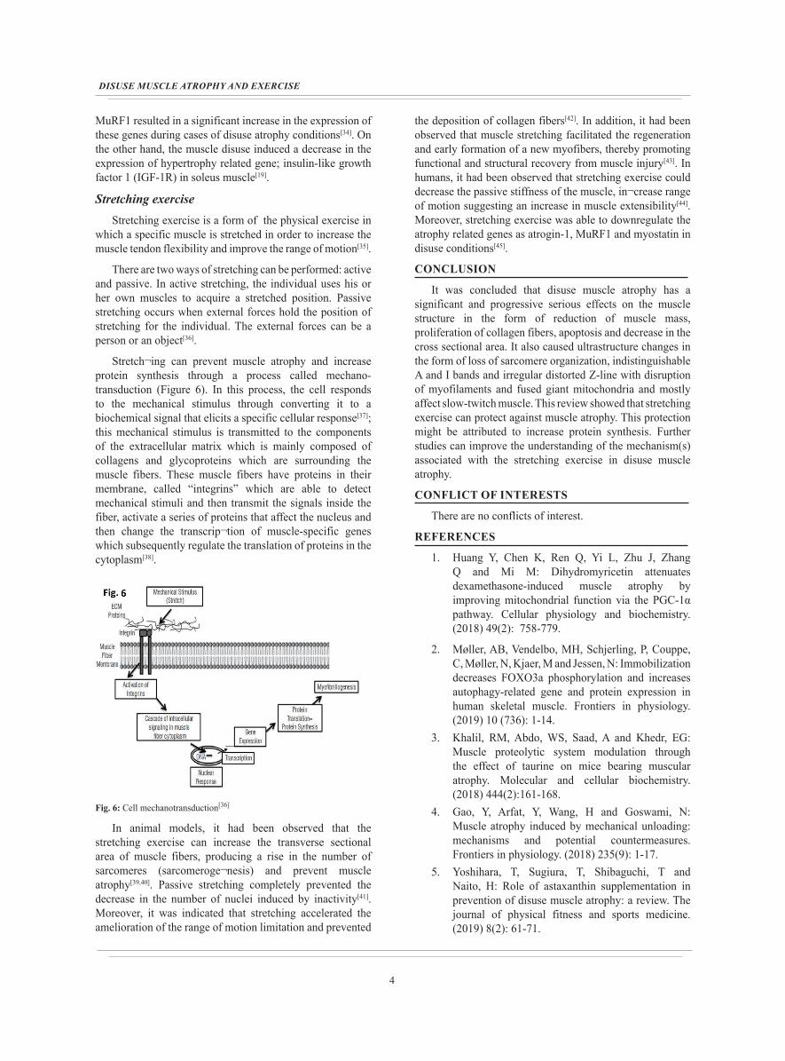
Ultrastructure of the normal skeletal muscle fibersIn transverse section, the skeletal0muscleofibers formed
of long cylindrical filament bundles called myofibrils. Each myofibril consists of repeated sections of sarcomeres which are the contractile units of the muscle fibers. Longitudinally sectioned skeletal muscle fibers show alternating dark (A) and light (I) bands. The light (I) band is bisected by a dense line called Z line. Sarcoplasmic reticulum and mitochondria are arranged in rows between the myofibrils[11] (Figure 3).
Fig. 3: The ultrastructure of skeletal muscle fibers[11]
Types of skeletal muscle fibersBased on various functional and structural characteristics,
the skeletal muscle fibers are divided into three type; Type I fibers, these fibers are also called slow twitch (slow oxidative) fibers and found in the postural muscles of the back. Type II A fibers are fast twitch (fast oxidative) fibers and found in leg muscles and type II B fibers fast twitch (fast glycolytic) fibers and found in extraocular muscles[12]. Disuse muscle atrophy
Disuse muscle atrophy is0a broad term including all instances of mechanical unloading of muscles. Human models of skeletal muscle disuse atrophy include cast immobilization, sedentary lifestyle, bed rest and space flight[13]. Hindlimb immobilization is the most widely used animal models for studying disuse atrophy because it is a simple technique to ruling out the concurrent presence of most of systemic alterations that occur in cases of sarcopenia and chronic disease induced muscle atrophy[14].
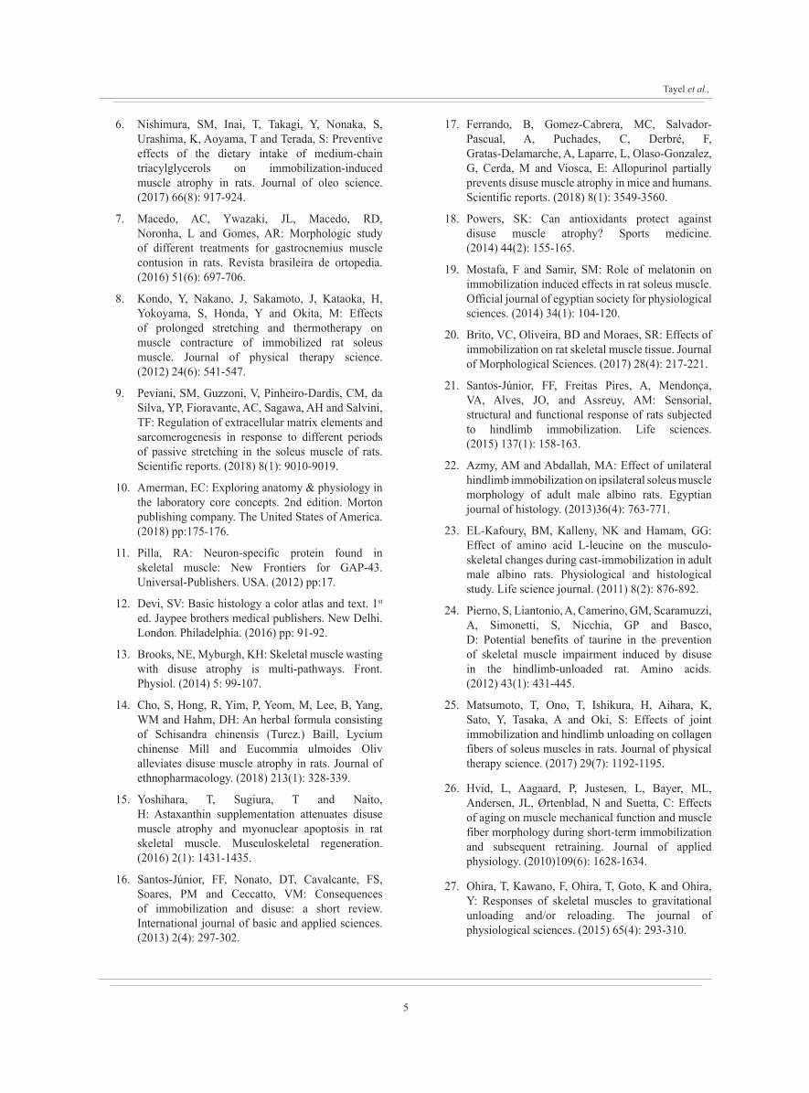
Increased production of reactive oxygen species in muscle tissue are important signaling molecules contributing to disuse muscle atrophy by promoting protease activation and depressing protein synthesis (Figure 4)[15]. The protein synthesis rate begins to decline six hours after the onset of immobilization[16].
Fig. 4: Mechanism of muscle atrophy[18]
3
Tayel et al.,
Protein degradation was mainly carried out through the following pathways: autophagy (lysosomal protease pathway), caspase-3 pathway, calpains pathway and the ubiquitin proteasome pathway. The last one play the major role in the pathogenesis muscle atrophy. This pathway includes two important critical muscle specific ubiquitin ligases; muscle atrophy F-box (atrogin) and muscle RING finger-1 (MuRF1)[17].
Effects of disuse muscle atrophy on the muscle massAs the mass of skeletal muscle tissue is determined by
the ratio of protein synthesis and degradation. The initial response of the muscle tissue to disuse is through decrease in protein synthesis and increase in its degradation which ultimately results in a great reduction in the muscle mass[19]. Different periods of immobilization can cause different degrees of atrophy. One day of immobilization can result in approximately 8% reduction in mass in the gastrocnemius muscle, whereas this reduction can reach 19% after three days and 20 to 30% after five days of limb immobilization[20]. Moreover, immobilization produced by application of external plastic casting induced a significant atrophy in soleus muscles (56% reduction) at days 14[21]. The loss of muscle mass during hindlimb immobilization was attributed to marked reduction in the mean fiber diameter and also due to the decrease in the number of sarcomeres[22].
Effect of disuse muscle atrophy on the structure and cross-sectional area of muscle fibers (CSA) (Figure 5)
Fig. 5: Showing shrunken muscle fibers (F) with apparent widening of the interstitial spaces between them (star). Splitting of muscle fibers (curved arrow), vacuoles in the sarcoplasm (arrow head) and pyknotic nuclei (arrow) of muscle fibers can be seen in disuse muscle atrophy. (H&E X 400)
The disuse muscle atrophy causes many alterations in the muscle fibers including sarcomere dissolution, endothelial degradation, connective tissue deposition between the muscle fibers, apoptotic myonuclei and a decrease in the capillary density[23]. These alterations lead to a reduction in the fibers' cross-sectional area[24]. In addition, 15 days of limb immobilization with a posterior ankle splint resulted in a significant decrease in the soleus muscle cross sectional
area when compared with the control group[17]. Moreover, it was reported that the reduction of soleus muscle weight and a decrease of cross sectional area are signs of atrophy[25]. In addition, there were ultrastructural alteration in the form of loss of sarcomere organization, indistinguishable A and I bands and irregular distorted Z-line with disruption of myofilaments and fused giant accumulated mitochondria in the inter myofibrillar and subsarcolemmal space[23].
Effect of disuse muscle atrophy on collagen fiber deposition
Increase of collagen fibers deposition accompanying disuse muscle atrophy is a response to loss of myofibers where fibroblasts replace the damaged area with subsequent formation of collagen fibers[9]. This increase in collagen fibers deposition due to immobilization induced a significant increase in the contracture of skeletal muscle[25].
Effect of disuse muscle atrophy on muscle functionThe effects of disuse models such as immobilization
on loss of muscle function and strength have been widely investigated[4]. It has been observed that hindlimb immobilization in rats for two weeks led to sensorial dysfunction (hyper thermal and hypo mechanical responsiveness) accompanied by systemic reduction in the protein and ions and concurrent with diminished contractile function[21]. Another study demonstrated that both contractile rate of force development and maximal isometric muscle strength are reduced significantly after 2 weeks of unilateral leg casting[26]. Similarly, electromyographic study of soleus muscle was reduced at by disuse muscle atrophy[27].
Apoptotic processes during disuse muscle atrophyApoptosis is a process involved in progression of disuse
muscle atrophy as concluded from studies of hindlimb immobilization in rabbits, mice and rats[28,29]. In prolonged muscular inactivity, oxidative stress has been suggested as a potential mechanism underlying apoptosis through activation of both intrinsic and extrinsic pathways with subsequent release of cytochrome c which activates caspases that specifically cleave amino acid sequences and are crucial for the execution of apoptosis[30]. After 14 days of disuse of skeletal muscle, rats exhibited a significant reduction in the nuclei size and number. There was an elevation in many markers for apoptosis such as Bcl-2, Bax protein and nuclear colocalization of Endo G (an apoptotic effector) in the nuclei of muscle fibers[31]. In addition, the apoptotic changes in disuse atrophy may be also attributed to the marked reduction in the number of blood capillaries with a marked deposition of collagen fibers around them that subsequently disturbs the blood flow to the muscle tissues[32].
Effect of disuse muscle atrophy on gene expressionMuscle atrophy F-box (Atrogin-1) and muscle specific
RING finger-1 (MuRF1) are two important muscle specific E3 ligases that play an important role in degradation of muscle proteins by the ubiquitin proteasome system[33]. Numerous studies indicated real time-PCR analysis for Atrogin-1 and
4
DISUSE MUSCLE ATROPHY AND EXERCISE
MuRF1 resulted in a significant increase in the expression of these genes during cases of disuse atrophy conditions[34]. On the other hand, the muscle disuse induced a decrease in the expression of hypertrophy related gene; insulin-like growth factor 1 (IGF-1R) in soleus muscle[19].
Stretching exerciseStretching exercise is a form of the physical exercise in
which a specific muscle is stretched in order to increase the muscle tendon flexibility and improve the range of motion[35].
There are two ways of stretching can be performed: active and passive. In active stretching, the individual uses his or her own muscles to acquire a stretched position. Passive stretching occurs when external forces hold the position of stretching for the individual. The external forces can be a person or an object[36].
Stretch¬ing can prevent muscle atrophy and increase protein synthesis through a process called mechano-transduction (Figure 6). In this process, the cell responds to the mechanical stimulus through converting it to a biochemical signal that elicits a specific cellular response[37]; this mechanical stimulus is transmitted to the components of the extracellular matrix which is mainly composed of collagens and glycoproteins which are surrounding the muscle fibers. These muscle fibers have proteins in their membrane, called “integrins” which are able to detect mechanical stimuli and then transmit the signals inside the fiber, activate a series of proteins that affect the nucleus and then change the transcrip¬tion of muscle-specific genes which subsequently regulate the translation of proteins in the cytoplasm[38].
Fig. 6: Cell mechanotransduction[36]
In animal models, it had been observed that the stretching exercise can increase the transverse sectional area of muscle fibers, producing a rise in the number of sarcomeres (sarcomeroge¬nesis) and prevent muscle atrophy[39,40]. Passive stretching completely prevented the decrease in the number of nuclei induced by inactivity[41]. Moreover, it was indicated that stretching accelerated the amelioration of the range of motion limitation and prevented
the deposition of collagen fibers[42]. In addition, it had been observed that muscle stretching facilitated the regeneration and early formation of a new myofibers, thereby promoting functional and structural recovery from muscle injury[43]. In humans, it had been observed that stretching exercise could decrease the passive stiffness of the muscle, in¬crease range of motion suggesting an increase in muscle extensibility[44]. Moreover, stretching exercise was able to downregulate the atrophy related genes as atrogin-1, MuRF1 and myostatin in disuse conditions[45].
CONCLUSION
It was concluded that disuse muscle atrophy has a significant and progressive serious effects on the muscle structure in the form of reduction of muscle mass, proliferation of collagen fibers, apoptosis and decrease in the cross sectional area. It also caused ultrastructure changes in the form of loss of sarcomere organization, indistinguishable A and I bands and irregular distorted Z-line with disruption of myofilaments and fused giant mitochondria and mostly affect slow-twitch muscle. This review showed that stretching exercise can protect against muscle atrophy. This protection might be attributed to increase protein synthesis. Further studies can improve the understanding of the mechanism(s) associated with the stretching exercise in disuse muscle atrophy.
CONFLICT OF INTERESTS
There are no conflicts of interest.
REFERENCES
1. Huang Y, Chen K, Ren Q, Yi L, Zhu J, Zhang Q and Mi M: Dihydromyricetin attenuates dexamethasone-induced muscle atrophy by improving mitochondrial function via the PGC-1α pathway. Cellular physiology and biochemistry. (2018) 49(2): 758-779.
2. Møller, AB, Vendelbo, MH, Schjerling, P, Couppe, C, Møller, N, Kjaer, M and Jessen, N: Immobilization decreases FOXO3a phosphorylation and increases autophagy-related gene and protein expression in human skeletal muscle. Frontiers in physiology. (2019) 10 (736): 1-14.
3. Khalil, RM, Abdo, WS, Saad, A and Khedr, EG: Muscle proteolytic system modulation through the effect of taurine on mice bearing muscular atrophy. Molecular and cellular biochemistry. (2018) 444(2):161-168.
4. Gao, Y, Arfat, Y, Wang, H and Goswami, N: Muscle atrophy induced by mechanical unloading: mechanisms and potential countermeasures. Frontiers in physiology. (2018) 235(9): 1-17.
5. Yoshihara, T, Sugiura, T, Shibaguchi, T and Naito, H: Role of astaxanthin supplementation in prevention of disuse muscle atrophy: a review. The journal of physical fitness and sports medicine. (2019) 8(2): 61-71.
5
Tayel et al.,
6. Nishimura, SM, Inai, T, Takagi, Y, Nonaka, S, Urashima, K, Aoyama, T and Terada, S: Preventive effects of the dietary intake of medium-chain triacylglycerols on immobilization-induced muscle atrophy in rats. Journal of oleo science. (2017) 66(8): 917-924.
7. Macedo, AC, Ywazaki, JL, Macedo, RD, Noronha, L and Gomes, AR: Morphologic study of different treatments for gastrocnemius muscle contusion in rats. Revista brasileira de ortopedia. (2016) 51(6): 697-706.
8. Kondo, Y, Nakano, J, Sakamoto, J, Kataoka, H, Yokoyama, S, Honda, Y and Okita, M: Effects of prolonged stretching and thermotherapy on muscle contracture of immobilized rat soleus muscle. Journal of physical therapy science. (2012) 24(6): 541-547.
9. Peviani, SM, Guzzoni, V, Pinheiro-Dardis, CM, da Silva, YP, Fioravante, AC, Sagawa, AH and Salvini, TF: Regulation of extracellular matrix elements and sarcomerogenesis in response to different periods of passive stretching in the soleus muscle of rats. Scientific reports. (2018) 8(1): 9010-9019.
10. Amerman, EC: Exploring anatomy & physiology in the laboratory core concepts. 2nd edition. Morton publishing company. The United States of America. (2018) pp:175-176.
11. Pilla, RA: Neuron-specific protein found in skeletal muscle: New Frontiers for GAP-43. Universal-Publishers. USA. (2012) pp:17.
12. Devi, SV: Basic histology a color atlas and text. 1st
ed. Jaypee brothers medical publishers. New Delhi. London. Philadelphia. (2016) pp: 91-92.
13. Brooks, NE, Myburgh, KH: Skeletal muscle wasting with disuse atrophy is multi-pathways. Front. Physiol. (2014) 5: 99-107.
14. Cho, S, Hong, R, Yim, P, Yeom, M, Lee, B, Yang, WM and Hahm, DH: An herbal formula consisting of Schisandra chinensis (Turcz.) Baill, Lycium chinense Mill and Eucommia ulmoides Oliv alleviates disuse muscle atrophy in rats. Journal of ethnopharmacology. (2018) 213(1): 328-339.
15. Yoshihara, T, Sugiura, T and Naito, H: Astaxanthin supplementation attenuates disuse muscle atrophy and myonuclear apoptosis in rat skeletal muscle. Musculoskeletal regeneration. (2016) 2(1): 1431-1435.
16. Santos-Júnior, FF, Nonato, DT, Cavalcante, FS, Soares, PM and Ceccatto, VM: Consequences of immobilization and disuse: a short review. International journal of basic and applied sciences. (2013) 2(4): 297-302.
17. Ferrando, B, Gomez-Cabrera, MC, Salvador-Pascual, A, Puchades, C, Derbré, F, Gratas-Delamarche, A, Laparre, L, Olaso-Gonzalez, G, Cerda, M and Viosca, E: Allopurinol partially prevents disuse muscle atrophy in mice and humans. Scientific reports. (2018) 8(1): 3549-3560.
18. Powers, SK: Can antioxidants protect against disuse muscle atrophy? Sports medicine. (2014) 44(2): 155-165.
19. Mostafa, F and Samir, SM: Role of melatonin on immobilization induced effects in rat soleus muscle. Official journal of egyptian society for physiological sciences. (2014) 34(1): 104-120.
20. Brito, VC, Oliveira, BD and Moraes, SR: Effects of immobilization on rat skeletal muscle tissue. Journal of Morphological Sciences. (2017) 28(4): 217-221.
21. Santos-Júnior, FF, Freitas Pires, A, Mendonça, VA, Alves, JO, and Assreuy, AM: Sensorial, structural and functional response of rats subjected to hindlimb immobilization. Life sciences. (2015) 137(1): 158-163.
22. Azmy, AM and Abdallah, MA: Effect of unilateral hindlimb immobilization on ipsilateral soleus muscle morphology of adult male albino rats. Egyptian journal of histology. (2013)36(4): 763-771.
23. EL-Kafoury, BM, Kalleny, NK and Hamam, GG: Effect of amino acid L-leucine on the musculo-skeletal changes during cast-immobilization in adult male albino rats. Physiological and histological study. Life science journal. (2011) 8(2): 876-892.
24. Pierno, S, Liantonio, A, Camerino, GM, Scaramuzzi, A, Simonetti, S, Nicchia, GP and Basco, D: Potential benefits of taurine in the prevention of skeletal muscle impairment induced by disuse in the hindlimb-unloaded rat. Amino acids. (2012) 43(1): 431-445.
25. Matsumoto, T, Ono, T, Ishikura, H, Aihara, K, Sato, Y, Tasaka, A and Oki, S: Effects of joint immobilization and hindlimb unloading on collagen fibers of soleus muscles in rats. Journal of physical therapy science. (2017) 29(7): 1192-1195.
26. Hvid, L, Aagaard, P, Justesen, L, Bayer, ML, Andersen, JL, Ørtenblad, N and Suetta, C: Effects of aging on muscle mechanical function and muscle fiber morphology during short-term immobilization and subsequent retraining. Journal of applied physiology. (2010)109(6): 1628-1634.
27. Ohira, T, Kawano, F, Ohira, T, Goto, K and Ohira, Y: Responses of skeletal muscles to gravitational unloading and/or reloading. The journal of physiological sciences. (2015) 65(4): 293-310.
6
DISUSE MUSCLE ATROPHY AND EXERCISE
28. Reilly, BD, and Franklin, CE: Prevention of muscle wasting and osteoporosis: the value of examining novel animal models. Journal of experimental biology. (2016) 219(17): 2582-2595.
29. Zhu, S, Nagashima, M, Khan, MA, Yasuhara, S, Kaneki, M and Martyn, JA: Lack of caspase-3 attenuates immobilization-induced muscle atrophy and loss of tension generation along with mitigation of apoptosis and inflammation. Muscle Nerve. (2013) 47: 711-721.
30. Hu, NF, Chang, H, Du, B, Zhang, QW, Arfat, Y, Dang, K and Gao, YF: Tetramethylpyrazine ameliorated disuse-induced gastrocnemius muscle atrophy in hindlimb unloading rats through suppression of Ca+2/ROS-mediated apoptosis. Applied physiology, nutrition and metabolism. (2017) 42(2): 117-127.
31. Wang, XD, Kawano, F, Matsuoka, Y, Fukunaga, K, Terada, M, Sudoh, M, Ishihara, A and Ohira, Y: Mechanical load-dependent regulation of satellite cell and fiber size in rat soleus muscle. Am. J. Physiol. Cell Physiol. (2006) 290(1): 981-989.
32. Santos-Júnior, FF, Nonato, DT, Cavalcante, FS, Soares, PM and Ceccatto, VM: Consequences of immobilization and disuse: a short review. International journal of basic and applied sciences. (2013) 2(4): 297-302.
33. Zhang, SF, Zhang, Y, Li, B and Chen, N: Physical inactivity induces the atrophy of skeletal muscle of rats through activating AMPK/FoxO3 signal pathway. European review for medical and pharmacological science. (2018) 22(1): 199-209.
34. Koike, TE, Watanabe, AY, Kodama, FY, Ozaki, GA, Castoldi, RC, Garcia, TA, Camargo, RC and Camargo Filho, JC: Physical exercise after immobilization of skeletal muscle of adult and aged rats. Revista Brasileira de medicina do esporte. (2018) 24(1): 60-63.
35. Manescu, C: The role of stretching exercises in bodybuilding. Bulletin of the Transilvania University of Brasov, Series IX: Sciences of human kinetics. (2013) 6(1): 93-102.
36. Nelson, A and kokkonen, J: Stretching anatomy. 2nd
ed. Human kinetics. USA. (2014) pp: VI- VII.
37. Dunn, SL and Olmedo, ML: Mechanotransduction: Relevance to physical therapist practice,
understanding our ability to affect genetic expression through mechanical forces. Physical therapy. (2016) 96(5): 712-721.
38. Sun, Z, Guo, SS and Fässler, R: Integrin-mediated mechanotransduction. Journal of Cell Biology. (2016) 215(4): 445-456.
39. Salvini, TF, Durigan, JL, Peviani, SM and Russo, TL: Effects of electrical stimulation and stretching on the adaptation of denervated skeletal muscle: implications for physical therapy. Brazilian journal of physical therapy. (2012) 16(3). 175-183.
40. Gomes ARS, Soares AG, Peviani SM, Nascimento RB, Moriscot AS, Salvini TF: The effect of 30 minutes of passive stretch of the rat soleus muscle on the myogenic differentiation, myostati. (2006) 87(2):241-246.
41. Tarakina, MV, Turtikova, OV, Nemirovskaya, TL, Kokontsev, AA and Shenkman, BS: Role of muscle progenitor cells in maintaining morphological characteristics of rat soleus muscle during gravitational unloading by means of passive stretch. Cell and tissue biology. (2008) 2(2): 176-183.
42. Kondo, Y, Nakano, J, Sakamoto, J, Kataoka, H, Yokoyama, S, Honda, Y and Okita, M: Effects of prolonged stretching and thermotherapy on muscle contracture of immobilized rat soleus muscle. Journal of physical therapy science. (2012) 24(6): 541-547.
43. Mori, T, Agata, N, Itoh, Y, Inoue-Miyazu, M, Mizumura, K.Sokabe, M and Kawakami, K: Post-injury stretch promotes recovery in a rat model of muscle damage induced by lengthening contractions. The journal of physiological sciences. (2018) 68(4): 483-492.
44. Batista LH, Vilar AC, de Almeida Ferreira JJ, Rebelatto JR, Salvini TF: Active stretching improves flexibility, joint torque, and functional mobility in older women. Ameican journal of physiology and medical rehabilitation. (2009) 88(10): 815-22.
45. Russo, TL, Peviani, S M, Durigan, J L, Gigo-Benato, D, Delfino, GB and Salvini, TF: Stretching and electrical stimulation reduce the accumulation of MyoD, myostatin and atrogin-1 in denervated rat skeletal muscle. Journal of muscle research and cell motility. (2010) 31(1): 45-57.
7
Tayel et al.,
الملخص العربى
التركيب النسيجي للعضلات الهيكلية في حالة ضمور العضلات الناتج عن عدم الاستخدام ودور التمارين في ذلك
سارة جمال عبد القوي طايل، فاطمة النبوية عبد الهادي الصفتي، نيفين محمد الشريف، منار علي فريد
قسم التشريح والأجنة، كلية الطب، المنوفية جامعة المنوفية، مصر
التأهيل. وهو يشير الناتج عن عدم الاستخدام من أهم الموضوعات البحثية في مجالات إعادة يعد ضمور العضلات هذه تحلل الوزن. وانعدام والشلل الحركة نقص حالات في الهيكلية للعضلات والوظيفي المورفولوجي التغيير إلى المراجعة عدداً من التغيرات الهيكلية والوظيفية في أنسجة العضلات الهيكليه في حالة عدم استخدام العضلات والتي ظهرت في فقدان كتلة العضلات، وانخفاض القطر العرضي لمعظم ألياف العضلات، والمسافات الواسعة بين ألياف العضلات، وزيادة ترسب ألياف الكولاجين وظهور نوى موت الخلايا المبرمج في ألياف العضلات بالإضافة إلى فقدان التنظيم العضلي حيث أن نطاقات A و I لا يمكن تمييزها، وخط Z مشوه غير منتظم مع وجود الميتوكوندريا المتراكمة العملاقة إلى جانب الخلل الوظيفي الحسي مثل الاستجابة الميكانيكية المنخفضة ووظيفة الانقباض المتناقصة. تلخص هذه المراجعة أيضًا الآليات الجزيئية للضمور الناتج عن عدم استخدام العضلات والذي يمكن أن ينتج عن تغيير في نشاط إنزيمات الإجهاد التأكسدي ويؤدي إلى زيادة تحلل البروتين وانخفاض في تخليق البروتين. علاوة على ذلك، تناقش المراجعة كفاءة الإجراءات المضادة المحتملة لممارسة تمارين التمدد كمحفز قوي جداً لنمو العضلات وتخليق البروتين العضلي لمواجهة التغيرات الهيكلية والوظيفية في الأنسجة العضلية الناتجة عن عدم الاستخدام. أظهرت المراجعة أن تمارين التمدد يمكن أن تزيد من المساحة المقطعية المستعرضة للألياف العضلية وتمنع ضمور العضلات. علاوة على
ذلك، أدى التمدد إلي منع ترسب نسيج الكولاجين الناتج عن ضمور العضلات.