+ All Categories
Home > Documents > Histology of the gills of Trachinotus carolinus (Carangidae

Histology of the gills of Trachinotus carolinus (Carangidae

Date post: 03-Feb-2022
Category:
Upload: others
View: 3 times
Download: 0 times
Share this document with a friend
12
Pan-American Journal of Aquatic Sciences (2011), 6(2):109-120 Histopathological alterations in gills of juvenile Florida pompano Trachinotus carolinus (Perciformes, Carangidae) following sublethal acute and chronic exposure to naphthalene THAÍS C. A. SANTOS 1* ; VICENTE GOMES 1 ; MARIA JOSÉ A. C. R. PASSOS 1 ; ARTHUR J. S. ROCHA 1 ; RENATO B. SALAROLI 2 & PHAN VAN NGAN 1 1 Universidade de São Paulo, Instituto Oceanográfico, Laboratório de Ecofisiologia de Animais Marinhos. Praça do Oceanográfico 191, 05508-900, São Paulo, SP, Brazil. *Corresponding author: [email protected] 2 Universidade de São Paulo, Departamento de Fitopatologia, Avenida Pádua Dias 11, 13418-900, Piracicaba, SP, Brazil. Abstract. Juvenile Florida pompanos (Trachinotus carolinus) were exposed to sublethal concentrations of naphthalene (0.15 ppm and 0.30 ppm) for 24 hours (acute) and 12 days (chronic). Control fish were maintained for the same periods in clean seawater and seawater with ethanol, which is a carrier to dissolve naphthalene. Gill samples from 56 fish were prepared for histological analysis and examined under optical and scanning electron microscopy. Alterations in the gills of fish exposed to naphthalene were semi-quantitatively ranked based on the severity of tissue lesions and comparisons were made with fish kept in clean water and water with ethanol. Fish of the control groups exhibited functionally normal gills, apart from sparse, slight alterations, such as the lifting of epithelial cells. Acute exposure to naphthalene caused slight to moderate alterations in the gills, whereas chronic exposure led to significant, progressively irreparable damage, especially at the greatest concentration. Chronic exposure resulted in a greater number and diversity of alterations. Hypertrophied epithelial cells, epithelial lifting, telangiectasia, fusion of secondary lamellae or their tips, rupture of lamellar epithelium, stasis and necrosis were the most common lesions. Naphthalene caused severe damage to the gills of the Florida pompano which was related to concentration and exposure time. Key words: Fish, marine pollution, PAH Resumo. Alterações histopatológicas em brânquias de juvenis de pampos Trachinotus carolinus (Perciformes, Carangidae) após exposições subletais aguda e crônica ao naftaleno. Pampos juvenis (Trachinotus carolinus) foram expostos a concentrações subletais de naftaleno (0,15 ppm e 0,30 ppm) por 24 horas (aguda) e 12 dias (crônica). Peixes do grupo controle foram mantidos pelo mesmo período em água do mar limpa e água do mar com etanol, usado como solvente do naftaleno. Amostras de brânquias de 56 peixes foram preparadas para análises histológicas e examinadas aos microscópios óptico e eletrônico. Alterações nas brânquias de peixes expostos ao naftaleno foram semi-quantitativamente classificadas segundo o grau de severidade das lesões dos tecidos e comparadas com os tecidos de peixes mantidos em água do mar e água do mar contendo etanol. Peixes dos grupos controle apresentaram brânquias funcionalmente normais, exceto por pequenas e esparsas alterações tais como descolamento de células epiteliais. Exposições agudas ao naftaleno causaram alterações branquiais leves a moderadas enquanto que exposições crônicas ocasionaram danos significativos, progressivamente irreparáveis, especialmente na maior concentração. Exposições crônicas resultaram em um maior número e maior diversidade de alterações. As lesões mais observadas foram hipertrofia das células epiteliais, descolamento epitelial, telangiectasia, fusão das lamelas secundárias ou das suas extremidades, ruptura do epitélio lamelar, aneurisma e necrose. O naftaleno causou danos severos às brânquias de pampos, relacionados à concentração e ao tempo de exposição. Palavras chave: Peixes, poluição marinha, PAH
Transcript

Pan-American Journal of Aquatic Sciences (2011), 6(2):109-120

Histopathological alterations in gills of juvenile Florida pompano

Trachinotus carolinus (Perciformes, Carangidae) following sublethal

acute and chronic exposure to naphthalene

THAÍS C. A. SANTOS1*

; VICENTE GOMES1; MARIA JOSÉ A. C. R. PASSOS

1;

ARTHUR J. S. ROCHA1; RENATO B. SALAROLI

2 & PHAN VAN NGAN

1

1 Universidade de São Paulo, Instituto Oceanográfico, Laboratório de Ecofisiologia de Animais Marinhos. Praça do

Oceanográfico 191, 05508-900, São Paulo, SP, Brazil. *Corresponding author: [email protected] 2 Universidade de São Paulo, Departamento de Fitopatologia, Avenida Pádua Dias 11, 13418-900, Piracicaba, SP,

Brazil.

Abstract. Juvenile Florida pompanos (Trachinotus carolinus) were exposed to sublethal concentrations

of naphthalene (0.15 ppm and 0.30 ppm) for 24 hours (acute) and 12 days (chronic). Control fish were

maintained for the same periods in clean seawater and seawater with ethanol, which is a carrier to

dissolve naphthalene. Gill samples from 56 fish were prepared for histological analysis and examined

under optical and scanning electron microscopy. Alterations in the gills of fish exposed to naphthalene

were semi-quantitatively ranked based on the severity of tissue lesions and comparisons were made with

fish kept in clean water and water with ethanol. Fish of the control groups exhibited functionally normal

gills, apart from sparse, slight alterations, such as the lifting of epithelial cells. Acute exposure to

naphthalene caused slight to moderate alterations in the gills, whereas chronic exposure led to significant,

progressively irreparable damage, especially at the greatest concentration. Chronic exposure resulted in a

greater number and diversity of alterations. Hypertrophied epithelial cells, epithelial lifting, telangiectasia,

fusion of secondary lamellae or their tips, rupture of lamellar epithelium, stasis and necrosis were the

most common lesions. Naphthalene caused severe damage to the gills of the Florida pompano which was

related to concentration and exposure time.

Key words: Fish, marine pollution, PAH

Resumo. Alterações histopatológicas em brânquias de juvenis de pampos Trachinotus carolinus

(Perciformes, Carangidae) após exposições subletais aguda e crônica ao naftaleno. Pampos juvenis

(Trachinotus carolinus) foram expostos a concentrações subletais de naftaleno (0,15 ppm e 0,30 ppm) por

24 horas (aguda) e 12 dias (crônica). Peixes do grupo controle foram mantidos pelo mesmo período em

água do mar limpa e água do mar com etanol, usado como solvente do naftaleno. Amostras de brânquias

de 56 peixes foram preparadas para análises histológicas e examinadas aos microscópios óptico e

eletrônico. Alterações nas brânquias de peixes expostos ao naftaleno foram semi-quantitativamente

classificadas segundo o grau de severidade das lesões dos tecidos e comparadas com os tecidos de peixes

mantidos em água do mar e água do mar contendo etanol. Peixes dos grupos controle apresentaram

brânquias funcionalmente normais, exceto por pequenas e esparsas alterações tais como descolamento de

células epiteliais. Exposições agudas ao naftaleno causaram alterações branquiais leves a moderadas

enquanto que exposições crônicas ocasionaram danos significativos, progressivamente irreparáveis,

especialmente na maior concentração. Exposições crônicas resultaram em um maior número e maior

diversidade de alterações. As lesões mais observadas foram hipertrofia das células epiteliais,

descolamento epitelial, telangiectasia, fusão das lamelas secundárias ou das suas extremidades, ruptura do

epitélio lamelar, aneurisma e necrose. O naftaleno causou danos severos às brânquias de pampos,

relacionados à concentração e ao tempo de exposição.

Palavras chave: Peixes, poluição marinha, PAH

110 T. SANTOS ET. AL.

Pan-American Journal of Aquatic Sciences (2011), 6(2):109-120

Introduction

The Florida pompano, Trachinotus carolinus

(Linnaeus 1766), is a member of the family

Carangidae and is found in abundance from the

coast of Massachusetts in the United States to the

coasts of Central and South America (Hoese &

Moore 1998). This species is usually found in the

surf zone of sandy beaches with strong wave action

and intense mixture process.

Fish play an important role in the food chain

of marine ecosystems (Du Preez et al. 1990) and are

a valuable source of proteins. Fish are also

considered good indicators of environmental quality

and are therefore receiving special attention in

ecotoxicological studies. These organisms can

absorb contaminants in the water. Those that inhabit

waters in the vicinities of urban areas may

frequently be exposed to sublethal concentrations of

pollutants.

Polycyclic aromatic hydrocarbons (PAHs)

are important constituents of petroleum, and

naphthalene has been one of the most intensively

studied PAHs because of its high toxicity, lower

sensitivity to photo-oxidation, high persistence in

water and low molecular weight (Vijayavel et al.

2004). PAHs accumulate rapidly in aquatic animals,

reaching greater concentrations than in the

surrounding environment, which affects normal vital

functions (Nagabhushanam et al. 1991, Elumalai &

Balasubramanian 1999). Laboratory studies have

shown that the presence of PAH metabolites in the

bile of organisms is correlated with the degree of

exposure (Collier & Varnasi 1991, Britvic et al.

1993, Upshall et al. 1993, Yu et al. 1995, Silva et al.

2006). Moreover, PAHs of lower molecular weight

are generally found in greater concentrations in fish

tissues (Swapan et al. 2000, Silva et al. 2006).

PAHs in fish organs are not directly

responsible for the death of the organism, but

sublethal concentrations may affect its functionality

and normal physiology by damaging biological

structures (Vargas et al. 1991, Santos et al. 2006).

PAHs can exert toxic effects at tissue concentrations

of only a few µg.g-1

(Pitot III & Dragan 1996). The

main mechanism behind the toxicity of polyaromatic

hydrocarbons is their direct binding to hydrophobic

sites of macromolecules, thereby disturbing their

normal function (Molven & Gooksoyr 1993, Santos

et al. 2006) and resulting in toxic effects to the fish

(Payne et al. 2003, Albers 2003). It has been

suggested that the concentration of PAHs frequently

found in many aquatic environments is a significant

risk factor with regard to various aspects of fish

health (Payne et al. 2003). PAHs have been reported

to cause structural damage to the respiratory

lamellae of the gills (DiMichele & Taylor 1978,

Correa & Garcia 1990, Prasad 1991, Nero et al.

2006). PAHs are also reported to have narcotic

action (Correa & Garcia 1990, Alkindi et al. 1996).

Naphthalene is one of the most intensively

studied PAHs due to its high toxicity, low sensitivity

to photo-oxidation, long persistence in water and

low molecular weight (Vijayavel et al. 2004). It is a

two-ring PAH and ubiquitous pollutant introduced

into the aquatic environment mainly as a result of

discharge from coal tar production and distillation

processes (ATSDR 1995) as well as from petroleum

products and by-product spillages (Irwin et al. 1997,

Pacheco & Santos 2001).

Gills are the major organ for osmotic

regulation, excretion and respiration in fish. The

gills of fish are located on each side of the head

beneath a gill-covering operculum and are composed

of finger-like filaments attached to a cartilaginous

gill bar. Numerous, delicate, leaf-like structures, the

lamellae, project from each filament and these

consist of minute capillaries covered by a single

layer of thin epithelial cells. The epithelium forms a

barrier between the fish’s blood and the surrounding

water.

Gills are generally considered a good tissue

indicator of the water quality and are appropriate for

the assessment of environmental impact (Mallatt

1985, Winkaler et al. 2001, Fanta et al. 2003). There

are few articles available on gill histology of fish

exposed to naphthalene (DiMichele & Taylor 1978,

Black et al. 1991, Spies et al. 1996, Schirmer et al.

1998, Ahmad et al. 2003). Moreover, studies on the

histopathology of different fish organs exposed to

contaminants are often carried out with freshwater or

brackish-water species (El-Sayed et al. 1995, Spies

et al. 1996, Dwivedi et al. 1997, Simonato et al.

2008). Histopathological studies are performed to

evaluate the direct effects of contaminants on fish in

laboratory bioassays (Schwaiger et al. 1992, 1997,

Ortiz-Delgado et al. 2007). However, despite its

broad range of distribution on coasts throughout the

Americas and its suitability for aquaculture, few

studies have reported the effects of pollutants on the

species T. carolinus (Hymel et al. 2002, Santos et al.

2006).

The aim of the present study was to describe

the effects of naphthalene on the gills of T.

carolinus, assessing alterations in the tissues and gill

function in relation to concentration and exposure

time. To our knowledge, this is the first investigation

on alterations of gill morphology in T. carolinus

exposed to acute and chronic sublethal

concentrations of naphthalene.

Gill histopathological alterations in pompano

Pan-American Journal of Aquatic Sciences (2011), 6(2):109-120

111

Materials and methods

Field sampling

Juvenile Florida pompanos (body weight: 1.6

± 1 g; body length: 6 ± 2 cm) were collected from

the Enseada beach in the city of Ubatuba, Brazil

(23°30’S; 45°07’W). This area was selected due to

the low concentrations of petroleum aromatic

hydrocarbons detected in water samples (Zanardi

1996). The fish were kept in 500-liter tanks filled

with filtered water (1μm) at a constant temperature

(24°C) and salinity (35) for at least 10 days to

minimize the stress of capture prior to exposure to

the pollutant. The water was 75% replaced daily.

The fish were fed with a dry commercial feed

containing 45% protein, as recommended for the

genus (Heilman & Spieler 1999). Food was

withdrawn 48 hours before beginning the

experiments. Throughout the experiment, the water

was filtered, salinity was maintained at 35 ± 1, the

ammonium level was kept lower than 0.01x10⁻³

mM.NH4/L and temperature was maintained at 24 ±

1 °C.

Exposure to naphthalene

Naphthalene (99% pure) was purchased from

Sigma Chemicals Co. (São Paulo, Brazil). A total of

56 fish were randomly divided into two batches. The

fish in one batch were submitted to acute 24-hour

exposure and those in the other batch were used for

chronic 12-day exposure. Each batch consisted of

four groups of seven fish: two groups as controls and

two for exposure to two different concentrations of

naphthalene (0.15 ppm and 0.30 ppm). The

concentrations of naphthalene were one-fifth and

one-tenth the lethal concentration (LC50-96h),

determined for this species in a previous study

(Santos et al. 2006). There were no mortalities

throughout the exposure period. One control group

was maintained in clean seawater (water control)

and the other in water with ethanol (solvent control),

which is the solvent used for naphthalene dilution.

Ethanol was added to the aquaria at the same

concentration of 0.05% for both the solvent control

and the fish exposed to naphthalene. DiMichele &

Taylor (1978) reported that ethanol is a good solvent

for naphthalene because it has no effect on fish.

The experiments were carried out in 40-liter

aquaria under controlled laboratory conditions. The

water in all the aquaria was partially changed (¾)

every twelve hours and the pollutant or the ethanol

was replaced. Naphthalene concentration was

monitored in some aquaria every 12 hours to

determine its diffusion into the air or biodegradation,

using the UV spectrophometric method described by

Neff & Anderson (1975). The monitoring of

naphthalene concentration in the experiments

revealed a maximal reduction of 22% in the water at

the end of twelve hours. These data [previously

published by Santos et al. (2006)] agree with those

described by Wakeham et al. (1983), who

established 19 hours as the half-life of naphthalene

in seawater. At the end of the exposure period, the

oxygen consumption and ammonium excretion of

the fish was determined to analyze the physiological

alterations caused by exposure to the pollutant.

These results were also published elsewhere (Santos

et al. 2006). Subsequently, the same fish were

promptly sacrificed by rupturing their central

nervous system without anesthesia.

Anesthesia for fish is usually delivered in the

water and the anesthetic agent is absorbed through

the gills. Under anesthesia, breathing is reduced and

fish may go through an excitement phase as

inhibitory neurons are depressed. Under the effects

of anesthesia, fish may become hypoxic, with low

blood oxygen levels (Bowser 2001). Fish that

become highly stressed may experience

hemorrhaging of the gills, which could damage the

tissue and incur undesirable lesions. Therefore, the

fish were quickly killed by rupturing their central

nervous system.

Body length and weight were measured before

the gills were dissected.

Histological analysis

Gill samples were immediately fixed in

Dietrich (10% paraformaldehyde, 30% absolute

alcohol, 2% acetic acid in distilled water),

dehydrated in graded ethanol and embedded in

historesin. Sections of 4 µm were stained with

hematoxylin and eosin (H&E). Alterations in the

structure of a central section of the two first gill

arches were semi-quantitatively evaluated by the

degree of tissue change (DTC), which is based on

the severity of the lesions according to the

methodology described by Poleksic & Mitrovic-

Tutundzik (1994) and Simonato et al. (2008). For

the calculation of DTC, the alterations in each gill

were classified in progressive stages of tissue

damage. First-stage lesions (I) are slight and would

be reversible with an improvement in the

environmental conditions; second-stage lesions (II)

are more severe, leading to effects on tissue

function; and third-stage lesions (III) are very

severe, with irreparable damage. The sum of the

number of lesion types within each of the three

stages multiplied by the stage coefficient represents

the numerical value of the DTC, based on the

formula DTC = (100

ΣI) + (101

ΣII) + (102

ΣIII), in

which I, II and III correspond to the sum of the

number of alterations found in stages I, II and III,

respectively. The DTC was obtained for the fish of

112 T. SANTOS ET. AL.

Pan-American Journal of Aquatic Sciences (2011), 6(2):109-120

all the experimental groups and used in the statistical

analysis to compare the mean degree of tissue

damage between groups.

DTC values between 0 and 10 indicate normal

gill function; values between 11 and 20 indicate

slight damage; values between 21 and 50 indicate

moderate changes; values between 50 and 100

indicate severe lesions; and values above 100

indicate irreversible damage to the organ (Poleksic

& Mitrovic-Tutundzik 1994, Simonato et al. 2008).

Scanning electron microscopy

Gill samples from each group fixed in

Diethich’s solution were dehydrated and critical

point dried using liquid CO2. Dried specimens were

mounted on aluminum stubs and sputter coated with

gold. Specimens were examined and photographed

using scanning electron microscopy (SEM) (Zeiss

LEO 435VPA).

Statistical analysis

Histological alterations in the gills of the 56 fish,

quantified as DTC values, were analyzed using the

non-parametric Kruskall-Wallis test to determine

differences between groups. The Mann-Whitney U-

Test for independent samples was used to determine

differences between the control and exposed groups.

The significance level was P < 0.05.

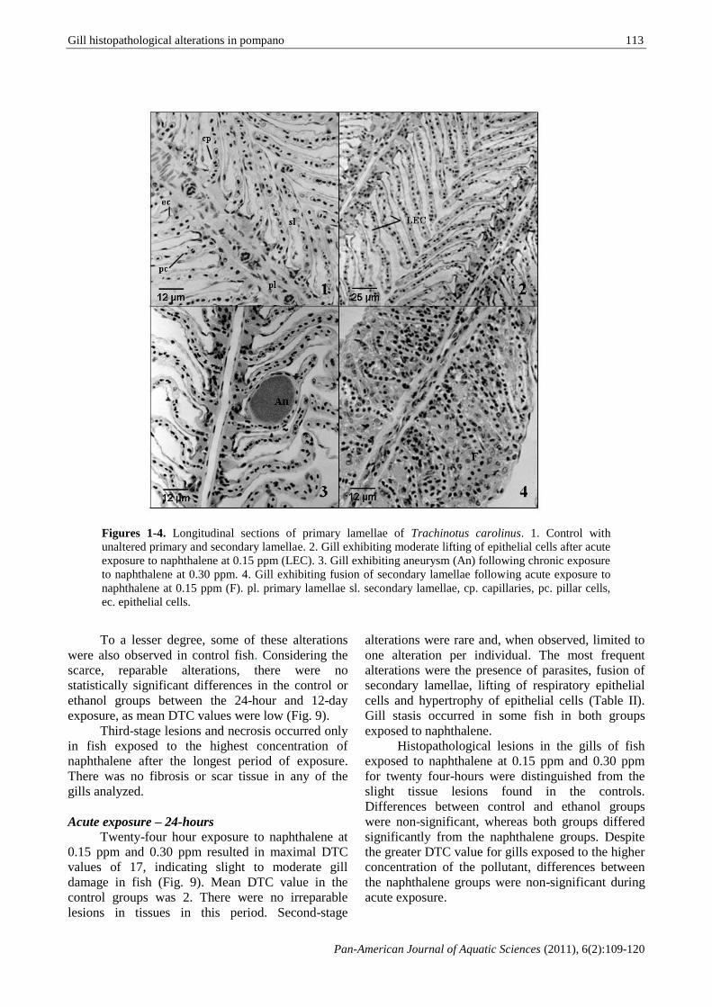
Results General remarks

The gill filaments of fish are straight and

secondary lamellae line both sides. The surface of

the gill lamellae in the control groups was covered

with epithelial cells running parallel (Fig. 1).

Using the criteria described by Poleksic &

Mitrovic-Tutundsic (1994) as reference, 21 different

types of lesions were identified in the gills of T.

carolinus (Table I), 14 of which were first-stage

lesions, five were second-stage lesions and two were

third-stage lesions. Hypertrophied epithelial cells,

epithelial lifting, telangiectasia, focal proliferation of

primary and secondary lamellar epithelial cells,

fusion of adjacent secondary lamellae or their tips,

rupture of the lamellar epithelium cells, stasis and

the presence of microscopic parasites were the most

common alterations in the fish exposed to

naphthalene (Figs 2, 4 and 5-8). A moderate

presence of aneurysm (Fig. 3) and necrosis of pillar

cells were also detected in fish exposed to

naphthalene, but at a lower percentage.

Telangiectasia (Fig. 6) consisted of dilatation of the

terminal blood vessel in secondary lamellae, in

which erythrocytes were easily recognized. Stasis or

aneurysm was determined by the congestion of

blood cells, thereby becoming a compact

homogenous mass (Fig. 3).

Table I. List of severity of gill lesions in Trachinotus carolinus.

(a) Hypertrophy and hyperplasia of gill epithelia Stage

Hypertrophy of respiratory epithelium I

Lifting of respiratory epithelial cells I

Leukocyte infiltration of gill epithelium I

Thinning of respiratory epithelium I

Rupture and peeling of lamellar epithelium II

Focal hyperplasia of epithelial cells I

Hyperplasia from base to approximately half the length of secondary lamellae I

Irregular (“chaotic”) hyperplasia of epithelial cells I

Fusion of tips of secondary lamellae I

Fusion of primary lamellae tips I

Uncontrolled thickening of proliferated tissue II

Fusion of several secondary lamellae I

Shortening of secondary lamellae I

Complete fusion of all the secondary lamellae II

(b) Blood vessel changes Stage

Lamellar telangiectasia I

Filament blood vessel enlargement I

Hemorrhages with rupture of epithelium II

Stasis II

(c) Gill parasites Stage

Presence of parasites I

(d) Terminal stages Stage

Scar tissue – fibrosis III

Necrosis III

Gill histopathological alterations in pompano

Pan-American Journal of Aquatic Sciences (2011), 6(2):109-120

113

Figures 1-4. Longitudinal sections of primary lamellae of Trachinotus carolinus. 1. Control with

unaltered primary and secondary lamellae. 2. Gill exhibiting moderate lifting of epithelial cells after acute

exposure to naphthalene at 0.15 ppm (LEC). 3. Gill exhibiting aneurysm (An) following chronic exposure

to naphthalene at 0.30 ppm. 4. Gill exhibiting fusion of secondary lamellae following acute exposure to

naphthalene at 0.15 ppm (F). pl. primary lamellae sl. secondary lamellae, cp. capillaries, pc. pillar cells,

ec. epithelial cells.

To a lesser degree, some of these alterations

were also observed in control fish. Considering the

scarce, reparable alterations, there were no

statistically significant differences in the control or

ethanol groups between the 24-hour and 12-day

exposure, as mean DTC values were low (Fig. 9).

Third-stage lesions and necrosis occurred only

in fish exposed to the highest concentration of

naphthalene after the longest period of exposure.

There was no fibrosis or scar tissue in any of the

gills analyzed.

Acute exposure – 24-hours

Twenty-four hour exposure to naphthalene at

0.15 ppm and 0.30 ppm resulted in maximal DTC

values of 17, indicating slight to moderate gill

damage in fish (Fig. 9). Mean DTC value in the

control groups was 2. There were no irreparable

lesions in tissues in this period. Second-stage

alterations were rare and, when observed, limited to

one alteration per individual. The most frequent

alterations were the presence of parasites, fusion of

secondary lamellae, lifting of respiratory epithelial

cells and hypertrophy of epithelial cells (Table II).

Gill stasis occurred in some fish in both groups

exposed to naphthalene.

Histopathological lesions in the gills of fish

exposed to naphthalene at 0.15 ppm and 0.30 ppm

for twenty four-hours were distinguished from the

slight tissue lesions found in the controls.

Differences between control and ethanol groups

were non-significant, whereas both groups differed

significantly from the naphthalene groups. Despite

the greater DTC value for gills exposed to the higher

concentration of the pollutant, differences between

the naphthalene groups were non-significant during

acute exposure.

114 T. SANTOS ET. AL.

Pan-American Journal of Aquatic Sciences (2011), 6(2):109-120

Figures 5-8. Frequent lesions found in Florida pompanos. 5. Lifting of epithelial cells (LEC) following

acute exposure to naphthalene at 0.30 ppm. 6 & 7. Gill exhibiting subepithelial hemorrhage

(telangiectasia) following chronic exposure to naphthalene at 0.15 ppm (T). 8. Parasite infestation

following chronic exposure to naphthalene at 0.15 ppm (P).

Chronic exposure – 12-days

After chronic exposure to naphthalene for

twelve days, the survival rate was 85%. The lowest

DTC value was 2 in both the control and ethanol

groups and the highest value was 134 in fish

exposed to naphthalene at 0.30 ppm (Fig. 9). After

chronic 12-day exposure to the pollutant, the

animals had higher mean DTC values. Only at the

highest concentration of naphthalene did the animals

exhibit irreparable third-stage lesions – necrosis of

the gill tissue. There was a greater quantity and

diversity of alterations in the chronic experiment, the

most common of which were lamellar epithelium

hypertrophy, fusion of lamellae and presence of

microscopic parasites, which were even found in the

control groups (Table II).

There were no significant differences in DTC

values between control and ethanol groups in

chronic exposure (Fig. 9). The naphthalene groups

differed significantly from control and ethanol

groups with regard to DTC. Moreover, there were

significant differences in DTC values between the

two naphthalene groups. The gills of fish submitted

to long-term exposure to naphthalene exhibited

extensive epithelial lifting and blood congestion

(aneurysm) in many areas of the secondary lamellae,

with the breakdown of the pillar cell system. Twelve

days of exposure to naphthalene resulted in a mean

DTC of 26 ± 7 at a concentration of 0.15 ppm and

125 ± 5 at 0.30 ppm.

Comparing the 24-hour and 12-day periods,

the longer the period of exposure to naphthalene led

to more severe and diversified damage to the gills.

Gill histopathological alterations in pompano

Pan-American Journal of Aquatic Sciences (2011), 6(2):109-120

115

Table II. Most frequently alterations observed at the gills of Trachinotus carolinus according to the exposure

time to naphthalene.

Exposure time Alterations most commonly observed Stage of lesion severity

24 hours presence of parasites I

(acute exposure) fusion of secondary lamellae I

lifting of respiratory epithelial cells I

hypertrophy of epithelial cells I

stasis II

12 days lamellar epithelium hypertrophy I

(chronic exposure) fusion of lamellae I

presence of microscopic parasites I extensive lifting of respiratory epithelial cells I

stasis (aneurysm) II

necrosis III

Scanning electron microscopy

The SEM examination confirmed the partial

fusion of many secondary lamellae in the fish

exposed to naphthalene (Figs 10-12). SEM of the

control fish revealed normal gill surfaces, with no

alterations (Fig. 13). Although not considered for the

DTC value, SEM analysis confirmed the

disarrangements in gill filaments with damage to the

structure of the secondary lamellae, clearly observed

in the fish exposed to naphthalene.

Figure 9. Degree of tissue change in Trachinotus carolinus used as controls and exposed to different

sublethal concentrations of naphthalene. Grey bars correspond to 24-hour acute exposure and dark bars

correspond to 12-day chronic exposure. Asterisks denote significant differences (P < 0.05) from control

groups. Control refers to clean-water group; ethanol refers to control group exposed to solvent of

naphthalene; 0.15 ppm and 0.30 ppm are naphthalene concentrations.

Discussion Histopathological studies are recommended

for the evaluation of fish health. Such studies allow

reliable assessments of biochemical responses in

animals exposed to a variety of environmental

stressors. Alterations in fish gills are among the

most commonly recognized responses to

environmental pollutants (Mallatt 1985, Laurent &

Perry 1991, Au 2004).

The method applied to the Florida pompano,

to calculate the degree of tissue change, enabled the

comparison of alterations in fish exposed to

naphthalene and control groups, and to correlate the

effects of exposure to gill functionality. Epithelial

116 T. SANTOS ET. AL.

Pan-American Journal of Aquatic Sciences (2011), 6(2):109-120

lifting was the first alteration detected. The

epithelium covering the secondary lamellae was

lifted away from the pillar cell system as a

continuous sheet, thereby increasing the diffusion

distance between water and blood. This process

affects respiration and ionic regulation by inhibiting

key transport processes (Mallatt 1985, Wood 2001,

Van Heerden et al. 2004).

Figures 10-13. Gill lamella with extensive lifting of epithelial cell layer observed under scanning electron

microscope. 10. Lamellae of fish exposed to 0.30 ppm of naphthalene after acute exposure. 11. Slight

lifting of epithelial cell layer in fish exposed to naphthalene at 0.15 ppm following exposure for 24 hours.

12. Extensive lifting of epithelial cell layer in fish exposed to 0.30 ppm of naphthalene following chronic

exposure. 13. Lamellae of control fish after 24 hours of experiment.

The histopathological lesions in both control

groups should be considered carefully. Background

knowledge on the control conditions is essential to

interpreting pathological responses (Poleksic &

Mitrovic-Tutundsic 1994). Despite a few sparse

lesions, the mean DTC values in the control groups

remained within the range expected for gills with

normal function. The slight lesions in both control

groups may be due to the stress of confinement, as

T. carolinus normally exhibits intense swimming

activity, which characterizes it as a species with a

high energy demand (Santos et al. 2006).

Although ethanol causes physiological

alterations in oxygen consumption and ammonia

excretion in fish (Vargas et al. 1991, Santos et al.

2006), there were no significant histological

differences between ethanol and clean-water

controls. The concentration of ethanol used in this

study, was not considered harmful to gill tissues.

As observed in the pompanos exposed to

naphthalene, a number of studies have demonstrated

that interstitial edema, or epithelial lifting, is one of

the most frequent lesions found in the gill epithelium

of fish exposed to pollutants (Mallatt 1985, Sola et

al. 1995, Bury et al. 1998, Figueiredo-Fernandes et

al. 2007). Epithelial lifting is one of the first

responses of gills even with exposure to low

concentrations of contaminants (Segers et al. 1984).

In studies on Lates calcarifer, exposed to acute and

subchronic cadmium, Thophon et al. (2003) also

described epithelial lifting in the fish gills.

The fusion of secondary lamellae, hyperplasia

and the lifting of epithelial cells, with the presence

of edema and an increase in mucus secretion, are the

Gill histopathological alterations in pompano

Pan-American Journal of Aquatic Sciences (2011), 6(2):109-120

117

main lesions described in fish exposed to petroleum

hydrocarbons (DiMichele & Taylor 1978,

Engelhardt et al. 1981, Correa & Garcia 1990,

Prasad 1991). All of these alterations affect gas

exchange. The complete fusion of secondary

lamellae is usually induced by high doses of

chemical compounds or is a final result of

hyperplasia in sublethal poisoning following chronic

exposure (Temmink et al. 1989). The non-specificity

of the histological gill alterations suggests

stereotyped physiological reactions of the gills to

stress and many alterations may plausibly be

considered defensive reactions (Mallatt 1985).

After 24 hours of exposure, the fish in the

control and ethanol groups exhibited normal gills

whereas those exposed to naphthalene exhibited

slight to moderate alterations affecting the normal

functioning of the respiratory organ. When fish are

exposed to contaminants, alterations in their normal

functions make them more susceptible to parasites

and illness (Ahmad et al. 2003). As in the present

study, Ahmad et al. (2003) also found that

naphthalene exposure had a harmful effect on cell

membranes in Anguilla anguilla, impairing cell

viability. The significant results of tissue change

impairing alteration on the normal function of gills

of fish exposed, even to low concentrations of

naphthalene as that of 0.15 ppm, indicates that even

low doses of pollutants can alter the tissue function

and, in spite of being reversible, alterations at the

cellular structure can result even in organism death.

Stasis, which restricts blood flow through the

lamellae, was evident in both periods in the groups

exposed to the greatest concentration of naphthalene.

This condition reduces both gas and ion exchange,

which is extremely harmful to the organism and may

lead to death.

After chronic exposure to naphthalene at a

concentration of 0.30 ppm, the fish exhibited the

greatest amount of tissue damage, mainly due to the

presence of focal necrosis in pillar cells. Necrosis in

fish gills is believed to reflect direct harmful effects

of irritants (Temmink et al. 1983) and cell death is

characteristic of irreparable damage. Thus, chronic

exposure to high doses of naphthalene affected the

vital organs of the fish and the longer the period of

exposure led to a higher degree of tissue change in

the gills. Rodrigues & Fanta (1998) also report

increasing tissue damage associated to exposure

time and pollutant concentration. Degenerative

lesions are frequently found in fish due to exposure

to high concentrations of pollutants and after chronic

exposure periods (Pacheco & Santos 2002,

Rodrigues & Fanta 1998).

Mallatt (1985) reviewed studies on the main

alterations induced by toxic substances in the

structure of fish gills and the most common of which

were necrosis, hyperplasia, hypertrophy, rupture of

gill tissue, lamellar fusion, hyper secretion and

proliferation of mucous cells, alteration in chloride

cells and vascularization. The author reports that

even lamella disorganization and first-stage lesions

indicate that the environment is not completely

favorable to the development and survival of fish.

The alterations found in the present study

proved to be a semi-quantitative response to the

action of naphthalene on the gills of T. carolinus.

The analysis methodology employed can be used in

laboratory to analyze the effects of exposure time

and dose of toxic substances on fish. Despite the

non-specificity of the lesions, histological analysis

on fish proved an efficient tool for the assessment of

environmental quality, as individual effects of

pollutants in nature - thousands of different

compounds - are relatively rare. Nonethelless, the

assessment of the effect of a single component is

very useful for toxicological studies as well as the

establishment of monitoring programs and

environmental laws.

Acknowledgements

The authors are grateful to the Oceanographic

Institute of the Universidade de São Paulo for the

facilities offered and the Brazilian fostering agency

Fundação de Amparo à Pesquisa do Estado de São

Paulo (FAPESP – 02/10566-7) for the scholarship

granted to the first author. We are also grateful for

the use of the Nap/Mepa (Esalq) scanning electron

microscope.

References

Ahmad, I., Pacheco, M. & Santos, M. A. 2003.

Naphthalene-induced differential tissue

damage association with circulating fish

phagocyte induction. Ecotoxicology and

Environmental Safety, 54(1): 7-15.

Albers, P. H. 2003. Petroleum and individual

polycyclic aromatic hydrocarbons. In:

Hoffman, D. J., Rattner, B.A., Burton Jr., G. A.

& Cairns Jr., J. (Eds.). Handbook of

Ecotoxicology. 2nd

ed. Lewis Publishers, Boca

Raton, Florida, pp. 341-371.

Alkindi, A. Y. A., Brown, J. A., Waring, C. P. &

Collins, J. E. 1996. Endocrine,

osmoregulatory, respiratory and

haematological parameters in flounder

exposed to the water soluble fraction of crude

oil. Journal of Fish Biology, 49(6): 1291–

1305.

118 T. SANTOS ET. AL.

Pan-American Journal of Aquatic Sciences (2011), 6(2):109-120

ATSDR (Agency for Toxic Substances and Disease

Registry). 1995. Toxicological profile for

naphthalene, 1-methylnaphthalene, and 2-

methylnaphthalene. Report of the U.S.

Department of Health and Human Services,

Public Health Service.

Au, D. W. T. 2004. The application of histo-

cytopathological biomarkers in marine

pollution monitoring: a review. Marine

Pollution Bulletin, 48(9-10): 817-834.

Black, M. C., Millsap, D. S. & McCarthy, J. F. 1991.

Effects of acute temperature-change on

respiration and toxicant uptake by rainbow-

trout, Salmo gairdneri (Richardson).

Physiological Zoology, 64(1): 145-168.

Bowser, P. 2001. Anesthetic option for fish. In:

Gleed, R. D. & Ludders, J. W. (Eds.). Recent

advances in veterinary anesthesia and

analgesia: Companion animals (available at

http://www.ivis.org, Document No.

A1412.0701).

Britvic, S., Lucic, D. & Kurelec, B. 1993. Bile

fluorescence and some early biological effects

in fish as indicators of pollution by

xenobiotics. Environmental Toxicology and

Chemistry, 12(4): 765-773.

Bury, N. R.; Li, J., Flik, G., Lock, R.A.C. &

Wendelaar-Bonga, S.E. 1998. Cortisol protects

against copper induced necrosis and promotes

apoptosis in fish gill chloride cells in vitro.

Aquatic Toxicology, 40(2): 193-202.

Collier, T. K. & Varnasi, U. 1991. Hepatic activities

of xenobiotic metabolizing enzymes and

biliary levels of xenobiotics in English sole

(Parophyrs vetulus) exposed to environmental

contaminants. Archives of Environmental

Contamination and Toxicology, 20(4): 462-

473.

Correa, M. & Garcia, H. I. 1990. Physiological

responses of juvenile white mullet, Mugil

curema, exposed to benzene. Bulletin of

Environmental Contamination and

Toxicology, 44(3): 428-434.

DiMichele, L. & Taylor, M. H. 1978.

Histopathological and physiological responses

of Fundulus heteroclitus to naphthalene

exposure. Journal of the Fisheries Research

Board of Canada, 35(8): 1060-1066.

Du Preez, H. H., McLachlan, A., Marais, J. F. K. &

Cockcroft, A. C. 1990. Bioenergetics of fishes

in a high-energy surf-zone. Marine Biology,

106(1): 1-12.

Dwivedi, H., Sari, R. & Wate, S. 1997. Long term

effects of DI-aromatic hydrocarbon in catfish

Heteropneustes fossilis. Journal of

Ecobiology, 9: 247-254.

El-Sayed, N. K., Salem, S. A., Moursy, A. &

Ibrahim, B. M. 1995. Acute and chronic

toxicity of some aromatic hydrocarbons on

Tilapia zillii. Bulletin of the National

Institute of Oceanography and Fisheries,

21: 613-630.

Elumalai, M. & Balasubramanian, M. P. 1999.

Influence of naphthalene on esterase activity

during vitellogenesis of marine edible crab,

Scylla serrata. Bulletin of Environmental

Contamination and Toxicology, 62(6): 743–

748.

Engelhardt, F. R., Wong, M. P. & Duey, M. E. 1981.

Hydromineral balance and gill morphology in

rainbow trout Salmo gairdneri, acclimated to

fresh and sea water, as affected by petroleum

exposure. Aquatic Toxicology, 1(3-4): 175-

186.

Fanta, E., Rios, F. S., Romão, S., Vianna, A. C. C. &

Freiberger, S. 2003. Histopathology of the fish

Corydoras paleatus contaminated with

sublethal levels of organophosphorus in water

and food. Ecotoxicology and Environmental

Safety, 54(2): 119-130.

Figueiredo-Fernandes, A., Ferreira-Cardoso, J. V.,

Garcia-Santos, S., Monteiro, S. M., Carrola, J.,

Matos, P. & Fontaínhas-Fernandes, A. 2007.

Histopathological changes in liver and gill

epithelium of Nile tilapia, Oreochromis

niloticus, exposed to waterborne copper.

Pesquisa Veterinária Brasileira, 27(3): 103-

109.

Heilman, M. J. & Spieler, R. E. 1999. The daily

feeding rhythm to demand feeders and the

effects of timed meal feeding on the growth of

juvenile Florida pompano, Trachinotus

carolinus. Aquaculture, 180(1-2): 53–64.

Hoese, H. D. & Moore, R. H. 1998. Fishes of the

Gulf of Mexico: Texas, Louisiana and

adjacent waters. 2nd

ed. Texas A&M

University Press, Texas, 422 pp.

Hymel, M. K., Baltz, D. M., Chesney, E. J., Tarr, M.

A. & Kolok, A. S. 2002. Swimming

performance of juvenile Florida pompano

exposed to ethylene glycol. Transactions of

the American Fisheries Society, 131(6):

1152-1163.

Irwin, R. J., VanMouwerik, M., Stevens, L., Seese,

M. D. & Basham, W. 1997. Environmental

contaminants encyclopedia. National Park

Service, Water Resources Division, Colorado,

114 pp.

Gill histopathological alterations in pompano

Pan-American Journal of Aquatic Sciences (2011), 6(2):109-120

119

Laurent, P. & Perry, S. F. 1991. Environmental

effects on fish gill morphology. Physiological

Zoology, 53(1): 4-25.

Linnaeus, C. 1766. Systema naturae per regna tria

naturae, secundum classes, ordines, genera,

species, cum characteribus, differentiis,

synonymis, locis. Laurentii Salvii,

Holmiae. Systema Naturae, 12: 1-532.

Mallatt, J. 1985. Fish gill structural changes induced

by toxicants and other irritants: a statistical

review. Canadian Journal of Fisheries and

Aquatic Sciences, 42(4): 630-648.

Molven, A. & Gooksoyr, A. 1993. Biological effects

and biomonitoring of organochlorines and

PAH in the marine environment. In:

Mervyn, R. (Ed.). Ecotoxicology Monitoring.

VCH, New York, pp 137-162.

Nagabhushanam, R., Machale, P. R., Kkatyayani, R.

V., Reddy, P. S. & Sarojini, R. 1991.

Erythrophoretic responses induced by

naphthalene in freshwater prawn, Caridina

rajadhari. Journal of Ecotoxicology and

Environmental Monitoring, 1: 185-191.

Neff, J. M. & Anderson, J. W. 1975. An ultraviolet

spectrophometric method for the determination

of naphthalene and alkyl naphthalenes in the

tissues of oil-contaminated marine animals.

Bulletin of Environmental Contamination

and Toxicology, 14(1): 122-128.

Nero, V., Farwell, A., Listen, A., Van Der Kraak,

G., Lee, L. E. J., T. Van Meerd, Mackinnon,

M. D. & Dixon, D. G. 2006. Gill and liver

histopathological changes in yellow perch

(Perca flavescens) and goldfish (Carassius

auratus) exposed to oil sands process-affected

water. Ecotoxicology and Environmental

Safety, 63(3): 365-377.

Ortiz-Delgado, J. B., Segner, H., Arellano, J. M. &

Sarasquete, C. 2007. Histopathological

alterations, EROD activity, CYP1A protein

and biliary metabolites in gilthead seabream

Sparus aurata exposed to Benzo(a)pyrene.

Histology and Histopathology, 22(4): 417-

432.

Pacheco, M. & Santos, M. A. 2001.

Biotransformation, endocrine and genetic

responses of Anguilla anguilla L. to petroleum

distillate products and environmental

contaminated waters. Ecotoxicology and

Environmental Safety, 49(1): 64–75.

Pacheco, M. & Santos, M. A. 2002.

Biotransformation, genotoxic and

histopathological effects of environmental

contaminants in European eel (Anguilla

anguilla L.). Ecotoxicology and

Environmental Safety, 53(3): 331-347.

Payne, J. F., Mathieu, A. & Collier, T. K. 2003.

Ecotoxicological studies focusing on marine

and freshwater fish. In: Douben, P. E. T.

(Ed.). PAHs: An Ecotoxicological

Perspective. Wiley, UK, pp. 191-224.

Pitot III, H. C. & Dragan, Y. P. 1996. Chemical

carcinogenesis. In: Klaasen, C. D. (Ed.).

Casarett and Doull’s Toxicology: The basic

science of poisons. McGraw Hill Publishing,

New York, pp. 241-320.

Poleksic, V. & Mitrovic-Tutundzic, V. 1994. Fish

gills as monitor of sublethal and chronic

effects of pollution. In: Müller, R. & Lloyd,

R. (Eds.). Sublethal and chronic effects of

pollutants on freshwater fish. Fishing News

Books, Oxford, pp 339-352.

Prasad, M. S. 1991. SEM study on the effects of

crude oil on the gills and airbreathing organs

of climbing perch Anabas testudineus.

Bulletin of Environmental Contamination

Toxicology, 47(6): 882-889.

Rodrigues, E. L. & Fanta, E. 1998. Liver

histopathology of the fish Brachydanio rerio

Hamilton-Buchman after acute exposure to

sublethal levels of the organophosphate

dimethoate 500. Revista Brasileira de

Zoologia, 15(2): 441-450.

Santos, T. C. A., Phan, V. N., Passos, M. J. A. C. R.

& Gomes, V. 2006. Effects of naphthalene on

metabolic rate and ammonia excretion of

juvenile Florida Pompano, Trachinotus

carolinus. Journal of Experimental Marine

Biology and Ecology, 335(1): 82-90.

Schirmer, K., Dixon, D. G., Greenberg, B. M. &

Bols, N. C. 1998. Ability of 16 priority PAHs

to be directly cytotoxic to a cell line from the

rainbow trout gill. Toxicology, 127(1-3): 129-

141.

Schwaiger, J., Bucher, F., Ferling, H., Kalbfus, W.

& Negele, R.D. 1992. A prolonged toxicity

study on the effects of sublethal concentrations

of bis(tri-n-butyltin)oxide (TBTO):

histopathological and histochemical findings

in rainbow trout (Oncorhynchus mykiss).

Aquatic Toxicology, 23(1): 31-48.

Schwaiger, J., Wanke, R., Adam, S., Pawert, M.,

Honnen, W. & Triebskorn, R. 1997. The use of

histopathological indicators to evaluate

contaminant-related stress in fish. Journal of

Aquatic Ecosystem Stress and Recovery,

6(1): 75-86.

Segers, J.H.L., Temmink, J.H.M., Van den Berg,

120 T. SANTOS ET. AL.

Pan-American Journal of Aquatic Sciences (2011), 6(2):109-120

J.H.J. & Wegman, R.C.C. 1984.

Morphological changes in gill of carp

(Cyprinus carpio L.) exposed to acutely toxic

concentrations of methyl bromide. Water

Research, 18(11): 1437-1441.

Silva, D. A. M., Buzitis, J., Krahn, M. M., Bícego,

M. C. & Pires-Vanin, A. M. S. 2006.

Metabolites in bile of fish from São Sebastião

Channel, São Paulo, Brazil as biomarkers of

exposure to petrogenic polycyclic aromatic

compounds. Marine Pollution Bulletin,

52(2): 175-183.

Simonato, J. D., Guedes, C. L. B. & Martinez, C. B.

R. 2008. Biochemical, physiological, and

histological changes in the neotropical fish

Prochilodus lineatus exposed to diesel oil.

Ecotoxicology and Environmental Safety, 69(1): 112-120.

Sola, F., Isaia, J. & Masoni, A. 1995. Effects of

copper on gill structure and transport function

in the rainbow trout, Oncorhynchus mykiss.

Journal of Applied Toxicology, 15(5): 391-

398.

Spies, R. B., Stegeman, J. J., Hinton, D. E., Woodin,

B., Smolowitz, R., Okihiro, M. & Shea, D.

1996. Biomarkers of hydrocarbon exposure

and sublethal effects in embiotocid fishes from

a natural petroleum seep in the Santa Barbara

Channel. Aquatic Toxicology, 34(3): 195-

219.

Swapan, C. D., Araki, T. & Fukushima, T. 2000.

Polycyclic aromatic hydrocarbons in fish

organs. Marine Pollution Bulletin, 40(10):

882–885.

Temmink, J. H. M., Bouwmeister, P., de Jong, P. &

Van Den Berg, J. 1983. An ultrastructural

study of chromate-induced hyperplasia in the

gill of rainbow trout (Salmo gairdneri).

Aquatic Toxicology, 4(2): 165-179.

Temmink, J. H. M., Field, J. A., Van Haastrecht, J.

C. & Merkelbach, R. C. M. 1989. Acute and

subacute toxicity of bark tannings in carp

(Cyprinus carpio). Water Research, 23(3):

341-344.

Thophon, S., Kruatrachue, M., Upatham, E. S.,

Pokethitiyook, P., Sahaphong, S. & Jaritkhuan,

S. 2003. Histopathological alterations of white

seabass, Lates calcarifer, in acute and

subchronic cadmium exposure.

Environmental Pollution, 121(3): 307-320.

Upshall, C., Payne, J. F. & Hellou, J. 1993.

Induction of MFO enzymes and production of

bile metabolites in rainbow trout

(Oncorhynchus mykiss) exposed to waste

crankcase oil. Environmental Toxicology

and Chemistry, 12(11): 2105-2112.

Van Heerden, D., Vosloo, A. & Nikinmaa, M. 2004.

Effects of short-term copper exposure on gill

structure, methallothionein and hypoxia-

inducible factor-1α (HIF-1α) levels in rainbow

trout (Oncorhynchus mykiss). Aquatic

Toxicology, 69(3): 271-280.

Vargas, M., Correa, M. & Chung, K. S. 1991.

Indicadores fisiológicos en la evaluación de la

toxicidad de hidrocarburos aromaticos.

Boletin del Instituto Oceanográfico de

Venezuela, 30(1): 57-64.

Vijayavel, K., Gomathi, R. D., Durgabhavani, K. &

Balasubramanian, M. P. 2004. Sublethal effect

of naphthalene on lipid peroxidation and

antioxidation status in the edible marine crab

Scylla serrata. Marine Pollution Bulletin,

48(5-6): 429-433.

Wakeham, S. G., Davis, A. C., Karas, I. L. 1983.

Mesocosm experiments to determine the fate

and persistence of volatile organic compounds

in coastal waters. Environmental Science &

Technology, 17(10): 611-617.

Winkaler, E. U., Silva, A. G., Galindo, H. C. &

Martinez, C. B. R. 2001. Biomarcadores

histológicos e fisiológicos para o

monitoramento da saúde de peixes de ribeirões

de Londrina, Estado do Paraná. Acta

Scientiarum, 23(2): 507-514.

Wood, C. M. 2001. Toxic responses of the gill. In:

Schlenck, D. & Benson, W. H. (Eds.). Target

organ toxicity in marine and freshwater

teleosts. Taylor & Francis, London, pp. 1-89.

Yu, Y., Wade, T. L., Frang, J., McDonald, S. &

Brooks, J. M. 1995. Gas chromatographic-

mass spectrometric analysis of polycyclic

aromatic hydrocarbon metabolites in Antarctic

fish (Notothenia gibberiforns) injectes with

diesel fuel arctic. Archives of Environmental

Contamination and Toxicology, 29(2): 241-

246.

Zanardi, E. 1996. Hidrocarbonetos no canal de São

Sebastião e na plataforma interna adjacente –

influência do derrame de maio de 1994.

Master’s Thesis. Universidade de São Paulo,

São Paulo, Brazil, 112 pp.

Received December 2010

Accepted April 2011

Published online November 2011


Recommended