Page 1
Histology Technician
Field description
Histology is an essential component to the art and science of pathology. The histology laboratory contributes a valuable service to help
pathologists provide patient diagnoses. Information gained through microscopic evaluation of tissue slides prepared by histology technicians
allows a pathologist to either identify or dismiss disease. The preparatory steps taken to ensure a quality diagnosis are key to this relationship.
Knowledge of the basic pathologic conditions, skills in the use of precision equipment, and performance of special stains enable histology
technicians to accurately demonstrate the morphology of tissue specimens. Knowledge of biology, chemistry, anatomy, physiology and medical
terminology is essential for the professional histology technician or technologist. In addition, attention to detail, good manual dexterity, and
above all, a concern for patient well-being are imperative characteristics of a good histology technician.
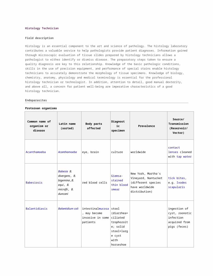
Endoparasites
Protozoan organisms
Common name of
organism or disease
Latin name
(sorted)Body parts affected
Diagnostic
specimenPrevalence
Source/
Transmission
(Reservoir/
Vector)
Acanthamoeba Acanthamoeba eye, brain culture worldwide
contact
lenses cleaned
with tap water
Babesiosis
Babesia B.
divergens, B.
bigemina,B.
equi, B. microfti, B.
duncani
red blood cells
Giemsa-stained
thin blood
smear
New York, Martha's Vineyard,
Nantucket (different species
have worldwide distribution)
tick bites, e.g. Ixodes
scapularis
Balantidiasis Balantidium coli
intestinalmucosa, may
become invasive in
some patients
stool
(diarrhea=ciliat
ed trophozoite;
solid
stool=large cyst
with horseshoe
shaped
nucleus)
ingestion of cyst,
zoonotic infection
acquired from pigs
(feces)
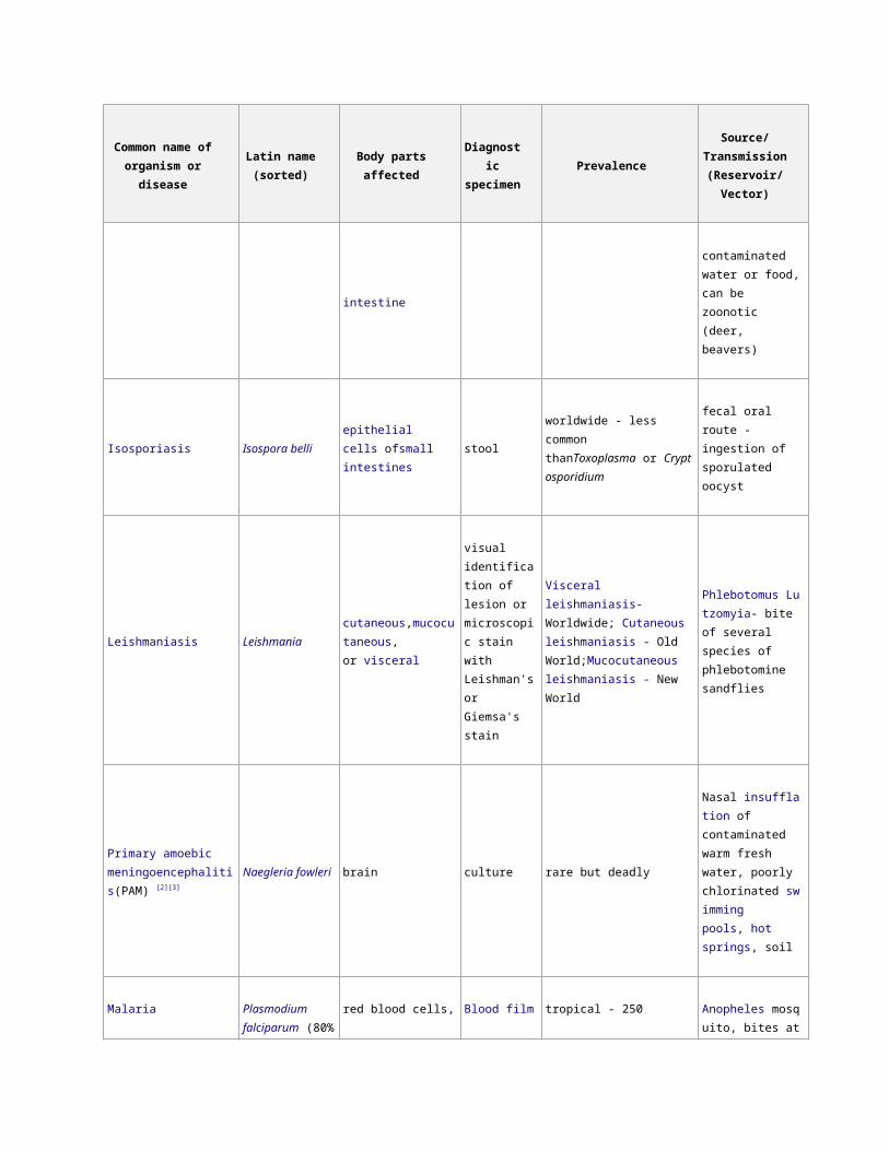
Blastocystosis Blastocystis intestinal direct
microscopy of
stool (PCR, anti
body)
2 - 20% of population [1] eating food
contaminated with
feces from an
infected human or
Page 2
Common name of
organism or disease
Latin name
(sorted)Body parts affected
Diagnostic
specimenPrevalence
Source/
Transmission
(Reservoir/
Vector)
animal
Coccidia, cryptosporidiosis Cryptosporidium intestines stool widespread
ingestion of oocyst
(sporulated), some
species are zoonotic
(e.g. bovine fecal
contamination)
DientamoebiasisDientamoeba
fragilisintestines stool
up to 10% in industrialized
countries
ingesting water or
food contaminated
with feces
AmoebiasisEntamoeba
histolytica
Intestines (mainly
Large, can go to
extraintestinal sites)
stool (fresh
diarrheic stools
have amoeba,
solid stool has
cyst)
areas with poor sanitation, high
population density and tropical
regions
fecal-oral
transmission of cyst,
not amoeba
Giardiasis Giardia lamblialumen of thesmall
intestinestool widespread
ingestion of cysts in
fecal contaminated
water or food, can be
zoonotic (deer,
beavers)
Isosporiasis Isospora belliepithelial cells ofsmall
intestinesstool
worldwide - less common
thanToxoplasma or Cryptospori
dium
fecal oral route -
ingestion of
sporulated oocyst
Leishmaniasis Leishmania cutaneou
s,mucocutaneous,
or visceral
visual
identification
of lesion or
microscopic
stain with
Leishman's or
Visceral leishmaniasis-
Worldwide; Cutaneous
leishmaniasis - Old
World;Mucocutaneous
leishmaniasis - New World
Phlebotomus Lutzom
yia- bite of several
species of
phlebotomine
sandflies
Page 3
Common name of
organism or disease
Latin name
(sorted)Body parts affected
Diagnostic
specimenPrevalence
Source/
Transmission
(Reservoir/
Vector)
Giemsa's stain
Primary amoebic
meningoencephalitis(PAM) [2][3]
Naegleria fowleri brain culture rare but deadly
Nasal insufflation of
contaminated warm
fresh water, poorly
chlorinated swimmin
g pools, hot springs,
soil
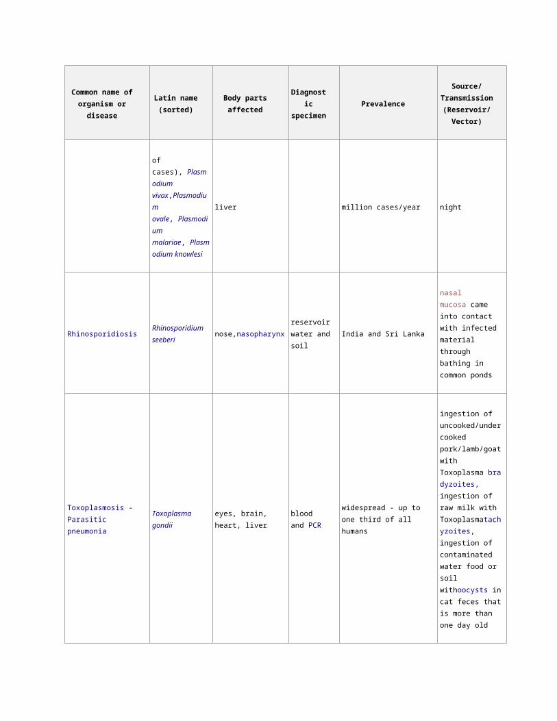
Malaria
Plasmodium
falciparum (80% of
cases), Plasmodium
vivax,Plasmodium
ovale, Plasmodium
malariae, Plasmodi
um knowlesi
red blood cells, liver Blood filmtropical - 250 million
cases/year
Anopheles mosquito,
bites at night
RhinosporidiosisRhinosporidium
seeberinose,nasopharynx
reservoir water
and soilIndia and Sri Lanka
nasal mucosa came
into contact with
infected material
through bathing in
common ponds
Toxoplasmosis -Parasitic
pneumonia
Toxoplasma gondii eyes, brain, heart, liver blood and PCR widespread - up to one third of
all humans
ingestion of
uncooked/undercook
ed pork/lamb/goat
with
Toxoplasma bradyzoi
tes, ingestion of raw
milk with
Toxoplasm
atachyzoites,
ingestion of
contaminated water
food or soil
withoocysts in cat
feces that is more
Page 4
Common name of
organism or disease
Latin name
(sorted)Body parts affected
Diagnostic
specimenPrevalence
Source/
Transmission
(Reservoir/
Vector)
than one day old
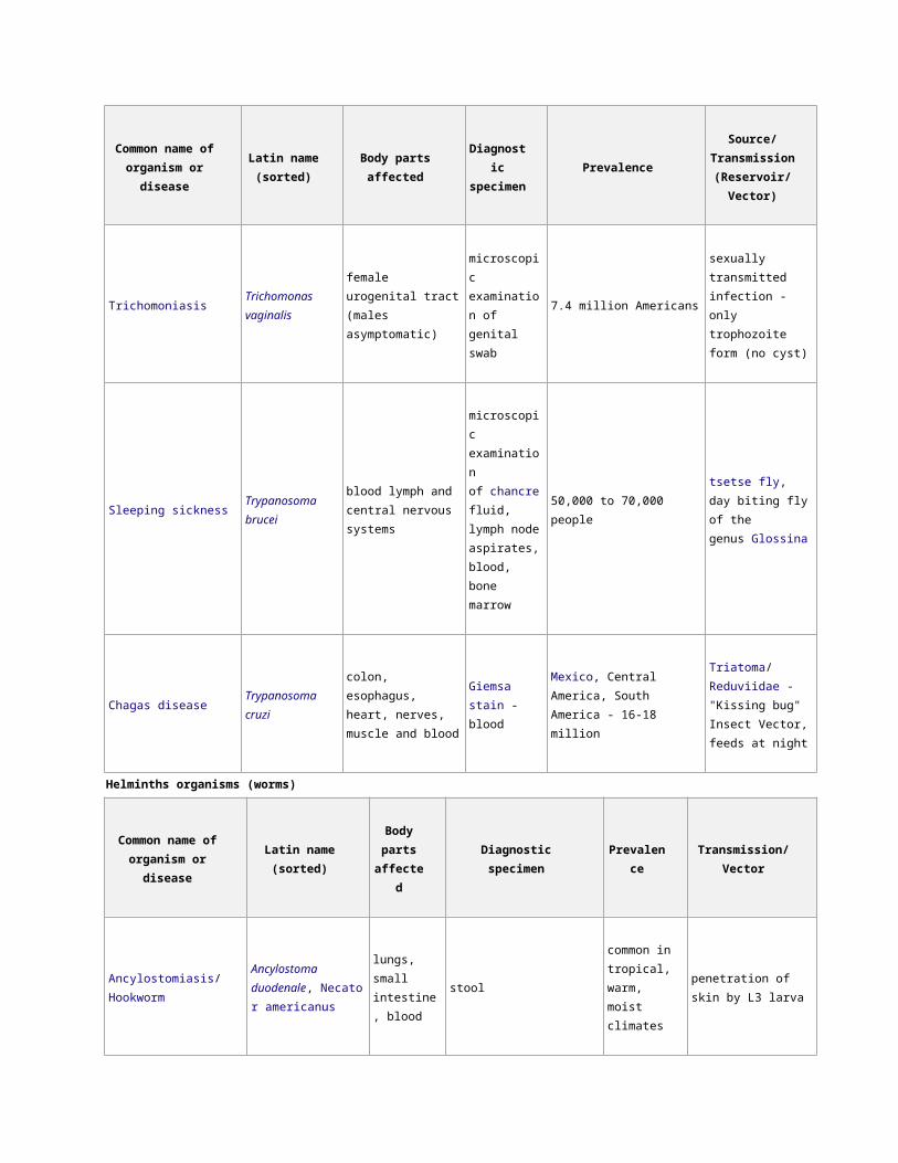
TrichomoniasisTrichomonas
vaginalis
female urogenital tract
(males asymptomatic)
microscopic
examination of
genital swab
7.4 million Americans
sexually transmitted
infection - only
trophozoite form (no
cyst)
Sleeping sicknessTrypanosoma
brucei
blood lymph and
central nervous systems
microscopic
examination
of chancre fluid
, lymph node
aspirates,
blood, bone
marrow
50,000 to 70,000 people
tsetse fly, day biting
fly of the
genus Glossina
Chagas disease Trypanosoma cruzi
colon, esophagus,
heart, nerves, muscle
and blood
Giemsa stain -
blood
Mexico, Central America, South
America - 16-18 million
Triatoma/Reduviidae
- "Kissing bug" Insect
Vector, feeds at night
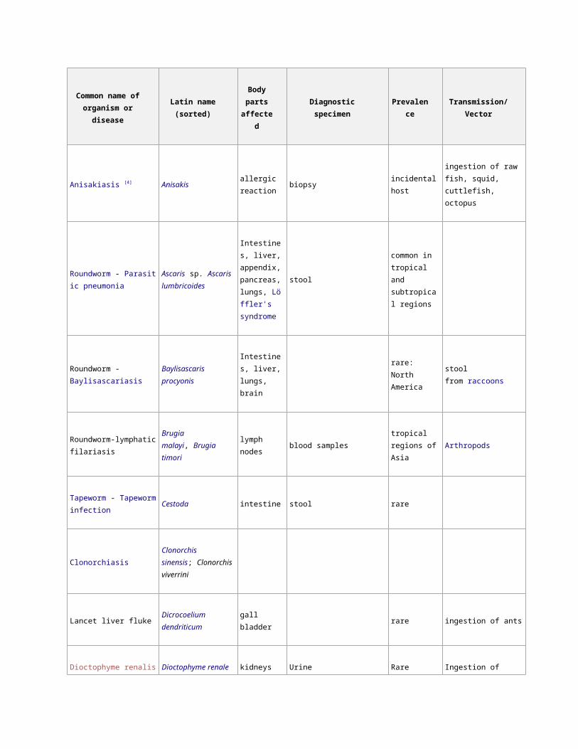
Helminths organisms (worms)
Common name of
organism or diseaseLatin name (sorted)
Body
parts
affected
Diagnostic specimen Prevalence Transmission/Vector
Ancylostomiasis/Hookworm
Ancylostoma
duodenale, Necator
americanus
lungs, small
intestine,
blood
stool
common in
tropical, warm,
moist climates
penetration of skin by L3
larva
Anisakiasis [4] Anisakisallergic
reactionbiopsy incidental host
ingestion of raw fish,
squid, cuttlefish, octopus
Roundworm - Parasitic
pneumonia
Ascaris sp. Ascaris
lumbricoides
Intestines,
liver,
stool common in
tropical and
Page 5
Common name of
organism or diseaseLatin name (sorted)
Body
parts
affected
Diagnostic specimen Prevalence Transmission/Vector
appendix,
pancreas,
lungs, Löffler's
syndrome
subtropical
regions
Roundworm -
BaylisascariasisBaylisascaris procyonis
Intestines,
liver, lungs,
brain
rare: North
Americastool from raccoons
Roundworm-lymphatic
filariasis
Brugia malayi, Brugia
timorilymph nodes blood samples
tropical regions
of AsiaArthropods
Tapeworm - Tapeworm
infectionCestoda intestine stool rare
Clonorchiasis
Clonorchis
sinensis; Clonorchis
viverrini
Lancet liver flukeDicrocoelium
dendriticumgall bladder rare ingestion of ants
Dioctophyme renalis
infectionDioctophyme renale
kidneys
(typically the
right)
Urine Rare
Ingestion of
undercooked or raw
freshwater fish
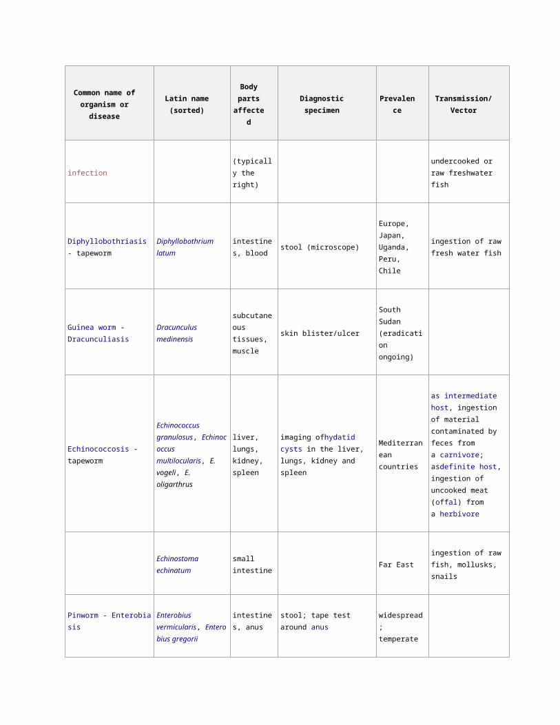
Diphyllobothriasis -
tapewormDiphyllobothrium latum
intestines,
bloodstool (microscope)
Europe, Japan,
Uganda, Peru,
Chile
ingestion of raw fresh
water fish
Guinea worm -
Dracunculiasis
Dracunculus medinensis subcutaneous
tissues,
skin blister/ulcer South Sudan
(eradication
Page 6
Common name of
organism or diseaseLatin name (sorted)
Body
parts
affected
Diagnostic specimen Prevalence Transmission/Vector
muscle ongoing)
Echinococcosis - tapeworm
Echinococcus
granulosus, Echinococc
us multilocularis, E.
vogeli, E. oligarthrus
liver, lungs,
kidney, spleen
imaging ofhydatid cysts in the
liver, lungs, kidney and spleen
Mediterranean
countries
as intermediate host,
ingestion of material
contaminated by feces
from a carnivore;
asdefinite host, ingestion
of uncooked meat (offal)
from a herbivore
Echinostoma echinatum small intestine Far Eastingestion of raw fish,
mollusks, snails
Pinworm - Enterobiasis
Enterobius
vermicularis, Enterobius
gregorii
intestines,
anusstool; tape test around anus
widespread;
temperate
regions
Liver fluke - Fasciolosis [5]
Fasciola
hepatica, Fasciola
gigantica
liver, gall
bladderstool
Fasciola
hepatica in
Europe, Africa,
Australia, the
Americas and
Oceania; Fascio
la
gigantica only
in Africa and
Asia, 2.4 million
people infected
by both species
freshwater snails
Fasciolopsiasis - intestinal
fluke [6]Fasciolopsis buski intestines stool or vomitus (microscope)
East Asia - 10
million people
ingestion of infested
water plants or water
(intermediate
host:amphibic snails)
Page 7
Common name of
organism or diseaseLatin name (sorted)
Body
parts
affected
Diagnostic specimen Prevalence Transmission/Vector
Gnathostomiasis [7]
Gnathostoma
spinigerum, Gnathosto
ma hispidum
subcutaneous
tissues (under
the skin)
physical examinationrare - Southeast
Asia
ingestion of raw or
undercooked meat (e.g.,
freshwater fish, chicken,
snails, frogs, pigs) or
contaminated water
Hymenolepiasis[8]
Hymenolepis
nana, Hymenolepis
diminuta
ingestion of material
contaminated by flour
beetles, meal worms,
cockroaches
Loa loa filariasis, Calabar
swellingsLoa loa filaria
Connective
tissue, lungs,
eye
blood
(Giemsa,haematoxylin,eosin st
ain)
rain forest of
West Africa -
12-13 million
people
Tabanidae - horse fly,
bites in the day
Mansonelliasis, FilariasisMansonella
streptocerca
subcutaneous
layer of skininsect
Metagonimiasis - intestinal
fluke
Metagonimus
yokogawaistool
Siberia,
Manchuria,
Balkan states,
Israel, Spain
ingestion of undercooked
or salted fish
River blindness
Onchocerca
volvulus, Onchocerciasi
s
skin, eye,
tissuebloodless skin snip
Africa, Yemen,
Central and
South America
near cool, fast
flowing rivers
Simulium/Black fly, bite
during the day
Chinese Liver Fluke
Opisthorchis
viverrini, Opisthorchis
felineus, Clonorchis
sinensis
bile duct
1.5 million
people in
Russia
consuming infected raw,
slightly salted or frozen
fish
Page 8
Common name of
organism or diseaseLatin name (sorted)
Body
parts
affected
Diagnostic specimen Prevalence Transmission/Vector
Paragonimiasis, Lung Fluke
Paragonimus
westermani; Paragonim
us
africanus; Paragonimus
caliensis;Paragonimus
kellicotti; Paragonimus
skrjabini; Paragonimus
uterobilateralis
lungs sputum, feces East Asia
ingestion of raw or
undercooked freshwater
crabs crayfishes or other
crustaceans
Schistosomiasis - bilharzia,
bilharziosis or snail fever (all
types)
Schistosoma sp.
Africa,
Caribbean,
eastern South
America, east
Asia, Middle
East - 200
million people
skin exposure to water
contaminated with
infected fresh water
snails
intestinal schistosomiasis Schistosoma mansoni
intestine,
liver, spleen,
lungs, skin
stool
Africa,
Caribbean,
South America,
Asia, Middle
East - 83 million
people
skin exposure to water
contaminated with
infected Biomphalaria fre
sh water snails
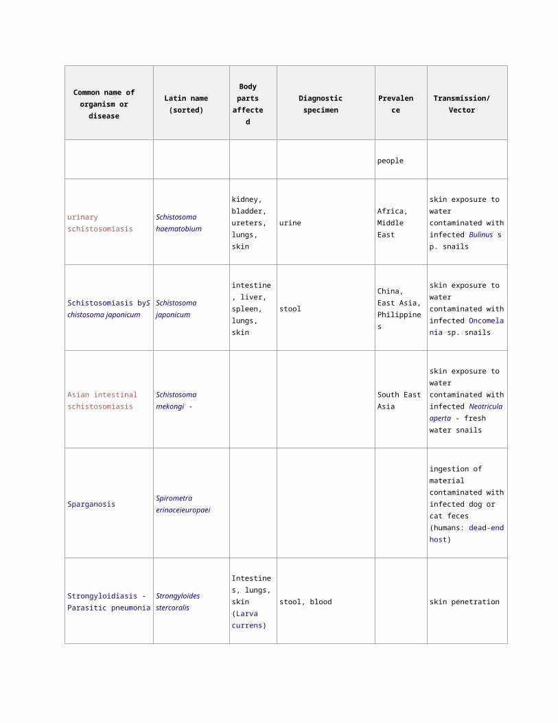
urinary schistosomiasisSchistosoma
haematobium
kidney,
bladder,
ureters, lungs,
skin
urineAfrica, Middle
East
skin exposure to water
contaminated with
infected Bulinus sp. snails
Schistosomiasis b
ySchistosoma japonicumSchistosoma japonicum
intestine,
liver, spleen,
lungs, skin
stool
China, East
Asia,
Philippines
skin exposure to water
contaminated with
infected Oncomelania sp.
snails
Asian intestinal
schistosomiasis
Schistosoma mekongi - South East Asia skin exposure to water
contaminated with
infected Neotricula
Page 9
Common name of
organism or diseaseLatin name (sorted)
Body
parts
affected
Diagnostic specimen Prevalence Transmission/Vector
aperta - fresh water
snails
SparganosisSpirometra
erinaceieuropaei
ingestion of material
contaminated with
infected dog or cat feces
(humans: dead-end host)
Strongyloidiasis - Parasitic
pneumonia
Strongyloides
stercoralis
Intestines,
lungs, skin
(Larva
currens)
stool, blood skin penetration
Beef tapeworm Taenia saginata Intestines stoolworldwide
distribution
ingestion of undercooked
beef
Pork tapeworm Taenia soliumingestion of undercooked
pork
ToxocariasisToxocara
canis, Toxocara cati
liver, brain,
eyes
(Toxocara
canis -Visceral
larva
migrans, Ocul
ar larva
migrans)
blood, ocular examinationworldwide
distribution
pica, unwashed food
contamined with
Toxocara eggs,
undercooked livers of
chicken
Trichinosis Trichinella
spiralis, Trichinella
britovi,Trichinella
nelsoni, Trichinella
nativa
muscle,
periorbital
region, small
intestine
blood more common
in developing
countries due
to improved
feeding
practices in
developed
ingestion of undercooked
pork
Page 10
Common name of
organism or diseaseLatin name (sorted)
Body
parts
affected
Diagnostic specimen Prevalence Transmission/Vector
countries.
Swimmer's itch
Trichobilharzia
regenti, Schistosomatid
ae
skin exposure to
contaminated water
(snails and vertebrates)
Whipworm
Trichuris
trichiura, Trichuris
vulpis
large
intestine,
anus
stool (eggs)common
worldwide
accidental ingestion of
eggs in dry goods such as
beans, rice, and various
grains or soil
contaminated with
human feces
ElephantiasisLymphatic
filariasisWuchereria bancrofti
lymphatic
system
thick blood smears stained
withhematoxylin.
Tropical and
subtropicalmosquito, bites at night
[edit]Other organisms
Common
name of
organism or
disease
Latin name (sorted)Body parts
affected
Diagnostic
specimenPrevalence Transmission/Vector
parasitic worm Archiacanthocephala
Halzoun
SyndromeLinguatula serrata nasopharynx
physical
examinationMid East
ingestion of raw or undercooked
lymph nodes (e.g., meat from
infected camels and buffalos)
MyiasisOestroidea, Calliphorida
e,Sarcophagidae
dead or living
tissue
Page 11
Common
name of
organism or
disease
Latin name (sorted)Body parts
affected
Diagnostic
specimenPrevalence Transmission/Vector
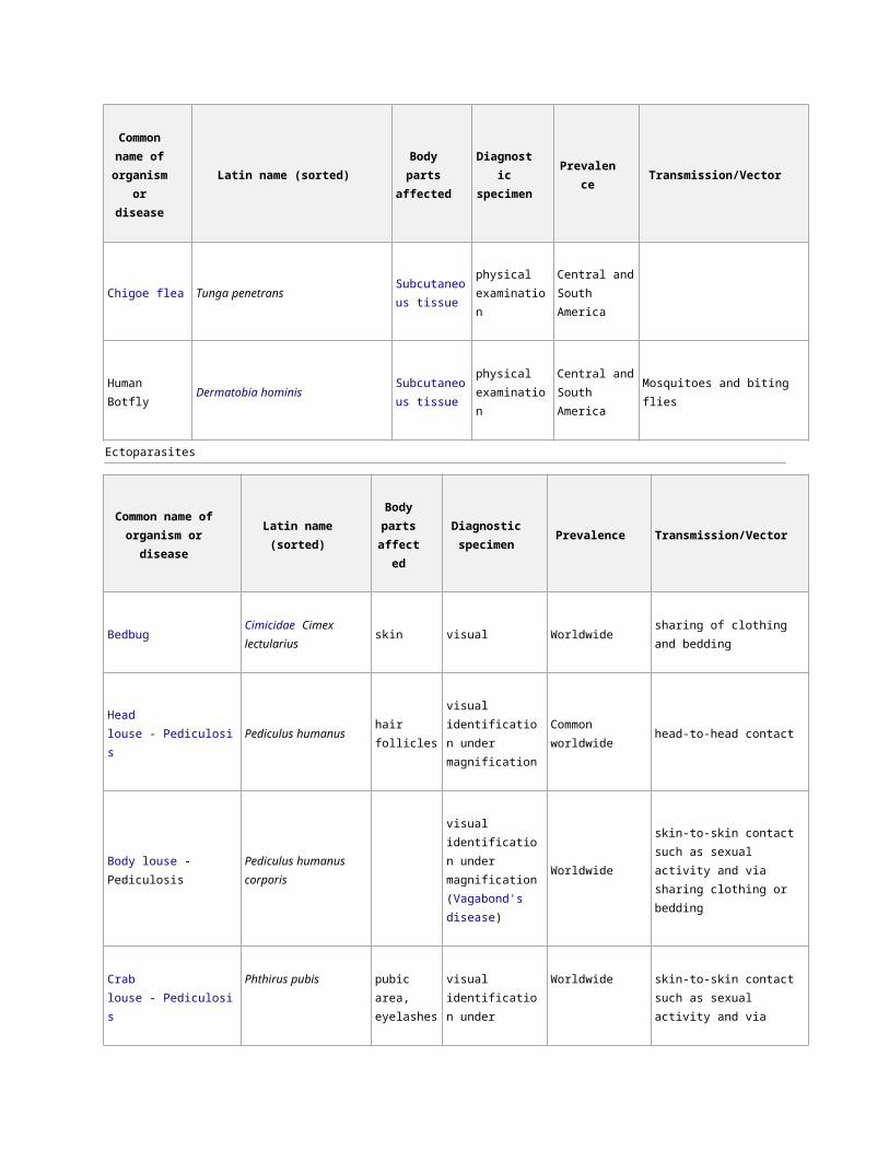
Chigoe flea Tunga penetransSubcutaneous
tissue
physical
examination
Central and
South America
Human Botfly Dermatobia hominisSubcutaneous
tissue
physical
examination
Central and
South AmericaMosquitoes and biting flies
Ectoparasites
Common name of
organism or diseaseLatin name (sorted)
Body
parts
affected
Diagnostic
specimenPrevalence Transmission/Vector
BedbugCimicidae Cimex
lectulariusskin visual Worldwide sharing of clothing and bedding
Head louse - Pediculosis Pediculus humanus hair folliclesvisual identification
under magnificationCommon worldwide head-to-head contact
Body louse - PediculosisPediculus humanus
corporis
visual identification
under magnification
(Vagabond's
disease)
Worldwide
skin-to-skin contact such as
sexual activity and via sharing
clothing or bedding
Crab louse - Pediculosis Phthirus pubispubic area,
eyelashes
visual identification
under magnificationWorldwide
skin-to-skin contact such as
sexual activity and via sharing
clothing or bedding
Demodex - DemodicosisDemodex
folliculorum/brevis/canis
eyebrow,
eyelashes
Microscopy of
eyelash or eyebrow
hair follicle
Pandemic,
worldwideprolonged skin-to-skin contact
Scabies Sarcoptes scabiei skin microscopy of Worldwide skin-to-skin contact such as
Page 12
Common name of
organism or diseaseLatin name (sorted)
Body
parts
affected
Diagnostic
specimenPrevalence Transmission/Vector
surface scrapingssexual activity and via sharing
clothing or bedding
Screwworm, Cochliomyia Cochliomyia hominivoraxskin and
woundsvisual
North America
(eradicated),
Central America,
North Africa
direct contact with fly
Flea, Siphonaptera Pulex irritans skinvisual identification
under magnificationWorldwide environment
Parasite life cycles can take a variety of forms, all involving the exploitation of one or more hosts. Those that must
infect more than one host species to complete their life cycles are said to have complex or indirect life cycles, while
those that infect a single species have direct life cycles.
If a parasite has to infect a given host in order to complete its life cycle, then it is said to be an obligate parasite of
that host; sometimes, infection is facultative—the parasite can survive and complete its life cycle without infecting
that particular host species. Parasites sometimes infect hosts in which they cannot complete their life cycles; these
are accidental hosts.
A host in which parasites have sexual reproduction is known as the definitive, final or primary host.
In intermediate hosts, parasites either do not reproduce or do so asexually, but the parasite always develops to a
new stage in this type of host. In some cases a parasite will infect a host, but not undergo any development, these
hosts are known as paratenic[1] or transport hosts. The paratenic host can be useful in making it more likely that the
parasite will be transmitted to the definitive host. For example the cat lungworm (Aelurostrongylus abstrusus) uses a
slug or snail as an intermediate host; the first stage larva enters the mollusk and develops to the third stage larva,
which is infectious to the definitive host—the cat. If a mouse eats the slug, the third stage larva will enter the mouse's
tissues, but will not undergo any development.
Parasitic Nutrition is a mode of heterotrophic nutrition where an organism (known as a parasite) lives on the body
surface or inside the body of another type of organism (known as a host). The parasite obtains nutrition directly from
the body of the host. Since these parasites derive their nourishment from their host, this symbiotic interaction is often
described as harmful to the host. Parasites are dependent on their host for survival, since the host provides nutrition
and protection. As a result of this dependence, parasites have considerable modifications to optimise parasitic
nutrition and therefore their survival.
Parasites are divided into two groups: endoparasites and ectoparasites. Endoparasites are parasites that live inside
the body of the host, whereas ectoparasites are parasites that live on the outer surface of the host and generally
attach themselves during feeding[1]. Due to the different strategies of endoparasites and ectoparasites they require
different adaptations in order to acquire nutrients from their host.
Page 13
Parasites require nutrients to carry out essential functions including reproduction and growth. Essentially, the
nutrients required from the host are carbohydrates, amino acids and lipids. Carbohydrates are utilised to generate
energy, whilst amino acids and fatty acids are involved in the synthesis of macromolecules and the production of
eggs[2]. Most parasites are heterotrophs, so they therefore are unable to synthesise their own 'food' i.e. organic
compounds and must acquire these from their host.
Endoparasitism
Endoparasites are parasites which live inside the body of the host. This group
includes helminths, trematodes and cestodes. Endoparasites are two groups of parasites: intercellular
and intracellular parasites. Intercellular parasites live in spaces within the host e.g. the alimentary canal, whereas
intracellular parasites live in cells within the host e.g. erythrocytes. Intracellular parasites typically rely on a third
organism, a vector, to transmit the parasite between hosts[3][1]. Rather than requiring adaptations to penetrate the
host, as ectoparasites do, endoparasites are in a nutrient-rich location so they instead have adaptations to maximise
nutrient absorption. Endoparasites have a readily available and renewable supply of nutrients inside the host, which
in some cases is pre-digested by the host, so mechanisms of nutrient absorption across their body surface is a
common feature[1]. As part of their life cycle strategy, endoparasites must also be able to transmit from within the host
body and survive the hostile environment within the host. Only by achieving this can they benefit from acquiring
nutrition in this way.
Microvilli structure which increases the surface area available for nutrient uptake in endoparasites
Endoparasites have various anatomical and biochemical adaptations, typically at the host-parasite interface, to
maximise nutrient acquisition. One such adaptation is the tegument, a metabolically active external cover which plays
an important role in the acquisition of nutrients from the host[2]. The parasite tegument is permeable to various organic
solutes and has transporters for the facilitated or active uptake of nutrients. Various studies have attempted to
characterise these transporters in a number of parasites e.g. the amino acid transporter molecules in protozoa [2][4][5].
Cestodes do not have a gut so the tegument is therefore critical for nutrient uptake. In cestodes the tegument is
highly efficient with spine-like microtriches, similar to microvilli, to increase the surface area available for nutrient
acquisition[6]. In many parasites the tegument structure has folds or microvilli to maximise the surface area available
for diffusion and uptake of nutrients. The tegument also commonly has additional organelles and features with
important functions in metabolism including the glycocalyx. The glycocalyx is a carbohydrate-rich layer which
enhances nutrient absorption and secretes enzymes to aid primary digestion[7].
Another important adaptation of endoparasites is the gut, which digests host macromolecules into soluble utilisable
products[2]. This feature is particularly important in endoparasites which are not located in the alimentary canal and
therefore the supply of nutrients is not pre-digested by the host. The gut lining typically has a layer of endodermal
cells which secrete proteolytic enzymes to aid digestion. Some endoparasites have both a gut and anus, some lack
an anus and some have neither i.e. those residing in the alimentary canal which instead diffuse pre-digested host
nutrients across their body surface[2].
Page 14
The relative importance of the tegument, gut and other adaptations involved in nutrient acquisition varies between
different endoparasitic species.
Tapeworms
Head of the pork tapeworm, Taenia solium ., with attachment structures to attach to the wall of the small intestine
Tapeworms are endoparasites which have numerous adaptations to enhance parasitic nutrition. Tapeworms live in
the small intestine of humans, providing an ideal location to access a readily available, rich source of pre-digested
nutrients[8]. Since nutrients in the small intestine are plentiful and pre-digested by the host, tapeworms do not require
a gut and instead have adaptations to maximise nutrient absorption. Tapeworms have a tegument which allows
nutrients to be absorbed directly from the host small intestine by diffusion. They also have anatomical adaptations in
the form of a scolex with hookers and suckers to allow the parasite to attach to the host small intestine wall,
preventing the tapeworm from being egested following peristalsis [2] . Tapeworms have a flattened body
with microtriches to maximise the surface area available for nutrient absorption and they additionally have various
transporter molecules. Tapeworms have to compete with the host epithelium for nutrients, so it is essential that they
compete more efficiently for nutrients. They also secrete enzymes to enhance host digestive enzymes e.g. pancreatic
α-amylase[2].
Schistosomes
Schistosomes, another type of endoparasite, also live inside the body of the host but instead these parasites acquire
their nutrients from host blood. Schistosomes are in direct contact with host blood, a rich source of amino acids, and
they therefore do not require penetrative structures to reach host nutrients. Schistosomes take blood up through the
negative pressure created by muscle contractions of their sucker and oesophagus[2]. They obtain amino acids from
host blood through a mechanism of haemoglobin degradation, which remains unresolved but is suggested to involve
a series of proteases. Mechanisms to overcome blood clotting are also employed[9][10]. Various studies have
attempted to characterise the components of the schistosome tegument, including transporter molecules suggested
to be involved in nutrient uptake. Such transporter molecules include schistosome alkaline phosphatase (SmAP) and
cathepsin B, which are suggested to be important in nutrient acquisition[11][12][13].
Malaria
Malaria, caused by the apicomplexan parasite Plasmodium falciparum, is an intracellular endoparasite. This parasite
relies on a third organism, in the form of an Anopheles mosquito vector. The host blood provides an ideal rich source
of glucose and amino acids to the parasite, particularly during blood stage infection
where Plasmodium infects erythrocytes [14] . In order to acquire essential nutrients Plasmodiumhas to compete with
both the vertebrate and insect host and therefore must be highly efficient, regulating uptake according to nutrient
availability[14]. Plasmodium, along with many other endoparasitic parasites, have numerous channels in their
parasitophorous vacuole membrane rendering it permeable to organic solutes to allow the uptake of necessary
nutrients. The Plasmodium falciparum hexose transporter (PfHT) is such a transporter, which is critical for the uptake
of glucose and fructose and therefore survival of the parasite[15]. These organic molecules have to cross three
membranes altogether; the plasma membrane of the erythrocyte, the parasitophorous vacuole membrane and
the Plasmodium plasma membrane, facilitated by transporters such as PfHT[16].
Ectoparasitism
Ectoparasites live on the outer surface of the host. This group includes ticks, leeches, mites and the tsetse fly.
Ectoparasites do not have a readily available source of nutrients available on the outer surface of the host so they
therefore require adaptations which enable them to gain access to host nutrients. This requires penetrative features
which can insert into the host, as well as the ability to secrete digestive enzymes and the presence of a gut to digest
Page 15
host-derived nutrients[1]. Ectoparasites also have a variety of parasite transporters and permeases to enable them to
acquire nutrition from their host, across numerous membranes. Many ectoparasites are known to be vectors of
pathogens, so they therefore transmit these pathogens during nutrient acquisition[17].
Tsetse flies, the insect vector of Trypanosoma brucei, the causative agent of African trypanosomiasis are an example
of an ectoparasite. These insects have specialised structures, known as a proboscis to pierce and draw nutrients
from their host. These then employ transport proteins to transport the essential nutrients across membranes,
ultimately from the host to the tsetse fly gut for digestion. Various permeases have been characterised including
those that import hexoses, carboxylates, and amino acids[18][19].
Another endoparasite is scabies, caused by Sarcoptes scabiei. Scabies, transmitted by female mites, depends on
nutrition from its host for survival. This endoparasite obtains nutrients by burrowing into the skin of the host. Studies
have also identified the presence of scabies mite inactivated protease paralogues (SMIPPs), which are believed to
compete with host proteases[20].
Effects on the host
As mentioned previously, parasitic nutrition is beneficial to the parasite but typically detrimental to the host since it
deprives the host of nutrients. This mode of nutrition has numerous side effects on the host including weight loss,
anaemia, obstruction of the intestine, damage to the host intestinal wall and in some cases transmission of serious
pathogens e.g. the ectoparasitic tsetse fly which transmits African trypanosomiasis[1][14][2][21].
Applications
Nutrient acquisition is an important component of parasite pathogenesis since it is critical to parasite survival [14].
Understanding the mechanisms of parasite nutrient acquisition therefore identifies novel targets which we can exploit
as a form of parasite control e.g. through knock-out or inhibition of crucial transporters or destruction of penetrative
anatomical features. Several studies have looked into nutrient acquisition as a method of parasite control, including
the development of vaccines against helminth parasites by targeting digestive proteases[22].
Parasitic nutrition in plants
Plants are typically autotrophic organisms meaning that they synthesise their own 'food' from inorganic compounds
by photosynthesis. Some plants however are unable to synthesise their own 'food' by photosynthesis and therefore
acquire nutrients by parasitic nutrition from other living plants. Plants which acquire their nutrients in this way are
known as parasitic plants [23] . These plants have modified root structures known as haustorium, which the parasitic
plants use to penetrate the vascular bundle of host plants and essentially 'steal' nutrients from host plants [24][1].
Parasitic plants include Dodder, Rafflesia and Broomrape.