HISTOLOGY AND HISTOPATHOLOGY (non-edited manuscript) ONLINE FIRST This is a provisional PDF only. Copyedited and fully formatted versión will be made available at final publication This article has been peer reviewed and published immdediately upon acceptance. Articles in “Histology and Histopathology” are listed in Pubmed. Pre-print author´s version ISSN: 0213-3911 e-ISSN: 1699-5848 Submit your article to this Journal (http://www.hh.um.es/Instructions.htm) Overexpression of EIF5A2 is associated with poor survival and aggressive tumor biology in gallbladder cancer Authors: Xin Zheng, Lei Gao, Bo-tao Wang, Ping Shen, Xiang-fei Yuan, Lan-Qiu Zhang, Lei Yang, Da-Peng Zhang, Qi Zhang and Xi-Mo Wang DOI: 10.14670/HH-18-186 Article type: ORIGINAL ARTICLE Accepted: 2019-11-20 Epub ahead of print: 2019-11-20
Overexpression of EIF5A2 is associated with poor survival and aggressive tumor biology in gallbladder cancer
Authors: Xin Zheng, Lei Gao, Bo-tao Wang, Ping Shen, Xiang-fei Yuan, Lan-Qiu Zhang, Lei Yang, Da-Peng Zhang, Qi Zhang and Xi-Mo Wang DOI:10.14670/HH-18-186Articletype:ORIGINALARTICLEAccepted:2019-11-20Epubaheadofprint:2019-11-20
HISTOLO
GY AND H
ISTOPATHOLO
GY
(non-e
dited
man
uscri
pt)
1
Overexpression of EIF5A2 is associated with poor survival and
aggressive tumor biology in gallbladder cancer
Xin Zheng a, b, #, Lei Gao a, c, #, Bo-tao Wang a, c, #, Ping Shen a, c, Xiang-fei
Yuan a, Lan-Qiu Zhang a, Lei Yang a, Da-Peng Zhang a, Qi Zhang a, *,
Xi-Mo Wang a, c *
Running title: The biological role of EIF5A2 in gallbladder carcinoma
a. Tianjin key Laboratory of Acute abdomen disease associated organ
injury and ITCWM Repair, Institute of Acute Abdominal Diseases,
Tianjin Nankai Hospital, Tianjin, 300100, China.
b. Department of General Surgery, The Second Affiliated Hospital of
Chengdu Medical College, China National Nuclear Corporation 416
Hospital, Chengdu,Sichuan, 610051, China.
c. Graduate School, Tianjin Medical University, Tianjin, 300070, China.
# These authors contributed equally to this work.
*Corresponding author.
*Correspondence to: Xi-Mo Wang and Qi Zhang, Tianjin key Laboratory of Acute abdomen disease associated organ injury and ITCWM Repair, Institute of Acute Abdominal Diseases, Tianjin Nankai Hospital, 6 Changjiang Road, Tianjin, 300100, China; Tel: +86 22 2743 5365; Email: [email protected] (X. Wang), [email protected] (Q. Zhang).
HISTOLO
GY AND H
ISTOPATHOLO
GY
(non-e
dited
man
uscri
pt)
2
Abstract
Gallbladder cancer (GBC) is a malignant tumor of the biliary tract. The main problem affecting the treatment of gallbladder cancer is late diagnosis and poor prognosis. EIF5A2 is one of two isoforms of the EIF5A family and is reported to be a new oncogenic protein in many human cancers. In this study, our results showed for the first time that EIF5A2 was overexpressed in GBC samples compared with non-tumor tissue. Overexpression of EIF5A2 was associated with lymph node metastasis, tumor differentiation, UICC (Union for International Cancer Control) staging, histological type, metastasis, and tumor size. Overexpression of EIF5A2 in gallbladder carcinoma tissues is also associated with poor prognosis in patients. The interference of EIF5A2 significantly inhibited the proliferation, cell cycle, migration and colony formation of GBC-SD cells in vitro. Our results suggest that EIF5A2 is a target oncogene and may be an important prognostic biomarker in the pathogenesis of gallbladder cancer.
The incidence of gallbladder cancer varies widely geographically, ethnically, and culturally, suggesting that genetic and environmental factors play a key role in the development of gallbladder cancer (Misra et al., 2003; Andia et al., 2008). Although the incidence of gallbladder cancer is relatively low, most patients are diagnosed at an advanced stage (Li, Zhang, et al., 2014). The lack of gallbladder serosa in the vicinity of the liver leads to liver invasion and metastasis, which is one of the main reasons for its poor prognosis (Hundal and Shaffer, 2014). There are currently no reliable tumor markers for the diagnosis of gallbladder cancer. Currently only two markers, carcinoembryonic antigen (CEA) and CA19-9, are most frequently elevated in advanced stages, but have lower specificity. Therefore, in most cases, they are not used for independent diagnosis of GBC (Srivastava et al., 2013). However, other tumor markers such as CA242, CA125, cancer antigen (CA), CEA (carcinoembryonic antigen), CA199, etc. have also been studied in the diagnosis of gallbladder cancer, but the results are highly inconsistent (Zur et al., 2012; He et al., 2013; Zhang et al., 2013). In addition, previous reports have shown that biomarkers such as CA242, Mac-2BP, CA15-3, RCAS1, and Fragments of cytokeratin19 (CYFRA211) are frequently found in the blood of cancer patients, showing a correlation with GBC, but with different sensitivities and specificities (Koopmann et al., 2004; Srivastava et al., 2013; Huang et al., 2015). Therefore, an understanding of the molecular mechanisms of gallbladder cancer metastasis and recurrence is essential for the development of effective adjuvant therapy.
Eukaryotic translation initiation factor 5A2 (EIF5A2) is a putative oncogene isolated from 3q26.2 using the chromosomal micro-separation-hybridization selection technique (Guan et al., 2001). EIF5A2 is rare in most normal tissues, but is evident in many malignancies, and the association of EIF5A2 with cancer has received considerable attention (Caraglia et al., 2013). The EIF5A2 gene is frequently amplified in different human malignancies, including pancreatic cancer (Griffin et al., 2007), gastric cancer (Guan et al., 2000; Takada et al., 2005), colon cancer (He et al., 2003), liver cancer (Tang et al., 2010), and breast cancer (Forozan et al., 2000). Among the several cancers just mentioned, elevated EIF5A2 is often associated with more severe disease states, increased likelihood of postoperative recurrence and metastasis, and decreased survival.
Here, our results indicate that EIF5A2 is up-regulated in many primary GBC tumors and is strongly associated with poor patient
HISTOLO
GY AND H
ISTOPATHOLO
GY
(non-e
dited
man
uscri
pt)
4
prognosis. The interference of EIF5A2 inhibits proliferation, tumor growth and migration of gallbladder cancer cells. Therefore, we believe that EIF5A2 is a regulator of GBC development, which may be a novel biomarker and potential therapeutic target for GBC patients.
Materials and methods
Patients and clinicopathological data
A total of 40 paraffin-embedded specimens of gallbladder cancer were collected from January 2014 to May 2017 at the Nankai Hospital in Tianjin, China. Another 40 cases of gallbladder cancer tissue chips were purchased from Shanghai OUTDO Biotechnology Company Ltd. (Shanghai, China). All the above gallbladder cancer tissue samples were confirmed by paraformaldehyde, paraffin embedding, sectioning, and HE staining. No patients received radiotherapy or chemotherapy related treatment before surgery. All the 80 patients with gallbladder cancer collected had follow-up information. This study was approved by the Clinical Research Ethics Committee of Tianjin Nankai Hospital.
Immunohistochemical analysis of gallbladder cancer tissues
Immunohistochemistry (IHC) uses the manufacturer's instructions for the SP-9001 system (ZSGB, China). For antigen retrieval,the antigen was repaired with sodium citrate antigen repair solution at 95 °C for 10 minutes, and then slowly cooled. The sections were incubated with rabbit monoclonal anti-EIF5A2 (Abcam, UK) diluted 1:200 and placed in a refrigerator at 4 °C overnight. Finally, an appropriate amount of freshly prepared 3, 5-diaminobenzidine (DAB) chromogenic solution was added and the staining was observed under the microscope (Leica, Germany). The staining evaluation of EIF5A2 adopted a semi-quantitative scoring standard (Ma et al., 2010), which includes staining intensity and percentage of positive cells. The scoring criteria are as follows: when the positive cells accounted for 0%-25%, it was 1 point, 26%-50% was 2 points, 51%-75% was 3 points, and >76% was 4 points; when the staining intensity is negative, it is 0, weak is 1 point, moderate is 2 points, and strong is 3 points. The multiplication of the two scores is the final score. The staining index ≥ 4 is divided into positive, and < 4 is divided into negative. Pathological section staining was independently assessed by two pathologists.
HISTOLO
GY AND H
ISTOPATHOLO
GY
(non-e
dited
man
uscri
pt)
5
Cell culture and EIF5A2 interference
GBC-SD cell lines were purchased from the Shanghai Institute of Cell Bank, Chinese Academy of Science (Shanghai, China). GBC-SD cell lines were cultured in RPMI 1640 medium (Gibco) containing 10% fetal bovine serum (FBS; Gibco). The GBC-SD cell line was incubated in a humidified incubator containing 5% carbon dioxide at 37 °C.
The double-stranded small interfering RNA (siRNA) were purchased from GenePharma company (Shanghai, China). GBS-SD cells were transfected with siRNA with siRNA Transfection Reagent (Polyplus transfection, France), according to the manufacturer’s instructions. The EIF5A2-specific siRNA target sequences were as follows: control-siRNA, 5’-UUCUCCGAACGUGUCACGUTT-3’, 5’-ACGUGACACGUUCGGAGAATT-3’; EIF5A2-siRNA-1, 5’-GUGGAGAUGUCAACUUCCATT-3’, 5’-UGGAAGUUGACAUCUCCACTT-3’; EIF5A2-siRNA-2, 5’-GCAUUCAAGAUGGUUACCUTT-3’, 5’-AGGUAACCAUCUUGAAUGCTT-3’; EIF5A2-siRNA-3, 5’-GGAUCUUAAACUGCCAGAATT-3’, 5’-UUCUGGCAGUUUAAGAUCCTT-3’. After 48h of si-RNA transfection, the gene silencing efficiency was detected by Western blot. Each experiment was repeated three times independently.
Western blot analysis
GBC-SD cells were lysed with cold RIPA buffer (Millipore, USA) supplemented with phosphatase, protease inhibitors, phenylmethanesulfonyl fluoride and aprotinin (Sigma, USA) after transfection of siRNA for 48h. Protein was extracted according to instructions and quantified by BCA protein concentration assay kit (Solarbio, China). 40µg of total protein was electrophoresed through 10% separation gel, then transferred onto polyvinylidene fluoride membrane (Millipore, USA) by semi-dry electrophoretic transfer cell (Bio-rad, USA). The membranes were blocked and probed with the primary antibodies against β-actin and EIF5A2 (1:1000 dilution) (Cell Signaling Technology, USA) overnight at 4℃. The membranes were incubated with horseradish peroxidase-conjugated secondary antibodies after three times of washing. The immunoreactivity was detected by chemiluminescent HRP substrate kit (Millipore, USA).
HISTOLO
GY AND H
ISTOPATHOLO
GY
(non-e
dited
man
uscri
pt)
6
Quantitative real-time PCR
Total RNA was extracted from GBC-SD cells transfected with siRNA by TRIzol reagent (Invitrogen, USA). 2µg of total RNA was reverse transcribed into cDNA using RevertAid First Strand cDNA Synthesis Kit (Thermo Scientific, USA). All primers were synthesized by Invitrogen (USA), and the primer sequences are as follows: β-actin (forward primer: 5’-ACCGGGCATAGTGGTTGGA-3’; reverse primer: 5’-ATGGTACACGGTTCTCAACATC -3’), EIF5A2 (forward primer: 5’- TGTCCTTCTACTCACAACATGGA -3’; reverse primer: 5’- CTCACGAACTTCACCAGTTTCT -3’). Real-time PCR was performed with GoTaq® qPCR Master Mix (Promega, USA) on ABI 7500 FAST Real-Time PCR System according to the manual. The relative expression of mRNA was calculated by 2-△△Ct.
Two dimensional colony formation assay and cell cycle
Plate clone formation experiment was performed by plating approximately 200-500 cells in 6-well culture dishes for 10-14 days. The cells in the 6-well plate were fixed with 4% paraformaldehyde and then stained with a 0.1% crystal violet solution. The formed cell colonies (≥ 50 cells) were counted under a microscope.
To examine cell cycle, after transfecting the cells with si-RNA for 48 h, the cells were fixed with a 90% ethanol solution overnight. The cell cycle was detected using a flow cytometer (ACEA Biosciences, Inc.), and the cells were stained using propidium iodine dye (Biolegend, San Diego, USA) according to the manufacturer's protocol.
Cell proliferation assay
Carboxyfluorescein succinimidyl ester (CFSE) is applicable to track cell division. Such dye-labeled cells can distribute the fluorescence equally into the daughter cells with every cell division. Fluorescence intensity of each generation of cells can be detected by flow cytometry. Subsequently, ModFit LT (Verity Software House, USA) software was used to determine the proportion of cells in each generation according to the fluorescence intensity of the initial CFSE-labeled cells, and calculate the proliferation index (PI).
GBC-SD cells were cultured to the logarithmic growth phase and starved overnight with serum-free medium. Next, the cells were collected
HISTOLO
GY AND H
ISTOPATHOLO
GY
(non-e
dited
man
uscri
pt)
7
and incubated with CFSE at the final concentration of 5µM for 10min. After termination of staining, the cells were centrifuged with medium containing 10% FBS. CFSE-labeled cells were then seeded into 6-well plates, while incubated with control-siRNA or EIF5A2-siRNA-3 for 48h. Finally, cell proliferation of two groups was detected by flow cytometry.
The MMT assay measures cell proliferation. Cell viability was detected at 24h, 48h, 72h and 96h using the MTT solution (Sigma, USA) according to the manufacturer’s protocol. The medium was detected using a spectrophotometer with a wavelength of 570 nm.
Wound healing and cell migration assay
Wound healing was assessed by measuring the rate at which cells move in the scratch area. The scratch areas were created by a 10-µl pipette tube. The area of wound was observed after 24h and 48h and pictures were taken under the microscope. Cell migration assay was verified using Transwell chamber (Corning Incorporated, USA). 5×10⁴ cells suspended in 100µl of RPMI 1640 medium with 10% FBS were added to the upper chamber. Then 600ul of RPMI 1640 medium with 20% FBS were added to the lower chamber. After 16h of culture, the cells were fixed with 4% paraformaldehyde and stained with crystal violet. The upper chamber cells were wiped with a cotton swab and three random fields were selected to capture the cells that have migrated through the membrane. The results were analyzed using the ImageJ software(NIH).
Statistical analysis
All experimental data were expressed as mean ± S.E (standard deviation). Statistical analysis was performed using GraphPad Prism Version 5.0 (GraphPad Inc., San Diego CA) and SPSS statistical analysis software (SPSS Inc., Chicago, IL, USA). The chi-square test was used to evaluate the correlation between EIF5A2 protein expression and clinicopathological features in GBC patients. Survival statistics were performed using the Kaplan-Meier method and survival curves were plotted, and the difference in survival rates among the subgroups was compared using the Log-rank test. Multivariate analysis of gallbladder cancer case data was performed using Cox regression analysis model. All data were derived from at least three independent experiments and a P value of less than 0.05 was considered statistically significant.
HISTOLO
GY AND H
ISTOPATHOLO
GY
(non-e
dited
man
uscri
pt)
8
Results
EIF5A2 is overexpressed in GBC tissues
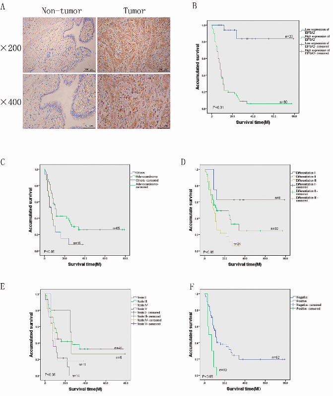
The expression of EIF5A2 in GBC tissues was investigated by IHC staining. Compared to the adjacent non-tumor tissues, the EIF5A2 was up regulated in tumors (Fig.1A).
The expression of EIF5A2 is associated with the clinicopathological variables of GBC patients.
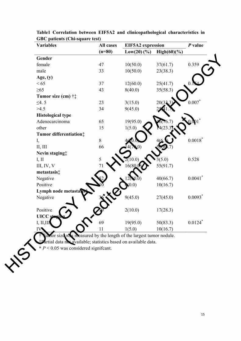
According to the statistical result from the 80 investigated patients, it was revealed that the expression of EIF5A2 was high in 75% (60/80) of the tumor tissues, and the level of EIF5A2 was associated with the degree of malignancy (Table 1). As shown in Table 1, high expression of EIF5A2 in GBC was significantly associated with tumor size (P=0.007), histological type (P=0.001), tumor differentiation (P=0.0018), metastasis (P=0.0041), lymph node metastasis (P=0.0093), and UICC staging (P=0.0124).
The expression of EIF5A2 protein is associated with survival and prognosis in GBC patients.
The Kaplan-Meier survival curves suggested that the accumulated survival rate of GBC patients with high level of EIF5A2 (n = 60, with a mean survival of 13.6 months) was poorer than those with low level (n = 20 ,with a mean survival of 69.3 months, P<0.05, Table 2, Fig. 1B).
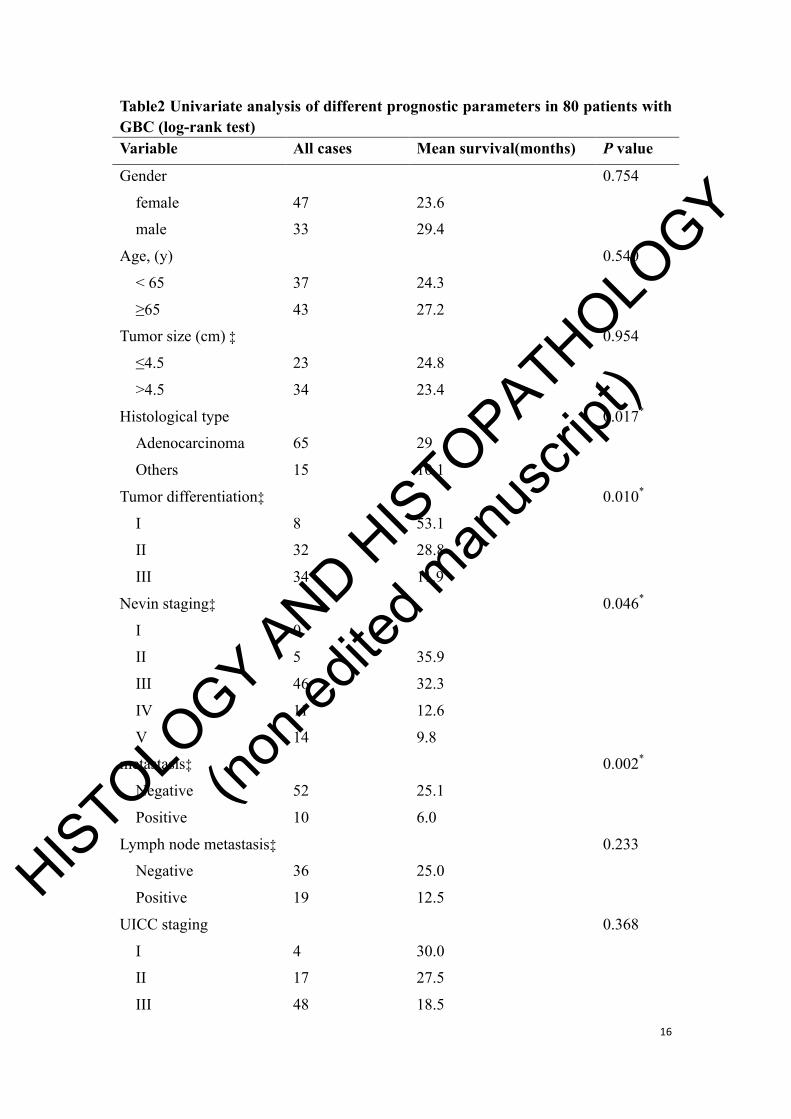
Univariate and multivariate analyses were performed to investigate the relationship between survival rates and clinicopathological prognostic features of GBC patients. Survival and prognosis was significantly associated with histological type (P<0.05, Table 2, Fig. 1C), tumor differentiation (P<0.05, Table 2, Fig. 1D), nevin staging (P<0.05, Table 2, Fig. 1E), and metastasis (P<0.05, Table 2, Fig. 1F). Subsequently, multivariate statistical analysis was performed using the Cox regression model to examine independent risk factors for each clinicopathological feature. The result demonstrated that a high level of EIF5A2 was an independent prognostic factor with low overall survival (P=0.036, Table 3) in GBC patients, like tumor differentiation and metastasis.
HISTOLO
GY AND H
ISTOPATHOLO
GY
(non-e
dited
man
uscri
pt)
9
Downregulation of EIF5A2 inhibits the growth of GBC cell lines.
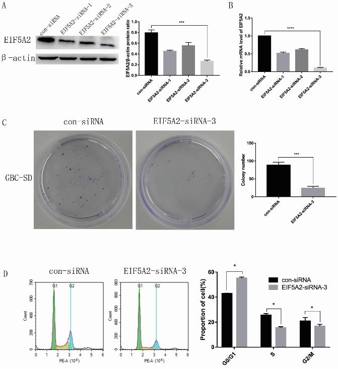
EIF5A2 was highly expressed and downregulated by siRNA in GBC cell line. Western blot and qPCR were used to detect the interference efficiencies of three siRNAs targeting EIF5A2. As shown in Figs 2A&2B, EIF5A2-siRNA-3 downregulated the expression of EIF5A2 effectively and was chosen for the following experiments. Subsequently, clone formation experiment was performed to evaluate the growth of GBC cell line, after the cells were transfected by EIF5A2-siRNA-3 or control-siRNA. The result demonstrated that the colony formation capability of EIF5A2 downregulated cells was attenuated compared to the control cells (Fig. 2C). To explore the mechanism of the above phenomenon, flow cytometry was used to analyze the change in the cell cycle after EIF5A2 was downregulated. According to the result, the percentage of S and G2/M phases was significantly reduced in EIF5A2 downregulated cells, but the percentage of the G1 phase was increased in them (Fig. 2D). These results indicated that the inhibition of cell growth resulted from G1 phase arrest induced by downregulating EIF5A2.
Downregulating EIF5A2 surpresses cell proliferation and cell migration
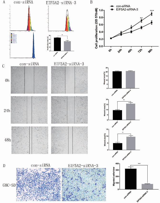
We further examined the effect of EIF5A2-siRNA-3 on GBC-SD cell lines by flow cytometry. After CFSE-labeled cells were treated with control-siRNA or EIF5A2-siRNA-3, the EIF5A2-siRNA-3 group cell proliferation and the proliferation index was significantly decreased compared with the control-siRNA group (Fig. 3A). As shown in Fig. 3B, the MTT assay revealed that the viability of GBC-SD cells was suppressed in the EIF5A2-siRNA-3 group compared with the control-siRNA group. In the wound healing assay, EIF5A2-siRNA-3 group significantly suppressed the wound healing at 24h and 48h compared with control-siRNA group (Fig. 3C). In addition, the transwell migration assays indicated that interference EIF5A2 markedly suppressed cell migration ability in the GBC-SD cell lines (Fig. 3D). These data were consistent with our previous conclusion that GBC patients with high EIF5A2 expression is significantly associated with metastasis.
HISTOLO
GY AND H
ISTOPATHOLO
GY
(non-e
dited
man
uscri
pt)
10
Discussion
Despite improvements in the diagnostic and therapeutic strategies of GBC, the prognosis of GBC patients is still unsatisfactory. EIF5A2 is considered a candidate oncogene, but there is currently no information on the role of EIF5A2 in gallbladder cancer. In the present study, we for the first time found that EIF5A2 was upregulated in GBC tissues compared with the non-tumor counterparts. Overexpression of EIF5A2 in GBC was signifcantly associated with tumor size, histological type, tumor differentiation, metastasis, lymph node metastasis, and UICC staging. Furthermore, the Kaplan-Meier survival curves suggested that the accumulated survival rate of GBC patients with high EIF5A2 expression was poorer than those with low EIF5A2 expression. In vitro experiments showed that the knockdown of EIF5A2 remarkably inhibited the proliferation, tumor growth and migration capacity of GBC-SD cells.
In cancers that have been studied, higher expression of EIF5A2 is associated with poorer clinical features and outcomes. Many malignant tumors, including esophageal squamous cell carcinoma, hepatocellular carcinoma, ovarian cancer, colorectal carcinoma, gastric cancer, lung cancer, and bladder cancer, have been reported to be associated with decreased survival (Chen et al., 2009; Yang et al., 2009; He et al., 2011; Zhu et al., 2012; Li, Fu, et al., 2014; Wang et al., 2014; Meng et al., 2015). These data indicate that knockdown of EIF5A2 can inhibit tumor growth and metastasis in a variety of cancers. Consistent with these findings, our results demonstrate EIF5A2 knockdown significantly inhibits GBC-SD cell proliferation and migration in vitro. A previous study reached a conclusion that EIF5A2 is one of the three genes that predict lymph node metastasis in gastric cancer (Marchet et al., 2007). Our clinicopathological data also showed that high EIF5A2 expression is significantly associated with metastasis and lymph node metastasis. The relationship between EIF5A2 and metastasis is also seen in esophageal cancer (Li, Fu, et al., 2014) and colorectal cancer (Xie et al., 2008). These data support the use of EIF5A2 as a prognostic indicator for various malignant diseases.
Compared with EIF5A1, another member of the EIF5A gene familywhich is widely expressed, EIF5A2 is expressed only in a few specific parts such as testis and adult brain. And the eIF5A2 isoform is not necessary for embryonic development and organism viability in adults (Meng et al., 2019). However, it is up-regulated in multiple malignant tumor tissues including GBC and is associated with poor prognosis. Therefore, EIF5A2 may be a novel biomarker and potential therapeutic
HISTOLO
GY AND H
ISTOPATHOLO
GY
(non-e
dited
man
uscri
pt)
11
target for GBC patients. But it is necessary to elucidate the mechanism regulating EIF5A2 expression and its downstream pathways.
Acknowledgments
The authors thank the National Natural Science Foundation of China (No.81602496), the National Natural Science Foundation of Tianjin (No.18JCQNJC11100; No.18JCQNJC13400) and Tianjin Municipal Science and Technology Commission (No.17ZXMFSY00220) for their great supports in financing this research.
Conflict of Interest
The authors declare no conflict of interest.
Figure legends Fig. 1 Immunohistochemical analysis of EIF5A2 in GBC tissues. (A) EIF5A2 expression level in the gallbladder tumor and adjacent non-tumor tissues. (B) Kaplan–Meier overall survival curve of GBC patients based on EIF5A2 expression (log-rank test). Overall survival of all patients with GBC: low expression, n=20 cases, mean time=69.3 months; high expression, n=60 cases, mean time=13.6 months. (C, D, E, and F) Survival curve for 80 GBC patients according to histological type, tumor differentiation, nevin staging and metastasis. Fig. 2 Inhibition of EIF5A2 expression by RNA interference and EIF5A2 promotes GBC cell growth. (A and B) The efficiency of EIF5A2 interference was evaluated by western blot and qPCR after GBC-SD cells were transfected with EIF5A2-siRNA; EIF5A2-siRNA-3 is considered to have the best down-regulation effect (***P < 0.001, ****P < 0.0001). All subsequent experiments were performed using EIF5A2-siRNA-3 in combination with control-siRNA. (C) Colony formation was inhibited in EIF5A2-siRNA-3 cell lines (***P < 0.001). (D) Flow cytometry was used to analyze the cell cycle of EIF5A2-siRNA-3 and control-siRNA cell lines (*P < 0.05). These data were reported as mean ± SD and three separate experiments were performed.
HISTOLO
GY AND H
ISTOPATHOLO
GY
(non-e
dited
man
uscri
pt)
12
Fig. 3 Silencing of EIF5A2 decreases cell proliferation and cell migration. (A) Effects of EIF5A2-siRNA-3 and control-siRNA cell lines on proliferation index after 48h transfection si-RNA, different colored peaks represent different generations (**P < 0.01). (B) The proliferative capacity of EIF5A2-siRNA-3 and control-siRNA cell lines were compared using MTT assays (*P < 0.05, **P < 0.01, ***P < 0.001). (C) The most representative photographs of wound healing were observed under the microscope at 0h, 24h and 48h (**P < 0.01). Relative wound area was shown in the bar graph. (D) Cell migration rates of EIF5A2-siRNA-3 and control-siRNA cell lines were compared using transwell migration assays (***P < 0.001). The graph shows the migration rates of EIF5A2-siRNA-3 and control-siRNA cell lines after 16h of culture on transwell. These data are reported as mean ± SD and three separate experiments were performed.
REFERENCS Andia M.E., Hsing A.W., Andreotti G. and Ferreccio C. (2008). Geographic variation of gallbladder
cancer mortality and risk factors in chile: A population-based ecologic study. Int. J. Cancer. 123, 1411-1416.
Caraglia M., Park M.H., Wolff E.C., Marra M. and Abbruzzese A. (2013). Eif5a isoforms and cancer: Two brothers for two functions? Amino Acids. 44, 103-109.
Chen W., Luo J.H., Hua W.F., Zhou F.J., Lin M.C., Kung H.F., Zeng Y.X., Guan X.Y. and Xie D. (2009). Overexpression of eif-5a2 is an independent predictor of outcome in patients of urothelial carcinoma of the bladder treated with radical cystectomy. Cancer Epidemiol. Biomarkers Prev. 18, 400-408.
Forozan F., Mahlamaki E.H., Monni O., Chen Y., Veldman R., Jiang Y., Gooden G.C., Ethier S.P., Kallioniemi A. and Kallioniemi O.P. (2000). Comparative genomic hybridization analysis of 38 breast cancer cell lines: A basis for interpreting complementary DNA microarray data. Cancer Res. 60, 4519-4525.
Griffin C.A., Morsberger L., Hawkins A.L., Haddadin M., Patel A., Ried T., Schrock E., Perlman E.J. and Jaffee E. (2007). Molecular cytogenetic characterization of pancreas cancer cell lines reveals high complexity chromosomal alterations. Cytogenet Genome Res. 118, 148-156.
Guan X.Y., Sham J.S., Tang T.C., Fang Y., Huo K.K. and Yang J.M. (2001). Isolation of a novel candidate oncogene within a frequently amplified region at 3q26 in ovarian cancer. Cancer Res. 61, 3806-3809.
Guan X.Y., Fu S.B., Xia J.C., Fang Y., Sham J.S., Du B.D., Zhou H., Lu S., Wang B.Q., Lin Y.Z., Liang Q., Li X.M., Du B., Ning X.M., Du J.R., Li P. and Trent J.M. (2000). Recurrent chromosome changes in 62 primary gastric carcinomas detected by comparative genomic hybridization. Cancer Genet. Cytogenet. 123, 27-34.
He C.Z., Zhang K.H., Li Q., Liu X.H., Hong Y. and Lv N.H. (2013). Combined use of afp, cea, ca125 and cal9-9 improves the sensitivity for the diagnosis of gastric cancer. BMC Gastroenterol. 13, 87.
He L.R., Zhao H.Y., Li B.K., Liu Y.H., Liu M.Z., Guan X.Y., Bian X.W., Zeng Y.X. and Xie D. (2011).
HISTOLO
GY AND H
ISTOPATHOLO
GY
(non-e
dited
man
uscri
pt)
13
Overexpression of eif5a-2 is an adverse prognostic marker of survival in stage i non-small cell lung cancer patients. Int. J. Cancer. 129, 143-150.
He Q.J., Zeng W.F., Sham J.S., Xie D., Yang X.W., Lin H.L., Zhan W.H., Lin F., Zeng S.D., Nie D., Ma L.F., Li C.J., Lu S. and Guan X.Y. (2003). Recurrent genetic alterations in 26 colorectal carcinomas and 21 adenomas from chinese patients. Cancer Genet. Cytogenet. 144, 112-118.
Huang L., Chen W., Liang P., Hu W., Zhang K., Shen S., Chen J., Zhang Z., Chen B., Han Y., Meng F., DeMorrow S., Yin X., Lai J. and Liang L. (2015). Serum cyfra 21-1 in biliary tract cancers: A reliable biomarker for gallbladder carcinoma and intrahepatic cholangiocarcinoma. Dig. Dis. Sci. 60, 1273-1283.
Hundal R. and Shaffer E.A. (2014). Gallbladder cancer: Epidemiology and outcome. Clin. Epidemiol. 6, 99-109.
Koopmann J., Thuluvath P.J., Zahurak M.L., Kristiansen T.Z., Pandey A., Schulick R., Argani P., Hidalgo M., Iacobelli S., Goggins M. and Maitra A. (2004). Mac-2-binding protein is a diagnostic marker for biliary tract carcinoma. Cancer. 101, 1609-1615.
Li M., Zhang Z., Li X., Ye J., Wu X., Tan Z., Liu C., Shen B., Wang X.A., Wu W., Zhou D., Zhang D., Wang T., Liu B., Qu K., Ding Q., Weng H., Ding Q., Mu J., Shu Y., Bao R., Cao Y., Chen P., Liu T., Jiang L., Hu Y., Dong P., Gu J., Lu W., Shi W., Lu J., Gong W., Tang Z., Zhang Y., Wang X., Chin Y.E., Weng X., Zhang H., Tang W., Zheng Y., He L., Wang H., Liu Y. and Liu Y. (2014). Whole-exome and targeted gene sequencing of gallbladder carcinoma identifies recurrent mutations in the erbb pathway. Nat. Genet. 46, 872-876.
Li Y., Fu L., Li J.B., Qin Y., Zeng T.T., Zhou J., Zeng Z.L., Chen J., Cao T.T., Ban X., Qian C., Cai Z., Xie D., Huang P. and Guan X.Y. (2014). Increased expression of eif5a2, via hypoxia or gene amplification, contributes to metastasis and angiogenesis of esophageal squamous cell carcinoma. Gastroenterology. 146, 1701-1713.e1709.
Ma Y., Ma L., Guo Q. and Zhang S. (2010). Expression of bone morphogenetic protein-2 and its receptors in epithelial ovarian cancer and their influence on the prognosis of ovarian cancer patients. J. Exp. Clin. Cancer Res. 29, 85.
Marchet A., Mocellin S., Belluco C., Ambrosi A., DeMarchi F., Mammano E., Digito M., Leon A., D'Arrigo A., Lise M. and Nitti D. (2007). Gene expression profile of primary gastric cancer: Towards the prediction of lymph node status. Ann. Surg. Oncol. 14, 1058-1064.
Meng Q.B., Peng J.J., Qu Z.W., Zhu X.M., Wen Z. and Kang W.M. (2019). Eukaryotic initiation factor 5a2 and human digestive system neoplasms. World J. Gastrointest Oncol. 11, 449-458.
Meng Q.B., Kang W.M., Yu J.C., Liu Y.Q., Ma Z.Q., Zhou L., Cui Q.C. and Zhou W.X. (2015). Overexpression of eukaryotic translation initiation factor 5a2 (eif5a2) correlates with cell aggressiveness and poor survival in gastric cancer. PLoS One. 10, e0119229.
Misra S., Chaturvedi A., Misra N.C. and Sharma I.D. (2003). Carcinoma of the gallbladder. Lancet Oncol. 4, 167-176.
Srivastava K., Srivastava A. and Mittal B. (2013). Potential biomarkers in gallbladder cancer: Present status and future directions. Biomarkers. 18, 1-9.
Takada H., Imoto I., Tsuda H., Sonoda I., Ichikura T., Mochizuki H., Okanoue T. and Inazawa J. (2005). Screening of DNA copy-number aberrations in gastric cancer cell lines by array-based comparative genomic hybridization. Cancer Sci. 96, 100-110.
Tang D.J., Dong S.S., Ma N.F., Xie D., Chen L., Fu L., Lau S.H., Li Y., Li Y. and Guan X.Y. (2010). Overexpression of eukaryotic initiation factor 5a2 enhances cell motility and promotes tumor
HISTOLO
GY AND H
ISTOPATHOLO
GY
(non-e
dited
man
uscri
pt)
14
metastasis in hepatocellular carcinoma. Hepatology. 51, 1255-1263. Wang F.W., Cai M.Y., Mai S.J., Chen J.W., Bai H.Y., Li Y., Liao Y.J., Li C.P., Tian X.P., Kung H.F.,
Guan X.Y. and Xie D. (2014). Ablation of eif5a2 induces tumor vasculature remodeling and improves tumor response to chemotherapy via regulation of matrix metalloproteinase 2 expression. Oncotarget. 5, 6716-6733.
Xie D., Ma N.F., Pan Z.Z., Wu H.X., Liu Y.D., Wu G.Q., Kung H.F. and Guan X.Y. (2008). Overexpression of eif-5a2 is associated with metastasis of human colorectal carcinoma. Hum. Pathol. 39, 80-86.
Yang G.F., Xie D., Liu J.H., Luo J.H., Li L.J., Hua W.F., Wu H.M., Kung H.F., Zeng Y.X. and Guan X.Y. (2009). Expression and amplification of eif-5a2 in human epithelial ovarian tumors and overexpression of eif-5a2 is a new independent predictor of outcome in patients with ovarian carcinoma. Gynecol. Oncol. 112, 314-318.
Zhang D., Yu M., Xu T. and Xiong B. (2013). Predictive value of serum cea, ca19-9 and ca125 in diagnosis of colorectal liver metastasis in chinese population. Hepatogastroenterology. 60, 1297-1301.
Zhu W., Cai M.Y., Tong Z.T., Dong S.S., Mai S.J., Liao Y.J., Bian X.W., Lin M.C., Kung H.F., Zeng Y.X., Guan X.Y. and Xie D. (2012). Overexpression of eif5a2 promotes colorectal carcinoma cell aggressiveness by upregulating mta1 through c-myc to induce epithelial-mesenchymaltransition. Gut. 61, 562-575.
Zur B., Holdenrieder S., Walgenbach-Brunagel G., Albers E. and Stoffel-Wagner B. (2012). Method comparison for determination of the tumor markers afp, cea, psa and free psa between immulite 2000 xpi and dimension vista 1500. Clin. Lab. 58, 97-105.
HISTOLO
GY AND H
ISTOPATHOLO
GY
(non-e
dited
man
uscri
pt)
15
Table1 Correlation between EIF5A2 and clinicopathological characteristics in GBC patients (Chi-square test) Variables All cases
(n=80) EIF5A2 expression P value Low(20)(%) High(60)(%)
Gender female 47 10(50.0) 37(61.7) 0.359 male 33 10(50.0) 23(38.3) Age, (y) < 65 37 12(60.0) 25(41.7) 0.154 ≥65 43 8(40.0) 35(58.3) Tumor size (cm) †‡ ≤4. 5 23 3(15.0) 20(33.3) 0.007* >4.5 34 9(45.0) 25(41.7) Histological type Adenocarcinoma 65 19(95.0) 46(76.7) 0.001* other 15 1(5.0) 14(23.3) Tumor differentiation‡ I, 8 4(20.0) 4(6.7) 0.0018* II, III 66 14(70.0) 52(86.7) Nevin staging‡ I, II 5 2(10.0) 3(5.0) 0.528 III, IV, V 71 16(80.0) 55(91.7) metastasis‡ Negative 52 12(60.0) 40(66.7) 0.0041* Positive 10 0(0.0) 10(16.7) Lymph node metastasis‡ Negative 36 9(45.0)
27(45.0)
0.0093*
Positive 19 2(10.0) 17(28.3) UICC staging I, II,III 69 19(95.0) 50(83.3) 0.0124* IV 11 1(5.0) 10(16.7) † Tumor size was measured by the length of the largest tumor nodule. ‡Partial data not available; statistics based on available data. * P < 0.05 was considered signifcant.
HISTOLO
GY AND H
ISTOPATHOLO
GY
(non-e
dited
man
uscri
pt)
16
Table2 Univariate analysis of different prognostic parameters in 80 patients with GBC (log-rank test) Variable All cases Mean survival(months) P value
Gender 0.754
female 47 23.6
male 33 29.4
Age, (y) 0.540
< 65 37 24.3
≥65 43 27.2
Tumor size (cm) ‡ 0.954
≤4.5 23 24.8
>4.5 34 23.4
Histological type 0.017*
Adenocarcinoma 65 29
Others 15 10.1
Tumor differentiation‡ 0.010*
I 8 53.1
II 32 28.8
III 34 11.9
Nevin staging‡ 0.046*
I 0
II 5 35.9
III 46 32.3
IV 11 12.6
V 14 9.8
metastasis‡ 0.002*
Negative 52 25.1
Positive 10 6.0
Lymph node metastasis‡ 0.233
Negative 36 25.0
Positive 19 12.5
UICC staging 0.368
I 4 30.0
II 17 27.5
III 48 18.5
HISTOLO
GY AND H
ISTOPATHOLO
GY
(non-e
dited
man
uscri
pt)
17
IV 11 8.1
EIF5A2 0.000*
Low expression 20 69.3
High expression 60 13.6
* P < 0.05 was considered signifcant. ‡Partial data not available; statistics based on available data. Table3 Multivariate analysis on overall survival (Cox regression model) Variable Hazard ratio 95%confidence interval P value