IJOI 24 iAOI CASE REPORT 14 HISTORY AND ETIOLOGY A 13-year-7-month-old female was referred for orthodontic consultation ( Figure 1 ). Her chief concern was the edentulous space for an unerupted permanent canine (Figure 2 and 3). A specific surgical plan to expose and retract the impacted canine was proposed. There was no contributory medical or dental history. Clinical examination revealed bilateral Class I molar relationship and minor crowding in both arches. The patient was treated to an acceptable result as documented in Figures 4-9, as will be subsequently discussed. DIAGNOSIS A pretreatment cone beam computed tomography ( CBCT ) scan indicated that the canine was horizontally impacted in a high labial position, about 14 mm away from the crest of the alveolar ridge (Figure 10). The crown of the horizontal impaction was apical to the adjacent teeth, and its cusp tip was oriented toward the labial. Skeletal: Skeletal Class I (SNA 81°, SNB 78°, ANB 3°) Mandibular plane angle (SN-MP 36°, FMA 31°) Dental: Bilateral Class I malocclusion The overbite and overjet were both 4 mm. Slight crowding in both the upper and lower arches Facial: Straight profile with acceptable lip position The IBOI Discrepancy Index (DI) was 12 as shown in the subsequent worksheet. █ Fig 1. Pretreatment facial photographs █ Fig 2. Pretreatment intraoral photographs █ Fig 3. Pretreatment study models iAOI Case Report Highly Positioned and Transalveolar Impacted Maxillary Canine
Transcript
IJOI 24 iAOI CASE REPORT
14
HISTORY AND ETIOLOGY A 13-year-7-month-old female was referred for orthodontic consultation (Figure 1). Her chief concern was the edentulous space for an unerupted permanent canine (Figure 2 and 3). A specifi c surgical plan to expose and retract the impacted canine was proposed. There was no contributory medical or dental history. Clinical examination revealed bilateral Class I molar relationship and minor crowding in both arches. The patient was treated to an acceptable result as documented in Figures 4-9, as will be subsequently discussed.
DIAGNOSIS
A pretreatment cone beam computed tomography (CB C T ) scan indicated that the canine was horizontally impacted in a high labial position, about 14 mm away from the crest of the alveolar ridge (Figure 10). The crown of the horizontal impaction was apical to the adjacent teeth, and its cusp tip was oriented toward the labial.Skeletal:Skeletal Class I (SNA 81°, SNB 78°, ANB 3°)Mandibular plane angle (SN-MP 36°, FMA 31°)
Dental:Bilateral Class I malocclusionThe overbite and overjet were both 4 mm.Slight crowding in both the upper and lower arches
Facial:Straight profi le with acceptable lip position
The IBOI Discrepancy Index (DI) was 12 as shown in the subsequent worksheet.
█ Fig 1. Pretreatment facial photographs
█ Fig 2. Pretreatment intraoral photographs
█ Fig 3. Pretreatment study models
iAOI Case ReportHighly Positioned and Transalveolar Impacted
Maxillary Canine
Highly Positioned and Transalveolar Impacted Maxillary Canine IJOI 24
15
█ Fig 4. Posttreatment facial photographs
█ Fig 5. Posttreatment intraoral photographs
SPECIFIC OBJECTIVES OF TREATMENT
Maxilla (all three planes):• A - P: Maintain• Vertical: Allow for normal expression of growth• Transverse: Maintain
Mandible (all three planes):• A - P: Allow for normal expression of growth• Vertical: Allow for normal expression of growth• Transverse: Maintain
Non-extract ion t reatment with a fu l l f i xed orthodontic appliance was indicated to align and level the dentition. In the initial stage of treatment, space was opened for the impacted canine and the patient was referred for surgical exposure (Figure 11). The closed eruption technique (CET) was selected due to the relatively high, horizontal position of the impacted tooth (above MGJ). After crown exposure, two buttons were bonded with power-chains (PC) attached, on the lingual and labial surface of the impacted canine (Figure 12). To prevent the █ Fig 6. Posttreatment study models
Dr. Billy Su, Director, Newtons Implant Center (right)Dr. Chris HN Chang, Director, Beethoven Orthodontic Center (middle)
Dr. Eugene W. Roberts, Consultant, International Journal of Orthodontics & Implantology (left)
IJOI 24 iAOI CASE REPORT
16
█ Fig. 7. Pretreatment pano and ceph radiographs
█ Fig 9. Superimposed tracings
█ Fig. 8. Posttreatment pano and ceph radiographs
Highly Positioned and Transalveolar Impacted Maxillary Canine IJOI 24
17
█ Fig. 12.
Bond buttons on the labial and buccal side of the impaction and use color-coded power chains as markers.
archwire from deforming and causing iatrogenic tooth movement, the traction anchorage was via an extra-alveolar miniscrew (2x12 mm, OrthoBoneScrew,
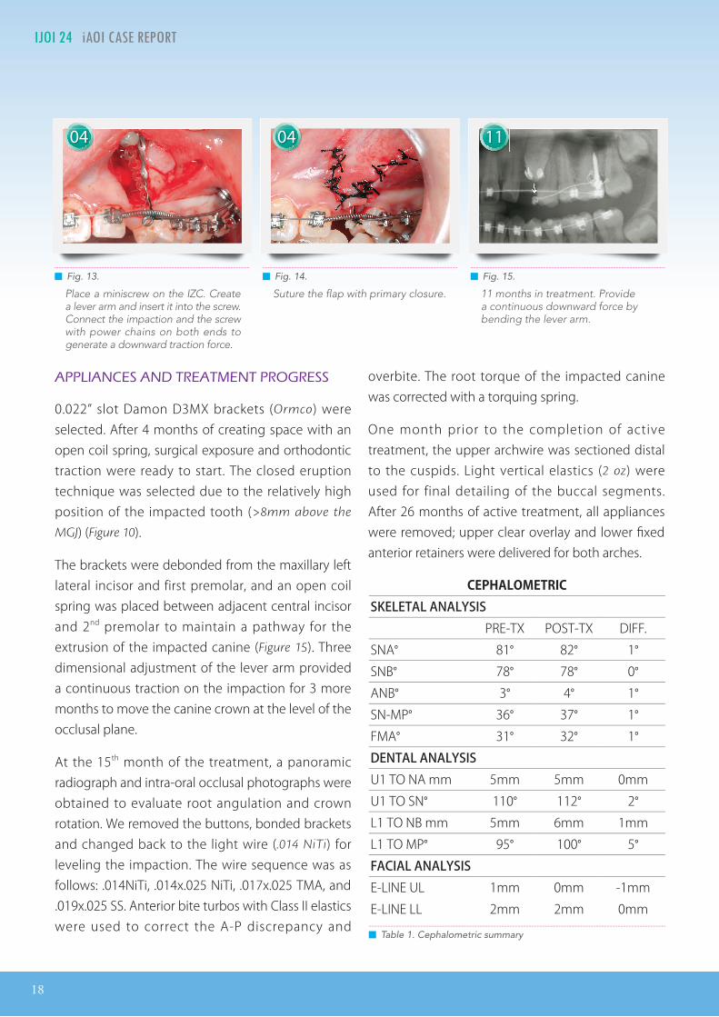
Newton’s A, Inc.) inserted in the infrazygomatic crest (Figure 13). Figure 14 shows primary closure of the wound. The PCs were activated with a lever arm of 17x25 SS (Figure 15) anchored by the miniscrew. The traction force was independent of the rest of the dentition; it was easily activated by adjusting the length of the PC chain and the lever arm in three dimensions (Figure 16). The gingival dynamics and third order control of the recovered canine, illustrated in Figures 17-21, will described in the discussion section.
Anterior bite turbos were used for correction of the 4 mm overbite (Figure 22). Class II elastics were used to resolve the sagittal occlusal discrepancy, and detailing bends produced the final occlusion. Fixed appliances were removed and the corrected dentition was retained with fixed anterior retainers on the lower arch and a clear overlay on the upper arch.
█ Fig. 10. A 13y 7m female with an impaction 14 mm away from the alveolar ridge.
Fig. 12.
04
04
█ Fig. 11.
A flap was used to expose the impaction and remove the bone until CEJ.
IJOI 24 iAOI CASE REPORT
18
CEPHALOMETRICSKELETAL ANALYSIS
PRE-TX POST-TX DIFF.
SNA° 81° 82° 1°
SNB° 78° 78° 0°
ANB° 3° 4° 1°
SN-MP° 36° 37° 1°
FMA° 31° 32° 1°
DENTAL ANALYSISU1 TO NA mm 5mm 5mm 0mm
U1 TO SN° 110° 112° 2°
L1 TO NB mm 5mm 6mm 1mm
L1 TO MP° 95° 100° 5°
FACIAL ANALYSISE-LINE UL 1mm 0mm -1mm
E-LINE LL 2mm 2mm 0mm
█ Table 1. Cephalometric summary
overbite. The root torque of the impacted canine was corrected with a torquing spring.
One month prior to the completion of active treatment, the upper archwire was sectioned distal to the cuspids. Light vertical elastics (2 oz) were used for final detailing of the buccal segments. After 26 months of active treatment, all appliances were removed; upper clear overlay and lower fi xed anterior retainers were delivered for both arches.
APPLIANCES AND TREATMENT PROGRESS
0.022” slot Damon D3MX brackets (Ormco) were selected. After 4 months of creating space with an open coil spring, surgical exposure and orthodontic traction were ready to start. The closed eruption technique was selected due to the relatively high position of the impacted tooth (>8mm above the
MGJ) (Figure 10).
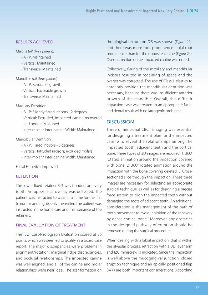
The brackets were debonded from the maxillary left lateral incisor and first premolar, and an open coil spring was placed between adjacent central incisor and 2nd premolar to maintain a pathway for the extrusion of the impacted canine (Figure 15). Three dimensional adjustment of the lever arm provided a continuous traction on the impaction for 3 more months to move the canine crown at the level of the occlusal plane.
At the 15th month of the treatment, a panoramic radiograph and intra-oral occlusal photographs were obtained to evaluate root angulation and crown rotation. We removed the buttons, bonded brackets and changed back to the light wire (.014 NiTi) for leveling the impaction. The wire sequence was as follows: .014NiTi, .014x.025 NiTi, .017x.025 TMA, and .019x.025 SS. Anterior bite turbos with Class II elastics were used to correct the A-P discrepancy and
04 04 11
█ Fig. 13.
Place a miniscrew on the IZC. Create a lever arm and insert it into the screw. Connect the impaction and the screw with power chains on both ends to generate a downward traction force.
█ Fig. 15.
11 months in treatment. Provide a continuous downward force by bending the lever arm.
█ Fig. 14.
Suture the flap with primary closure.
Highly Positioned and Transalveolar Impacted Maxillary Canine IJOI 24
19
RESULTS ACHIEVED
Maxilla (all three planes): • A - P: Maintained • Vertical: Maintained • Transverse: Maintained
Mandible (all three planes): • A - P: Favorable growth • Vertical: Favorable growth • Transverse: Maintained
The lower fixed retainer 3-3 was bonded on every tooth. An upper clear overlay was delivered. The patient was instructed to wear it full time for the fi rst 6 months and nights only thereafter. The patient was instructed in the home care and maintenance of the retainers.
FINAL EVALUATION OF TREATMENT
The IBOI Cast-Radiograph Evaluation scored at 26 points, which was deemed to qualify as a board case report. The major discrepancies were problems in alignment/rotation, marginal ridge discrepancies, and occlusal relationships. The impacted canine was well aligned, and all of the canine and molar relationships were near ideal. The scar formation on
the gingival texture on #23 was shown (Figure 25), and there was more root prominence labial root prominence than for the opposite canine (Figure 24). Over-correction of the impacted canine was noted.
Collectively, fl aring of the maxillary and mandibular incisors resulted in regaining of space and the overjet was corrected. The use of Class II elastics to anteriorly position the mandibular dentition was necessary, because there was insufficient anterior growth of the mandible. Overall, this difficult impaction case was treated to an appropriate facial and dental result with no iatrogenic problems.
DISCUSSION
Three dimensional CBCT imaging was essential for designing a treatment plan for the impacted canine to reveal the relationships among the impacted tooth, adjacent teeth and the cortical bone. Three types of 3D images are required: 1. 360º rotated animation around the impaction covered with bone. 2. 360º rotated animation around the impaction with the bone covering deleted. 3. Cross-sectioned slice through the impaction. These three images are necessary for selecting an appropriate surgical technique, as well as for designing a precise force system to align the impacted tooth without damaging the roots of adjacent teeth. An additional consideration is the management of the path of tooth movement to avoid inhibition of the recovery by dense cortical bone.1 Moreover, any obstacles in the designed pathway of eruption should be removed during the surgical procedure.
When dealing with a labial impaction, that is within the alveolar process, retraction with a 3D lever arm and IZC miniscrew is indicated, Since the impaction is well above the mucogingival junction, closed eruption technique and an apically positioned flap (APF) are both important considerations. According
IJOI 24 iAOI CASE REPORT
20
to Dr. Kokich’s article in 2004, closed eruption technique is more appropriate than an APF when the impaction is in a high position, such as for the present case, which is 14mm apical to the alveolar crest.
Four types of surgical techniques are applicable for recovering impactions. The open window technique is usually the most ideal for palatal impactions with the potential to spontaneously erupt into oral cavity. Since the palate is covered by keratinized gingiva, there is little consideration for the impact on soft tissue. Closed eruption technique and APF are commonly adopted procedures for buccal and labial impactions. The choice of technique depends on the position of the impaction and the conditions of the soft tissue. However, one of the main problems with the closed eruption technique is the difficulty in tension release. APF shares a similar challenge in wound closure. The apical repositioning makes it diffi cult to have precise control of the fl ap margin.
To prevent the archwire deformation and unwanted tooth movement, the traction force was produced by a lever arm made of 17x25 SS anchored with a miniscrew (2x12 mm, Stainless Steel, OrthoBoneScrew,
Newton’s A, Inc.) that is inserted in the infrazygomatic crest (Figure 16). At subsequent visits, the device was reactivated by cutting loops off the power chain
or adjusting the lever arm. The adjacent lateral incisor and first premolar were not bonded nor engaged on the arch wire until the canine had been moved between them.2,3,4 This method allows the vulnerable, adjacent teeth to act as free bodies. They are free to move out of the way of tooth movement if they are contacted by the erupting canine, and thereby iatrogenic root resorption.
During the orthodontic treatment, three stages of soft tissue change can be observed in the process of rapid traction of an impaction: Stage 1. Gingival collar redness;Stage 2. The red patch;Stage 3. Keratinization. 5
Stage 1. Gingival collar redness:During the initial period of rapid forced eruption, a collar of redness is often observed around the gingival margin. This is the color of the non-keratinized epithelium tissue inside of the periodontal pocket (Figure 17). At this stage the pocket depth tends to be high and can be misinterpreted as poor healing or gingival inflammation. Patients should be instructed to maintain proper oral hygiene and this is in fact a normal condition.
Stage 2. The red patch:As the impaction continues to erupt occlusally, a margin of immature-appearing tissue, “the red patch”, appears coronal to the
15 16
█ Fig 16. 2x12 mm OBS with 3D lever arm █ Fig. 18.
16 months in treatment. Distal-rotated canine was noted. Light round wire .014 NiTi was engaged into bracket.
█ Fig. 17.
15 months in treatment. Stage 1. Gingival collar redness. Deep probing depth was noted and careful monitor of oral hygiene was necessary.
Highly Positioned and Transalveolar Impacted Maxillary Canine IJOI 24
21
original gingival margin. At this stage the probing depth reduces from around 5mm to 3mm, which is in the normal range. However, the color of the gingival margin still tends to be more reddish than surrounding tissue and is easily distinguished from the keratinized gingiva (Figure 19).
Stage 3. Keratinization:When the impacted tooth moves into an ideal position, the surrounding gingival t issue completes is progression of proliferation and maturation. The keratinization of gingiva takes about 28-42 days (Figure 20).
The orifice of the gingival sulcus is bound by the tooth surface on one side and sulcular epithelium on the other, the bottom of the sulcus is frequently bordered by epithelium on all sides. The soft tissue wall of the gingival sulcus is lined coronally with nonkeratinizing epithelium, which is deemed the sulcular epithelium. For teeth that are erupted in the presence of soft tissue pocket appear to move coronally for a considerable distance before the gingival margin follows. Concurrently, the pocket depth is reduced and an immature appearing tissue, ‘‘red patch’’, that is sulcular epithelium, has peeled off the tooth surface directly. The nonkeratinized tissue remains erythematous for about 28 to 42 days, until keratinization occurs and it assumes the
appearance of normal gingiva.6 It is imperative that all periodontal pathology be under control before any orthodontic treatment is attempted. A chemical agent, such as chlorhexidine, can be used during the active phase of orthodontic treatment to reduce bacterial plaque accumulation, thereby improving the gingival condition and possibly reducing the acute inflammatory process. As a result, patients with a history of periodontal compromise are placed on 0.12% chlorhexidine gluconate (CH) mouthrinse during the course of active eruption of an impaction.
In the 22th month of treatment, insufficient lingual root torque was noted on the recovered impaction. When the impaction forcefully erupted, the root was more labially positioned because of the relatively dense bone on the palatal surface. Hence, it is important to monitor the torque of the impacted tooth in the finishing stage. Torquing springs are useful auxiliaries (Figure 21). Root angulation and torque control, for the transalveolar impacted canine, present significant challenges. There are several methods for solving these problems: 1. Bond the canine with a high torque bracket (or inverted
low torque bracket). 2. Torque the segmental wire within the canine bracket; 3. add a torquing spring3. The most effi cient and eff ective method to generate favorable torque expression is the use of an
2019 22
█ Fig. 19.
19 months in treatment. Stage 2. The red patch. As the impaction erupted downward to the occlusal level, the ‘‘red patch’’, appeared coronal to the original gingival margin and probing depth was back to normal 3 mm.
█ Fig. 21.
22 months in treatment. Use a single torquing spring (18X25) to increase lingual root torque.
█ Fig. 20.
20 months in treatment. Stage 3. Keratinization. The surround gingival tissue started the progression of proliferation and maturation and the whole process took 28~42 days.
IJOI 24 iAOI CASE REPORT
22
individual root torquing spring. It can be used earlier with light rectangular archwires such as .014x.025 copper NiTi, or the wire segment in the canine slot of the heavy rectangular wire can be rounded with a handpiece in the late stage of treatment.
Class II elastics combined with anterior Bite Turbos were applied for molar extrusion and to bring the mandible forward. These mechanics significantly improved the 4mm overjet and overbite (Figure 22).
The initial DI score was only 10, indicating a minor to moderate malocclusion for an ABO case report7. However, it is necessary to also assess the complexity of a canine impaction, especially when it is 14 mm away from alveolar crest. In this regard, six points were added under the item of the additional treatment complexities to reflect the difficulty for management of a high transalveolar impaction. The fi nal DI score was 16. The IBOI CRE score was 26 points, with most of the points refl ecting problems in alignment and marginal ridge discrepancies (Figures 26 and 27). Pre-fi nish casts, obtained about six months before the end of active treatment, would have been helpful in identifying and resolving these fi nishing problems.
To more thoroughly evaluate the recovery and alignment of an impacted canine, the record of gingival esthetic change after impaction-related surgery is indicated.8 The quality of a prosthesis can also be evaluated by the measurement of the white esthetic score. Because of the scar formation, inadequate lingual root torque over impacted canine and disharmony of the curvature of gingival margin over adjacent lateral incisor, 4 points were assessed on the pink esthetic score. The Pink & White esthetic score worksheet9 provide a broader array of clinical parameters for patients with impacted teeth. The Pink and White Esthetic Score worksheet is demonstrated at the end of this article.
█ Fig. 24.
Occlusal view of anterior teeth showed different root torque of bilateral canine. Absence of cingulum indicated insufficient lingual root torque.
Fig. 24.
█ Fig. 23.
Post-treatment intra-oral frontal photo. Uneven gingival margin level was noted on the upper anterior area. Gingivectomy procedure with diode laser was recommended to patient.
█ Fig. 22.
23 months in treatment. Class II elastics combined with anterior BT were applied for molar extrusion and to bring the mandible forward, which significantly improved the 4mm overjet and overbite.
█ Fig. 25.
Buccal view of active treatment for 26 months. Scar formation over mucosa area of impacted canine was noted.
24
Highly Positioned and Transalveolar Impacted Maxillary Canine IJOI 24
23
CONCLUSION
High, transalveolar impactions are difficult canines to recover. Selection of an appropriate surgical technique depends on the height (position) of the impaction and the conditions of the adjacent soft tissue. 3D imaging is particularly helpful for labial impactions or for those deeply embedded in the alveolar process. In this case, we chose the closed eruption technique and used a 3D lever arm to move the impaction occlusally. The adjacent teeth should not be bonded until the canine erupts to prevent iatrogenic root resorption. Moving a transalveolar impaction into the maxillary arch usually results in inadequate lingual root torque on the recovered canine. This third order problem should be addressed with a torqueing auxiliary druing orthodontic fi nishing.
ACKNOWLEDGMENT
Thanks to Ms. Tzu Han Huang for proofreading this article.
REFERENCES 1. Kokich VG. Surgical and orthodontic management of impacted
maxillary canine. Am J Orthod Dentofacial Orthop 2004; 126:278-83.
2. Niagara K. et al. Impacted maxillary central incisor, canine, and second molar with 2 supernumerary teeth and an odontoma. Am J Orthod Dentofacial Orthop 2009; 135:390-9.
3. Tseng SP, Chang CH, Roberts WE. High maxillary canine impaction with mesial and labial displacement. News & Trends in Orthodontics 2010; 18:36-44
4. Chang CH. Advanced Damon Course No. 9: Impacted and transposed teeth, Beethoven Podcast Encyclopedia in Orthodontics 2011, Newton’s A Ltd, Taiwan
5. Theo M, Ilan S. Forced eruption and implant site development: Soft tissue response. Am J Orthod Dentofac Orthop 1997; 112:596-606.
6. Goldman H, Cohen DW. Healing in periodontal surgical wounds. In: Periodontal therapy 5th ed. St Louis : CV Mosby; 1973. p. 898.
7. Chang CH. Advanced Damon Course No. 4,5 : DI & CRE Workshop (1)(2)., Podcast Encyclopedia in Orthodontics 2011, Newton’s A Ltd, Taiwan.
8. Su B, Chang CH, Roberts WE. Class II Malocclusion Complicated by a Complex Impacted Cuspid; Introduction of the iSAS Method for More Clinical Assessment. News & Trends in Orthodontics 2010; 19:32-46.
9. Belser C, Linda G, Francesca V et al. Outcome Evaluation of Early Placed Maxillary Anterior Single-Tooth Implants Using Objective Esthetic Criteria: A Cross-Sectional, Retrospective Study in 45 Patients With a 2- to 4- Year Follow-Up Using Pink and White Esthetci Scores. J Periodontol 2009; 80:140-151.
Marginal ridge discrepancy over 1 st and 2nd molars was noted.
IJOI 24 iAOI CASE REPORT
24
Highly Positioned and Transalveolar Impacted Maxillary Canine IJOI 24
25
Total Score:
Case # Patient
10
22
2 22
2
22 2 2221
11
0 0
0
0
1111
2
113
1 1
1
����� Alignment/Rotations
Marginal Ridges
Buccolingual Inclination
Overjet
Occlusal Contacts
Occlusal Relationships
Interproximal Contacts
INSTRUCTIONS: Place score beside each deficient tooth and enter total score for each parameter in the white box. Mark extracted teeth with “X”. Second molars should be in occlusion.