Honokiol Decreases Lung Cancer Metastasisthrough Inhibitionof theSTAT3SignalingPathwayJing Pan1,2, Yongik Lee1,2, Qi Zhang1,2, Donghai Xiong1,2, Tina C.Wan2,3, Yian Wang1,2, andMing You1,2
Abstract
Lung cancer is the leading cause of cancer death in theUnited States. Metastasis to lymph nodes and distal organs,especially brain, leads to severe complications and death.Preventing lung cancer development and metastases is animportant strategy to reduce lung cancer mortality. Honokiol(HNK), a natural compound present in the extracts of mag-nolia bark, has a favorable bioavailability profile and recentlyhas been shown to readily cross the blood–brain barrier. In thecurrent study, we evaluated the antimetastatic effects of HNKin both the lymph node and brain mouse models of lungtumor metastasis. We tested the efficacy of HNK in preventing18 H2030-BrM3 cell (brain-seeking human lung tumor cells)migration to lymph node or brain. In an orthotopic mousemodel, HNK significantly decreased lung tumor growth com-
pared with the vehicle control group. HNK also significantlyreduced the incidence of lymph node metastasis and theweight of mediastinal lymph nodes. In a brain metastasismodel, HNK inhibits metastasis of lung cancer cells to thebrain to approximately one third of that observed in controlmice. We analyzed HNK's mechanism of action, which indi-cated that its effect is mediated primarily by inhibiting theSTAT3 pathway. HNK specifically inhibits STAT3 phosphory-lation irrespective of the mutation status of EGFR, and knock-down of STAT3 abrogated both the antiproliferative and theantimetastatic effects of HNK. These observations suggest thatHNK could provide novel chemopreventive or therapeuticoptions for preventing both lung tumor progression and lungcancer metastasis. Cancer Prev Res; 10(2); 133–41. �2016 AACR.
IntroductionLung cancer is the leading cause of cancer death worldwide.
Brain metastasis is one of the most intractable clinical problemsassociated with lung cancer and is a major cause of lung cancermortality (1, 2). It is estimated that approximately 10%ofpatientspossess brainmetastases at the time of their lung cancer diagnosis,whereas 40% to50%ofpatients developbrainmetastasis during atypical course of lung cancer disease (2). Because of the difficultyof drug transport through the blood–brain barrier (BBB), the onlyavailable therapies to address central nervous system (CNS)metastases include whole brain/CNS irradiation or surgical resec-tion in eligible patients with non-EGFR–mutant lung cancer.Patients with EGFR-mutant disease are treated with anti-EGFRagents (2). All of these therapies are purely palliative and elicitsignificant toxicities. Therefore, naturally occurring agents thatproduce little or no toxicity, and that can be delivered systemically
to the original tumor site and to the brain, may prove highlyefficacious for lung cancer treatment.
Honokiol (HNK) is a key bioactive compound present in theextracts of magnolia bark. The extracts have been used as a folkremedy in Asian countries to treat gastrointestinal disorders,cough, anxiety, stroke, and allergic diseases for centuries. HNKhas a favorable bioavailability profile in rodents with a sus-tained plasma concentration in mice and a 2-compartmentpharmacokinetic profile in rats (3, 4). Our recent study mea-suring oxygen consumption rate in whole intact cells demon-strated that HNK may directly target mitochondria, leading torapid and persistent inhibition of mitochondrial respiration.This results in the induction of apoptosis in lung cancer cellsand ultimately attenuates lung squamous cell carcinoma (SCC)growth in the N-nitroso-tris-chloroethylurea–induced murinemodel of lung SCC (5). Remarkably, HNK has been shown toreadily cross both the BBB (6, 7) and blood–cerebrospinal fluidbarrier to inhibit brain tumor growth in rodent models (6),which prompted us to develop the hypothesis that HNK maynot only inhibit lung tumorigenesis but also suppress lungcancer brain metastasis.
Several mechanisms of action have been suggested for HNK asan antitumor agent, including induction of apoptosis by causingmitochondrial dysfunction and endoplasmic reticulum stress (8),cell-cycle arrest (9), and inhibition of tumor invasion via down-regulation of EGFR, NF-kB, Ras/ERK, PI3K/AKT, and Akt/mTORpathways (10–13). One key mechanism of action for HNK is theinduction of apoptosis through a mitochondrial-dependentmechanism (5, 7). We demonstrated that HNK suppresses mito-chondrial respiration and increases the generation of reactiveoxygen species in mitochondria, leading to the induction ofapoptosis in lung cancer cells (5). Recently, the mitochondrialproteins SIRT3 andGrp78 (an apoptosis-associated protein) have
1Cancer Center, Medical College of Wisconsin, Milwaukee, Wisconsin. 2Depart-ment of Pharmacology & Toxicology, Medical College of Wisconsin, Milwaukee,Wisconsin. 3Cardiovascular Center, Medical College of Wisconsin, Milwaukee,Wisconsin.
Note: Supplementary data for this article are available at Cancer PreventionResearch Online (http://cancerprevres.aacrjournals.org/).
J. Pan, Y. Lee, and Q. Zhang contributed equally to this article.
Corresponding Author: Ming You, Department of Pharmacology and Toxicol-ogy and Cancer Center, Medical College of Wisconsin, 8701 WatertownPlank Road, Milwaukee, WI 53226. Phone: 414-805-8228; Fax: 414-805-8281;E-mail: [email protected]
been suggested as possible targets of HNK. Interestingly, STAT3 isa major downstream mediator of these pathways and is alsoknown to play a major role in regulating mitochondrial activity(14–18). STAT3 is a well-known oncogene that can be regulatedby receptor tyrosine kinases (RTK), G-protein–coupled receptors,and interleukin families via phosphorylation. PhosphorylatedSTAT3 undergoes dimerization and translocalization in eitherthe nucleus or mitochondria to mediate its activity, resulting inenhanced cell proliferation, invasion, and survival for manycancer types (19, 20).
In the current study, we evaluated the ability of HNK toprevent lung cancer metastasis to lymph nodes and brain usingwell-established murine models. We also explored the poten-tial role of RTKs as targets of HNK in the inhibition of lungcancer brain metastasis. We found that a major effect of HNK isthe inhibition of STAT3 phosphorylation. We also determinedthe role of STAT3 in mediating the anticancer effects of HNK inlung cancer by knocking down endogenous STAT3 in the brainmetastatic lung cancer cell lines (PC9-BrM3 and H2030-BrM3).Our results showed that HNK significantly inhibits STAT3Tyr705
and STAT3Ser727 phosphorylation in both cell lines. STAT3knockdown abrogated the antiproliferative and antiinvasiveeffects of HNK. Understanding this novel mechanism of actionfor HNK may lead to the development of a new class ofchemopreventive agents that not only inhibit lung cancerlocally (5), but also have the potential to inhibit distal metas-tasis, which could directly benefit patients.
Materials and MethodsCell culture and reagents
Brain metastatic lung cancer cell lines, PC9-BrM3 and H2030-BrM3, were generously provided by Dr. Joan Massagu�e (CancerBiology and Genetics Program, Memorial Sloan Kettering CancerCenter, New York, NY), and were not authenticated by theauthors. Both cell lines were maintained in RPMI1640 medium(Invitrogen) supplemented with 10% FBS (Invitrogen) at 37�C ina humidified atmosphere of 5%CO2 and air. HNKwas purchasedfrom Sigma-Aldrich.
Cell proliferationFor cell proliferation assays, cells were seeded onto 96-well
tissue culture plates at 2–3� 103 cells per well. Twenty-four hoursafter seeding, cellswere exposed to various concentrations ofHNKfor 48 hours, while the control cells received medium only. Theplates were incubated at 37�C and 5% CO2, and cell growth wasmonitored by IncuCyte (Essen BioScience). Data analysis wasconducted using IncuCyte 2011A software. All assays were per-formed in triplicate.
Transwell invasion assayCell invasion was determined using Boyden chamber trans-
wells that were precoated with a growth factor reduced Matrix(Thermo Fisher Scientific). Transwell invasion assays were per-formed as described in the manufacturer's protocol. Briefly, 3 �105 cells were seeded into each transwell containing serum-freeRPMI1640 media and 10 mmol/L HNK. Bottom wells containedRPMI1640 media supplemented with 10% FBS and 10 mmol/LHNK. After 36 hours, cells that had invaded through the transwellwere fixed with 10% formalin, stained with 5% crystal violet in70% ethanol, and counted in three randomly selected areas of
each transwell using an inverted tissue culture microscope at�10magnification. The results were normalized to controls.
RTK assayH2030-BrM3 cells, treated either with DMSO (vehicle control)
orHNK at various concentrations for 6 hours, were lysedwith 200mL of 1� NP40 lysis buffer containing proteinase inhibitor cock-tails (Thermo Fisher Scientific), sheared 10 times with a 28-gaugeneedle, spun at 16,000 � g for 30 minutes, and normalized byprotein concentration as determined by the Bradford method.Normalized lysate was resolved in PathScan RTK Signaling Array,and the signaling arraywas examined by Li-COROdyssey infraredimaging system (Li-Cor).
Western blot analysisCells were lysed with 200 mL of RIPA buffer containing pro-
teinase inhibitor cocktails (Thermo Fisher Scientific), sheared 10times with a 28-gauge needle, spun at 16,000� g for 30 minutes,normalized by protein concentration as determined by the Brad-ford method, and boiled for 5 minutes. Normalized lysate wasresolved by 4% to 12% SDS-PAGE (Thermo Fisher Scientific) andimmunoblotted with indicated antibodies; p-EGFR (#3777S), p-STAT3 (#9134S), p-AKT (#4060S), EGFR (4267S), STAT3(9139S), and AKT (9272S), which were all purchased from CellSignaling Technology. Actin (SC-8432)was purchased fromSantaCruz Biotechnology.
Endogenous STAT3 knockdownLentiviral particles against STAT3 were purchased from Santa
Cruz Biotechnology. PC9-BrM3 and H2030-BrM3 cells wereinfected with lentiviral particles using 8 mg/mL polybrene, andthe infected cells were selected by treatment with puromycin(2 mg/mL) for 3 days.
KINOMEscan HNK binding assayDirect interaction between HNK and candidate RTKs was
examined via the KINOMEscan binding assay from DiscoveRx.
RNA sequencing and pathway analysisWe conducted an RNA sequencing (RNA-seq) study of human
lung tumor metastases in mouse brains. Three brain metastaseswere sampled from mice without HNK treatment and anotherthree brain metastases were obtained from mice treated withHNK. Total RNA samples were extracted from these six samplesusing Qiagen RNeasy Mini Kit. We used NEBNext Ultra RNALibrary Prep Kit from Illumina to construct the RNA-seq librariesfor these samples. Whole transcriptome analysis of the RNA-seqlibrary samples was performed using HiSeq 2500 sequencingplatforms (Illumina). The experiment was single-end with 50nucleotides read length. Coverage for the samples ranged from15 million to 32 million reads per sample. To identify andunequivocally separate graft (human) and host (mouse) reads,processed sample reads were sequentially aligned to both graft[complete hg19 human genome (UCSC version, February 2009)]and host [complete mm9 mouse genome (UCSC version, July2007)] genomes using Bowtie-TopHat (21, 22). Read counts wereobtained using HTseq (23). Data normalization and differentialexpression analysis were performed using the statistical algo-rithms implemented in EdgeR Bioconductor package (24). FDR,corrected P values of less than 0.05, was used as criteria for
Pan et al.
Cancer Prev Res; 10(2) February 2017 Cancer Prevention Research134
significantly regulated genes. We used a strategy that efficientlyseparates human lung tumor sequence data from xenograftmouse (mice with genetically human tumors) sequence data intoseparate microenvironment and tumor expression profiles (25,26). Using this tool, we obtained more accurate RNA expressionprofiles for both metastatic human lung tumors and mousestromal cells.
Brain metastases mouse modelAnimal procedures were in accordance with the Medical Col-
lege of Wisconsin Institutional Animal Care and Use Committee.For lung cancer brain metastasis study, 4- to 6-week-old femaleNOD/SCIDmicewere used. Brain-seekingH2030-BrM3 cells (2�105) were suspended in 0.1 mL PBS and injected into the leftventricle (LV) under ultrasound guidance (ECHO 707, GE). Oneday after engraftingH2030-BrM3 cells into the arterial circulation,mice were randomly grouped into vehicle treatment group andHNK treatment group (10 mg/kg b.w.). Mice were treated witheither solvent control (0.1% DMSO in corn oil) or HNK by oralgavage for 4 weeks, metastasis was monitored over time bybioluminescence with an IVIS 200 Xenogen and confirmed withex vivo luminescence, GFP fluorescence followed by hematoxylin
and eosin (H&E), and GFP staining. For analysis of lung tumorlymph nodemetastases, H2030-BrM3 (104) cells were suspendedin a 1:2 mixture of PBS and growth factor reduced Matrigel (BDBiosciences) and injected into the lung. HNK treatment wasinitiated one day after orthotopic injection of tumor cells by oralgavage.
In vivo lung cancer orthotopic modelWe used an orthotopic model of lung adenocarcinoma cells
(H2030-BrM3 cells) in athymic nude mice to evaluate the inhib-itory effect of HNK on lung tumor growth and lymph nodemetastasis. Five-week-old male athymic nude mice were used forthe experiments. Mice were anesthetized with isoflurane andplaced in the right lateral decubitus position. A total of 1 �106 H2030-Br3 cells in 50 mg of growth factor reduced Matrigelin 50 mL of RPMI1640 medium were injected into the left lungthrough the left rib cage as described previously (27). One dayafter injection, mice in the HNK group were treated with 2 or 10mg/kg b.w. HNK, once a day, 5 days per week for four consecutiveweeks. Tumor growth and metastases phenotype was monitoredover time by bioluminescence with an IVIS 200 Xenogen. Micewere euthanized at the endpoint; tissues were immediately fixed
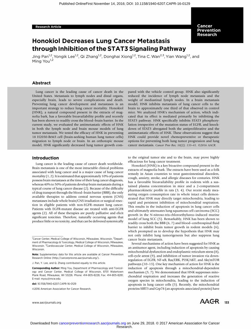
Effects of HNK on lung cancer cell proliferation and invasion. A and B, PC9-BrM3 and H2030-BrM3 lung cancer cell lines were treated with HNK (0, 10, 20, 30, and50 mmol/L) for 96 hours. ns, not significant. Cell proliferation was measured using the IncuCyte Live Imaging System as indicated in Materials and Methods.HNK inhibited proliferation of PC-9BrM3andH2030-BrM3 cells in a dose- and time-dependentmanner. PC9-BrM3 (IC50¼ 28.4mmol/L) andH2030-BrM3 (IC50¼ 25.7mmol/L; � , P < 0.05; ��, P < 0.005). C, HNK significantly inhibited the invasion of PC9-BrM3 and H2030-BrM3 cells even at the lowest dose of 10 mmol/L whencompared with PC9-BrM3 and H2030-BrM3 cells treated with the vehicle control DMSO (��� , P < 0.001). Number of invaded cells per field (%)was normalized to the DMSO control group. D, Representative images of invaded PC9-BrM3 and H2030-BrM3 cells in the absence and presence of HNK.Invasion of both PC9-BrM3 and H2030-BrM3 cells was inhibited by HNK in a dose-dependent manner.
STAT3 Abrogates the Anticancer Effects of HNK
www.aacrjournals.org Cancer Prev Res; 10(2) February 2017 135
in optimal cutting temperature and frozen in liquid nitrogen forsubsequent Western blot and immunohistochemical analyses.
In vivo imaging systemIn vivo imaging system (IVIS) consists of a highly sensitive,
charge-coupled digital camera with accompanying advancedcomputer software for image data acquisition and analysis. Thissystem captures photons of light emitted by reagents or cells thathave been coupled or engineered to produce bioluminescence inthe living animal. The substrate luciferin was injected into theintraperitoneal cavity ofmice at a dose of 150mg/kg b.w. (30mg/mL luciferin), approximately 10 minutes before imaging. Micewere anesthetized with isoflurane/oxygen and placed on animaging stage. Photons emitted from the lung region were quan-tified using Living Image software (Xenogen Corp.).
HistopathologyMouse brains were fixed in 10% zinc formalin solution over-
night and stored in 70% ethanol for histopathology. Serial tissuesections (5-mm each) were made and stained with H&E or GFPand examined histologically under a light microscope to assessseverity of tumor development.
Statistical analysisData were analyzed by one-way ANOVA. � P < 0.05, �� P < 0.01,
and ��� P < 0.001 were considered statistically significant.
ResultsHNK inhibits proliferation and invasion of brain metastaticlung cancer cells in vitro
Previous research in our laboratory and in other laboratoriesdemonstrated the anticancer effect of HNK in many cancertypes, including lung (4, 5, 9, 11, 12, 28). To evaluate the effects
of HNK in brain metastatic lung cancer, we examined the effectsof HNK on the proliferation and invasion of PC9-BrM3 andH2030-BrM3 brain metastatic lung cancer cells. Initially, PC9-BrM3 and H2030-BrM3 cells were treated with various con-centrations of HNK for 96 hours to examine the antiprolifera-tive effects of the compound. HNK effectively inhibited bothPC9-BrM3 and H2030-BrM3 cell proliferation in a dose- andtime-dependent manner (IC50 for PC9-BrM3 is 28.4 mmol/L,for H2030-BrM3 is 25.7 mmol/L; Fig. 1A). We also examined theeffects of HNK on the invasion of PC9-BrM3 and H2030-BrM3cells in the Boyden chamber (Fig. 1C and D). As shownin Fig. 1C and D, HNK significantly inhibited the invasion ofboth PC9-BrM3 and H2030-BrM3 cell lines in a dose-depen-dent manner. The doses of HNK required to reduce the invasionof PC9-BrM3 and H2030-BrM3 cells were much lower (Fig. 1Cand D) than those required to inhibit cell proliferation (Fig. 1Aand B). On the basis of previous research (5–7, 29–31) and ourcurrent data, HNK is not only an effective chemopreventive/chemotherapeutic agent, but could also be an effective agent toprevent or inhibit invasion of lung cancer.
HNK inhibitsmetastasis of lung tumor cells to lymphnodes in alung orthotopic mouse model
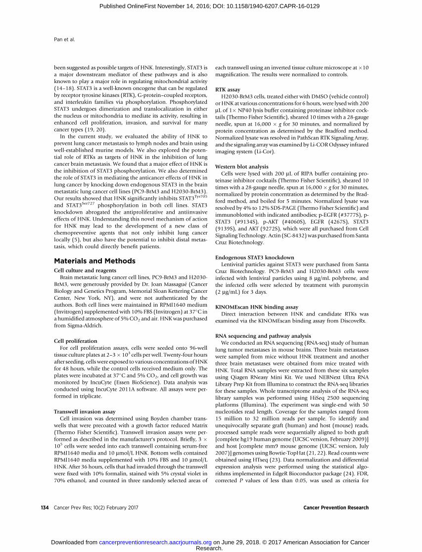
In themice implanted orthotopically withH2030-BrM3 cells inthe left lung, lung tumors grew and spread within the lung andthen to the mediastinum. The incidence of tumor formation was100%.Representative bioluminescence images of lung orthotopicxenografts are shown in Fig. 2A.Mice did not showany observableside effect when treated with HNK. Higher doses of HNK signif-icantly decreased lung tumor growth when compared with thevehicle control group (Fig. 2B). Comparing with the controlgroup, HNK, at the higher dose (10 mg/kg b.w.), significantlyreduced the incidence of mediastinal adenopathy. The incidenceof mediastinal lymph node metastasis in the control group was
A B
***
Control
HNK
2 mg/kg 10 mg/kg 4.0¥109
3.0¥109
2.0¥109
1.0¥109
00 10 20 30
Days post injection
Ctrl0.0
0.1
0.2
0.3
Wei
ght o
f med
iast
inal
lym
ph n
odes
(g)
0.4
HNK-low HNK-high
CtrlHNK (10 mg/kg b.w.)HNK (2 mg/kg b.w.)
Tota
l bio
lum
ines
cenc
eflu
x (p
/s)
***C
Figure 2.
HNK inhibits orthotopic lung tumor growth inmice injected with H2030-BrM3 cells. A,Representative luciferase images from micetreated with either gavage control (Ctrl) orHNK. B, Quantification of bioluminescenceimaging signal intensity in the control (Ctrl)and HNK-treated groups at different timepoints after the injection of H2030-BrM3cells. Quantified values are shown in totalflux. C, The weight of the mediastinal lymphnodeswas significantly lower comparedwiththe no-treatment controls. The incidence oflymphatic metastasis was significantly lowerin the high-dose HNK treatment group (2/6)compared with that of the no-treatmentcontrols (7/7). ��� , P < 0.001.
Pan et al.
Cancer Prev Res; 10(2) February 2017 Cancer Prevention Research136
100%; in the high-dose HNK treatment group, only 2 of 6 micehad lymphatic metastasis. The high-dose HNK also significantlydecreased the weight of mediastinal lymph nodes over 80%compared with control group (Fig. 2C).
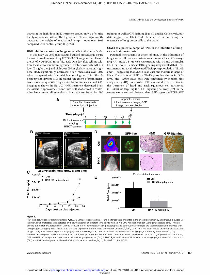
HNK inhibits metastasis of lung cancer cells to the brain in vivoIn this assay, we used an ultrasound-guided procedure to insure
the injection of brain-seeking H2030-BrM3 lung cancer cells intothe LV of NOD/SCID mice (Fig. 3A). One day after cell inocula-tion, themicewere randomly grouped to vehicle control andHNKlow- (2mg/kg b.w.) and high-dose (10mg/kg b.w.) groups. High-dose HNK significantly decreased brain metastasis over 70%when compared with the vehicle control group (Fig. 3B). Atnecropsy (28 days post-LV injection), the extent of brain metas-tases was also quantified by ex vivo bioluminescence and GFPimaging as shown in Fig. 3C. HNK treatment decreased brainmetastasis to approximately one third of that observed in controlmice. Lung tumor cell migration to brain was confirmed by H&E
staining, as well as GFP staining (Fig. 3D and E). Collectively, ourdata suggest that HNK could be effective in preventing themetastasis of lung cancer cells to the brain.
STAT3 as a potential target of HNK in the inhibition of lungcancer brain metastasis
Potential mechanisms of action of HNK in the inhibition oflung cancer cell brain metastasis were examined via RTK assays(Fig. 4A). H2030-BrM3 cells were treated with 10 and 20 mmol/LHNK for 6 hours. PathScan RTK signaling array revealed thatHNKtreatment dramatically decreased STAT3phosphorylation (Fig. 4Band C), suggesting that STAT3 is at least one molecular target ofHNK. The effects of HNK on STAT3 phosphorylation in PC9-BrM3 and H2030-BrM3 cells were confirmed by Western blotanalysis (Fig. 4D). Previously, HNK was found to be effective inthe treatment of head and neck squamous cell carcinoma(HNSCC) via targeting the EGFR signaling pathway (32). In thecurrent study, we also observed that HNK targets the EGFR–AKT
Establish brain metsmodel by LV injection
Endpoint: Ex vivobioluminescence image, GFP
image, tissue collection
1913710
Bioluminescenceimaging
HNK Treatment
Time (days)
A
B D
EC
BL
Ctr
lH
NK
In vivo brain mets grow along time
Ctrl4.0¥108
3.0¥108
2.0¥108
1.0¥108
4.0¥108
6.0¥108
8.0¥108
2.0¥108
0Ctrl
Ctrl
HNK (10 mg/kg b.w.)
HNK (10 mg/kg b.w.)
Ex vivo bioluminescence
0 5 10 15 20 25 30Days post cardiac injection
Tota
l flu
x (p
/s)
Tota
l flu
x (p
/s)
HNK (10 mg/k.g. b.w.)
GFP-live H&E GFP Staining
2328
27
Figure 3.
HNK inhibits lung cancer brain metastasis. A, H2030-BrM3 cells expressing GFP and luciferase were engrafted in the arterial circulation by an ultrasound-guided LVinjection. Brain metastasis was detected by bioluminescence at different time points with an IVIS 200 Xenogen monitor (Xenogen; exposure time, 1 minute;binning 8; no filter; f/stop16; field of view 12.5 cm). B, Corresponding grayscale photographs and color luciferase images are superimposed and analyzed withLivingImage (Xenogen). Mets, metastases. Data are expressed as normalized photon flux (photons/s/cm2). After final IVIS scan, mouse brain was dissected andimaged using Maestro Multi-Spectral Imaging System for GFP signal. C, Quantification of bioluminescence imaging signal intensity in the control (Ctrl)and HNK-treated group at different time points after the injection of H2030-BrM3 cells. Quantified values are shown in total flux. D, Representative luciferase,GFP, and H&E IHC images from mice treated with either gavage control (Ctrl) or HNK. E, Quantification of bioluminescence imaging signal intensity in the control(Ctrl) and HNK-treated group at the end of study via ex vivo Live Imaging. � , P < 0.05; ��� , P < 0.001.
STAT3 Abrogates the Anticancer Effects of HNK
www.aacrjournals.org Cancer Prev Res; 10(2) February 2017 137
signaling pathway in PC9-BrM3 cells, which harbor an EGFRmutation, but not in H2030-BrM3 cells, which harbor Krasmutations. In addition, we examined the interaction betweenHNK and multiple RTKs via the KINOMEScan binding assay. Asshown in Supplementary Fig. S1,HNKdidnot binddirectly to anyof the RTKs tested.
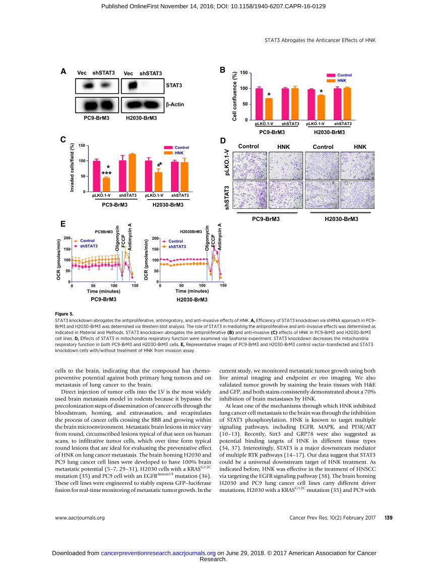
STAT3 knockdown decreases the anticancer effects of HNK inlung cancer
shRNA knockdown was used to demonstrate the role of STAT3in mediating the effects of HNK in lung cancer. Knockdown ofSTAT3 in PC9-BrM3 and H2030-BrM3 cells was validated byWestern blot analysis (Fig. 5A). STAT3 knockdown decreases theantiproliferative (Fig. 5B) and antiinvasive (Fig. 5C and D) effectsof HNK in both PC9-BrM3 and H2030-BrM3 cell lines. HNKtreatment (20 mmol/L) for 48 hours inhibited the proliferation ofPC9-BrM3 vector control cells by 30% and H2030-BrM3 vectorcontrol cells by 20% but had no significant effect on the prolif-eration of STAT3 knockdown PC9-BrM3 or H2030-BrM3 cells(Fig. 5B). In addition, HNK treatment (10 mmol/L) significantlyinhibited the invasionof bothPC9-BrM3andH2030-BrM3vectorcontrol cells but had no effect on the invasion of STAT3 knock-downPC9-BrM3orH2030-BrM3 cells (Fig. 5C andD). Finally,weexamined the effects of STAT3 on mitochondrial respiratoryfunction in PC9-BrM3 and H2030-BrM3 cells. As shown in Fig.5E, STAT3 knockdown significantly decreased mitochondrialrespiratory function in both PC9-BrM3 and H2030-BrM3 celllines. The anticancer effects of HNK, therefore, could be throughinhibition of STAT3-mediated mitochondrial functions in lungcancer cells that have metastasized to the brain.
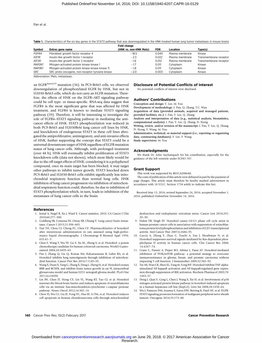
RNA-seq analysis showed that the expressions of key genesimportant to the activation of STAT3 pathway weredownregulated in themetastatic lung tumors byHNK treatment
The differentially expressed genes identified by RNA-seq anal-ysis software were subjected to IPA analysis (Ingenuity Pathway
Analysis; http://www.ingenuity.com/products/ipa) to identifythe most significant oncogenic pathways in metastatic lungtumors changed by HNK treatment. Our genome-wide RNA-seqscan showed that STAT3 pathway is the top downregulated oneamong the oncogenic pathways that were significantly down-regulated in the HNK-treated human lung tumor metastases inmouse brains (Fig. 4A). In addition, RNA-seq analyses identifiedthat six key genes involved in the activation of STAT3 pathwayswere significantly downregulated inmetastatic lung tumors in vivouponHNK treatment (Table 1). They were FGFR4, IGF1R, IGF2R,MAP2K1, MAP3K11, and SRC. These matched the findings fromour functional studies and supported that the anti–lung cancerrole of HNK was mediated via the STAT3 signaling pathway.
DiscussionOne of the common sites for metastases of lung cancers is the
brain. Currently available therapies to address CNS metastasesinclude whole brain/CNS irradiation or surgical resection ineligible patients, treatment with anti-EGFR agents in patientswhose tumors contain EGFR mutations, as well as using next-generation ALK TKI that is brain-penetrable such as PF-06463922, to control CNS metastases in lung cancer patients(2, 33). However, these treatment options are available onlyafter the diagnoses of brain metastases, and in many cases,metastatic lesions remain undiagnosed for long periods or theyare not amenable to treatment with chemo/radiotherapy orsurgery. Therefore, it is necessary to develop prevention strat-egies to inhibit metastases from primary tumors. Recently,we demonstrated the ability of HNK to potently inhibit thedevelopment of lung tumors in mice (5). Analysis of HNK'smechanism of action suggests that its effect is primarily medi-ated by inducing apoptosis through a mitochondria-dependentmechanism (5, 7, 34). Here, through the use of the well-characterized brain metastases murine model, we report thatHNK exerts inhibitory effects on the metastasis of lung cancer
p-STAT3
HNK
p-EGFR
p-ATK
AKT
EGFR
STAT3
Actin
HNK
H2030-BrM3PC9-BrM3
p-STAT3p-STAT3
STAT3
0.0 1.0 2.52.01.50.5
IGF1R
EGFR
PI3K/AKT
NF-kB
-Log10 (P) value
p-VEGFRp-Tie/TEKp-PDGFR
p-TrkBp-TrkA
Fluorescence intensity0 20 40 60 80
ControlHNK 10 mmol/LHNK 20 mmol/L
A B
C D
Figure 4.
HNK targets STAT3 phosphorylation viainhibition of multiple RTKs. A, Pathway analysisbased on RNA-seq revealed that HNK inhibitsmultiple RTK pathways, including EGFR, as wellas the IGF1R and NF-kB, PI3K/AKT, and STAT3signaling pathway. Notably, STAT3 signalingpathway was the most significantly affectedpathway by HNK. B, H2030-BrM3 cells weretreated with HNK, and cell lysates wereexamined using RTK signaling arrays. STAT3phosphorylation is downregulated by HNK in adose-dependent manner. C, Effects of HNK onthe phosphorylation of STAT3, as well as othersignaling pathways, such as TrkA/B, PDGFR, Tie/TEK, and VEGFR signaling pathways. D, Thephosphorylation of EGFR and AKT isdownregulated by HNK only in the EGFR-mutantPC9-BrM3 cell line, but not in the Kras-mutantH2030-BrM3 cell line. STAT3 phosphorylation isdownregulated by HNK in both cell lines.
Pan et al.
Cancer Prev Res; 10(2) February 2017 Cancer Prevention Research138
cells to the brain, indicating that the compound has chemo-preventive potential against both primary lung tumors and onmetastasis of lung cancer to the brain.
Direct injection of tumor cells into the LV is the most widelyused brain metastasis model in rodents because it bypasses theprecolonization steps of dissemination of cancer cells through thebloodstream, homing, and extravasation, and recapitulatesthe process of cancer cells crossing the BBB and growing withinthe brainmicroenvironment.Metastatic brain lesions inmice varyfrom round, circumscribed lesions typical of that seen on humanscans, to infiltrative tumor cells, which over time form typicalround lesions that are ideal for evaluating the preventative effectof HNK on lung cancer metastasis. The brain homing H2030 andPC9 lung cancer cell lines were developed to have 100% brainmetastatic potential (5–7, 29–31), H2030 cells with a KRASG12C
mutation (35) and PC9 cell with an EGFRDexon19 mutation (36).These cell lines were engineered to stably express GFP–luciferasefusion for real-timemonitoring ofmetastatic tumor growth. In the
current study, we monitored metastatic tumor growth using bothlive animal imaging and endpoint ex vivo imaging. We alsovalidated tumor growth by staining the brain tissues with H&EandGFP, and both stains consistently demonstrated about a 70%inhibition of brain metastases by HNK.
At least one of the mechanisms through which HNK inhibitedlung cancer cellmetastasis to the brainwas through the inhibitionof STAT3 phosphorylation. HNK is known to target multiplesignaling pathways, including EGFR, MAPK, and PI3K/AKT(10–13). Recently, Sirt3 and GRP78 were also suggested aspotential binding targets of HNK in different tissue types(34, 37). Interestingly, STAT3 is a major downstream mediatorof multiple RTK pathways (14–17). Our data suggest that STAT3could be a universal downstream target of HNK treatment. Asindicated before, HNK was effective in the treatment of HNSCCvia targeting the EGFR signaling pathway (38). The brain homingH2030 and PC9 lung cancer cell lines carry different drivermutations, H2030 with a KRASG12C mutation (35) and PC9 with
PC9-BrM3 H2030-BrM3
pLK
O.1
-Vsh
STAT
3
HNK
pLKO.1-V0
50
100
Cel
l con
fluen
ce (%
) 150
pLKO.1-VshSTAT3 shSTAT3
Control HNKControl
shSTAT3Control
shSTAT3Control
HNKControl
HNKControl
H2030BrM3
PC9-BrM3 H2030-BrM3
D
A B
PC9-BrM3 H2030-BrM3
shSTAT3 shSTAT3
STAT3
b-Actin
Vec Vec
C
Time (minutes)0
0
50
100
150
200
OC
R (p
mol
es/m
in)
50 100
PC9BrM3
150Time (minutes)
00
50
100
150
200
OC
R (p
mol
es/m
in)
50 100 150
Olig
omyc
inFC
CP
Ant
imyc
in A
Olig
omyc
inFC
CP
Antim
ycin
AE
****
* *
PC9-BrM3 H2030-BrM3
PC9-BrM3 H2030-BrM3
pLKO.1-V0
50
100
Inva
ded
cells
/fiel
d (%
) 150
pLKO.1-VshSTAT3 shSTAT3
* *
Figure 5.
STAT3 knockdown abrogates the antiproliferative, antimigratory, and anti-invasive effects of HNK. A, Efficiency of STAT3 knockdown via shRNA approach in PC9-BrM3 and H2030-BrM3 was determined via Western blot analysis. The role of STAT3 in mediating the antiproliferative and anti-invasive effects was determined asindicated in Material and Methods. STAT3 knockdown abrogates the antiproliferative (B) and anti-invasive (C) effects of HNK in PC9-BrM3 and H2030-BrM3cell lines. D, Effects of STAT3 in mitochondria respiratory function were examined via Seahorse experiment. STAT3 knockdown decreases the mitochondriarespiratory function in both PC9-BrM3 and H2030-BrM3 cells. E, Representative images of PC9-BrM3 and H2030-BrM3 control vector–transfected and STAT3knockdown cells with/without treatment of HNK from invasion assay.
STAT3 Abrogates the Anticancer Effects of HNK
www.aacrjournals.org Cancer Prev Res; 10(2) February 2017 139
an EGFRDexon19 mutation (36). In PC9-BrM3 cells, we observeddownregulation of phosphorylated EGFR by HNK, but not inH2030-BrM3 cells, which do not carry an EGFR mutation. There-fore, the effects of HNK on the EGFR–AKT signaling pathwaycould be cell type- or tissue-specific. RNA-seq data suggest thatFGFR4 is the most significant gene that was affected by HNKtreatment, and FGFR4 is known to mediate STAT3 signalingpathway (39). Therefore, it will be interesting to investigate therole of FGFR4–STAT3 signaling pathway in mediating the anti-cancer effects of HNK. STAT3 phosphorylation was reduced inboth PC9-BrM3 and H2030BrM lung cancer cell lines by HNK,and knockdown of endogenous STAT3 in these cell lines abro-gated the antiproliferative, antimigratory, and anti-invasive effectsof HNK, further supporting the concept that STAT3 could be auniversal downstream target ofHNK regardless of EGFRmutationstatus of lung cancer cells. Although, with prolonged treatment(over 48 h), HNK will eventually inhibit proliferation of STAT3knockdown cells (data not shown), which most likely would bedue to the off-target effects of HNK, considering it is a polyphenolcompound, once its main target has been blocked, it may targetother pathways to inhibit tumor growth. STAT3 knocked downPC9-BrM3 and H2030-BrM3 cells exhibit significantly less mito-chondrial respiratory function than normal lung cells. HNKinhibition of lung cancer progression via inhibition of mitochon-drial respiratory function could, therefore, be due to inhibition ofSTAT3 phosphorylation which, in turn, leads to inhibition of themetastases of lung cancer cells to the brain.
Disclosure of Potential Conflicts of InterestNo potential conflicts of interest were disclosed.
Authors' ContributionsConception and design: Y. Lee, M. YouDevelopment of methodology: J. Pan, Q. Zhang, T.C. WanAcquisition of data (provided animals, acquired and managed patients,provided facilities, etc.): J. Pan, Y. Lee, Q. ZhangAnalysis and interpretation of data (e.g., statistical analysis, biostatistics,computational analysis): J. Pan, Y. Lee, Q. Zhang, D. XiongWriting, review, and/or revision of the manuscript: J. Pan, Y. Lee, Q. Zhang,D. Xiong, Y. Wang, M. YouAdministrative, technical, or material support (i.e., reporting or organizingdata, constructing databases): Y. Lee, Y. WangStudy supervision: M. You
AcknowledgmentsWe thank Dr. John Auchampach for his contribution, especially for the
guidance of the left ventricle under ECHO 707.
Grant SupportThis work was supported by R01CA208648.The costs of publication of this articlewere defrayed inpart by the payment of
page charges. This article must therefore be hereby marked advertisement inaccordance with 18 U.S.C. Section 1734 solely to indicate this fact.
Received May 13, 2016; revised September 24, 2016; accepted November 1,2016; published OnlineFirst November 14, 2016.
References1. Jemal A, Siegel R, Xu J, Ward E. Cancer statistics, 2010. CA Cancer J Clin
2010;60:277–300.2. Goldberg SB, Contessa JN, Omay SB, Chiang V. Lung cancer brain metas-
tases. Cancer J 2015;21:398–403.3. Tsai TH, Chou CJ, Cheng FC, Chen CF. Pharmacokinetics of honokiol
after intravenous administration in rats assessed using high-perfor-mance liquid chromatography. J Chromatogr B Biomed Appl 1994;655:41–5.
4. Chen F, Wang T, Wu YF, Gu Y, Xu XL, Zheng S, et al. Honokiol: a potentchemotherapy candidate for human colorectal carcinoma. World J Gastro-enterol 2004;10:3459–63.
5. Pan J, Zhang Q, Liu Q, Komas SM, Kalyanaraman B, Lubet RA, et al.Honokiol inhibits lung tumorigenesis through inhibition of mitochon-drial function. Cancer Prev Res 2014;7:1149–59.
6. WangX,DuanX, YangG, Zhang X,Deng L, ZhengH, et al.Honokiol crossesBBB and BCSFB, and inhibits brain tumor growth in rat 9L intracerebralgliosarcoma model and human U251 xenograft glioma model. PLoS One2011;6:e18490.
7. Lin JW, Chen JT, Hong CY, Lin YL, Wang KT, Yao CJ, et al. Honokioltraverses the blood-brain barrier and induces apoptosis of neuroblastomacells via an intrinsic bax-mitochondrion-cytochrome c-caspase proteasepathway. Neuro Oncol 2012;14:302–14.
8. Chen YJ, Wu CL, Liu JF, Fong YC, Hsu SF, Li TM, et al. Honokiol inducescell apoptosis in human chondrosarcoma cells through mitochondrial
dysfunction and endoplasmic reticulum stress. Cancer Lett 2010;291:20–30.
9. Hahm ER, Singh SV. Honokiol causes G0-G1 phase cell cycle arrest inhuman prostate cancer cells in association with suppression of retinoblas-tomaprotein level/phosphorylation and inhibitionof E2F1 transcriptionalactivity. Mol Cancer Ther 2007;6:2686–95.
10. Garcia A, Zheng Y, Zhao C, Toschi A, Fan J, Shraibman N, et al.Honokiol suppresses survival signals mediated by Ras-dependent phos-pholipase D activity in human cancer cells. Clin Cancer Res 2008;14:4267–74.
11. Crane C, Panner A, Pieper RO, Arbiser J, Parsa AT. Honokiol-mediatedinhibition of PI3K/mTOR pathway: a potential strategy to overcomeimmunoresistance in glioma, breast, and prostate carcinoma withoutimpacting T cell function. J Immunother 2009;32:585–92.
12. Tse AK,WanCK, Shen XL, YangM, FongWF.Honokiol inhibits TNF-alpha-stimulated NF-kappaB activation and NF-kappaB-regulated gene expres-sion through suppression of IKK activation. Biochem Pharmacol 2005;70:1443–57.
13. Deng J, Qian Y, Geng L, Chen J, Wang X, Xie H, et al. Involvement of p38mitogen-activated protein kinase pathway in honokiol-induced apoptosisin a human hepatoma cell line (hepG2). Liver Int 2008;28:1458–64.
Table 1. Characteristics of the six key genes in the STAT3 pathway that was downregulated in the HNK-treated human lung tumor metastases in mouse brains
Symbol Entrez gene nameFold change(HNK vs. non-HNK Mets) FDR Location Type(s)
15. De Simone V, Franze E, Ronchetti G, Colantoni A, Fantini MC, Di Fusco D,et al. Th17-type cytokines, IL-6 and TNF-alpha synergistically activateSTAT3 and NF-kB to promote colorectal cancer cell growth. Oncogene2015;34:3493–503.
16. Zhou J, Wulfkuhle J, Zhang H, Gu P, Yang Y, Deng J, et al. Activation ofthe PTEN/mTOR/STAT3 pathway in breast cancer stem-like cells isrequired for viability and maintenance. Proc Natl Acad Sci U S A2007;104:16158–63.
17. Yau CY, Wheeler JJ, Sutton KL, Hedley DW. Inhibition of integrin-linked kinase by a selective small molecule inhibitor, QLT0254,inhibits the PI3K/PKB/mTOR, Stat3, and FKHR pathways and tumorgrowth, and enhances gemcitabine-induced apoptosis in humanorthotopic primary pancreatic cancer xenografts. Cancer Res 2005;65:1497–504.
18. Zhang Q, Raje V, Yakovlev VA, Yacoub A, Szczepanek K, Meier J, et al.Mitochondrial localized Stat3 promotes breast cancer growth via phos-phorylation of serine 727. J Biol Chem 2013;288:31280–8.
19. Lin L, Liu A, Peng Z, Lin HJ, Li PK, Li C, et al. STAT3 is necessary forproliferation and survival in colon cancer-initiating cells. Cancer Res2011;71:7226–37.
20. Yang H, Yamazaki T, Pietrocola F, Zhou H, Zitvogel L, Ma Y, et al. STAT3inhibition enhances the therapeutic efficacy of immunogenic chemother-apy by stimulating type 1 interferon production by cancer cells. Cancer Res2015;75:3812–22.
21. Langmead B, Trapnell C, Pop M, Salzberg SL. Ultrafast and memory-efficient alignment of short DNA sequences to the human genome.Genome Biol 2009;10:R25.
23. Anders S, Pyl PT, HuberW.HTSeq–a Python framework towork with high-throughput sequencing data. Bioinformatics 2015;31:166–9.
24. RobinsonMD,McCarthyDJ, SmythGK. edgeR: a Bioconductor package fordifferential expression analysis of digital gene expression data. Bioinfor-matics 2010;26:139–40.
25. Bradford JR, Farren M, Powell SJ, Runswick S, Weston SL, Brown H,et al. RNA-seq differentiates tumour and host mrna expressionchanges induced by treatment of human tumour xenografts withthe VEGFR tyrosine kinase inhibitor cediranib. PLoS One 2013;8:e66003.
26. Rossello FJ, Tothill RW, Britt K, Marini KD, Falzon J, Thomas DM, et al.Next-generation sequence analysis of cancer xenograft models. PLoS One2013;8:e74432.
27. Nguyen DX, Chiang AC, Zhang XH, Kim JY, Kris MG, Ladanyi M, et al.WNT/TCF signaling through LEF1 and HOXB9 mediates lung adenocar-cinoma metastasis. Cell 2009;138:51–62.
28. Arora S, Singh S, Piazza GA, Contreras CM, Panyam J, Singh AP. Honokiol:a novel natural agent for cancer prevention and therapy. Curr Mol Med2012;12:1244–52.
29. Arora S, Bhardwaj A, Srivastava SK, Singh S, McClellan S, Wang B, et al.Honokiol arrests cell cycle, induces apoptosis, andpotentiates the cytotoxiceffect of gemcitabine in human pancreatic cancer cells. PLoS One 2011;6:e21573.
30. Nagalingam A, Arbiser JL, Bonner MY, Saxena NK, Sharma D. Honokiolactivates AMP-activated protein kinase in breast cancer cells via an LKB1-dependent pathway and inhibits breast carcinogenesis. Breast Cancer Res2012;14:R35.
31. Singh T, Katiyar SK. Honokiol inhibits non-small cell lung cancer cellmigration by targeting PGE(2)-mediated activation of beta-catenin signal-ing. PLoS One 2013;8:e60749.
32. Park EJ, Min HY, Chung HJ, Hong JY, Kang YJ, Hung TM, et al. Down-regulation of c-Src/EGFR-mediated signaling activation is involved in thehonokiol-induced cell cycle arrest and apoptosis in MDA-MB-231 humanbreast cancer cells. Cancer Lett 2009;277:133–40.
33. Awad MM, Shaw AT. ALK inhibitors in non-small cell lung cancer: crizo-tinib and beyond. Clin Adv Hematol Oncol 2014;12:429–39.
34. Martin S, Lamb HK, Brady C, Lefkove B, Bonner MY, Thompson P, et al.Inducing apoptosis of cancer cells using small-molecule plant compoundsthat bind to GRP78. Br J Cancer 2013;109:433–43.
35. Phelps RM, Johnson BE, IhdeDC, Gazdar AF, CarboneDP,McClintock PR,et al. NCI-navymedical oncology branch cell line data base. J Cell BiochemSuppl 1996;24:32–91.
36. Koizumi F, Shimoyama T, Taguchi F, Saijo N, Nishio K. Establishment of ahumannon-small cell lung cancer cell line resistant to gefitinib. Int J Cancer2005;116:36–44.
37. Pillai VB, Samant S, Sundaresan NR, Raghuraman H, Kim G, Bonner MY,et al. Honokiol blocks and reverses cardiac hypertrophy in mice byactivating mitochondrial Sirt3. Nat Commun 2015;6:6656.
38. Singh T, Gupta NA, Xu S, Prasad R, Velu SE, Katiyar SK. Honokiol inhibitsthe growth of head and neck squamous cell carcinoma by targetingepidermal growth factor receptor. Oncotarget 2015;6:21268–82.
39. Tateno T, Asa SL, Zheng L, Mayr T, Ullrich A, Ezzat S. The FGFR4-G388Rpolymorphism promotes mitochondrial STAT3 serine phosphorylation tofacilitate pituitary growth hormone cell tumorigenesis. PLoSGenet 2011;7:e1002400.
www.aacrjournals.org Cancer Prev Res; 10(2) February 2017 141
2017;10:133-141. Published OnlineFirst November 14, 2016.Cancer Prev Res Jing Pan, Yongik Lee, Qi Zhang, et al. the STAT3 Signaling PathwayHonokiol Decreases Lung Cancer Metastasis through Inhibition of
Updated version
10.1158/1940-6207.CAPR-16-0129doi:
Access the most recent version of this article at:
Material
Supplementary
1
http://cancerpreventionresearch.aacrjournals.org/content/suppl/2016/11/12/1940-6207.CAPR-16-0129.DCAccess the most recent supplemental material at: