215 Hormonal and Metabolic Response to Operative Stress in the Neonate DAVID J. SCHMELING, M.D. AND ARNOLD G. CORAN, M.D. From the Section of Pediatric Surgery, Mott Children’s Hospital and University of Michigan Medical School, Ann Arbor, Michigan Reprint requests: Dr. Arnold Coran, Pediatric Surgery, F7516 Mott Children’s Hospital, University of Michigan, Ann Arbor, MI 48109- 0245. ABSTRACT. It is evident from this review that newborns, even those born prematurely, are capable of mounting an endocrine and metabolic response to operative stress. Unfor- tunately, many of the areas for which a relatively well-charac- terized response exists in adults are poorly documented in neonates. As is the case in adults, the response seems to be primarily catabolic in nature because the combined hormonal changes include an increased release of catabolic hormones such as catecholamines, glucagon, and corticosteroids coupled with a suppression of and peripheral resistance to the effects of the primary anabolic hormone, insulin. (Journal of Paren- teral and Enteral Nutrition 15: 215-238, 1991) It is apparent that adult patients demonstrate a cata- bolic response to the stresses induced by operative or accidental trauma. It seems that the degree of this cata- bolic response may be quantitatively related to the extent of the trauma or the magnitude of associated complica- tions such as infection. The host response to infection, traumatic injury, or major operative stress is character- ized by such events as fever, pituitary, and stress hor- mone elaboration, mineral redistribution, and increased acute phase protein synthesis. 1 The beneficial effects of this stress response are in providing alternate energy sources to meet metabolic demands as well as to provide essential building blocks for synthetic activities occurring in the postoperative period. It has been suggested that the hyperglycemic response is essential in supplying the increased glucose requirements of injured tissue.’ In addition, the proteo- lytic component of the stress response provides the nec- essary amino acid components for repairative protein synthesis and production of acute phase reactants by the liver. The changes in metabolic patterns induced by the stress response are satisfied in part by increased lipolysis and ketogenesis to provide an alternate source of meta- bolic fuel for tissues such as the brain and skeletal muscle. Additionally, the observed gluconeogenesis may aid in maintaining the glucose supply for vital organs principally dependent on glucose.3,4 However, this met- abolic response has also been shown to potentiate many adverse conditions in the postoperative period and to further exacerbate the stress response. Examples of this include a hypermetabolic state with attendant increased oxygen consumption, increased energy requirements, in- creased temperature, elevated cardiac output, and altered or impaired inflammatory or immune responsiveness. Numerous investigators have demonstrated that adult patients exposed to severe degrees of traumatic stress are subjected to greatly increased rates of complications such as cardiac or pulmonary insufficiency, myocardial infarction, impaired hepatic and/or renal function, gas- tric stress ulcers, and sepsis. Furthermore, evidence ex- ists to suggest that this response may be life threatening if the induced catabolic activity remains excessive or unchecked for a prolonged period. Moyer et al5 were able to identify with a great degree of certainty the patients who were likely to succumb based on a single analysis of a variety of plasma-borne substrates, obtained up to 9 days prior to death. It is apparent that modulating or blunting the catabolic response induced by the stress state may have beneficial effects. In studies of postoperative pain management, improved pain control resulted in reduction of postop- erative nitrogen loss and in shortened periods of conva- lescence following operation.6, 7 It is evident from this review that human newborns, even those born prematurely, are capable of mounting an endocrine and metabolic response to operative stress. Unfortunately, many of the areas for which a relatively well-characterized response exists in adults, are poorly documented in neonates. As is the case in adults, the response seems to be primarily catabolic in nature be- cause the combined hormonal changes include an in- creased release of catabolic hormones such as catechol- amines, glucagon, and corticosteroids coupled with a suppression of and peripheral resistance to the effects of the primary anabolic hormone, insulin. The catecholamines may be the agents of primary importance in this response and, thus, may modulate the remaining components of the hormonal response to stress as well as the metabolic changes including an inhibition of insulin release, marked hyperglycemia, and a breakdown of the neonate’s stores of nutrients (car- bohydrate, protein, and fat). These reactions result, ul- timately, in the release of glucose, nonesterified fatty acids, ketone bodies, and amino acids. Although these metabolic byproducts are necessary to meet the body’s altered energy needs in a time of increased metabolic demands, it is not difficult to imagine that a severe or
Transcript
215
Hormonal and Metabolic Response to Operative Stress in the Neonate
DAVID J. SCHMELING, M.D. AND ARNOLD G. CORAN, M.D.
From the Section of Pediatric Surgery, Mott Children’s Hospital and University of Michigan Medical School, Ann Arbor, Michigan
Reprint requests: Dr. Arnold Coran, Pediatric Surgery, F7516 MottChildren’s Hospital, University of Michigan, Ann Arbor, MI 48109-0245.
ABSTRACT. It is evident from this review that newborns,even those born prematurely, are capable of mounting anendocrine and metabolic response to operative stress. Unfor-tunately, many of the areas for which a relatively well-charac-terized response exists in adults are poorly documented inneonates. As is the case in adults, the response seems to be
primarily catabolic in nature because the combined hormonalchanges include an increased release of catabolic hormonessuch as catecholamines, glucagon, and corticosteroids coupledwith a suppression of and peripheral resistance to the effectsof the primary anabolic hormone, insulin. (Journal of Paren-teral and Enteral Nutrition 15:215-238, 1991)
It is apparent that adult patients demonstrate a cata-bolic response to the stresses induced by operative oraccidental trauma. It seems that the degree of this cata-bolic response may be quantitatively related to the extentof the trauma or the magnitude of associated complica-tions such as infection. The host response to infection,traumatic injury, or major operative stress is character-ized by such events as fever, pituitary, and stress hor-mone elaboration, mineral redistribution, and increasedacute phase protein synthesis. 1The beneficial effects of this stress response are in
providing alternate energy sources to meet metabolicdemands as well as to provide essential building blocksfor synthetic activities occurring in the postoperativeperiod. It has been suggested that the hyperglycemicresponse is essential in supplying the increased glucoserequirements of injured tissue.’ In addition, the proteo-lytic component of the stress response provides the nec-essary amino acid components for repairative proteinsynthesis and production of acute phase reactants by theliver. The changes in metabolic patterns induced by thestress response are satisfied in part by increased lipolysisand ketogenesis to provide an alternate source of meta-bolic fuel for tissues such as the brain and skeletalmuscle. Additionally, the observed gluconeogenesis mayaid in maintaining the glucose supply for vital organsprincipally dependent on glucose.3,4 However, this met-abolic response has also been shown to potentiate manyadverse conditions in the postoperative period and tofurther exacerbate the stress response. Examples of thisinclude a hypermetabolic state with attendant increasedoxygen consumption, increased energy requirements, in-creased temperature, elevated cardiac output, and alteredor impaired inflammatory or immune responsiveness.Numerous investigators have demonstrated that adultpatients exposed to severe degrees of traumatic stress
are subjected to greatly increased rates of complicationssuch as cardiac or pulmonary insufficiency, myocardialinfarction, impaired hepatic and/or renal function, gas-tric stress ulcers, and sepsis. Furthermore, evidence ex-ists to suggest that this response may be life threateningif the induced catabolic activity remains excessive orunchecked for a prolonged period. Moyer et al5 were ableto identify with a great degree of certainty the patientswho were likely to succumb based on a single analysis ofa variety of plasma-borne substrates, obtained up to 9days prior to death.
It is apparent that modulating or blunting the catabolicresponse induced by the stress state may have beneficialeffects. In studies of postoperative pain management,improved pain control resulted in reduction of postop-erative nitrogen loss and in shortened periods of conva-lescence following operation.6, 7
It is evident from this review that human newborns,even those born prematurely, are capable of mountingan endocrine and metabolic response to operative stress.Unfortunately, many of the areas for which a relativelywell-characterized response exists in adults, are poorlydocumented in neonates. As is the case in adults, theresponse seems to be primarily catabolic in nature be-cause the combined hormonal changes include an in-creased release of catabolic hormones such as catechol-amines, glucagon, and corticosteroids coupled with asuppression of and peripheral resistance to the effects ofthe primary anabolic hormone, insulin.The catecholamines may be the agents of primary
importance in this response and, thus, may modulate theremaining components of the hormonal response to
stress as well as the metabolic changes including aninhibition of insulin release, marked hyperglycemia, anda breakdown of the neonate’s stores of nutrients (car-bohydrate, protein, and fat). These reactions result, ul-timately, in the release of glucose, nonesterified fattyacids, ketone bodies, and amino acids. Although thesemetabolic byproducts are necessary to meet the body’saltered energy needs in a time of increased metabolicdemands, it is not difficult to imagine that a severe or
216
prolonged response would be very detrimental to a pre-viously ill neonate with limited reserves of nutrients andalready high metabolic demands imposed by rapidgrowth, organ maturation and adaptation to the post-natal environment. Preliminary investigations by Anandet al outlined in this review indicate that alterations inanesthetic technique with the addition of agents such ashalothane and fentanyl may be able to significantly bluntthis catabolic response. In addition, it seems that mod-ulation of the immune response may also greatly affectthe postoperative catabolic response. It is hopeful thatfuture developments and the acquisition of more detailedknowledge of the response will allow us to modify thestress response in postoperative neonates in order tofurther decrease their mortality and morbidity.
Postoperative or posttraumatic morbidity and mortal-ity have, in high-risk adult patients, been correlated with,and may be precipitated by, the magnitude and durationof the endocrine and metabolic response to the stressfulevent. Specifically, complications such as severe weightloss, cardiopulmonary insufficiency, thromboembolicdisorders, gastric stress ulcers, impaired immunologicfunction, prolonged convalescence, and death have beenrelated to aspects of the hormonal and metabolic re-sponse to surgical or traumatic stress.5° 8These hormonal and metabolic responses to operative
stress in the adult have been the subject of laboratoryand clinical investigation for the past century; however,similar responses in newborn infants are not as welldocumented. The aim of this paper is to review availableliterature concerning hormonal and metabolic responsesto operative stress in human neonates in order to presenta concise, complete, and up-to-date compilation of cur-rent knowledge for all those caring for infants undergoingsurgery. Metabolic complications or aberrations inducedby operative stress may upset the delicate metabolicbalance of a neonate already involved in the process ofadaptation to its postnatal environment. In addition, thenormal neonatal reserves of metabolic nutrients, namelycarbohydrate, protein, and fat are limited and the energy-consuming processes of rapid growth and maturation areoccurring simultaneously with the additional demandsproduced by an operation. This hypothesis is supportedby experimental data which demonstrate a higher mor-bidity and mortality occurring in neonates than in olderchildren or adults subjected to similar procedures.9, 10 Forthese reasons, knowledge of specific aspects of the neo-natal stress response may be of magnified importance incomparison to similar responses in the adult in an oth-erwise stable environment. Knowledge of this responseis imperative for those providing care to these infants.
HISTORICAL BACKGROUND
Justus von Liebig (1848), a German organic chemist,is credited with being the first to recognize the processof metabolism, which he aptly defined as &dquo;The sum ofchemical changes of materials under the influence ofliving cells.&dquo;&dquo; This definition remains accurate today.The interest in metabolic changes following surgical
trauma began in 1872, when Joseph Bauer documentedincreased nitrogen elimination from the body following
hemorrhage.&dquo; Subsequently, J. D. Malcolm, in 1893,postulated an increased metabolic rate following abdom-inal surgery as the explanation for his observation ofincreased urea excretion following operation. 13 Experi-mental support for these observations was provided byAub and WU14 who developed a feline laboratory modelfor traumatic shock and demonstrated a marked post-shock decline in basal metabolic rate as well as a rise inthe nonprotein nitrogen, urea, creatinine, and glucoselevels in blood.
Claude Bernard&dquo; was centrally involved in early stud-ies of mammalian metabolism with particular interest inthe role of the central nervous system (CNS) in metabolicregulation. He was able to produce glycosuria and adiabetic condition in dogs through CNS manipulationand, in 1855, he postulated a central role for an adrenalgland-derived substance in the control of blood glucose.&dquo;Subsequently, Bernard published his classic treatise onstress-induced physiologic changes, in which he demon-strated an increase in blood glucose associated with asimultaneous depletion of hepatic glycogen stores as aresult of hemorrhage and trauma.17 Brown- Sequard&dquo;, &dquo;confirmed Bernard’s hypothesis by successfully demon-strating the presence of adrenaline in the secretions ofthe adrenal gland. Further early observations on post-operative changes included Harold Pringle et al’s2° ob-servations in 1905 that surgical operations were fre-
quently followed by oliguria. G. H. Evans21 in 1911 dem-onstrated that salt retention was common in the
postoperative period.W. B. Cannon22 further focused attention on the en-
docrine response to injury when delivering The ShattuckLecture of 1917. He described a condition of wound shockwhich produced a marked increase in sympathetic nerv-ous system activity. He described stimulation of theoutput of adrenaline-like substances and a significantincrease in blood sugar as a result of these changes.Cannon later introduced the idea of &dquo;homeostasis&dquo; torepresent the constancy of the cellular environment, andhe proposed that operative or traumatic injury posed athreat to the body’s &dquo;Homeostatic&dquo; mechanism.23, 24
Cuthbertson,25 in 1929, characterized a catabolic re-sponse to injury consisting of increased losses of nitro-gen, sulfur, and phosphorus in the urine. He was the firstto propose that skeletal muscle was being catabolizedafter injury and coined the term &dquo;The catabolic responseto injury,&dquo; which he felt accounted for the changes heobserved in the urine.26 He subsequently demonstratedthat diets high in protein and energy content were ca-pable of diminishing the posttraumatic nitrogen lossesbut were unable to completely abolish this response.2’A significant therapeutic advance was made in 1936
with the publication of the classic paper by Hayes andColler28 which demonstrated that the postoperative useof intravenous fluids was associated with maintenanceof normal water exchange.Hans Selye29 in 1946 described a &dquo;general adaptation
syndrome&dquo; in response to stress and demonstrated thatthis adaptive process was associated with hypercalcemia,acidosis, and a negative nitrogen balance.
Francis Moore 31 is well known for his important con-
217
tributions to the field of postsurgical metabolism. Histextbook on this subject remains a classic resource.
Among his most important contributions is the demon-stration that surgical stress causes decreased carbohy-drate utilization, a marked increase in fat oxidation, anda net nitrogen loss. Moore3° characterized the responseto surgery as occurring in four phases: 1) adrenergic-corticoid phase, 2) the corticoid-withdrawal phase, 3) thespontaneous-anabolic phase, and 4) the fat-gain phase.Hayes and Coller28 demonstrated that postoperative
cation excretion was determined primarily by the mag-nitude of the adrenocortical response, and that postop-erative water excretion was controlled by vasopressinsecretion. As more sophisticated physiologic and bio-chemical assays were developed, advances in hormonalmetabolism and physiology were greatly facilitated. ThusSandberg and his colleagues3l in 1954 were able to dem-onstrate a marked increase in 17-hydroxycorticosteroidsintraoperatively, with a smaller rise noted immediatelyupon induction of anesthesia. These early investigationsinto the hormonal response to operative stress culmi-nated with the 1959 observations of Hume and Egdahl32which established the hypothalamus as the center ofcontrol for initiation of the hormonal response to surgicaland nonsurgical stress.
Investigations of normal neonatal metabolism origi-nated with Albert von Bezold’s studies of a stillborn fetus(1857 and 1858). 33,34 Camerer and Soldner’s investiga-tions at the turn of the century are among the earlieststudies of neonatal metabolism.35 In 1916, Yllpo35 docu-mented the presence of an &dquo;acidotic condition&dquo; in new-born infants. His findings were substantiated in studiesby Marples and Lippard 36,37 in 1932 to 1933 who dem-onstrated that normal premature and full term infantsare prone to develop acidosis. W. M. Marriott, 31 in the1919 Harvey lecture, described the many disastrous ef-fects of dehydration on infants.
Early investigations suggested that the physiologicaldisturbances associated with operative stress in the new-born infant were the same as in the adult and differedonly in the degree of change. 31,10 In order to facilitatethe acquisition of knowledge in this area and to optimizeperioperative care of surgical neonates, Peter Rickhamcarried out extensive investigations in newborns modeledafter those done in adults by Moore and Ball. His 1957monograph &dquo;The Metabolic Response To Neonatal Sur-gery&dquo; remains a classic work in this area.41 As a result ofhis studies and alterations in perioperative care based onthese, Rickham noted that the mortality for major sur-gical procedures performed on neonates decreased from76% in 1949 to about 25% in 1952.Since the publications of Moore and Rickham, a great
number of investigators have been involved in the studyof postoperative hormonal and metabolic changes in bothadults and neonates. We have attempted to summarizethe current knowledge of these responses in the followingsections.
ENDOCRINE RESPONSE TO SURGERY
Suits and Bottsford42 outlined a neuroendocrine reflexwhich is set in motion by significant stress; components
of this reflex include an afferent arc consisting of thosestimuli that initiate the metabolic responses. and anefferent arc which leads to volume restoration and energysubstrate production. The sequence is initiated by sur-gical stress affecting the neuroendocrine reflex directlythrough a nervous system signal to the CNS and indi-rectly through the elaboration of catecholamines, themajor mediators of the hypermetabolic response, andadrenocorticoids,adrenocorticoids, major augmentors ofthis response. Components of the afferent arc involvedin such a system are nociceptors, chemoreceptors andbaroreceptors, all of which are capable of sending signalsto the hypothalamus where they become integrated intothe physiologic response seen in the stress state.The efferent arc is described as originating in the
hypothalamus with efferent limbs traveling through thebrainstem autonomic regions and the pituitary. Thesebrainstem autonomic areas then send efferent fibers viathe parasympathetic and sympathetic nervous systemsto the periphery affecting neuromuscular junctions inthe circulatory system and receptors at end organs, whichstimulate the release of peripheral hormones. The pitui-tary response leads to increased adrenocorticotrophichormone (ACTH), vasopresin (ADH), growth hormone,and prolactin release.Neonates have well-developed neural pathways for
pain;43 in fact, the density of nociceptive nerve endingsin the skin of newborns is at least equivalent to that inadult skin. These receptors have been noted to be presentthroughout fetal cutaneous and mucosal surfaces by the20th week of gestation.44 Thus, the initial component ofthe proposed afferent arc is present early in fetal devel-opment and the capacity for initiating a stress responseis present.
Endorphins
Numerous investigators have examined the role ofelevated levels of endorphins in the postsurgical stressresponse of adult patients.45-51 These studies have docu-mented the importance of these opioid substances andof hypothalamic receptors for these substances in theinitiation of the postsurgical stress response. The hypo-thalamus is capable of modulating a wide variety ofhormonal secretions through its action on the pituitarygland. There is growing evidence that they are closelylinked with other factors released simultaneously fromthe pituitary such as ACTH. 52 There is also experimentalevidence that beta endorphin may be capable of gener-ating a sympathetic nervous system response in the
hypothalamic nuclei resulting in a marked increase incatecholamine secretion .5:’ The endorphins may also playa role in the control of the release of the pancreatichormones, glucagon, and insulin .5’ The precise source ofthese endorphins is unknown with the hypothalamus,pituitary, and adrenal glands all contributing to theirproduction.
In addition to the above-mentioned roles, it is generallyaccepted that these substances (endogenous opioids)cause some degree of analgesia. Recent studies havedemonstrated that encephalins, catecholamines, and ste-roid hormones are released simultaneously from the ad-
218
renal gland in response to stressful stimuli. byIt is possible that another of the major effects of these
opioid substances liberated during the stress response isto act as mediators of immune responsiveness in thestress state. It is known that components of the immuneand endocrine systems share a common embryonic originin the neural crest.56 In addition, it has been demon-strated that these two systems may share receptors aswell as signal molecules. 57,511 Some experimental studieshave demonstrated a role for opiate antagonists in theshock state with resultant reversal of the hemodynamicand biological sequelae of shock, suggesting that endog-enous opioid generation may indeed impair the ability ofthe host to mount an effective immune response. 59,60Present experimental evidence using a variety of opioidcompounds in in uitro systems of lymphocyte and neu-trophil function have demonstrated that either stress-mediated endogenous or exogenous opioids are capableof altering neutrophil and lymphocyte function, thusaltering host immune defenses. 61The importance of immune responsiveness in primar-
ily mediating posttraumatic stress responses was sup-ported in a recent study by Michie and co-workers, 62 whodemonstrated that infusion of the macrophage-derivedcytokine, tumor necrosis factor (TNF), was capable ofinitiating a significant stress response in otherwise
healthy humans. These responses were similar to thoseproduced by injection of endotoxin in healthy volunteers.Hormonal alterations such as elevated ACTH and, sub-sequently, cortisol levels were marked and were attrib-uted to TNF’s ability to directly influence pituitary hor-mone release as well as elaboration of stress hormones.TNF was also capable of inducing significant formationof the acute phase reactant, C-reactive protein. Theseauthors even postulated an immunoregulatory feedbackloop which is active in controlling the magnitude of theTNF-mediated stress response, indicating that elevatedcirculating levels of TNF were capable of stimulatingACTH release, which results in an increase in circulatingglucocorticoids, which may subsequently inhibit furthersynthesis of TNF by macrophageS.63 Tumor necrosisfactor has also been heralded by Beutler and Ceramis4 asa general mediator of inflammatory and catabolic proc-esses, capable of effecting stress responses from the
hypothalamus and adrenal cortex. The role of the TNFin mediating the neonatal or adult response to thestresses induced by operation is unknown but it is con-ceivable that this may be yet another important mediatorin the postoperative response observed.
Increased beta endorphin, as well as ACTH levels,have been documented in cord blood at the time of
delivery, which clearly produces a significant physiolog-ical stress.65. 66 These substances have been demonstratedto be elevated in neonatal blood following periods ofstress67,68 as well as in amniotic fluid during periods offetal distress.s9 Neonates subjected to increased stress atthe time of delivery (breech presentation or vacuumextraction) have documented further elevations in cordblood beta endorphin levels, suggesting a maturation ofthe capacity for stress related to release of this hor-mone.’° Altered beta endorphin levels have also been
demonstrated in neonatal septic shock and have beenconsidered to play a significant role in this state.71 Thereis some evidence to suggest that altered beta endorphinlevels may be related to neonatal apnea and even thesudden infant death syndrome.72, 73 No data on the effectof operative stress on beta endorphin levels in neonateshave been reported.
Pituitary Hormones
Upon initiation of the stress response, in addition tostimulation of endorphin release as mentioned above,there is also liberation of anterior and posterior pituitaryhormones. This has been demonstrated in both clinicaland experimental models with elevations of ACTH,growth hormone, prolactin, and arginine vasopressinbeing documented.32,43,74-75 The role of these elevatedhormone levels in mediating the metabolic response tosurgery is, however, not well established. The literaturecontains no studies documenting alterations in the levelsof these hormones in the postoperative neonate.Boix-Ocha et al&dquo; recently published a study examining
the cortisol response to operative stress in neonates, aswell as the response to exogenously administered ACTH.He demonstrated significant increases in cortisol levelsfollowing ACTH administration and noted that this re-sponse was similar to the increases noted in postopera-tive neonates. This study provides indirect evidence forthe role of pituitary-derived ACTH in the hormonal/metabolic response to operative stress in the neonate.
Catecholamines
Studies in adult patients have repeatedly documentedsubstantial rises in serum adrenaline and noradrenalinelevels in response to operative or traumatic stress. 78-81Investigations of catecholamine responses in neonatesare also numerous. Nakai and Yamadag2 in 1978 studiedcatecholamine secretion in normal neonates and in neo-nates subjected to the stress of birth asphyxia (apgar 5-7). They demonstrated significant (approximately two-fold) rises in adrenaline and noradrenaline levels after anormal birth. In addition a further significant increasein noradrenaline (2-fold) was noted following asphyxia;however, no additional significant rise in adrenaline con-centration was noted. They attributed these rises to
responses by the adrenal medulla and sympathetic nerv-ous system to the combined stresses of labor and as-phyxia. They concluded that noradrenaline was the dom-inant amine secreted by fetal and neonatal chromaffintissue during stress and postulated that extramedullarychromaffin tissues (sympathetic nervous system) of thefetus play a significant role in this response. Talbert etal83 in 1967 studied neonates undergoing bilateral in-
guinal herniorrhaphy and documented rises, though notstatistically significant, in epinephrine levels, presum-ably secondary to the operative stress. The same statis-tically insignificant rise was also demonstrated with nor-epinephrine. The absence of significant increases in thesetwo hormones was attributed to the relatively minorsurgical stress of inguinal herniorrhaphy. They also ex-pressed methodological concern regarding the difficulty
219
of measuring circulating catecholamines systemically.Their concerns centered around using a relatively insen-sitive assay, and the knowledge that these molecules maybe rapidly cleared from the systemic circulation intomore localized peripheral tissues where they exert theireffects.Anand et al$4-86 have reported extensively on fetal
metabolic and hormonal responses to operative stressand anesthetic management. They have published sev-eral studies on the catecholamine response to surgery. In1985, they reported their studies on various metabolicand hormonal parameters in neonates undergoing sur-gery and correlated these findings with a quantitativemeasure of the amount of stress encountered in the formof a surgical stress score.84 They documented a highlysignificant increase in the plasma adrenaline and nor-adrenaline concentrations at the end of surgery. Theypostulated that the catecholamine response was impor-tant in the initiation of the hyperglycemic response notedin postoperative neonates. In addition, they suggestedthat elevated catecholamines in postoperative neonatesmay also play a role in hepatic gluconeogenesis by pro-viding significant amounts of substrate in the form ofplasma lactate and pyruvate from the breakdown ofglycogen in peripheral tissue.86A subsequent study again demonstrated significant
increases in plasma adrenaline and noradrenaline con-centrations by the end of an operation, but, by 6 hourspostoperatively, the levels of adrenaline and noradrena-line had returned to preoperative values, in both termand preterm infants.85 They noted that the pattern ofchange in noradrenaline levels was similar to that ofadult patients, however the increases in adrenaline dur-ing surgery contrasted with the data available from adultsubjects in whom adrenaline levels may fall or remainunchanged during surgery and rise only during the post-operative period.These findings were confirmed in a study documenting
a significant rise in plasma adrenaline concentrationsduring patent ductus arteriosus (PDA) ligation; however,this response was abolished by fentanyl anesthesia.&dquo;Significant increases in plasma noradrenaline concentra-tion following surgery were also noted. Fentanyl hadlittle effect on noradrenaline production in the immedi-ate postoperative period, but a diminution of noradren-aline levels was noted in the fentanyl group at 24 hourspostoperatively.
In another study comparing anesthetic techniques withor without halothane the most prominent difference inthe hormonal responses between the halothane and non-halothane groups was the changes in the catecholamineconcentrations during operation. Neonates in the groupthat received unsupplemented nitrous oxide (withouthalothane) showed changes in adrenaline and noradren-aline concentrations that were two to three times greaterthan the changes seen in the group receiving halothane. 17The ability to ablate these stress-related responses byeither halothane or fentanyl anesthesia suggests thatthese reactions are initiated by nociceptive stimuli duringsurgery.The metabolic effects of these hormonal changes are
multiple. Adrenaline not only stimulates hepatic glucoseproduction and causes a sustained decrease in peripheralglucose utilization, it also stimulates glucagon secretionand suppresses the release of insulin. Arteriovenous cath-eterization in adult patients has demonstrated that therelease of adrenaline during surgery causes an increasedproduction of lactate and pyruvate through glycogeno-lysis in skeletal muscle.88
In addition to the metabolic effects outlined above, thephysiologic response to the effects of simultaneous sym-pathetic nervous discharge with norepinephrine releaseperipherally, as well as adrenomedullary secretion ofboth epinephrine and norepinephrine, include: increasedminute ventilation, increased cardiac output and heartrate, elevated blood pressure, redistribution of blood flowwith splanchnic vasoconstriction, dilatation of the skel-etal muscle vasculature, and central nervous systemstimulation.
Catecholamines are ultimately also capable of 1) ex-tracellular fluid volume restoration and maintenance and2) energy substrate production. They may stimulate betaadrenergic receptors in the renal juxtaglomerular appa-ratus (JGA) causing renin secretion while also decreasingglomerular filtration rate (GFR) in response to decreasedextracellular fluid volume (as a result of external lossesin the form of hemorrhage or internal redistribution offluids). Renin may then act on angiotensinogen to con-vert it to angiotensin I.
Pancreatic Hormones
Of the numerous hormones secreted by the endocrinepancreas, glucagon and insulin have received the mostattention with regard to postsurgical and posttraumaticmetabolic regulation. Numerous studies have demon-strated correlations of levels of these hormones withserum glucose as well as with adrenaline concentrations.
Insulin
One of the earliest reports correlating insulin levelswith postoperative metabolism came from Ross et al89and appeared in the Lancet in 1966. This group demon-strated significant decreases in plasma insulin levelsintraoperatively, but noted a significant increase abovepreoperative levels during the postoperative period. Inaddition, the patients in these studies were noted to havea reduced tolerance to parenterally administered glucosein the postoperative period, in spite of their elevatedinsulin levels. These early findings have been confirmedby numerous other investigators.3, 79, so, 90, 91
Finley et al,80 in contrast, noted a significant drop inarterial serum insulin levels in postoperative adults whohad undergone major operative procedures. This decreasewas attributed to increased activity of the sympatheticnervous system and was thought to be mediated by analpha adrenergic mechanism.92 They proposed that theend result of this presumably norepinephrine-mediatedresponse was regulation of protein metabolism aftertrauma by changing the ratio of glucagon to insulin, withthe resultant increase in hepatic glucose productionthrough increased extraction of glycogenic amino acids
220
derived from protein catabolism, a process which is me-diated by elevated glucagon levels.93Neonatal insulin levels and glucose metabolism have
been studied experimentally by Mayor ad Cuezva,94 whoreviewed changes during four stages of the perinatalperiod (late fetal, presuckling, suckling, and weaning) ina rat model. They documented the important role ofinsulin along with glucocorticoids in mediating hepaticglycogen deposition. They postulated that insulin aidedin this function by activating glycogen synthesis in thelate fetal stage of development. They also demonstratedthat insulin mediates fetal rat liver lipogenesis, as in theadult, but only very late in gestation. They hypothesizedthat the relatively blunted effects of fetal insulin onglycogen synthesis and lipogenesis were due to an 11,000dalton, biologically less active form of insulin being pres-ent in the fetus. These authors94 noted that in the neo-natal &dquo;presuckling phase&dquo; plasma-insulin levels were
very high at delivery and then rapidly declined. Theyalso postulated that these elevated insulin concentra-tions could potentially antagonize the effects of cate-cholamines on inducing hepatic glycogenolysis.Anand and Aynsley-Green95 have examined the insulin
response to operative stress extensively in human neo-nates and have documented no significant change in theplasma-insulin levels of preterm neonates undergoingligation of PDA. They did, however, note a significantdecrease in the molar insulin/glucose ratio at the end ofsurgery. This was due to significant increases in bloodglucose. The total lack of insulin secretion in responseto postsurgical hyperglycemia may be due to either adecreased responsiveness of beta cells in the prematurepancreas, which has been documented previously, or adirect inhibition of insulin secretion by the adrenalinewhich is released during surgery.96,97
In a subsequent study by this group, again no signifi-cant alteration in insulin levels was documented by theend of surgery in a group of neonates.85 They did, how-ever, observe significant elevations in insulin concentra-tions by 6-, 12-, and 24-hours postoperatively in termneonates, similar to the adult response. Preterm neo-nates, in contrast, had insulin levels which remainedunchanged during the postoperative period. They againpostulated that this may be due to a decreased respon-siveness of beta cells in the premature pancreas and&dquo;may explain the tendency of preterm neonates to de-velop a greater hyperglycemia than term neonates duringand after surgery.&dquo; They again suggested that the lack ofan insulin response to the hyperglycemia developed dur-ing surgery was most likely due to a direct inhibition ofinsulin secretion by intraoperative adrenaline release.The total lack of an insulin response in preterm neonatesmay not only result in unopposed catabolism during thepostoperative period, but, through the development of agreater hyperglycemia, may precipitate a rapid increasein plasma osmolality during surgery.These findings were substantiated in a 1985 study.&dquo;
Support for the inhibitory effect of catecholamines oninsulin secretion was provided in 1987 by this groupthrough evaluation of infants undergoing ligation of PDAwith or without fentanyl added to a standard anesthetic
regimen.86 They demonstrated that the fentanyl-treatedinfants had a diminished catecholamine response and anincrease in the insulin/glucagon ratio, relative to thenonfentanyl group.
Glucagon
Numerous experimental animals as well as adult hu-man studies have demonstrated significant elevations inplasma glucagon concentrations in the postoperativeperiod. 1, 81, 98-’Oo Experimentally, the infusion of glucagonleads to an increase in glucose production, which in anexperimental model of the metabolic response to stress,was potentiated by adrenaline infusion and sustained fora prolonged period by cortiso1.101 In addition, glucagonacts on skeletal muscle to cause amino acid mobilization,ultimately releasing three carbon amino acids (primarilyalanine) which stimulate gluconeogenesis, increase ureaproduction, replenish hepatic cell mass, and lead to theproduction of acute phase reactant glycoproteins.lo2
Again, little comprehensive data regarding the role ofglucagon in the postoperative stress response to neonatesexists. Three studies from Anand and his colleagues84-86demonstrated no significant alteration in plasma gluca-gon levels during or soon after surgery; however, by 24hours postoperatively, a significant decrease from thepreoperative values in term neonates had occurred, incontrast to the above-mentioned adult studies whereelevations were noted. They noted strong positive cor-relations between blood glucose concentrations andplasma glucagon levels at 6 hours postoperatively. Thesignificance of the neonatal glucagon response in con-trast to the adult response is unknown. Anand et al85pointed out that they studied only a small (n = 7) numberof patients and suggested that further confirmatory stud-ies and examinations of the mechanism and implicationsof this decrease are needed. No clear cut explanation forthe marked difference between the adult and neonatalresponses exists.
Adrenocortical Hormones
Adrenocorticoids, initially considered to be the majormediators of the posttraumatic metabolic response, arecurrently thought to play a permissive or subsidiaryrole
Glucocorticoids are involved in restoration of extra-cellular fluid volume via a shift of fluid between intra-cellular and extracellular spaces, through their effect onextracellular osmolality. Glucocorticoids, however, maketheir greatest contribution by participating in substrateproduction. Cortisol acts directly on adipose tissue tocause lipolysis and the release of free fatty acids.looAdditionally, through its mobilization of amino acidsfrom skeletal muscle, its stimulation of glucagon produc-tion and its augmentation of catecholamine-induced he-patic glycolysis, cortisol also influences the hypergly-cemic state.The effects of surgery on secretion of adrenal cortico-
steroid hormones has received much attention.31, 32, l03-l0~
It is well established that glucocorticoid hormones, pri-marily cortisol, are crucial in the metabolic response to
221
surgical stress in the adult, modulating the breakdownof proteins and leading to the release of gluconeogenicamino acids from skeletal muscle. In an experimentalmodel Hulton et a181 were able to attenuate the skeletalmuscle proteolysis initiated by cortisol by blocking thepostoperative rise in catecholamines and cortisol withepidural anesthesia. Solomon et al contributed a land-mark article concerning steroid biosynthesis and metab-olism in the fetus and placenta in 1967.1°8 Unfortunately,our understanding of the adrenocortical response to op-erative stress in the neonate has progressed little sincethese early studies. Anand et al demonstrated that theadrenocortical responses of premature babies were char-acterized by the diminished secretion of the final prod-ucts of steroid biosynthesis and an increased secretionof the precursor steroid hormones.g6 This was attributedto the immaturity of the steroid biosynthetic process asoutlined by Solomon et all
Cortisol
Circumcision is an operation frequently performedduring the neonatal period. Physiologic as well as behav-ioral effects of circumcision have become the subject ofnumerous recent studies.lo9-113 These studies have dem-onstrated that the neonate is capable of a significantcortisol response to the stress induced by circumcision,as early as in the first 6 hours of life. That these dataparalleled the response to exogenous ACTH administra-tion by earlier investigators114 led to the conclusion thatthe circumcision-induced response is likely secondary toendogenous ACTH production and that an intact hypo-thalamic-pituitary axis does exist and is capable of gen-erating a stress response in these young infants. Theability to block the cortisol response using the techniqueof dorsal nerve block suggests that the cortisol responsenoted with circumcision is mediated at least in partthrough afferent nerve pathways.115A more detailed look at adrenocortical responses was
outlined in a recent article by Boix-Ocha et al involvinga 7-year study of infants and neonates undergoing oper-ative stress or chemical (ACTH administration) stress.&dquo;A number of significant observations were noted: neo-nates did not demonstrate the normal adult circadiancycle of plasma cortisol levels (this difference was pos-tulated to be secondary to neuroendocrinological imma-turity), and the cortisol response to surgical or biochem-ical (ACTH) stimulation was age dependent, with neo-nates mounting a quantitatively lesser response thaninfants. Neonates less than 9 days of age released signif-icantly lesser amounts of cortisol in response to surgicalstress. The results were interpreted as follows: &dquo;Cortisolliberation is a defense mechanism of the organism inorder to mobilize stored energy reserves and form newones. When faced with a grave situation that requires anurgent response, cellular metabolism is activated. Thesparse energy stores of our patients require prompt stim-ulation. Therefore, as shown in our study, the responseis earlier in the neonate than in the infant, according totheir reserves.&dquo; By comparing postsurgical stress toACTH stimulation, the transmission factor (afferentconduction pathway for pain or the mature neuroendo-
crine system) was eliminated and the capacity of theadrenal reaction was demonstrated. Both infants andneonates were shown to have an adequate capacity forresponse. The factor of age or maturation of the trans-mission system seemed most important. They suggestedthat the neuroendocrinological maturation of the re-
sponse seems to became stabilized after 9 to 10 days.Differences noted between infants and neonates in-cluded : 1) the chronological appearance of the maximalcortisol peak, which depends on the energy reserves ofeach group and 2) the intensity of the response, whichdepends on the adrenal’s capacity to respond.A number of interesting observations have thus been
made and a few qualitative and quantitative differencesbetween children and adults have been noted, indicatingthe need for further study in this area.
Aldosterone
Aldosterone functions by acting on the ascending limbof the loop of Henle as well as the distal tubules andcollecting ducts of the kidney leading to increased sodiumreabsorption, and secondarily, water reabsorption. Theseactions lead to volume restoration as well as decreasedsodium bicarbonate excretion producing a net increasein potassium and hydrogen ion excretion with a resultingalkalosis. Aldosterone also acts on cardiac and vascularsmooth muscle producing a pressor effect. In addition,aldosterone is known to augment the action of ADH, thesecond major (catecholamine-stimulated) pathway forvolume restoration.
In adult patients undergoing major surgical proce-dures, plasma aldosterone concentrations have beenfound to increase within minutes following surgery andto remain elevated for up to 24 hours postsurgery.1 16Enquist et all&dquo; found that the aldosterone response tosurgery could be inhibited by intravenous saline givenduring the surgical procedure.117 These authors proposedthat infusion of saline inhibits the renin release seen
during surgery and thus decreases the aldosterone re-sponses.No data regarding the aldosterone response in post-
operative neonates was found in our review of the liter-ature.
Growth Hormone
Growth hormone levels are increased in the posttrau-matic state. Either by direct action or via somatomedins,growth hormone inhibits the action of insulin, decreasingglucose uptake in muscle and increasing free fatty acidoutput by stimulating lipolysis in adipose tissue. Growthhormone also increases peripheral uptake to amino acids,but this effect is not realized unless the patient is fed.Growth hormone release has been observed in adults
subjected to operative stress, and the quantity of growthhormone release was proportional to the degree ofstress.118Ward et all’9 in a recent study, demonstrated that
administration of growth hormone following gastrointes-tinal surgery in adult patients resulted in a postoperativeprotein synthesis rate 2Q9 o higher than in controls. The
222
protein breakdown rate was 170% higher than controlsand the net relative increase in synthesis to breakdownincreased by 39%. They suggested that the increased netsynthesis rate was due to a greater efficiency of proteinmetabolism, and thus, that growth hormone administra-tion was capable of improving the efficiency of proteinmetabolism after surgery. In addition to the observationof diminished oxidation of protein, these investigatorsdemonstrated increased fat oxidation in patients receiv-ing growth hormone, which is consistent with otherstudies which have documented the capacity of growthhormone to increase lipolysis and free fatty acid oxida-tion. 120One study recently demonstrated a postoperative rise
in the level of growth hormone following open heartsurgery in infants.&dquo;’
Renin-Angiotensin SystemThe renin-angiotensin axis plays a crucial role in
maintaining perfusion following hypovolemia or injury.Renin is released by the juxtaglomerular apparatus ofthe afferent renal arterioles in response to decreased
perfusion pressure, increased sympathetic stimulation,and decreased sodium chloride delivery to the distal renaltubules. Renin acts upon angiotensinogen, produced inthe liver, to convert it to angiotensin I. The synthesisand release of angiotensinogen are increased by ACTH,cortisol, estrogens, and angiotensin II. Angiotensin Iexerts its major effects after conversion to angiotensinII. There are three important actions of angiotensin IIin the metabolic response to trauma: 1) it is a very potentvasoconstrictor and chronotropic as well as inotropicagent, 2) it may act centrally to effect an indirect stim-ulation of ACTH and ADH secretion and sympatheticneurotransmission, and 3) it may also act directly byfunctioning as the primary stimulus to the secretion ofaldosterone from the zona glomerulosa of the adrenalcortex.
Adult patients have been demonstrated to produce a3-fold increase in plasma renin activity following anoperation.122 These changes in plasma renin activity havebeen found to be clearly correlated with blood pressurechanges during surgery.123 This alteration in renin activ-ity appears to be a transient phenomena, with return tonormal levels shortly after surgeryNo studies detailing the plasma renin response to
operative stress in newborns were discovered during oursearch of the literature; however an increase in plasmarenin activity has been observed to follow the stress ofvenipuncture in a group of full-term neonates.125 A sig-nificant increase in plasma renin activity was notedwithin 5 minutes after venipuncture with return to basallevels 60 minutes thereafter. These findings are some-what difficult to interpret as there were no significantchanges in the plasma levels of cortisol, epinepherine, ornorephinephrine following venipuncture. As has beenoutlined in earlier sections, these hormones have beenconsistently demonstrated to be increased following op-erative stress in other newborn studies. It remains pos-sible that renin responses may be triggered by a degreeof stress lower than the threshold required to induce the
release of catecholamines or cortisol, although this seemsunlikely. Certainly this area is ripe for further study.
Antidiuretic Hormone (ADH)
In addition to increasing renal water absorption, ADHis a potent vasopressor. It also has a direct sympatheticactivity effect on the pancreatic islet cells, causing glu-cagon release and insulin suppression.No data regarding the actual measurement of the ADH
response in the stressed situation was found in either theadult or pediatric literature. However, recent studies byCoran and Drongowski 126 measuring total body waterand extracellular fluid volume in postoperative neonatessuggest water retention occurs in the early postoperativeperiod in newborns, and ADH has been postulated to beinvolved in this process.
Thyroid Hormones
Finley and others80, 12’° 12g have demonstrated that op-erative trauma results in a fall in serum active tri-
iodothyronine and a rise in inactive tri-iodothyronine(reverse T3) levels.Hasselgren et al129, in an experimental model of sepsis
in thyroidectimized rats, demonstrated reduced serumlevels of T3 but maintained or increased muscle concen-trations of the hormone, suggesting that increased T3uptake by muscle may be one mechanism explaining the&dquo;Low T3 Syndrome&dquo; in sepsis. These studies furthersupport a role for thyroid hormone in the metabolicalterations in muscle protein metabolism during sepsisalthough the exact mechanisms remain to be elucidated.It is possible that such alterations may also be presentin the postoperative state.
It is known that shortly after birth there is a surge inTSH levels, followed by a remarkable change in thyroidhormone economy from preferential formation of reverseT3 to T3. A number of critically ill neonates have beenrecognized to have a low T4/T3 syndrome. No dataspecifically concerning changes in thyroid hormones inpostoperative neonates are available.
In conclusion, the endocrine response of adult patientsto surgical trauma is characterized mainly by a substan-tial increase in the circulating concentration of the cat-abolic hormones and a decrease in the plasma concentra-tion of the global anabolic hormone, insulin. The mag-nitude and duration of this response, particularly withrespect to changes in the plasma concentrations of cor-tisol, catecholamines, glucagon, growth hormone, andvasopressin appears to be proportional to the extent ofthe surgical injury. In addition, changes in the bloodconcentrations of some of these hormones may be pro-longed in patients with postoperative complications. Ithas been documented that these hormonal changes mayhave profound effects on metabolic homeostasis, circu-latory hemodynamics, immunocompetence, renal ho-meostasis, and gastrointestinal physiology as well as
having behavioral and psychological effects on patientsundergoing surgery. The adjustments in fuel metabolisrr.during and after surgery resulting from these hormona:changes will be discussed in the following section.
223
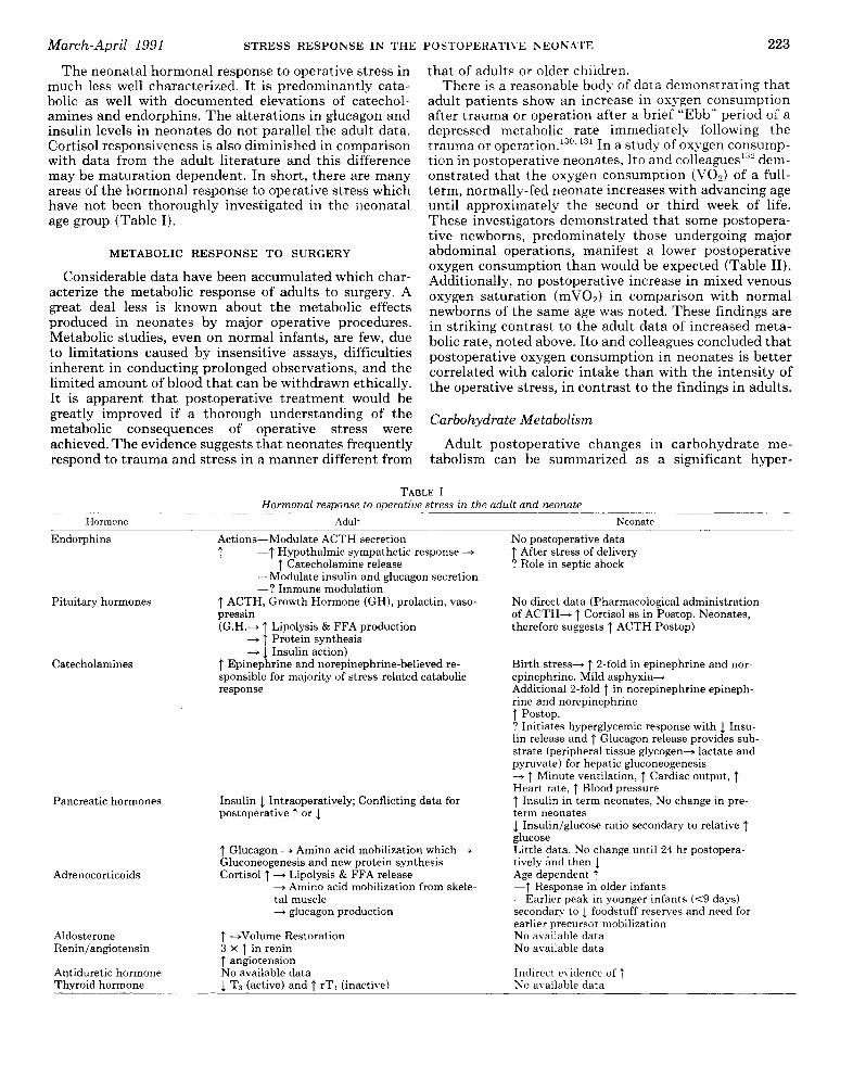
The neonatal hormonal response to operative stress inmuch less well characterized. It is predominantly cata-bolic as well with documented elevations of catechol-amines and endorphins. The alterations in glucagon andinsulin levels in neonates do not parallel the adult data.Cortisol responsiveness is also diminished in comparisonwith data from the adult literature and this differencemay be maturation dependent. In short, there are manyareas of the hormonal response to operative stress whichhave not been thoroughly investigated in the neonatalage group (Table I).
METABOLIC RESPONSE TO SURGERY
Considerable data have been accumulated which char-acterize the metabolic response of adults to surgery. Agreat deal less is known about the metabolic effects
produced in neonates by major operative procedures.Metabolic studies, even on normal infants, are few, dueto limitations caused by insensitive assays, difficultiesinherent in conducting prolonged observations, and thelimited amount of blood that can be withdrawn ethically.It is apparent that postoperative treatment would begreatly improved if a thorough understanding of themetabolic consequences of operative stress were
achieved. The evidence suggests that neonates frequentlyrespond to trauma and stress in a manner different from
that of adults or older children.There is a reasonable body of data demonstrating that
adult patients show an increase in oxygen consumptionafter trauma or operation after a brief ‘‘Ebb&dquo; period of adepressed metabolic rate immediately following thetrauma or operation. 130.131 In a study of oxygen consump-tion in postoperative neonates, Ito and colleagues 132 dem-onstrated that the oxygen consumption (V02) of a full-term, normally-fed neonate increases with advancing ageuntil approximately the second or third week of life.These investigators demonstrated that some postopera-tive newborns, predominately those undergoing majorabdominal operations, manifest a lower postoperativeoxygen consumption than would be expected (Table II).Additionally, no postoperative increase in mixed venousoxygen saturation (m V02) in comparison with normalnewborns of the same age was noted. These findings arein striking contrast to the adult data of increased meta-bolic rate, noted above. Ito and colleagues concluded thatpostoperative oxygen consumption in neonates is bettercorrelated with caloric intake than with the intensity ofthe operative stress, in contrast to the findings in adults.
Carbohydrate Metabolism
Adult postoperative changes in carbohydrate me-tabolism can be summarized as a significant hyper-
TABLE IHormonal response to operative stress in the adult and neonate
224
TABLE IIMetabolic response to operative stress in the adult and neonate
glycemic response both during and after surgery. Thiseffect may be the result of both an increase in glu-cose production as well as a diminution in peripheralglucose utilization, with a relative decrease in insulinconcentrations. 3,79,87,89,91,103,133-135
Pioneering work early in this century by Benedict andTalbot, who monitored the respiratory quotients (RQ) ofnormal newborn babies, demonstrated that as much as80% of the energy requirements is fulfilled by caloriesderived from fat.136 This is interesting in light of the factthat carbohydrates provide the main source of energy inthe fetus. However, soon after birth and even beforefeeding is started, a rapid fall in glycogen reserves hasbeen demonstrated. 117 In addition, the blood glucose con-centration is also known to fall in the early postnatalperiod.138 An increase of plasma free fatty acids (FFA)and ketone bodies has been documented to occur con-current with these changes in glucose and glycogen,adding support to the importance of fat-derived caloriesin the newborn as he/she changes his/her major meta-bolic foodstuff. 139,140
Unfortunately operations on neonates are frequentlyaccompanied by periods of starvation which may beprolonged, especially if the gastrointestinal tract is in-volved. The advent of hyperalimentation has aided some-what in altering this pattern. It is known that depot fataccounts for 10-15% of the body weight of the normalhuman neonate, and, as stated above, this may providethe main source of energy during the period of starvationsoon after birth. 141,142
Glucose
In 1968, intravenous glucose tolerance tests were per-formed on 14 newborn babies being operated upon for
abnormalities of the alimentary tract.143 The authorsobserved that six of these 14 infants had a greatly reducedtolerance to glucose administered by intravenous infu-sion. They noted a constant rate of glucose disappearancewhich was unrelated to the absolute glucose concentra-tion, in contrast to data in older children and adultswhose rate of disappearance varies with the rate ofadministration. Elphick and Wilkinson postulated asexplanations for these observations: 1) babies may beless able than adults to form glycogen from glucose, 2)there may be a temporary increased insulin dependencyin the newborn, and 3) the uptake of glucose by thetissues may be reduced by high circulating concentra-tions of hormones such as adrenaline and growth hor-mone. These authors also noted depression of the con-centration of free fatty acids after the injection of glu-cose, which suggested that the administered glucose mayhave had a fat-sparing action even when the Kt values(percent clearance of administered glucose from bloodper minute) were low. They concluded that the prolongeduse of parenteral glucose solutions might, in some cases,lead to severe hyperglycemia and that there is markedvariability between infants in their capacity to handleinfused glucose.
Elphick and Wilkinson also demonstrated a postop-erative increase in the blood glucose concentration toapproximately two times preoperative levels in newbornsbut noted that the glucose concentration returned tonormal within 12 hours. 143,144 This is in contrast to datafrom adult surgical patients where blood glucose levelsmay remain high for several days. These authors notedthe similarity of their findings to those of Pinter97 and
225
proposed that the elevation in blood glucose noted in thepostoperative period may be due to either increased
production or decreased utilization of glucose or a com-bination of the two. In an earlier study of glucose toler-ance testing in postoperative infants a diminished glu-cose utilization had been demonstrated by these inves-tigators.143 In attempting to explain this relativeintolerance, they cited the type of anesthesia as oneimportant contributory factor. The mechanism postu-lated was a direct effect by endogenous catecholaminesresulting in altered glucose metabolism with variationsin anesthetic methods effecting the degree of the cat-echolamine response. This concept has been confirmedin experimental studies with newborn rabbits and pup-pies.145, 146 The conclusion from these experiments wasthat endogenous sources of energy were capable of sup-plying a sufficient number of calories to satisfy therequirements of normal infants during starvation sec-ondary to congenital anomalies and after the surgicalcorrection of these anomalies but at significant metaboliccost to the patient.
In evaluating starvation, a condition which is fre-
quently linked with operative stress in newborns, Elphickand Wilkinson144 were unable to document hypoglycemiain normal birth weight infants starved for up to a week.They postulated that the glucose sparing action of freefatty acids was responsible and suggested a relationshipbetween maintenance of a normal blood sugar duringstarvation and body fat stores.
In a study utilizing stable carbon isotopes, Kalhan etap47 examined glucose turnover, systemic glucose pro-duction rate, and recycling of glucose carbon as an indi-cator of gluconeogenesis. Their study included six normalnewborn infants ranging in age from 2 hours to 3 days.The human fetus is known to be dependent upon themother for it’s glucose needs and no glucose productionhas been demonstrated in intrauterine life.148 There ishowever, the potential for fetal gluconeogenesis. Thepresence of key gluconeogenic enzymes in fetal liver
specimens has been documented.149 Kalhan et a114’° 14aconcluded from their stable isotope studies that gluco-neogenesis is not expressed in utero. However, duringthe perinatal period when the fetus’s neonate’s placentalor maternal supply of substrate including glucose is
abruptly interrupted, the newborn demonstrates a nor-mal capacity for systemic glucose production in order tomeet its metabolic needs. Their studies, however, suggestthat the source of the available glucose is chiefly fromthe process of glycogenolysis rather than gluconeogene-sis. These authors did demonstrate that gluconeogenesisvia the Cori cycle may be possible out as early as 2 hoursof life. They also noted that the contribution of recycledcarbon to systemic glucose production does not increaseduring the neonatal period and that glycogenolysis con-tinues to play the key role in maintaining adequateglucose availability for metabolic needs. They postulatedthat this predominant role of glycogenolysis over gluco-neogenesis may be the result of the ready availability ofsufficient glycogen stores due to the frequent feeding ofneonates. It is not difficult to imagine that this systemmay be interfered with by the stresses placed on an
infant by operation and interruption of dietary intake aswell as alteration in gastrointestinal function.
Unfortunately similar stable isotope studies to eluci-date stress-induced changes in postoperative glucose ho-meostatic mechanisms in neonates are nonexistent. Ithas been documented through elaborate arteriovenouscatheterization studies in adult patients with major in-jury and sepsis that there is increased splanchnic pro-duction of glucose in these states. 151 Concomitant in-creased uptake of gluconeogenic amino acids (primarilyalanine) and increased production of glucose and ureaimplicate increased gluconeogenesis rather than glyco-genolysis as the source of the glucose generated. Exoge-nous glucose sources were found by these investigatorsto diminish the observed gluconeogenic response in nor-mal control subjects but not in septic or postoperativepatients.Thus the available evidence in adult patients suggests
that increased glucose production from the splanchnictissues may contribute substantially to the hypergly-cemic response to surgical stress. Elphick’s studies show-ing altered glucose tolerance, however, also suggest a rolefor decreased glucose utilization in this state. Thus thehyperglycemic response is, in all likelihood, complex andmultifactorial. Not only the ability to utilize glucose inperipheral tissues in an impaired state, but also themechanism of utilization may be altered. In an experi-mental model of skin healing utilizing &dquo;carbon -labeledglucose to assess the various pathways of glucose metab-olism in wounded tissue resulting in ATP production, Imand Hoopes demonstrated a marked increase in glyco-lytic capacity (Embden-Meyerhof Pathway), as well asincreased activity of the pentose shunt and decreasedactivity of the Krebs cycle. Their wounded skin modelwas characterized by increased glucose utilization andlactate production. Seventy percent of ATP producedwas through the Embden-Meyerhof pathway in woundedtissue, rather than through the Krebs cycle as in normalskin.’Another postulated mechanism for the observed post-
surgical hyperglycemia and increase in blood lactate andpyruvate concentrations is the elevated adrenaline levelin response to the operative stress, resulting in activationof the Cori cycle. Thus, although the precise mechanismfor the hyperglycemic response is not clear, the clinicalimplications of significant hyperglycemia in a neonateare important. Significant changes in plasma osmolalitycan result from alterations in glucose levels. It has beendocumented in newborns that an increase in plasmaosmolality of greater than 25 mOsmol/kg over a periodof 4 hours can have profound detrimental effects on therenal cortex and cerebral cortex and may even precipitateintracranial hemorrhage in these infants. 151,152
Pyruvate, Lactate, Alanine
In addition to the marked postoperative hypergly-cemia, a number of investigators have demonstratedincreases in blood lactate and pyruvate concentrationsin postoperative adult patients. 151. 15-! Arteriovenous cath-eterization studies in adults have demonstrated thatadrenaline release during surgery increases lactate and
226
pyruvate production as a result of glycogen breakdownin peripheral tissues.8’
In addition, it is well known that injured tissues sur-rounding the surgical wound derive their energy mainlyfrom glycolysis and this may contribute to the increasedlactate production after surgery.’,’ 3
Other factors involved in the increased lactate levelsnoted include tissue hypoperfusion and hypoxia duringoperation.’ These changes may be related to anesthesiaor may be secondary to hypotension as a result of exces-sive blood loss or altered circulatory patterns duringsurgery. 155 Double isotope turnover studies in normalneonates have demonstrated that many metabolites areremoved from the circulation by the liver and are usedas substrates for hepatic gluconeogenesis. 147 It is evidentfrom the preceding discussion, however, that this maynot be the case in the stressed neonate.The significance of elevated blood alanine concentra-
tions in newborns is much less clear. Although alanineis known to be the key gluconeogenic amino acid inadults, some studies have documented hypoalaninemiain newborn infants receiving glucagon.&dquo;’, I&dquo; This effectwas postulated to be secondary to an increased splanch-nic utilization of alanine for glucagon-stimulated gluco-neogenesis. In a subsequent study of the relationships ofneonatal plasma levels of alanine, glucagon, and insu-lin,158 however, no correlation was observed betweenchanges in alanine and glucose concentrations, furtherclouding the role of gluconeogenic substrates and theprocess of gluconeogenesis in the hyperglycemic re-
sponse.In their 1987 study of the effects of fentanyl on post-
operative metabolic changes in neonates, Anand et a186demonstrated increases in blood lactate and pyruvateconcentrations during surgery in the nonfentanyl groupbut noted no similar changes in the fentanyl-treatedpatients. Twenty-four hours postoperatively blood lac-tate and pyruvate values had fallen below preoperativelevels in the nonfentanyl group of infants. Quantitativeblood levels of total gluconeogenic substrates (measuredas the sum of the blood concentrations of lactate, pyru-vate, alanine, and glycerol) in the nonfentanyl group ofbabies also increased substantially during surgery butfell by 24 hours postoperatively. These changes in thepostoperative period were attributed to the utilization ofthese substrates for gluconeogenesis with excess glucoseproduction in the nonfentanyl neonates. The differencesbetween the fentanyl and nonfentanyl groups were pos-tulated to be due to a blunting of the stress-inducedcatecholamine response in the fentanyl group with re-sultant diminution of catecholamine-induced postoper-ative changes.An earlier study from Anand’s group provides support
for this concept.84 Significant increases in blood concen-trations of lactate, pyruvate, total ketone bodies (aceto-acetate and hydroxybutyrate), and glycerol were notedduring surgery in their experimental group, which con-sisted of both term and preterm neonates. In this study,the levels of blood lactate remained elevated until 12hours after surgery, whereas all other metabolites meas-ured returned to preoperative levels by 6 hours postop-
eratively. No significant changes were seen in bloodconcentrations of the gluconeogenic amino acid alanineduring or after surgery. Levels of blood lactate showed ahigh degree of correlation with plasma adrenaline con-centrations at the end of surgery and 6 hours after
surgery. There was also a significant correlation betweenblood glycerol levels and plasma adrenaline and nor-adrenaline at the end of surgery.
In examining the response of a subgroup of six termand preterm neonates matched for degree of surgicalstress and anesthetic technique, some interesting find-ings were noted. No significant differences in blood glu-cose, pyruvate, total ketone bodies, or glycerol levels werenoted between these two groups of infants either beforeor after surgery. Preterm neonates did, however, dem-onstrate a significant rise in blood lactate concentrationsduring surgery whereas no similar change was noted inthe subgroup of term infants.
In summarizing their observations, these investigatorssuggested that the importance of the changes noted intheir study may be in the provision of substrates forhepatic gluconeogenesis in the postoperative period. Thesignificant hyperlactatemia noted during surgery in thepremature infants was postulated to be due to deficiencyof the key hepatic gluconeogenic enzymes, although sep-arate studies by Kalhan and Marsac do not support thishypothesis.148, 149
It is conceivable that the greater degree of hyperlac-tatemia in preterm neonates may possibly be related toless rich glycogen stores in their skeletal muscles incomparison with term neonates, with resultant increaseddependence on gluconeogenesis for substrate provisionin the face of an immature gluconeogenic mechanism.However, the rise in blood lactate levels may also be dueto tissue hypoxia caused by changes in peripheral circu-lation during anesthesia and surgery.From the above discussion, it is apparent that the
hyperglycemic response to surgery may result from acombination of increased production and decreased uti-lization of glucose. Many of the hormonal changes af-fecting the hyperglycemic response have been describedin the previous sections. These hormonal changes arecapable of inducing glycogenolysis as well as gluconeo-genesis following surgery. These responses are accom-panied by a decreased rate of glucose utilization partic-ularly during the surgical procedure itself. The relativecontributions of each of these mechanisms may dependon a variety of factors including the degree of surgicaltrauma as well as particulars of the anesthetic manage-ment. In addition nutritional supplementation seems toplay a modulating role.
Protein Metabolism
Acute malnutrition as a result of insufficient nutrientintake or the increased metabolic demands of illness ortrauma leads to increased catabolism of muscle proteinand a negative nitrogen balance. These changes, alongwith rapid utilization of energy substrate stores at a timewhen nutritional intake is often reduced, will drasticallyaffect the ability to heal wounds, combat infection, andhave sufficient muscular strength to breathe adequately,
227
all resulting in increased morbidity and mortality.Even the well-nourished may experience periods of de-bility after the injury of major surgery, which may relateto the reduction of protein reserves and energy stores.160Major operative stress in adult patients results in a
negative nitrogen balance. A compilation of factors ac-counts for this result. Among those well-documentedfactors are increased protein breakdown and decreasedprotein synthesis in extrahepatic tissues. In addition,there is increased utilization of amino acids for alternatepurposes such as gluconeogenesis, synthesis of acutephase reactants by the liver, as well as for synthesis ofcomponents of the healing process in injured tissues.Patients experiencing trauma or sepsis have been dem-onstrated to have rapid onset of muscle wasting, proteindepletion, and elevated urea excretion. 161,162 Therefore,an increased supply of amino acids is made available
during sepsis or trauma for energy production by gluco-neogenesis and oxidation. These additional amino acidsalso satisfy the requirements of the liver and other vis-ceral tissues for greatly accelerated synthesis of the pro-teins essential to immunologic defense, healing of
wounds, and maintenance of functions in the vital or-gans. The adult response to starvation is characterizedby sacrifice to visceral protein to furnish amino acids, asare needed, for gluconeogenesis and other purposes,whereas in stressful situations such as trauma or sepsis,muscle protein is degraded and the liver increases its
protein content. 163 Important as this metabolic responsemay be to survival, prolonged mobilization of amino acidsleads to devastating muscle weakness. In some patientsmuscle weakness is so great that ventilation is insuffi-cient to overcome the respiratory insufficiency associatedwith the traumatic event. Depletion of protein is also
accompanied by deterioration of cellular structure, in-sufficient production of acute phase reactants, and re-duced synthesis of other necessary proteins. Under suchconditions, patients are prone to perish from overwhelm-ing infection, culminating in multisystem failure.164The sick infant is particularly susceptible to the ad-
verse metabolic effects that a major illness or surgicaloperation may impose. Perioperative protein metabolicand nutritional status must be given special considera-tion in this population due to smaller body size, rapidgrowth, highly variable fluid requirements, and the im-maturity of certain organ systems. These factors, pluslow caloric reserves in the premature infant and sickchild, make an adequate caloric and amino acid intakeparticularly important. Consequently, the infant whosenutritional needs are not met, as a result of functionalor organic disorder of the gastrointestinal tract, can veryrapidly develop protein-calorie malnutrition and associ-ated complications. 165The most important clinical consequence of a catabolic
stress reaction is felt to be increased protein breakdownafter surgery. 166 The consequences, as outlined above,could be particularly deleterious in a postoperative neo-nate whose nutritional status is already tenuous.Adult urinary nitrogen excretion is increased following
major surgery and may remain elevated for as long as 5days postoperatively.167 Johnston’s study suggests that
an adult patient’s nitrogen losses are equivalent to 500 gof lean muscle tissue per day.&dquo; An important determi-nant of the magnitude and duration of the postoperativenitrogen loss appears to be the severity of surgicalstress. ’ There is some evidence in adult patients thatthe availability of ketone bodies as a metabolic fuel forperipheral tissues may result in a decreased need foramino acid oxidation in extrahepatic tissues (mainlyskeletal muscle) and may ultimately result in a decreasednitrogen loss and sparing of muscle protein sources. 169
In an elaborate study of muscle protein degradation innonoperated premature infants, Ballard et allss examinedcorrelations between energy input, nitrogen retention,weight gained and subsequent survival. They demon-strated that approximately 5% of total muscle proteinwas degraded daily. In addition the total and fractionalrates of protein breakdown demonstrated significant re-verse correlations with nitrogen retention but had norelationship to total energy input. Not surprisingly, pro-tein degradation was higher than average in infants whowere losing weight at the time of the balance study, andlower in infants who demonstrated weight gain. Proteindegradation was also higher in infants who died within2 weeks of the study. It was unclear whether this in-creased degradation in preterminal infants was relatedto events which stimulated muscle proteolysis, such assepsis, or was due to the underlying nitrogen status ofthe patients.&dquo;O Significantly, myofibrillar protein break-down was not different between infants fed orally andthose receiving total parenteral nutrition. These inves-tigators commented that the effects of nitrogen andenergy status on muscle protein degradation in prema-ture infants are different from changes reported in adulthumans or adult rats. To explain these findings theypostulated that the very limited energy reserves of thepremature infant may be responsible for the differencesobserved. They were unable to demonstrate any corre-lation between energy input in the premature infantsand rates of muscle protein breakdown (in contrast tolarge increases in total muscle protein breakdown seenin rats subjected to total energy restriction, and a slightdecrease in muscle protein breakdown in long-term fast-ing in obese adult humans).171,172 They attempted toexplain the differences between their results and thoseof other studies mentioned above on the basis of the sizeof the fat reserves, since there is evidence that ketonemia
produced by fat mobilization is accompanied by a lowerrate of muscle protein breakdown; and, since the pre-mature infant clearly has very little adipose tissue, thiswould explain the difference.169 Ballard et all&dquo; also dem-onstrated an increase in muscle protein degradation inpremature infants by demonstrating a negative nitrogenbalance or minimal retention of nitrogen daily. Theypostulated that this may be due to increased proteinbreakdown as a result of a demand for amino acids,which cannot be met simply by a decrease in proteinsynthesis. They state, however, that the response ob-served &dquo;is surely catastrophic if prolonged for any lengthof time, thus arguing forcibly that a substantial nitrogensupply to the premature infants should be maintained.&dquo;They also noted that the ratio of muscle protein deg-
228
radation to total body protein degradation was 7%, incontrast to a value of 30% found in adults. They attrib-uted this difference to the very small pool of muscleprotein in premature infants.173 In addition they specu-lated on the tissue sites of the remaining 93% of theprotein degradation in premature infants and postulatedthat organs which account for greater relative ratios ofneonatal body weight such as brain, liver, or skin maycontribute significantly to total body protein degrada-tion. The result of this visceral protein breakdown couldbe disastrous.
Colle and Paulsen, in a 1959 study of postoperativeinfants, demonstrated urinary nitrogen losses of 200 to300 mg/kg/24 h in contrast to 80 mg/kg/24 h in normalinfants.&dquo;’, 1’5 These losses were, however, transient andnot sustained.Duffy and Pencharz176 studied the effects of postop-
erative amino acid intake on urinary nitrogen losses andwhole body protein synthesis in 18 neonates. They con-cluded that a nitrogen intake of about 450 mg/kg/dshould meet the needs of a neonate in the immediatepostoperative period. This is higher than the data ofZlotkin suggests.&dquo;’ These investigators (Duffy andPencharzl’6) also documented an improved nitrogen bal-ance (the relationship between nitrogen intake and ni-trogen retention) in association with an increased nitro-gen intake. This improved balance was attributed to areduction in the fraction of amino nitrogen flux comingfrom the breakdown of endogenous protein. They wereunable to demonstrate any increase in skeletal musclebreakdown postoperatively in the infants studied bymeasuring urinary creatinine and 3-methyl histidine ex-cretion. Finally, this group was able to show postopera-tive nitrogen accretion, even during the 3 days immedi-ately postsurgery, but noted that nitrogen utilization maybe partially impaired postoperatively. On the basis ofthese studies, they recommended a nitrogen intake of450 mg/kg/d with a nonprotein energy intake of 85 to 90kcal/kg/d.Rickham4l in the late 1950’s demonstrated a postop-
erative increase in nitrogen excretion in neonates, butadded that, as in adults, this increase is no greater thanthat found when the patient is starved. Rickham alsonoted that a crude protein infusion in the form of plasmaresulted in rapid utilization of the infused protein. There-fore, he felt that postoperative plasma infusions servedthe double purpose of maintaining a normal plasmavolume and of restoring the plasma protein concentra-tion.
Winthrop et al demonstrated, in a prospective evalu-ation of pediatric trauma patients, significant increasesin basal metabolic rate (BMR), whole-body protein turn-over, protein synthesis, and urinary nitrogen excretion. 178These patients were found to have a negative nitrogenbalance due to the fact that protein breakdown increasedrelatively more than protein synthesis. The increase inprotein breakdown/turnover, synthesis, and nitrogen ex-cretion was found to have greatly exceeded the increasenoted in BMR (93%, 82%, and 56% us 14% increase inBMR) in these young (less than 10 years old) posttraumapatients. They were unable to demonstrate a correlation
between BMR and whole-body protein turnover, sug-gesting that changes in energy expenditure and proteinmetabolism following injury may be mediated by differ-ent mechanisms. They concluded that the metabolicresponse of pediatric patients to multiple trauma differsfrom that of adults and noted that pediatric traumapatients need not only increased caloric intake but, moreimportantly, a significant increase in protein intake inan attempt to optimize the balance between proteinsynthesis and breakdown. Thus, the differences fromadults include a much smaller change in total energyexpenditure in children and the lack of correlation be-tween an increased metabolic rate and whole-body pro-tein turnover.Minor or moderate surgical stress is capable of signif-
icantly decreasing plasma concentrations of total aminoacids. 179,180 Primarily responsible for this decrease is thereduction in the plasma concentrations of gluconeogenicamino acids, especially alanine.181 As mentioned earlier,Stjernstrom et al87 demonstrated, through catheteriza-tion studies of muscle vascular beds, that peripheralrelease of gluconeogenic amino acids accompanies ab-dominal operations. Therefore, postoperative productionof gluconeogenic amino acids may be a result of skeletalmuscle catabolism.&dquo;&dquo;, 1113 Gluconeogenesis then can takeplace as these amino acids are selectively taken up in thesplanchnic, hepatic and renal tissues.150,184 In contrastto the decreased systemic levels of gluconeogenic aminoacids noted above, there is evidence for elevations ofbranched chained amino acid (BCAAs) concentrationsin blood and within skeletal muscle. 179,180,185,186 This isin contrast to the decreased levels documented in pa-tients with liver disease. 187,111
Therefore, stress-induced muscle protein catabolismresults in the release of gluconeogenic amino acids whichare rapidly cleared from the blood to be metabolized bythe liver and splanchnic tissues, as well as release ofBCAAs into the circulation. These BCAAs are not nor-
mally metabolized in the liver but rather, initially, inmuscle and other peripheral tissues, where anabolism isdiminished in the stressed state, resulting in minimalutilization of these BCAAs186, 189, 190
In addition to being indicators of increased proteolyticactivity or altered protein metabolism there may be afunctional role for the alterations in amino acid patterns.For example, it has been suggested that arginine mayhave an immunoregulatory effect as well as an effect onpromoting nitrogen retention and wound healing.’9’ Ar-ginine may be important because of its effects in aug-menting immune responsiveness and in diminishing pro-tein catabolism.192, 193 Arginine is also known to stimulatesecretion of pituitary and pancreatic hormones.194,195Any of these roles may be important in the postoperativestressed state.
Studies by Clowes et ap96 have demonstrated a nearlyidentical relationship between the amino acid composi-tion of hydrolyzed muscle protein and the molar propor-tion of amino acids released into the bloodstream duringsepsis or following trauma, implicating the breakdown ofmuscle protein as the source of these amino acids. Asignificant breakthrough in the understanding of this
229
proteolytic response was made when Clowes et al197 dem-onstrated a circulatory peptide capable of inducing mus-cle proteolysis in sepsis and trauma. This group sug-gested that the proteolysis-inducing factor they isolatedfrom plasma was not one of the hormones usually se-creted in stressful situations; they postulated that it maybe a product released from leukocytes or macrophages inassociation with the activation of complement in thepresence of infection or tissue damage associated withtrauma or operation. This was substantiated by the ob-servation that septic patients’ plasma could induce pro-teolytic changes when incubated with normal skeletalmuscle. They reiterated that the increased supply ofamino acids is made available in sepsis or trauma notonly for energy production or oxidation, but, more im-portantly, to satisfy the requirements of the liver andother visceral tissues for a greatly accelerated synthesisof the proteins essential for immunological defense, heal-ing of wounds, and maintenance of function in vital
organs.Finley et al examined the effect of major operative
trauma on skeletal muscle metabolism in adult patientsreceiving a constant preoperative infusion of nutrients.8°They noted significant postoperative decreases in theplasma concentrations of the following amino acids: tau-rine, threonine, serine, glycine, alanine, citralline, amino-N-butyrate, methionine, histidine, arginine, glutamine,glutamate, and branched-chain amino acids includingvaline and isoleucine. At the same time there was apostoperative increase in amino acid release from a fore-arm muscle bed which was made up in large part by anincrease in the glycogenic amino acids, serine, threonine,glycine and alanine, by a marked increased in the releaseof branched-chain amino acids, and by an efflux oftaurine, methionine, phenylalanine, lysine, and arginine.They suggested that the visceral production of glucose isquantitatively matched by the net uptake of glucoseprecursors across the splanchnic bed.198 They noted thatinfused nutrients suppressed visceral gluconeogenesis inthese patients. Their results showed that new glucoseproduction was lower than what was observed in a fastingman suffering from trauma.199 This group (Finley) spec-ulated that the increased release of proline, methionine,arginine and phenylalanine from muscle may be relatedto higher requirements by healing wounds and an in-creased demand for the precursors of catecholamines.Hulton et a1,81 in 1985, demonstrated, in an animal
model, that hormonal blockade of the catabolic responsesto surgery by phentolamine and propanolol inhibited netskeletal muscle protein catabolism without alteringwhole-body nitrogen loss. This may prove clinically use-ful in that hormonal blockade may attenuate the post-traumatic catabolic response, preventing acceleratedskeletal muscle breakdown and body protein loss. Totalbody nitrogen loss in this study was, however, unaffectedindicating that skeletal muscle was spared at the expenseof other sources of amino nitrogen. The investigatorsspeculated that these other amino acids were derivedfrom the viscera. It is possible that hormonal blockadecould prevent the obligatory loss of skeletal muscle pro-tein in critically ill patients. Simultaneous nutritional
support might then provide the amino acids necessaryfor acute phase protein sy nthesis, gluconeogenesis, andwound repair.Warner et al,2°O in an experimental animal model,
demonstrated that infusion of catabolic hormones (glu-cagon, epinephrine, and corticosterone) resulted in in-creased amino acid uptake in the liver; however, thesecatabolic hormones had no effect on amino acid uptakein skeletal muscle. Total plasma amino acids were re-duced in the hormone-infused animals. They concludedthat stress-induced elevations of catabolic hormones are,at least in part, responsible for the augmented liveramino acid uptake but that these catabolic hormones arenot responsible for the reduced muscle amino acid uptakecharacteristic of sepsis or severe trauma. 211Moyer et al studying critically ill adult patients who
were septic or posttraumatic, examined concentrationsof various plasma substances in an effort to identifyplasma profiles reflective of patients’ prognosis. Theywere able to identify numerous amino acid fractionalconcentrations and patterns which had specific predic-tive value.
Studies of postoperative nitrogen balance in term neo-nates originate with Rickham in 1957. Since that time,several investigators have substantiated a strongly neg-ative nitrogen balance in response to surgical stress andhave demonstrated that this may persist for 72 to 96hours.41, 1’4° 202-204 These studies have demonstrated thatthe severity of the surgical stress is correlated with thedegree of nitrogen loss. In addition, it has been notedthat nitrogen loss postoperatively is greater in the neo-natal age group than in older infants subjected to similardegrees of surgical stress. 205,206 In a recent study of neo-nates undergoing either major or minor operative pro-cedures, a direct relationship was noted between thedegree of stress and the quantity of nitrogen loss.207
Zlotkin 117 in 1984, published a study assessing post-operative nitrogen balance in term neonates who hadreceived parenteral nutrition containing 300 to 600 mg/kg/24 h of nitrogen. These authors demonstrated nitro-gen retention in these infants and correlated this with
increasing nitrogen intake. They calculated that a nitro-gen intake of 280 mg/kg/24 h would be required toduplicate the nitrogen accretion rate of breast-fed in-fants. This study may be criticized on a methodologicalbasis due to the fact that these neonates had undergonevariable degrees of surgical stress and thus representeda nonhomogeneous group. In addition, the studies werecarried out at 7 to 9 days postoperatively, which is a timewhen the major hormonal and metabolic alterations in-duced by operative stress have returned to baseline. Asubsequent study of 18 preterm neonates undergoing avariety of surgical procedures examined early (0-72 hpostoperatively) changes in nitrogen retention, proteinsynthesis, and protein turnover.176 The authors demon-strated a strong correlation between nitrogen intake andnet nitrogen retention and concluded that the improvednitrogen utilization in the infants receiving the greatestquantity of parenterally administered amino acids wasdue to a reduced breakdown of endogenous proteins.
In a recent study, different levels of amino acid intake
230
were evaluated with regard to nitrogen retention, ratiosof whole body amino acid nitrogen flux, and proteinsynthesis and breakdown ratios in 18 infants during the72 hours immediately following surgery. 1-,6 One group ofinfants received 2.3 ± 0.4 g of amino acid/kg/d and theother 3.9 ± 0.5 g/kg/d. They demonstrated no differencesin amino acid flux or in synthesis and breakdown ofprotein. However, the group receiving the higher aminoacid intake had significantly greater net protein synthe-sis ratios. This improved nitrogen utilization in this
group was again achieved principally by a reduction inendogenous protein breakdown. There were no differ-ences between the two groups in urinary creatinine or 3-methylhistidine excretion. Since these two parametersreflect skeletal muscle protein turnover, the differencesbetween groups in nitrogen retention and protein turn-over appear to be mediated through visceral proteinsparing.Another marker of endogenous protein breakdown
which has received widespread attention is the molar 3-methylhistidine :creatinine ratio (3MH:Cr).’O’ The ra-
tionale for use of the 3 MH:Cr ratio is the belief that 3-methylhistidine originates from the breakdown of skel-etal muscle actin and myosin. A significant series ofassumptions accompany use of this ratio. First, thismolecule is excreted quantitatively in the urine followingits liberation from myofibrils. Second, the contributionof nonskeletal muscle (skin and gastrointestinal muscle)to the total 3 MH pool is negligible. Additionally, thisamino acid is not metabolized further and is not used forde novo protein synthesis following its liberation into thecirculation. Numerous studies have demonstrated thatthe urinary 3MH:Cr ratios correlate closely with the netnitrogen balance in preterm and term neonates. 168,208-210In addition, preterm neonates stressed by severe clinicalillness and manifesting a negative nitrogen balance andweight loss at the time of study have also demonstrateda markedly raised 3MH:Cr ratio.211 Additional studies inpostoperative term neonates have demonstrated a signif-icant increase in the 3MH:Cr ratio and in nitrogen lossin the first 72 hours after surgery.2&dquo; This finding waslater confirmed by the same investigators in pretermneonates undergoing surgery. 86 Reduction of the surgicalstress responses in preterm and term neonates by usingdifferent anaesthetic techniques such as halothane sup-plementation or fentanyl was found to inhibit these
changes in the urinary 3MH:Cr ratio.86, 212The most important clinical consequence of the cata-
bolic stress reaction has been said to be the increased
protein breakdown after surgery.211 During the first fewdays after birth human infants lose weight before resum-ing the rapid weight gain associated with intrauterinedevelopment. 166 Although much of the weight loss is
water, some of it reflects the breakdown of carbohydrateand lipid stores and the relatively high rate of total bodyand muscle protein degradation which may further con-tribute to the catabolic state in these infants.173,213 Theprevious study 166 failed to demonstrate any correlationbetween energy input in the premature infant and therate of muscle protein breakdown, in contrast to thefindings of Zlotkin 177 and Duffy and Pencharz. 176 Ballard
et al felt this may be due in part to the size of the fatreserves, since there is evidence that ketonemia producedby fat mobilization is accompanied by a lower rate ofmuscle protein breakdown.166 However, they pointed outthat the preterm human neonate has relatively little
adipose tissue and may be expected to demonstrate in-creased muscle protein catabolism when energy intake isminimal. They concluded by stating &dquo;Nevertheless weconsider it is not possible at the present time to reconcileall of the findings on energy restriction in such a waythat an expected response on muscle protein breakdowncan be stated.&dquo;
Pinter97 in 1973 examined the ratio of nonessential toessential amino acids in 17 infants undergoing surgery.He demonstrated a fall in the ratio during surgery witha gradual postoperative rise. Alpha-amino nitrogen dem-onstrated a slight, insignificant rise during surgery andthen remained at a constant level. Postoperatively, apronounced fall in alpha-amino nitrogen was observed.Due to the rise in the combined concentrations of theessential amino acids leucine, valine, and methionine,the nonessential:essential amino acid plasma ratio de-creases. In addition, the changes in the alpha-aminonitrogen level indicate a marked redistribution of thecirculating free amino acids. The rise in blood ureanitrogen (BUN) observed in the early postoperativeperiod is probably due to several factors, such as en-hanced protein breakdown, hemoconcentration, and oli-guria, which are well-known consequences of surgery.
In a later study of 29 infants undergoing a moderatedegree of surgical stress for correction of various congen-ital anomalies, Pinter215 was unable to demonstrate astatistically significant change in the ratio of nonessen-tial :essential amino acids. Also, no significant change inthe alpha-amino-nitrogen level was noted. They did sug-gest, however, that the plasma-free amino acid pool didchange dramatically both during and after surgery.
Therefore, from the adult studies outlined it can beconcluded that the negative nitrogen balance seen follow-ing moderate surgical stress is mainly due to a decreasein the rate of protein synthesis, while the rate of proteinbreakdown is unaltered or slightly increased. However,protein metabolism in patients exposed to severe degreesof surgery, trauma, or sepsis is characterized by a massivebreakdown of tissue proteins, with protein synthesisrates being unaltered, decreased, or in some cases,slightly increased.
It is in this latter group of critically ill patients thatthe therapeutic manipulation of protein metabolism mayprovide the greatest clinical benefit in terms of reductionof morbidity and mortality. The neonatal surgical dataindicate a higher degree of muscle protein degradationthan seen in adults, resulting in a strongly negativenitrogen balance in the postoperative period. It seemsthat the provision of adequate amounts of amino acids,either enterally or parenterally, is capable of, at leastpartially, ablating this degradation of endogenous pro-tein.
FAT METABOLISM
The postoperative state in adult patients produces acatabolic response which in addition to the already men-
231
tioned changes in carbohydrates and protein metabolismalso results in the mobilization of nonesterified fattyacids (NEFA’s) from adipose tissues as well as increasedformation of ketone bodies. These changes may be ofprime importance in providing an endogenous energysource in the posttraumatic state.Two decades ago Allison et aF16 documented increased
plasma concentrations of nonesterified fatty acids asso-ciated with a decreased glucose tolerance in a group ofpatients suffering burn injuries. Subsequent studies bythis same group in postoperative patients also demon-strated an increase in plasma nonesterified fatty acids.21’This increase was noted both preoperatively and intra-operatively. The preoperative increase was attributed tothe catabolic stimulus provided by the emotional stressof anticipating an operation. An increase of nonesterifiedfatty acids following trauma was confirmed in 1974; andthe extent of the response was correlated with the sever-ity of trauma.218The importance of the contribution of fat to energy
supply in a stressed state was illustrated by Kinney etal219 in a 1970 study in which they demonstrated, byindirect calorimetry, that as much as 75 to 90% of
postoperative energy requirements were supplied by fatmetabolism and the remainder was provided by protein.It may be necessary for these nonesterified fatty acids toundergo conversion by the liver to ketone bodies prior totheir utilization as an energy source. 220
Lipolysis of stored triglycerides and the control ofadipocyte lipolysis are important in mobilization of lipidin the injured patient. Lipolysis in the adipocyte is car-ried out by the enzyme hormone sensitive lipase(HSL).221 This enzyme complex (HSL) is affected by anumber of other circulating hormones, including thecatecholamines.
Forse et al, 222 in an in vitro study, noted that in traumathe beta-adrenergic responsiveness of adipocytes and thecatecholamine receptors on these cells were significantlydecreased. This may be interpreted as desensitization ofthe beta receptors with down regulation and indicatesincreased in vivo lipolysis early after injury. After 4 days,these changes had returned to normal.Wolfe et al,223 in a study of patients suffering from
severe burn injury, in which he utilized stable isotopetracers, demonstrated changes in the substrate cycleinvolving the simultaneous breakdown and synthesis ofstored triglycerides (triglyceride-fatty acid cycle). Therates of triglyceride-fatty acid and glycolytic-gluconeo-genic cycling were elevated in these patients by 450 and250%, respectively. These investigators concluded thatincreased substrate cycling contributes to the increasedthermogenesis and energy expenditure seen in severeburns and that increased triglyceride-fatty acid cyclingis due to beta-adrenergic stimulation. Therefore, theincreased metabolic rate observed may be secondary toincreased substrate cycling and not solely caused byincreased rates of protein synthesis.224Because the stress response associated with surgery
causes an elevation of plasma nonesterified fatty acidsand decreased insulin secretion, one would expect anincreased production of ketone bodies in response to
operative stress. However, several studies have shownthat the levels range from no change to a mild elevationto a substantial increase.225-228 It has been demonstratedthat patients who remain normoketonemic after majorsurgery are likely to manifest an increased nitrogen lossin comparison with patients who are hyperketonemicpostoperatively.169 Studies in trauma patients suggestthat the lack of ketogenesis is due to postinjury vaso-pressin release, the degree of which is directly propor-tional to the severity of injury.214, 220 Therefore, vasopres-sin may exacerbate protein catabolism and muscle wast-ing by suppressing ketogenesis in patients subjected tosevere trauma, major surgical stress, or sepsis.
In the human baby, depot fat accounts for 10 to 15%of body weight.141 From metabolic balance data, Hugheset all&dquo; calculated that only about 8% of body proteinwas catabolized when a 3-kg neonate was starved for 12days, yet 39% of the baby’s fat was used up. Because ofthis low metabolic conversion of protein, the ability toreduce peripheral glucose utilization would be of advan-tage to the starving neonate. Whether or not high freefatty acid (FFA) turnover can result in reduced periph-eral glucose uptake remains to be confirmed. Glycerolreleased from adipose tissue during lipolysis could be asource for supplementation or maintenance of bloodglucose levels.
In their experimental study of perinatal rats, Mayorand Cuezva noted that, in the suckling period, the oxi-dation of fatty acids, ketone body utilization, and activegluconeogenesis supply the bulk of energy and carboncomponents required to support the rapid growth rateduring this period.94 This metabolic process begins withan increase in the insulin/glucagon ratio that occurs withthe change to a carbohydrate-rich diet, which then ini-tiates the induction of lipogenesis at weaning.Anand et al demonstrated an increase in blood levels
of total ketone bodies and glycerol during surgery inneonates.84 They believed this increase was a reflectionof catecholamine-stimulated lipolysis and ketogenesis.They noted a strong correlation between serum levels ofglycerol and adrenaline and noradrenaline at the end ofan operation. In addition to their use as an energy source,they postulated that the ketone bodies in peripheraltissues, through the formation of citrate and the inhibi-tion of phosphofructokinase, may also further inhibit theperipheral utilization of glucose and contribute to thepostoperative hyperglycemia seen in neonates. 95,230 in astudy of the effectiveness of improved anesthetic man-agement through the use of halothane, this group dem-onstrated that concentrations of ketone bodies increased
during surgery in the group not receiving halothane butwere unchanged in the group receiving halothane, witha significant difference at the end of the operation. 212Plasma concentrations of nonesterified fatty acids (NE-FAs) were significantly higher in the group not receivinghalothane than in the other group at the end of, and 6hours after, operation. These responses indicate a greaterdegree of lipolysis, probably mediated by the release ofcatecholamines in the nonhalothane group and facili-tated by the decrease in the ratio of insulin to glucagonduring surgery in the group that did not receive halo-
232
thane. In addition, halothane suppresses the catecholam-ine response, which results in decreased lipolysis anddecreased formation of NEFAs.
Further data from this group have shown that, al-
though there is a marked hyperglycemia and mobilizationof gluconeogenic substrates during and after surgery, itis likely that these substrates are utilized in the periop-erative period.230 They postulate that the primary sourcesof energy in the surgical neonate are provided by themobilization of nonesterified fatty acids (NEFAs) fromadipose tissue and their conversion to ketone bodies inliver cells. Despite this potential physiological impor-tance, fat metabolism in surgical neonates and infantshas received little study. Pinter97 reported a substantialincrease in plasma NEFA values during surgery with afurther significant increase postoperatively, whereas El-phick and Wilkinson144 found no significant changes inNEFAs in the perioperative period. In the latter study,a decrease in plasma triglycerides was documented post-operatively, whereas the plasma concentration of lipo-proteins, phospholipids, and cholesterol were unchangedduring and after surgery. These responses could be al-tered, at least partially, by starvation since the neonatesin both the above studies received no nutritional supportfor variable periods before and during the study. Recentstudies in term neonates have shown that circulatingconcentrations of NEFAs, glycerol, and total ketonebodies increased significantly during surgery, but hadreverted to preoperative values by 6 hours postopera-tively.85 The significant increase in free fatty acids, glyc-erol, and total ketone bodies during surgery is indicativeof lipolysis and ketogenesis, mediated by intraoperativecatecholamine release, as evidenced by the strong corre-lation between blood glycerol concentrations and plasmaadrenaline and noradrenaline levels at the end of surgery.An earlier study in older infants undergoing inguinalherniorrhaphy documented a significant increase in
plasma NEFA values during surgery and concluded thatthis was indicative of lipolysis in response to the stressinduced by surgery.83 In contrast, in studies of neonatesundergoing cardiac operations, blood concentrations oftotal ketone bodies were found to be decreased signifi-cantly at the end of surgery and these remained belowpreoperative values at 6 hours postoperatively.231 Thesechanges were attributed to the effects of cardiopulmo-nary bypass (CPB) and two mechanisms were postulatedto account for this: 1) the substantial increase in bloodglycerol values noted may have been caused by the hep-arinization of blood just prior to CPB, secondary toheparin-induced activation of lipoprotein lipase and sub-sequent breakdown of plasma triglycerides (in additionto the glycerol produced from lipolysis in adipose tissue),and 2) the markedly decreased hepatic circulation anddecreased metabolic rate during CPB with deep hypo-thermia and circulatory arrest (DHCA) would preventthe utilization of this glycerol for gluconeogenesis andthe conversion of circulating NEFA’s into ketone bodies.
In addition to serving as an energy source, studies ofglycerol turnover in newborn infants have shown that75% of glycerol formed from lipolysis enters the gluco-neogenic pathway in the neonatal liver and contributes
to 5% of hepatic glucose production.232 The oxidation offree fatty acids by the neonatal liver may further stimu-late postoperative gluconeogenesis through the genera-tion of ATP to support gluconeogenesis, the productionof acetyl-CoA which activates pyruvate carboxylase, andthe provision of reducing equivalents for glyceraldehyde-3-phosphate dehydrogenase.233
In a study on PDA ligation in term and preterminfants, there was a significant rise in blood levels oflactate, pyruvate, total ketone bodies and glycerol by theend of the operative procedures, but by 6 hours postop-eratively the levels of all these metabolites had revertedto their preoperative levels.95
In nonoperated newborn babies who have not yet beenfed, the respiratory quotient and the blood glucose levelfall while the serum concentration of free fatty acids(FFA) rises, indicating a rapid change from carbohydrateto fat metabolism soon after birth.~43 In addition, liverand muscle glycogen reserves are reduced and the rate ofdisappearance of glucose administered by intravenousinfusion is decreased. 138,234-237 It also appears that proteinis less easily utilized for energy purposes during starva-tion at this time.238, 239 All this indicates that fat, ratherthan protein or carbohydrate, is being used for energyproduction in the newborn.
In a recent study, various surgical procedures led tovariable changes in the plasma concentration of free fattyacids.144 In addition, 4 to 24 hours after surgery, theplasma triglyceride level fell by an average of 25%, butlater rose. In contrast, in this study during starvation,plasma FFA concentrations rose during the first 2 daysof life and were very high between days 3 and 5. Plasmatriglycerides, cholesterol, phospholipids, and total ester-ified fatty acids also increased after birth. These resultssuggest that during starvation in the neonate there israpid mobilization of fat from adipose tissue stores anda reduction in the peripheral utilization of glucose. Thereis no evidence to suggest any impairment of fat mobili-zation or metabolism even after 7 days of starvation.After surgery, however, even though there is more rapidmobilization of fat, the rate of utilization is greater thanthe rate of mobilization, resulting in variable and evenreduced levels of the various lipids. These results ledthese authors to speculate that babies of normal birth-weight may be more able to cope with starvation andsurgical injury than is generally realized through thisrapid mobilization of stored fat.
Pinter 17 demonstrated that the average plasma FFAlevel showed a slight but significant increase at the endof surgery. At the 6th and 12th postoperative hour thisincrease was already more pronounced. The plasma FFAlevel, however, showed great individual variation. Al-though, in most cases, a marked increase in plasma FFAconcentration occurred, the average increase, because ofthe significantly different initial levels, did not achievestatistical significance. At 24 hours postoperatively, theincrease still persisted. As an explanation for the occa-sional absence of an increase in FFA postoperatively,Pinter 17 postulates that pronounced hyperglycemia di-rectly or indirectly inhibits the mobilization of FFA. Thefact that, after surgery, a rapid fall in blood glucagon
233
concentration is accompanied by a rise in plasma FFAlevels lends support to this explanation.
Pinter215, in a study of 29 newborns being operatedupon for congenital anomalies, described their metaboliccharacteristics between the lst and 7th postoperativeday. In these investigations, a decreased free fatty acidlevel was observed between the 2nd and 7th days,whereas during the operation, as well as on the first
postoperative day, a well-defined increase in the FFAlevel took place, which might have been caused by theresponse to the anesthetic and surgery (increased releaseof catecholamines and steroids, metabolic effects of an-esthetics, hypoxia, hypothermia, acidosis, etc). Althoughthe FFA level showed a tendency to decrease postopera-tively, it still remained higher than the preoperativevalue. This pattern of fat metabolism can be explainedby two facts: 1) in the postoperative period the complexhormonal and metabolic changes evoked by surgery arereturning to the preoperative level, and 2) the state ofhypoalimentation. These combined hormonal and met-abolic processes, which are also typical of the adaptationto extrauterine life, explain why it is difficult to find areciprocal relationship between glucose and FFA metab-olism.24o° 241 Elphick also failed to demonstrate a relation-ship between glucose and FFA levels in newborn infantsfollowing surgery.242
Studies in adults have indicated that the concentrationof circulating free fatty acids varies in response to sur-gical stress.243 Talbert et al’s83 study examined infantsundergoing bilateral inguinal hernia repair. Eleven of 13patients demonstrated a significant elevation in free fattyacid levels following surgery. The plasma-free fatty acidshave been identified as the major metabolite from themobilization of body adipose tissue/depot fat to be usedas an energy source. 240,244 The hydrolysis of triglyceridesis the major biochemical reaction in fat stores for theproduction of energy precursors. Mobilization of fattyacids is mediated by three central mechanisms: meta-bolic, hormonal, and neural. 141 Under conditions of star-vation a net release of FFA from the peripheral fat depotsis observed. Various hormones have been demonstratedto be active in the regulation of fatty acid mobilization.246Among the most important of these hormonal regulatorsare the catecholamines. These compounds have beenrecognized as potent stimulants of FFA mobilization. 241Concomitant increases in the rate of glycerol productionverify the fact that elevations in plasma-free fatty acidsare due to an absolute increase in the rate of hydrolysisof triglycerides. Animal experiments have emphasizedthe importance of this mechanism in producing postop-erative elevations in plasma FFAs.248 The importance ofthe innervation of fat stores in facilitating free fatty acidmobilization has been verified by experiments with in-nervated and denervated tissues.245 The sympatheticnervous system is a critical component of this process.Since norepinephrine is the chemical mediator at the
postganglionic sympathetic nerve ending, the final mech-anism of action may be similar to that observed followingthe parenteral administration of this compound. Theimportance of this system as a mechanism for mobilizingfree fatty acids has been documented in adults during
emotional stress.249 It is evident that circulating levels offree fatty acids are regulated by a variety of factors, manyof which also participate in the infant’s response tostress. The importance of this composite action is sug-gested in Talbert’s experiments in which he demon-strated an increase in plasma FFA in the absence of adiscernible increase in circulating catecholamines. Pre-vious investigators have demonstrated an elevation ofFFA in adults following cholecystectomy and inguinalherniorrhaphy.240 These results substantiate the sensitiv-ity of free fatty acid mobilization to the stimulus ofsurgical trauma and suggest the usefulness of the plasma-free fatty acid level as an index of the infant’s stressresponse.
In addition to the outlined mobilization of body fatstores, renewal of these stores has been suggested.Winthrop et all‘’8 showed that body fat increased post-operatively from day 0 to day 7 from 12.9 ± 0.6% to 14± 0.06% (p < 0.05) in 13 full-term infants undergoingsurgery at approximately 10 days of life. Although thisis a small but statistically significant increase in the bodyfat, the magnitude of the change falls within the rangeof experimental error for anthropometry. In this study,fat accounted for almost 60% of the new solid tissuesynthesized, which is in agreement with Fomon et al’sfigure of 56.6%250
REFERENCES
1. Bessey PQ, Watters JM, Aoki TT, Wilmore DW: Combinedhormonal infusion stimulates the metabolic response to injury.Ann Surg 200:264-281, 1984
2. Im MJC, Hoopes JE: Energy metabolism in healing skin wounds.J Surg Res 10:459-466, 1970
3. Wilmore DW: Glucose metabolism following severe injury. JTrauma 21:705-707, 1981
4. Elliott M, Albert KGMM: The hormonal and metabolic responseto surgery and trauma. IN: New aspects of clinical nutrition.Kleinberger G, Deutsch E. (eds). Karger, Basel, 1983 pp 247-270
5. Moyer E, Cerra F, Chenier R, et al: Multiple systems organ failure.VI. Death predictors in the trauma-septic state—the most criticaldeterminants. J Trauma 21:862-869, 1981
9. Rackow H, Salanitre E, Green LT: Frequency of cardiac arrestassociated with anesthesia in infants and children. Pediatrics28:697-704, 1961
10. Schweiss JF, Pennington DG: Anesthetic management of neo-nates undergoing pallative operations for congenital heart defects.Clev Clin Q 48:153-165, 1981
11. Liebig J: Die organisuhe chemie. IN: Ihrer anwendung auf phy-siologie und pathologie. Braunschvweig, Wiley and Putnam, NewYork, 1848
12. Bauer J: Ilbur Zerretzungsvorgange in thierkirper unter demkinflusse von blutentziehunger. Zeitschr Biol 8:567-603, 1872
13. Malcolm JD: The physiology of death from traumatic fever; astudy in abdominal surgery. Lancet 1:408-410, 460-462. 519-521,1893
r 14. Aub JC, Wu H: Studies in experimental traumatic shock II:Chemical changes in the blood. Am J Physiol 54:416-424, 1920
15. Bernard C: Chiens rendus diabetiques. Compt Rend Soc De Biol1:60-63, 1849
16. Bernard C: Lecons de physiologie experimentelle au college de
234
France. Paris, 185517. Bernard C: Lecons de physiologie operatoire. JB Balliere Et Fils.
Paris, 1879, pp 64018. Brown-Sequard: Des effets produits chez l’homme par des injec-
tions soucutoanees d’un liquide retire des testicules frais de cobageet de chien. Compt Rend Soc De Biol 9:415-454, 1889
19. Brown-Sequard: Des effets produits chez l’homme par des injec-tions soucutoanees d’un liquid retire des testicules frais de cobageet de chien. Lancet 2:105-107, 1889
20. Pringle H, Maunsell RCB, Pringle S: Clinical effects of etheranaesthesia on renal activity. Br Med J 2:542-543, 1905
21. Evans GH: The abuse of normal salt solutions. JAMA 57:2126-2127, 1911
22. Cannon WB: The Shattuck Lecture. The physiological factorsconcerned in surgical shock. Boston Med Surg J CLXXVI:859-867, 1917
23. Cannon WB: A consideration of the nature of wound shock.JAMA 70:611-617, 1918
24. Cannon WB: The Wisdom of the Body. Norton and Co., NewYork, 1932
25. Cuthbertson DP: The influence of prolonged muscular rest onmetabolism. Biochem J 23:1328-1345, 1929
26. Cuthbertson DP: Observations on the disturbance of metabolism
produced by injury to the limbs. Q J Med 1:233-246, 193227. Cuthbertson DP, Munro AN: A study of the effect of overfeeding
on the protein metabolism of man. Biochem J 31:694-705, 193728. Hayes MH, Coller FA: The neuroendocrine control of water and
electrolyte excretion during surgical anesthesia. Surg GynecolObstet 95:142-149, 1952
29. Selye H: The general adaptation syndrome and the diseases ofadaptation. J Clin Endocrinol 6:117-230, 1946
30. Moore FD, Ball MR: The Metabolic Response to Surgery. CharlesC Thomas Co., Springfield, 1952
31. Sandberg AA, Elk-Nes K, Sammels LT, et al: The effects ofsurgery on the blood levels and metabolism of 17-hydroxy-corti-costeroids in man. J Clin Invest 33:1509-1516, 1954
32. Hume DM, Egdahl RH: The importance of the brain in theendocrine response to injury. Ann Surg 150:697-712, 1959
33. von Bezold A: Ztschr Wissensch Zool 8:487, 185734. von Bezold A: Ztschr Wissensch Zool 9:240, 185835. Ylppo A: Neugebofenen-, Hunger-und intoxikatations acidosis in
ihren Beziehungen zueinander. Ztschr Kindrh 14:268-448, 191636. Marples E, Lippard VW: Acid-base balance of newborn infants.
II. Consideration of the low alkaline reserve of normal new-borninfants. Am J Dis Child 44:31-39, 1932
37. Marples E, Lippard VW: Acid-base balance of newborn infants.III. Influence of cowsmilk on the acid base balance of the bloodof new-born infants. Am J Dis Child 45:294-306, 1933
38. Marriott WM: Some phases of the pathology of nutrition in
infancy. Harvey Lecture 15:121-151, 191939. Moore RM: Acute intestinal obstruction in infants and children:
Physiological and pathological considerations. Mississippi Doctor23:554-556, 1946
40. Santulli TV: Intestinal obstruction in the newborn infant. JPediatr 44:317-337, 1954
41. Rickham PP: The metabolic response to neonatal surgery. Har-vard University Press, Cambridge, 1957
42. Suits GS, Bottsford JE Jr: The metabolic response to trauma.Resident Staff Phys 33:21-29, 1987
43. Anand KJS, Hickey PR: Pain and its effects in the human neonateand fetus. N Engl J Med 317:1321-1329, 1987
44. Valman HB, Pearson JF: What the fetus feels. Br Med J 280 :233-234, 1980
45. Anand KJS: The stress response to surgical trauma: From phys-iological basis to therapeutic implications. Proc Food Nutr Sci10:67-132, 1986
46. Atwelt SF, Kuhar MJ: Distribution and physiological significanceof opoid receptors in the brain. Br Med Bull 39:47-52, 1983
47. Dubois M, Pickar D, Cohen M, et al: Surgical stress in humans isaccompanied by an increase in plasma B-endorphin immunoreac-tivity. Life Sci 29:1249-1254, 1981
48. Cohen MR, Pickar D, Dubois M: Role of the endogenous opioidsystem in the human stress response. Psychiatr Clin North Am6:457-471, 1983
49. Pickar D, Cohen MR, Dubois M: The relationship of plasma
cortisol and B-endorphin immunoreactivity to surgical stress andpostoperative analgesic requirement. Gen Hosp Psychiatry 5:93-98, 1983
50. Cohen MR, Pickar D, Dubois M, et al: Stress-induced plasmabeta-endorphin immunoreactivity may predict postoperativemorphine usage. Psych Res 6:7-12, 1982
51. McIntosh TK, Bush HL, Palter M, et al: Prolonged disruption ofplasma beta-endorphin dynamics following surgery. J Surg Res38:210-215, 1985
52. Guillemin R, Vargo T, Rossier J, et al: Beta-endorphin andadrenocorticotropin are secreted concomitantly by the pituitarygland. Science 197:1367-1369, 1971
53. Van Loon GR, Appel NM, Ho D: B-endorphin induced stimula-tion of central sympathetic outflow: B-endorphin increasesplasma concentrations of epinephrine, norepinephrine, and do-pamine in rats. Endocrinology 109:46-53, 1981
54. Feldman M, Kiser RS, Unger RH, et al: Beta-endorphin and theendocrine pancreas. N Engl J Med 308:349-353, 1983
55. Hanbauer I, Kelly GD, Saiani L, et al: [Met5]-enkephalin-likepeptides of the adrenal medulla: Release by nerve stimulationand functional implications. Peptides 3:469-473, 1982
56. Bockman DE, Kirby ML: Neural crest interactions in the devel-opment of the immune system. J Immunol 135(suppl):766-768,1985
57. Smith EM, Harbour-McMenamin D, Blalock JE: Lymphocyteproduction of endorphins and endorphin-mediated immunoregu-latory activity. J Immunol 135(Suppl):779-782, 1985
58. Roszman TL, Jackson JC, Cross RJ, et al: Neuroanatomic andneurotransmitter influences on immune function. J Immunol135(Suppl):769-772, 1985
59. Holadam JW: Opiate antagonists in shock and trauma. Am JEmerg Med 2:8-12, 1983
60. Amaral JF, Gann DS: The effects of naloxone on glucose uptakeand metabolism in the isolated perfused hindlimb of the rat. JTrauma 25:1119-1125, 1985
62. Michie HR, Spriggs DR, Manogue KR, et al: Tumor necrosisfactor and endotoxin induce similar metabolic responses in hu-man beings. Surgery 104:280-286, 1988
63. Beutler B, Krochin N, Milsark IW, et al: Control of cachectin(tumor necrosis factor) synthesis: Mechanisms of endotoxin re-sistance. Science 232:977-980, 1986
64. Beutler B, Cerami A: Cachectin: More than a tumor necrosisfactor. N Engl J Med 316:379-385, 1987
65. Winters AJ, Oliver C, Colston C, et al: Plasma ACTH levels inthe human fetus and neonate as related to age and paturition. JClin Endocrinol Metab 39:269-273, 1974
66. Arai K, Yanaihara T, Okinaga S: Adrenocorticotropic hormonein human fetal blood at delivery. Am J Obstet Gynecol 125:1136-1140, 1976
67. Hindmarsh KW, Sankaran K: Endorphins and the neonate. CanMed Assoc J 132:331-334, 1985
68. Hindmarsh KW, Sankaran K, Watson VG: Plasma beta-endor-phin concentrations in neonates associated with acute stress. DevPharmacol Ther 7:198-204, 1984
69. Gautray JP, Jolivet A, Vielh JP, et al: Presence of immunoassay-able beta-endorphin in human amniotic fluid: Elevation in casesof fetal distress. Am J Obstet Gynecol 129:211-212, 1979
70. Puolakka J, Kaupila A, Leppaluoto J, et al: Elevated beta-endor-phin immunoreactivity in umbilical cord blood after complicateddelivery. Acta Obstet Gynecol Scand 61:513-514, 1982
71. Furman WL, Menke JA, Barson WJ, et al: Continuous naloxoneinfusion in two neonates with septic shock. J Pediatr 105:649-651, 1984
72. Sankaran K, Hindmarsh KW, Watson VG: Plasma beta endor-phin concentration in infants with apneic spells. Am J Perinatol1:331-334, 1984
73. Kuich TE, Zimmerman D: Could endorphins be implicated insudden-infant-death-syndrome. N Engl J Med 304:973, 1981
74. DeMaria EJ, Lilly MP, Gann DS: Potentiated hormonal responsesin a model of traumatic injury. J Surg Res 43:45-51, 1987
75. Newsome HH, Rose JC: The response of adrenocorticotrophichormone and growth hormone to surgical stress. J Clin Endocri-
235
nol 33:481-487, 197176. Gann DS, Lilly MP: The neuroendocrine response to multiple
trauma. World J Surg 7:101-118, 198377. Boix-Ocha J, Martinez Ibanez V, Potau N, et al: Cortisol response
to surgical stress in neonates. Pediatr Surg Int 2:267-270, 198778. Halter JB, Pflug AE, Porte D: Mechanism of plasma catecholam-
ine increases during surgical stress in man. J Clin EndocrinolMetab 45:936-944, 1977
79. Watters JM, Bessey PQ, Dinarello CA, et al: Both inflammatoryand endocrine mediators stimulate host response to sepsis. ArchSurg 121:179-190, 1986
80. Finley RJ, Inculet RI, Pace R, et al: Major operative traumaincreases peripheral amino acid release during the steady-stateinfusion of total parenteral nutrition in man. Surgery 99:491-499, 1986
81. Hulton N, Johnson DJ, Smith RJ, et al: Hormonal blockademodifies posttraumatic protein catabolism. J Surg Res 39:310-315, 1985
82. Nakai T, Yamada R: The secretion of catecholamines in newbornbabies with special reference to fetal distress. J Perinatol Med6:39-45, 1978
83. Talbert JL, Karmen A, Graystone JE, et al: Assessment of theinfants response to stress. Surgery 61:626-633, 1967
84. Anand KJS, Brown MJ, Causon RC, et al: Can the human neonatemount an endocrine and metabolic response to surgery? J PediatrSurg 20:41-48, 1985
85. Anand KJS, Brown MJ, Bloom SR, et al: Studies on the hormonalregulation of fuel metabolism in the human newborn infant
undergoing anesthesia and surgery. Hormone Res 22:115-128,1985
86. Anand KJS, Sippell WG, Aynsley-Green A: Randomized trial offentanyl anaesthesia in preterm babies undergoing surgery: Ef-fects on the stress response. Lancet 8527:243-248, 1987
87. Anand KJS, Sippel WG, Schofield NM, et al: Does halothaneanaesthesia decrease the metabolic and endocrine stress re-
sponses of newborn infants undergoing operation? Br Med J296:668-672, 1988
88. Stjernstrom H, Jorfeldt L, Wiklund L: The influence of abdominalsurgical trauma upon the turnover of some blood-borne metabo-lites in the human leg. JPEN 5:207-214, 1981
89. Ross H, Johnston IDA, Welborn TA, et al: Effect of abdominaloperation on glucose tolerance and serum levels of insulin, growthhormone, and hydrocortisone. Lancet 2:563-566, 1966
90. Giddings AEB: The control of plasma glucose in the surgicalpatient. Br J Surg 61:787-792, 1974
91. Wright PD, Henderson K, Johnston IDA: Glucose utilization andinsulin secretion during surgery in man. Br J Surg 61:5-8, 1974
92. Halter JB, Beard JC, Porte D: Islet function and stress hypergly-caemia: Plasma glucose and epinephrine interaction. Am J Phys-iol 10:E47-E52, 1984
93. Mallette LE, Exton JH, Park DR: Effects of glucagon on aminoacid transport and utilization in the perfused rat liver. J BiolChem 244:5724-5728, 1979
94. Mayor F, Cuezva JM: Hormonal and metabolic changes in theperinatal period. Biol Neonate 48:185-196, 1985
95. Anand KJS, Aynsley-Green A: Metabolic and endocrine effectsof surgical ligation of patent ductus arteriosus in the humanpreterm neonate: Are there implications for further improvementof postoperative outcome? Mod Probl Paediatr 23:143-157, 1985
96. Sperling MA: Integration of fuel homeostasis by insulin andglucagon in the newborn. Mongr Pediatr 16:39-58, 1982
97. Pinter A: The metabolic effects of anesthesia and surgery in thenewborn infant. Zeitschr Kinderchir 12:149-162, 1973
98. Russell RCG, Walker CJ, Bloom SR: Hyperglucagonaemia in thesurgical patient. Br Med J 1:10-12, 1975
99. Shamoon H, Hendler R, Sherwin RS: Synergistic interactionsamong antiinsulin hormones in the pathogenesis of stress hyper-glycemia in humans. J Clin Endocrinol Metab 52:1235-1241, 1981
100. Wilmore DW: Hormonal responses and their effect on metabo-lism. Symposium on response to infection and injury. II. SurgClin North Am 56(5):999-1018, 1976
101. Eigler N, Sacca L, Sherwin RS: Synergistic interactions of phys-iologic increments of glucagon, epinephrine and cortisol in thedog. J Clin Invest 63:114-123, 1979
102. Wolfe BM, Culebras JM, Aoki TT, et al: The effect of glucagon
on protein metabolism in normal man. Surgery 86:248-257. 1979103. Alberti KGMM, Batstone GF. Foster KJ. et al: Relative role of
various hormones mediating the metabolic response to injury.JPEN 4:141-146, 1980
104. Mohler JL, Michael KA, Freedman AM, et al: The serum andurinary cortisol response to operative trauma. Surg Gynecol Ob-stet 161:445-449, 1985
105. Helmreich ML, Jenkins D, Swan H: The adrenal cortical responseto surgery. II. Changes in plasma and urinary corticosteroid levelsin man. Surgery 41:895-909, 1957
106. George JM, Reier CE, Lanese RR, et al: Morphine anesthesiablocks cortisol and growth hormone response to surgical stress inhumans. J Clin Endocrinol Metab 38:736-741, 1974
107. Lines JG, Loder RE, Millar RA: Plasma cortisol responses duringneurosurgical and abdominal operations. Br J Anaesth 43:1136-1144, 1971
108. Solomon S, Bird CE, Ling W, et al: Formation and metabolismof steroids in the fetus and placenta. Recent Progr Horm Res23:297-347, 1967
109. Stang HJ, Gunnar MR, Snellman L, et al: Local anesthesia forneonatal circumcision. Effects on distress and cortisol response.JAMA 259:1507-1511, 1988
110. Williamson PS, Williamson ML: Physiologic stress reduction bya local anesthetic during newborn circumcision. Pediatrics 71:36-40, 1983
111. Williamson PS, Evans ND: Neonatal cortisol response to circum-cision with anesthesia. Clin Pediatr 25:412-415, 1986
112. Talbert LM, Kraybill EN, Potter HD: Adrenal cortical responseto circumcision in the neonate. Obstet Gynecol 48:208-210, 1976
113. Gunnar MR, Malone S, Vance G, et al: Coping with aversivestimulation during the neonatal period: Quiet sleep and plasmacortisol levels during recovery from circumcision. Child Dev56:824-834, 1985
114. Hillman DA, Giroud CJ: Plasma cortisone and cortisol levels atbirth and during the neonatal period. J Clin Endocrinol Metab25:243-248, 1965
115. Kirya C, Ertthmann MW: Neonatal circumcision and peniledorsal nerve block: A painless procedure. J Pediatr 96:998-1000,1978
116. Cochrane JPS: The aldosterone response to surgery and the
relationship of this response to postoperative sodium retention.Br J Surg 65:744-747, 1978
117. Enquist A, Blichert-Toft M, Olgaard K, et al: Inhibition of aldo-sterone response to surgery by saline administration. Br J Surg65:224-227, 1978
118. Wright PD, Johnston IDA: The effects of surgical operation ongrowth hormone levels in plasma. Surgery 77:479-486, 1975
119. Ward HC, Halliday S, Sim AJW: Protein and energy metabolismwith biosynthetic human growth after gastrointestinal surgery.Ann Surg 206:56-61, 1987
120. Kostyo JL, Keagan CR: The biology of growth hormone. Phar-macol Ther 2:591-604, 1976
121. Milne EMG, Elliott MJ, Pearson DT, et al: The effect on inter-mediary metabolism of open-heart surgery with deep hypothermiaand circulatory arrest in infants of less than 10 k body weight.Perfusion 1:29-40, 1986
122. Robertson D, Michelakis AM: Effect of anesthesia and surgeryon plasma renin activity in man. J Clin Endocrinol Metab 34:831-836, 1972
123. Bailey DR, Miller ED, Kaplan JA, et al: The renin-angiotensinaldosterone system during cardiac surgery with morphine-nitrousoxide anesthesia. Anesthesiology 42:538-544, 1975
124. Brandt MR, Olgaard K, Kehlet H: Epidural analgesia inhibits therenin and aldosterone response to surgery. Acta Anesth Scand23:267-272, 1979
125. Fiselier T, Monnens L, Moerman E, et al: Influence of the stressof venipuncture on basal levels of plasma renin activity in infantsand children. Int J Pediatr Nephrol 4:181-185, 1983
127. Brandt M, Kehlet H, Skovsted L, et al: Rapid decrease in plasmatriiodothyronine during surgery and epidural analgesia independ-ent of afferent neurogenic stimuli and of cortisol. Lancet 2:1333-1336, 1976
128. Prescott RWG, Yeo PPB, Watson MJ, et al: Total and free
236
thyroid hormone concentrations following elective surgery. J ClinPathol 32:321-324, 1979
129. Hasselgren P, Chen I, James JH, et al: Studies on the possiblerole of thyroid hormone in altered muscle protein turnover duringsepsis. Ann Surg 206:18-24, 1987
131. Cuthbertson DP: Protein metabolism in relation to energy needs.Metabolism 8:787-808, 1959
132. Ito T, Iyomasa Y, Inoue T: Changes in the postoperative minimaloxygen consumption of the newborn. J Pediatr Surg 11:495-503,1976
133. Bromage PR, Shibata HR, Willoughby HW: Influence of pro-longed epidural blockade on blood sugar and cortisol response tooperations upon the upper part of the abdomen and thorax. SurgGynecol Obstet 132:1051-1056, 1971
134. Kusaka M, Ui M: Activation of the cori cycle by epinephrine. AmJ Physiol 232:E145-E155, 1977
135. Mills NL, Beaudet RL, Isom OW, et al: Hyperglycaemia duringcardiopulmonary bypass. Ann Surg 177:203-205, 1973
136. Benedict FG, Talbot FB: The physiology of the newborn infant.Character and amount of the catabolism. Carnegie Inst Pub No.233, Washington, 1915
137. Sheeley HJ: Glycogen reserves and their changes at birth and inanoxia. Br Med Bull 17:137-143, 1961
138. Cornblath M, Ganzon AF, Nicolopoulos D, et al: Studies ofcarbohydrate metabolism in the newborn infant. III. Some factorsinfluencing the capillary blood sugar and the response to glucagonduring the first hours of life. Pediatrics 27:378-389, 1961
139. Novak M, Melichar V, Hahn P, et al: Levels of lipids in the bloodof newborn infants and the effect of glucose administration.Physiol Bohemoslov 10:488-492, 1961
140. Persson B, Gentz J: The pattern of blood lipids, glycerol andketone bodies during the neonatal period, infancy and childhood.Acta Pediatr Scand 55:353-362, 1966
141. Widdowson EM, Spray CM: Chemical development in utero. ArchDis Child 26:205-214, 1951
142. Hughes EA, Stevens LH, Wilkinson AW: Some aspects of star-vation in the newborn baby. Arch Dis Child 39:598-604, 1964
143. Elphick MC, Wilkinson AW: Glucose intolerance in newborninfants undergoing surgery for alimentary tract anomalies. Lancet539-541, 1968
144. Elphick MC, Wilkinson AW: The effects of starvation and sur-gical injury on the plasma levels of glucose, free fatty acids, andneutral lipids in newborn babies suffering from various congenitalanomalies. Pediatr Res 15:313-318, 1981
145. Elphick MC: The effect of starvation and injury on the utilizationof glucose in newborn rabbits. Biol Neonate 17:399-409, 1971
146. Pinter A, Schafer J: Metabolic effects of anesthesia and surgerynewborn: blood glucose, plasma free fatty acids, free amino acidand blood lactate level in newborn puppies. Acta Paediatr AcadSci Hung 14:85-90, 1973
147. Kalhan SC, Bier DM, Savin SM, et al: Estimation of glucoseturnover and 13C recycling in the human newborn by simultaneous[1-13C] glucose and [6.6-2H2] glucose tracers. J Clin EndocrinolMetab 50:456-460, 1980
148. Kalahan SC, C’Angelo LJ, Savin S, et al: Glucose production inpregnant women at term gestation: Sources of glucose for humanfetus. J Clin Invest 63:388-394, 1979
149. Marsac C, Saudubray JM, Moncion A, et al: Development ofgluconeogenic enzymes in the liver of human newborns. BiolNeonate 28:317-325, 1976
150. Gump FE, Long CL, Geiger JW, et al: The significance of alteredgluconeogenesis in surgical catabolism. J Trauma 15:704-712,1975
151. Finberg L: Dangers to infants caused by changes in osmolalconcentration. Pediatrics 40:1031-1034, 1967
152. Arant BS, Gooch WM: Effects of acute hyperglycemia on thecentral nervous system of neonatal puppies. Pediatr Res 12:549,1978
153. Bent JM, Paterson JL, Mashiter K, et al: Effects of high-dosefentanyl anesthesia on the established metabolic and endocrineresponse to surgery. Anesthesia 39:19-23, 1984
154. Walsh ES, Traynor C, Paterson JL, et al: Effect of differentintraoperative fluid regimenson circulating metabolites and in-
sulin during abdominal surgery. Br J Anaesth 55:135-140, 1983155. Bunker JP: Metabolic acidosis during anesthesia and surgery.
Anesthesiology 23:107-123, 1962156. Felig P, Pozefsky T, Marliss E, et al: Alanine: Key role in
gluconeogenesis. Science 167:1003-1004, 1970157. Reisner SH, Aranda JV, Colle E, et al: The effect of intravenous
glucagon on plasma amino acids in the newborn. Pediatr Res7:184-191, 1973
158. DeLamater PV, Sperling MA, Fiser RH, et al: Plasma alanine:Relation to plasma glucose, glucagon and insulin in the neonate.J Pediatr 85:702-706, 1974
159. Studley HO: Percentage of weight loss. A basic indicator of
surgical risk in patients with chronic peptic ulcer. JAMA 106:458-460, 1936
161. Cuthbertson DP: The disturbance of metabolism produced bybony and nonbony injury, with notes on certain abnormal con-ditions of bone. Biochem J 24:1244-63, 1930
162. Duke JH, Jorgensen SB, Broell JR, et al: Contribution of proteinto caloric expenditure following injury. Surgery 68:168-174, 1970
163. Ryan NT: Metabolic adaptations for energy production duringtrauma and sepsis. Surg Clin North Am 56:1073-1090, 1976
164. McMenamy RH, Birkhahn R, Oswald G, et al: Multiple systemsorgan failure: I. The basal state. J Trauma 21:99-114, 1981
165. Coran, AG: Nutrition of the surgical patient. IN Pediatric Surgery,4th ed, Welch KJ (ed). Chapter 10, Yearbook Publishers, 1986
166. Ballard FJ, Tomas FM, Pope LM, et al: Muscle protein degrada-tion in premature human infants. Clin Sci 57:535-544, 1979
167. Fleck A: Protein metabolism after surgery. Proc Nutr Soc 39:125-132, 1980
168. Johnston IDA: Endocrine aspects of the metabolic response tosurgical operation. Ann R Coll Surg Engl 35:270-286, 1964
169. Rich AJ, Wright PD: Ketosis and nitrogen excretion in under-nourished surgical patients. JPEN 3:350-354, 1979
170. Wannemacher RW, Dinterman RE: Total body protein catabo-lism in starved and infected rats. Am J Clin Nutr 30:1510-1511,1977
171. Ogata ES, Foung SKH, Holliday MA: The effects of starvationand refeeding on muscle protein synthesis and catabolism in theyoung rat. J Nutr 108:759-765, 1978
172. Marliss EB, Murray FT, Nakhooda AF: The metabolic responseto hypocaloric protein diets in obese men. J Clin Invest 62:468-479, 1978
173. Tomas FM, Ballard FJ, Pope LM: Age-dependent changes -in therate of myofibrillar protein degradation in humans as assessed by3-methylhistidine and urinative excretion. Clin Sci 56:341-346,1979
174. Colle, Paulsen: Response of the newborn infant to major surgery.I. Effects on water, electrolyte, and nitrogen balance. Pediatrics23:1063-1084, 1959
175. Hansen JDL, Smith CA: Effects of withholding fluid in theimmediate postnatal period. Pediatrics 12:99-113, 1953
176. Duffy B, Pencharz P: The effects of surgery on the nitrogenmetabolism of parenterally fed human neonates. Pediatr Res20:32-35, 1986
178. Winthrop AL, Jones PJH, Schoeller DA, et al: Changes in thebody composition of the surgical infant in the early postoperativeperiod. J Pediatr Surg 22:546-549, 1987
179. Johnston IDA, Dale G, Craig RP, et al: Plasma amino acidconcentrations in surgical patients. JPEN 4:161-164, 1980
180. Vinnars E, Bergstrom J, Furst P: Influence of the postoperativestate on the intracellular free amino acids in human muscle tissue.Ann Surg 182:665-671, 1975
181. Elia M, Ilic V, Bacon S, et al: Relationship between the basalblood alanine concentration and the removal of an alanine loadin various clinical states in man. Clin Sci 58:301-304, 1980
182. Karl IE, Garber AJ, Kipnis DM: Alanine and glutamine synthesisand release from skeletal muscle. J Biol Chem 251:844-860, 1976
183. Muhlbacher F, Kapadia CF, Colpoys MF, et al: Effects of gluco-corticoids on glutamine metabolism in skeletal muscle. Am JPhysiol 10:E75-E83, 1984
41:251-256, 1985 185. Dale G, Young G, Latner AL, et al: The effect of surgical operation
on venous plasma amino acids. Surgery 81:295-301, 1977186. Wedge JH, DeCampos R, Kerr A: Branched-chain amino acids,
nitrogen excretion, and injury in man. Clin Sci Mol Med 50:393-399, 1976
187. Fischer JE, Yoshimura N, Aguire A, et al: Plasma amino acids inpatients with hepatic encephalopathy. Am J Surg 127:40-47, 1974
188. Ansley JD, Issacs JW, Rikkers LF, et al: Quantitative tests onnitrogen metabolism in cirrhosis in relation to other manifesta-tions of liver disease. Gastroenterology 75:570-579, 1978
189. McMenamy RM, Shoemaker WC, Richmond JE, et al: Uptakeand metabolism of amino acids by the dog liver perfused in situ.Am J Physiol 202:407-414, 1962
190. Elia M, Farrell R, Iilc V, Smith R, et al: The removal of infusedleucine after injury, starvation, and other conditions in man. ClinSci 59:275-283, 1980
192. Saito H, Trocki O, Wang S, et al: Metabolic and immune effectsof dietary arginine supplementation after burn. Arch Surg122:784-789, 1987
193. Sitren HS, Fisher H: Nitrogen retention in rats fed on dietsenriched with arginine and glycine: I. Improved N retention aftertrauma. Br J Nutr 37:195-208, 1977
194. Barbul A, Rettura G, Levenson MS, et al: Wound healing andthymotropic effects of arginine: A pituitary mechanism of action.Am J Clin Nutr 37:786-794, 1983
195. Mulloy AL, Kari FW, Visek WJ: Dietary arginine, insulin secre-tion, glucose tolerance and liver lipids during repletion of protein-depleted rats. Horm Metab Res 14:471-475, 1982
196. Clowes JHA Jr, Randall HT, Cha CJ: Amino acid and energymetabolism in septic and traumatized patients. JPEN 4:195-205,1980
197. Clowes JHA, George BC, Villee CA, et al: Muscle proteolysisinduced by a circulating peptide in patients with sepsis or trauma.N Engl J Med 308:545-552, 1983
198. Garber AJ, Menzel PH, Boden G, et al: Hepatic ketogenesis andgluconeogenesis in humans. J Clin Invest 54:981-989, 1971
199. Wilmore DW, Goodwin CW, Aulick LH, et al: Effect of injuryand infection on visceral metabolism and circulation. Ann Surg192:491-504, 1980
200. Warner BW, James JH, Hasselgren PO, LaFrance R, Fischer JE:Effect of catabolic hormone infusion on argan amino acid uptake.J Surg Res 42:418-424, 1987
201. Hasselgren PO, James JH, Fischer JE: Inhibited muscle aminoacid uptake in sepsis. Ann Surg 203:360-365, 1986
202. Knutrud O: The Water and Electrolyte Metabolism in the New-born Child after Major Surgery. Scandinavian University Books,Universitetsforlaget, Oslo, Norway, 1965
203. Hughes EA, Stevens LH, Toms DA, et al: Esophageal atresia:Metabolic effects of operation. Br J Surg 52:403-410, 1965
204. Wilkinson AW, Hughes EA, Stevens LH: Neonatal duodenalobstruction. The influence of treatment on the metabolic effectsof operation. Br J Surg 52:410-424, 1965
205. Sukarochano K, Motai Y, Slim M, et al: Postoperative proteinmetabolism in pediatric surgery. Surg Gynecol Obstet 121:79-90,1965
206. Grewal RS, Mampilly J, Misra TR: Postoperative protein metab-olism and electrolyte changes in pediatric surgery. Int Surg51:142-148, 1969
207. Greenwall MJ, Kettlewell MGW, Gough MH: Nitrogen require-ment for postoperative parenteral nutrition in neonates. ActaTherap 9:5-9, 1983
208. Young VR, Munro HN: N T-methylhistidine (3-methylhistidine)and muscle protein turnover: An overview. Fed Proc Fed Am SocExp Biol 37:2291-2300, 1978
209. Burgoyne JL, Ballard FJ, Tomas FM, et al: Measurements ofmyofibrillar protein breakdown in newborn human infants. ClinSci 63:421-427, 1982
210. Anand KJS: Metabolic and Endocrine Effects of Surgery andAnesthesia in the Human Newborn Infant. D Phil Thesis, Uni-versity of Oxford, 1985
211. Seashore JH, Huszar G, Davis EM: Urinary 3-methylhistidine/creatinine ratio as a clinical tool: Correlation between 3-methyl-
histidine excretion and metabolic and clinical states in healthyand stressed premature infants. Metabolism 30:959-969, 1981
212. Anand KJS, Sippell WG, Schofield NM, et al: Does halothaneanaesthesia decrease the metabolic and endocrine stress responseof newborn infants undergoing operation? Br Med J 296:668-672, 1988
213. Pencharz PB, Steffee WP, Cochran W, et al: Protein metabolismin human neonates: Nitrogen balance studies, estimated obliga-tory losses of nitrogen and whole-body turnover of nitrogen. ClinSci Mol Med 52:485-498, 1977
214. Williamson DH, Farrell R, Kerr A, et al: Muscle-protein catabo-lism after injury in man, as measured by urinary excretion of 3-methylhistidine. Clin Sci and Molecular Med 52:527-533, 1977
215. Pinter A: Metabolic changes in newborn infants following surgicaloperations. Acta Pediatr Acad Sci Hungary 16:171-180, 1975
216. Allison SP, Hinton P, Chamberlain MJ: Intravenous glucosetolerance, insulin and free fatty acid levels in burned patients.Lancet II:1113-1116, 1968
217. Allison SP, Tomlin PJ, Chamberlain MJ: Some effects of anaes-thesia and surgery on carbohydrate and fat metabolism. Br JAnaesth 41:588-593, 1969
218. Meguid MM, Brennan MF, Aoki TT, et al: Hormone-substrateinterrelationships following trauma. Arch Surg 109:776-783, 1974
219. Kinney JM, Duke JH, Long CL, et al: Tissue fuel and weight lossafter injury. J Clin Pathol 23:65-72, 1970
220. Williamson DH: Regulation of ketone body metabolism and theeffects of injury. Acta Chir Scand 52(Suppl 507):22-29, 1981
222. Forse RA, Leibel R, Askanazi J, et al: Adrenergic control ofadipocyte lipolysis in trauma and sepsis. Ann Surg 206:744-751,1987
223. Wolfe RR, Herndon DN, Jahoor F, et al: Effect of severe burninjury on substrate cycling by glucose and fatty acids. N Engl JMed 317:403-408, 1987
224. Wolfe RR, Herndon DN, Peters EJ, et al: Regulation of lipolysisin severely burned children. Ann Surg 206:214-221, 1987
225. Cooper GM, Holdcroft A, Hall GM, et al: Epidural analgesia andthe metabolic response to surgery. Can Anaesth Soc J 26:381-
385, 1979226. Foster KJ, Alberti KGMM, Binder C, et al: Lipid metabolites and
nitrogen balance after abdominal surgery in man. Br J Surg66:242-245, 1979
227. Oppenheim WL, Williamson DH, Smith R: Early biochemicalchanges and severity of injury in man. J Trauma 20:135-140,1980
228. Kehlet H, Brandt MR, Hansen AP, et al: Effect of epiduralanalgesia on metabolic profiles during and after surgery. Br JSurg 66:543-546, 1979
229. Williamson DH: The production and utilization of ketone bodiesin the neonate: IN Jones: Biochemical development of the fetusand neonate, Jones (ed). Elsevier, Amsterdam, 1982, pp 621-650
230. Anand KJS: Hormonal and metabolic functions of neonates andinfants undergoing surgery. Curr Opin Cardiol 1:681-689, 1986
231. Anand KJS, Yacoub MH, Sippel WG, et al: Endocrine control ofglucose homeostasis during cardiopulmonary bypass and cardiacsurgery in full-term neonates. J Endocrinol 104:140, 1985
232. Bougneres PF, Karl IE, Hillman LS, et al: Lipid transport in thehuman newborn: Palmitate and glycerol turnover and the contri-bution of glycerol to neonatal hepatic glucose output. J ClinInvest 70:262-270, 1982
233. Williamson JR: IN Gluconeogenesis, Its Regulation in Mamma-lian Species, Hanson RW, Mehlman MA (eds). Wiley, New York,1976, pp 165-220
234. Shelley HJ: Carbohydrate reserves in the newborn infant. Br MedJ 1:273-275, 1964
235. Heard CRC, Stewart RJC: Protein malnutrition and disorders ofthe endocrine glands: Biochemical changes. Acta EndocrinolSuppl 51:1277, 1960
236. Baird JD, Farquhar JW: Insulin-secreting capacity in newborninfants of normal and diabetic women. Lancet 1:71-74, 1962
237. Bowie MD, Mulligan PB, Schwartz R: Intravenous glucose toler-ance in the normal newborn infant: The effects of a double doseof glucose and insulin. Pediatrics 31:590, 1963
238. Hahn P, Kddovsky O: Utilization of Nutrients during Postnatal
238
Development. Oxford. 1966, p 78239. McCance RA, Strangeways WMB: Protein catabolism and oxygen
consumption during starvation in infants, young adults, and oldmen. Br J Nutr 8:21-32, 1954
240. Dole VP: A relation between nonesterified fatty acids in plasmaand the metabolism of glucose. J Clin Invest 35:150-154, 1956
241. Randle PJ, Garland PB, Hales CN, et al: The glucose fatty-acidcycle: Its role in insulin sensitivity and the metabolic disturbancesof diabetes mellitus. Lancet 1:785-789, 1963
242. Elphick MC: Some aspects of fat and carbohydrate metabolismin the newborn. PhD Thesis, London, 1972
243. Wadstrom LB: Plasma lipids and surgical trauma: A methodolog-ical, experimental and clinical study. Acta Chir Scand Suppl238:1-19, 1959
244. Gordon RS, Cherkes A: Unesterified fatty acid in human bloodplasma. J Clin Invest 35:206-212, 1956
246. Vaughan M, Steinberg D: Effect of hormones on lipolysis andesterification of free fatty acids during incubation of adiposetissue in vitro. J Lipid Res 4:193-199, 1963
247. Steinberg D: Fatty acid mobilization- Mechanisms of regulationand metabolic consequences. IN The Control of Lipid Metabo-lism, Grant JK (ed). New York, Academic Press, Inc., 1963, pp111-143
248. Carlson LA, Liljedahl SO: Lipid metabolism and trauma. ActaMed Scand 173:25-34, 1963
249. Bogdonoff MD, Estes EH, Trout D: Acute effect of physiologicstimuli upon plasma nonesterified fatty acid level. Proc Soc ExpBiol Med 100:503-504, 1959
250. Fomon SJ, Haschke F, Zeigler EE, et al: Body composition ofreference children from birth to age 10 years. Am J Clin Nutr35:1169-1175, 1982