HOTAIR expression and prognostic impactin acute myeloid leukemia patientsRawda Ahmed Alaa Eldin* , Amany Ahmed Osman, Mona Fathey Abdel Fattah Hassan,Shereen Abdel Monem Ibrahim and Yasmin Nabil El-Sakhawy
Abstract
Background: Acute myeloid leukemia (AML) is a disorder characterized by a rapid onset of symptoms attributableto bone marrow failure due to clonal proliferation of primitive hematopoietic stem cells or progenitor cells.Epigenetic abnormalities play an important role in the development and progression of acute leukemia. Long non-coding ribonucleic acid (lncRNA) plays an important role in epigenetic regulation. Homeobox (Hox) transcriptantisense intergenic RNA (HOTAIR) is a lncRNA which has been determined to be a negative prognostic indicator invarious solid-tumor patients. However, its role in hematopoietic tumors as AML is to be assessed. This study aimedat measuring lncRNA HOTAIR expression level on bone marrow (BM) mononuclear cells in newly diagnosed AMLpatients and correlating its expression with their outcome and different prognostic variables. This provides newprospective for a novel marker involved in development and progression of AML which can be used as adiagnostic marker and a target of therapy. The current study included 65 subjects divided into 35 newly diagnosedAML adult patients (before initiation of chemotherapy) and 30 non-leukemic adult patients who are candidates forBM aspiration for causes other than hematological malignancies as immune thrombocytopenic purpura andhypersplenism as controls. HOTAIR expression was measured on BM mononuclear cells by quantitative reversetranscription polymerase chain reaction (qRT-PCR).
Results: HOTAIR expression was found to be significantly upregulated in AML patients (probability (p) value =0.000) and it can be used as a diagnostic biomarker of AML as confirmed by a significant difference between casesand controls using receiver operating characteristic curve (ROC) analysis. However, it was not significantly correlatedwith event free survival (EFS) or prognostic variables.
Conclusion: This study showed that the expression of HOTAIR is upregulated in de novo AML patients and can beused as a diagnostic marker. However, highly expressed HOTAIR is not associated with poor prognosis.
BackgroundAML is characterized by abnormal proliferation of undif-ferentiated and non-functional hematopoietic cells (theleukemic blasts) in the bone marrow. AML develops bythe accumulation of multiple genetic and epigenetic alter-ations in hematopoietic progenitors that are subjected toclonal evolution, with the result of considerable inter- andintra-individual heterogeneity of AML clones [1].AML exemplifies the impact of epigenetic disruption
in cancer, as while AML typically exhibits a low muta-tional burden, these alterations frequently directly or in-directly target epigenetic modulators. Comprehensivecataloging of the AML genome has revealed a high fre-quency of epigenetic changes that are frequently linkedto treatment resistance and poor patient outcome [2].Epigenetics is defined as a stably heritable phenotype
resulting from changes in a chromosome without alter-ations in the deoxyribonucleic acid (DNA) sequence [3].The epigenetic machinery is composed principally ofthree interconnected components: DNA methylation,histone post-translational modifications, and non-codingRNAs (ncRNAs). Based on their size, ncRNAs can be di-vided into two main groups: short-chain ncRNAs andlncRNAs [4]. LncRNAs are a type of functional RNAswith a transcript of more than 200 nucleotides in length,lacking the protein-encoding ability, but can regulate theprotein-coding genes at different levels [5].HOTAIR, a recently discovered lncRNA, is a polyade-
nylated RNA with 2158 nucleotides and 6 exons whichis transcribed from the antisense strand of the Homeo-box C (HOXC) gene cluster located between HomeoboxC11 (HoxC11) and Homeobox C12 (HoxC12) onchromosome 12q13.13. It plays a critical role in variousareas of cancer, such as proliferation, survival, migration,drug resistance, and genomic stability [6]. HOTAIR hasbeen shown to exert oncogenic and metastatic potentialin several solid tumors and it has been shown to be asignificant predictor for worse prognosis as in urologicalcancers, head and neck neoplasms, cancers of the digest-ive system, and several female cancers, for example, cer-vical, ovarian, and endometrial cancers [7].HOTAIR plays a role in the myelopoiesis through
modulation of gene expression in the HOXA cluster, soit may also function in malignant hematopoiesis [8].Upregulated expression of HOTAIR was found to fa-
cilitate the growth of lymphoma cells in lymphoma pa-tients, and it was demonstrated that patients with higherexpression levels of HOTAIR possessed lower survivalprobabilities than those with lower expression levels ofHOTAIR. In addition, it was found that HOTAIRknockdown inhibited cell viability, induced cell apop-tosis, and suppressed cell cycle progression, implyingthat HOTAIR might also act as an oncogene in lymph-oma [9]. Upregulated HOTAIR expression is also
incriminated in chronic myeloid leukemia (CML) pro-gression and may play a role in resistance to imatinibwhich is a tyrosine kinase inhibitor used as an oralchemotherapy for treatment of leukemia especially CML[10]. However, there are still few studies about its levelof expression and its role as a prognostic marker ofleukemia.
Aim of the workThe aim of this study is to measure the expression levelof lncRNA HOTAIR expression level on BM mono-nuclear cells in newly diagnosed AML patients (beforeinitiation of chemotherapy) and to correlate its expres-sion level with different prognostic variables in order todevelop a new diagnostic biomarker of AML that maybe also used as a target of therapy.
MethodsStudy design and settingCase-control study of patients with AML. The patientswere recruited from hematology unit, Clinical PathologyDepartment during the years 2018-2019.
SubjectsThe current study was conducted on 65 subjects (35newly diagnosed AML adult patients (before initiation ofchemotherapy) and 30 adult controls). The diagnosis ofAML was based on World Health Organization (WHO),2016 AML diagnostic criteria [11]: blast cells constitutes> 20% of all BM nucleated cells, morphology, especiallypresence of Auer rods, immunophenotyping (IPT) to de-tect lineage specific cluster of differentiation (CD)markers and myeloperoxidase. Classification of AML ac-cording to the French-American-British (FAB) classifica-tion criteria was done [12].Follow-up of the patients by BM blast % was done at
day 28 and afterwards till either the end of the study orlast contact with the patient (at least for 1 year from thestart of the study). Accordingly, patients were classifiedas responders (BM blasts % less than 5% at day 28 ofstarting chemotherapy), non-responders (BM blasts %more than 5% at day 28 of starting chemotherapy), andrelapsed (BM blasts % increased more than 5% duringfollow up) patients.Thirty non-leukemic adult patients who are candidates
for bone marrow aspiration for causes other thanhematological malignancies as immune thrombocytope-nic purpura and hypersplenism were studied as controls.
Inclusion criteriaAll included patients are adults newly diagnosed AMLpatients.
Eldin et al. Egyptian Journal of Medical Human Genetics (2021) 22:36 Page 2 of 11
Exclusion criteriaCases of transformation of myelodysplastic syndrome(MDS), acute transformation of CML, relapsed ortherapy-related AML, and other types of malignant tu-mors were excluded from this study.
Ethics approval and consent to participateThe approval of the study was taken from the Institu-tional Ethics Committee (Ethical Committee’s reference
number: 138/2018; 27 May 2018). Written informedconsent was taken from all patients who were invited toparticipate in the research.All patients were subjected to full history taking, thor-
ough clinical examination, complete blood count (CBC)on Sysmex-XN 1000TM using peripheral blood (PB)samples with examination of Leishman-stained PB films,BM aspiration with examination of Leishman-stainedBM smears, IPT carried on BM blasts/blast equivalentcells using a standard panel of monoclonal antibodiesusing Navios flowcytometer (Coulter, Electronics,Hialeah, FL, USA) and cytogenetic analysis carried onBM samples, if feasible, searching for translocation(t)(15;17), t(8;21), t(9;22), inversion (inv)16, and duplica-tion of X chromosome using Laika. HOTAIR expressionlevel on BM mononuclear cells was done on BM sam-ples by qRT-PCR using (Biometra, Germany) for reversetranscription and (Rotor-Gene, Germany) for real timePCR. This was performed on fresh samples taken fromboth controls and patients at time of diagnosis before
Table 1 Demographic, clinical, and hematological data of AMLpatients and control groups
Parameter Control group Patients’ group
n = 30 n = 35
Age (years) Mean ± SD 52.53 ± 16.29 41.77 ± 18.53
Range 16-77 14-75
Sex Female 24 (80.0%) 19 (54.3%)
Male 6 (20.0%) 16 (45.7%)
HSM No HSM 10 (33.3%) 25 (71.4%)
HSM 20 (66.7%) 10 (28.6%)
TLC (×103/uL) Median (IQR) 5.2 (3.7-8.86) 26.4 (1.87-69.4)
Range 0.6-15.3 0.3-376.9
Hb (g/dL) Mean ± SD 8.90 ± 3.14 8.15 ± 1.81
Range 2.23-14.4 3-14.2
PLT (×103/uL) Median (IQR) 39 (13-100) 26 (13-55)
Range 6-750 4-234
BM blasts % Mean ± SD 1.89 ± 0.89 67.74 ± 23.65
Range 1-3 21-97
n number, SD standard deviation, IQR interquartile range, uL microliter, g/dLgram per deciliter, HSM hepatosplenomegaly, TLC total leucocytic count, Hbhemoglobin, PLT platelets, BM bone marrow
Table 2 Cytogenetic data of AML patients
Cytogenetics AML patients’ group
n = 35
Unknown 30 (85.7%)
Known 5 (14.3%)
t(15;17) 2 (40.0%)
t(8;21) 1 (20.0%)
t(9;22) 1 (20.0%)
xx 1 (20.0%)
AML acute myeloid leukemia, n number, t translocation
Eldin et al. Egyptian Journal of Medical Human Genetics (2021) 22:36 Page 3 of 11
initiation of chemotherapy. Another sets of samples weretaken from patients at day 28 after treatment and duringfollow-up for assessment of BM blasts %. No storedsamples were used.
SamplingThree milliliters (mL) of venous blood were asepticallycollected from each patient, dispensed in a K2-ethylenediamine tetraacetic acid (K2-EDTA) tube to be used forCBC and Leishman-stained PB smear. BM aspirationwas done for all subjects and several BM smears werespread to be examined by Leishman stain. In total, 1.5mL of BM aspiration was drawn and dispensed intosterile K2-EDTA vacutainer tube to be used for IPT. Atotal of 0.5 mL BM samples were drawn and dispensedinto heparin anticoagulated tubes for cytogenetic ana-lysis. In total, 1.5 mL of BM aspiration was drawn anddispensed into sterile K2-EDTA vacutainer tube to beused for measurement of HOTAIR expression level onBM mononuclear cells by qRT-PCR.
These samples were taken from both controls and pa-tients at time of diagnosis before initiation of chemo-therapy. Another sets of BM aspiration samples weretaken from patients at day 28 after treatment and duringfollow-up for BM blasts % assessment.
HOTAIR expression level on BM mononuclear cellsmeasurement by qRT-PCRRibonucleic acid (RNA) extractionRNA extraction was done using QIAamp RNA BloodMini Kit, Qiagen (04/2010), catalog (cat.) number (no.):(52304) (http://www.qiagen.com). RNA species longerthan 200 bases bind to the QIAamp silica-based mem-brane using a specialized high-salt buffering system.Erythrocytes are lysed and leukocytes are recovered bycentrifugation then lysed using highly denaturing condi-tions that immediately inactivate ribonucleases (RNases),allowing the isolation of intact RNA. Homogenization ofthe lysate is done by a brief centrifugation through a
Table 3 HOTAIR expression in both AML patients and control groups
Parameter Control group Patients group pvalue
Significance(page 19,line 10)
n = 30 n = 35
HOTAIR (folds of control) Median (IQR) 0.87 (0.62-1.4) 4.23 (3.6-5.5) 0.000 HS
Range 0.14-3.25 1.11-16.11
Mann-Whitney testn number, IQR interquartile range, p value probability value, HS highly significant
Fig. 2 Demonstration of difference of HOTAIR expression level between AML patients and control groups
Eldin et al. Egyptian Journal of Medical Human Genetics (2021) 22:36 Page 4 of 11
QIAshredder spin column. Ethanol is added to adjustbinding conditions and the sample is applied to theQIAamp spin column. RNA is bound to the silica mem-brane during a brief centrifugation step. Contaminantsare washed away and total RNA is eluted in 30 microli-ters (uL) or more of RNase-free water for direct use.
Integrated removal of genomic DNA contamination andcomplementary DNA (cDNA) synthesisThis was done using QuantiTect® Reverse Transcription,Qiagen (1056039 03/2009), cat. no.: (204654, 204754)(http://www.qiagen.com).It comprises 2 main steps: elimination of genomic
DNA and reverse transcription (RT) [13] (http://www.qiagen.com).
Elimination of genomic DNA The purified RNA sam-ple is briefly incubated in genomic DNA Wipeout Bufferat 42 °C for 2 min to effectively remove contaminatinggenomic DNA, the RNA sample is then used directly inreverse transcription.
Reverse transcription The entire reaction takes place at42 °C and is then inactivated at 95 °C. Quantiscript re-verse transcriptase has a high affinity for RNA and is op-timized for efficient and sensitive cDNA synthesis from10 picograms (pg) to 1 microgram (ug) of RNA. Thishigh RNA affinity, in combination with quantiscript RTbuffer, enables high cDNA yields. RT primer mix en-sures cDNA synthesis from all regions of RNA tran-scripts, even from 5′ regions. This allows high yields ofcDNA template for real-time PCR analysis regardless ofwhere the target region is located on the transcript.
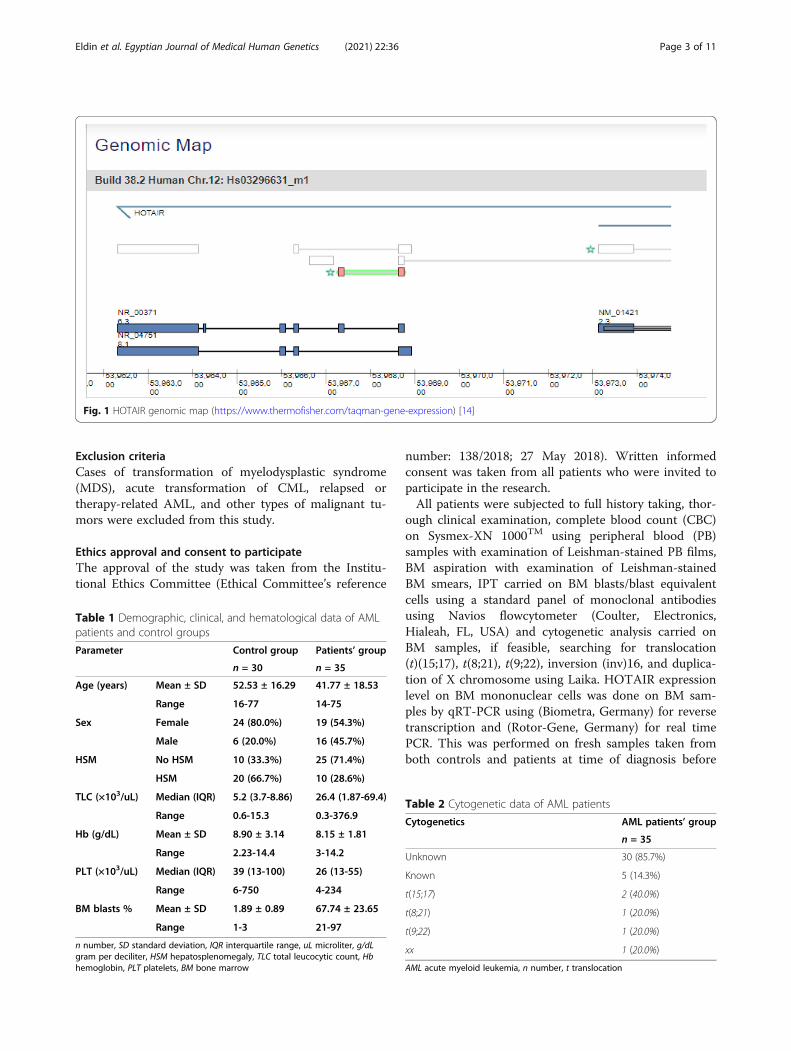
qRT-PCRHOTAIR TaqManTM Gene Expression Assay, Thermo-Fisher, cat. no.: (4448892) (Fig. 1) (https://www.thermofisher.com/taqman-gene-expression/) with primersequence (Forward 5′-GCA GTA GAA AAA TAG ACATAG GAGA-3′, Reverse 5′-AAT GAT AGG GAC ACATCG GGG AAC T-3) (https://www.ncbi.nlm.nih.gov/pmc/articles/PMC4774541/) was used.TaqManTM Gene Expression Master Mix, Thermo-
thermofisher.com), was used for qRT-PCR whichcontains AmpliTaq Gold®, DNA Polymerase, UP (UltraPure), Uracil-DNA Glycosylase (UDG), deoxyribonucleo-tide triphosphates (dNTPs) with deoxyuridine triphos-phate (dUTP), ROX™ Passive Reference and Buffercomponents optimized for sensitivity, precision, specifi-city, and duplexing. The PCR reaction exploits the 5′nuclease activity of AmpliTaq® Gold DNA Polymerase,UP (Ultra Pure) to cleave a TaqMan® probe during PCR.The TaqMan probe contains a reporter dye at the 5′end of the probe and a quencher dye at the 3′ end ofthe probe. Cleavage of the probe separates the reporterdye and the quencher dye, resulting in increased
fluorescence of the reporter. Accumulation of PCR prod-ucts is detected directly by monitoring the increase influorescence of the reporter dye. The nuclease activity isfork-like and structure dependent. When the probe is in-tact, the proximity of the reporter dye to the quencherdye results in suppression of the reporter fluorescenceprimarily by Förster-type energy transfer, the probe spe-cifically anneals to the target. The 5′ to 3′ nucleolyticactivity of the AmpliTaq Gold, UP enzyme cleaves theprobe between the reporter and the quencher only if theprobe hybridizes to the target. The probe fragments arethen displaced from the target, and polymerization ofthe strand continues. The 3′ end of the probe is blockedto prevent extension of the probe during PCR. Thisprocess occurs in every cycle, and it does not interferewith the exponential accumulation of product. Increasedfluorescence signal is detected only if the target se-quence is complementary to the probe and if it is ampli-fied during PCR.Housekeeping gene GABDH was used as an endogen-
ous control to normalize the amount of total mRNA ineach sample of HOTAIR between different samples.HOTAIR expression was measured as fold of controlusing the equation of ΔRn = (Rn+)−(Rn−), (where Rn+= emission Intensity of Reporter PCR with templateEmission Intensity of Passive Reference and Rn− =Emission Intensity of Reporter PCR without template orearly cycles of a real-time Emission Intensity of Passive
Table 6 Correlation of HOTAIR with age, laboratory data, andEFS (months) of the studied patients
Cases only HOTAIR
Correlation (r) p value
Age (years) 0.013 0.943
TLC (×103/microliter (uL)) 0.356 0.036
Hb (gram(g)/dL) 0.071 0.686
PLT (×103/uL) 0.218 0.208
BM blasts % -0.007 0.968
EFS -0.304 0.076
Spearman correlation coefficientr correlation coefficient, uL microliter, g/dL gram per deciliter, HSMhepatosplenomegaly, TLC total leucocytic count, Hb hemoglobin, PLT platelets,BM bone marrow, EFS event free survival (page 21, line 36)
Fig. 3 Significant correlation between HOTAIR expression and TLC in AML patients
Eldin et al. Egyptian Journal of Medical Human Genetics (2021) 22:36 Page 6 of 11
Reference reaction). Then the results is calculated as2−ΔΔCT (where CT= cycle threshold).
Statistical analysisDescriptive and analytical statistical procedures wereconducted. Data entry and statistical analysis of collecteddata was performed using Statistical Package for SocialScience (SPSS version 23.0). Comparison between twogroups regarding qualitative data was performed usingchi-square test (X2). The comparison between two inde-pendent groups regarding quantitative data with para-metric distribution was done by using independent t test(t) while comparison between two independent groupsregarding quantitative data with non-parametric distri-bution was done by using Mann-Whitney test. Parame-ters correlations (correlation coefficient “r”) wereperformed by using Spearman’s correlation. Kruskal-Wallis test and post HOC analysis were performed forcomparison among groups regarding quantitative non-parametric data. Receiver operating characteristic (ROC)as a graphical plot was done to determine the best cut-off value for HOTAIR as a diagnostic marker for AML.Kaplan-Meier analysis with log-rank test was used to as-sess the relation of HOTAIR with relapse free survival(event free survival (EFS)). Regarding power of signifi-cance, the probability level (p value) was considered sig-nificant (S) if p value was < 0.05, non-significant (NS) ifp value was ≥ 0.05 and highly significant (HS) if p valuewas <0.01.
ResultsDemographic, clinical, hematological, and cytogeneticdata are shown in Tables 1 and 2.HOTAIR was expressed in all AML patients ranging
from 1.11-16.11 (median: 4.23; interquartile range (IQR):3.6-5.5) (Table 3). HOTAIR expression was higher inAML patients group than control group and this differ-ence was statistically highly significant with p value=0.00 (Table 3 and Fig. 2).As regards FAB subtypes, highest levels of HOTAIR
expression were seen in M0-1 (median: 16.11, IQR:16.11-16.11) followed by M4 (median: 4.34, IQR: 3.84-4.64), M5 AML (median: 4.53, IQR: 4.03-5.5), and mixedphenotype acute leukemia (AL) (median: 4.96, IQR: 4.1-9.57) than other FAB classification subtypes. The leastexpression was seen in M3 (median: 3.32, IQR: 2.38-4.23). However, statistical analysis revealed no significant
relation between HOTAIR expression and FAB subtypes(p value = 0.408) (Table 4).There was no statistically significant relation between
HOTAIR expression and each of age and sex (p value=0.943 and 0.476 respectively) in AML patients. Inaddition there was no statistically significant relation be-tween HOTAIR expression and hepatosplenomegaly(HSM) among AML patients (p value= 0.956) (Table 5).As regards the correlation between HOTAIR expression
and hematological data among AML group, there was apositive significant correlation between HOTAIR expres-sion and total leucocytic count (TLC) (p = 0.036). How-ever. there was no statistical correlation between AMLpatients and hemoglobin (Hb) or platelets (PLT) (p value= 0.686, p value = 0.208 respectively). As regards BM blastcell count, there was no statistically significant correlationbetween HOTAIR expression and blasts count amongAML cases (p value = 0.968) (Table 6 and Fig. 3).Receiver operating characteristic (ROC) curve was
used to assess the best cut-off point with best sensitivityand specificity for the diagnosis of AML. This study re-vealed that HOTAIR expression value of 3.25 turned tobe the best cut-off value that could discriminate betweenAML patients and control group. The diagnostic
Table 7 Diagnostic performance of HOTAIR expression in AML patients
Cut-off point AUC Diagnostic accuracy Sensitivity Specificity +PV −PV
3.25a (folds of control) 0.978 97.8% 88.57 100.00 100.0 88.2
AUC area under the curve, PV predictive valuea3.25-folds of control: The cut-off concluded from receiver operating characteristic curve (ROC) for HOTAIR to differentiate between AML patients and controls(Fig. 4)
Fig. 4 Receiver operating characteristic curve (ROC) for HOTAIR todifferentiate between AML patients and controls
Eldin et al. Egyptian Journal of Medical Human Genetics (2021) 22:36 Page 7 of 11
performance was evident by area under the curve (AUC)of 0.978 with a diagnostic accuracy of 97.8%, diagnosticsensitivity of 88.57%, specificity of 100%, positive pre-dictive value (PV) of 100%, and negative predictive valueof 88.2% (Table 7 and Fig. 4).According to patient response to chemotherapy at day
28, most patients with higher HOTAIR expression are non-responsive (BM blast% > 5% at day 28 of starting chemo-therapy) (median: 5.03 and 4.6; IQR: 3.78-8 and 3.47-5.03respectively); nevertheless, the association betweenHOTAIR expression and response to chemotherapy wasfound to be statistically insignificant (p = 0.195) (Table 5).Follow-up of the patients was done after day 28 till ei-
ther the end of the study or last contact with the patientto find patients who were relapsed. In this study, 11AML cases out of 35 AML cases were relapsed. Ten of
AML relapsed cases had HOTAIR expression > 3.25-folds of control, while one of the 11 relapsed cases hadHOTAIR expression < 3.25-folds of control; however,this was statistically insignificant. Kaplan-Meier analysisby log-rank test shows that cases with HOTAIR > 3.25-folds of control have lower mean EFS (11.0 ± 0.828) thancases with HOTAIR < 3.25-folds of control who havehigher EFS (8.87 ± 0.816). This was statistically insignifi-cant (p value = 0.694) (Table 8 and Fig. 5). There was nosignificant association between HOTAIR expression andstandard prognostic factors in AML (Tables 9 and 10).Interestingly, there was an M0-1 FAB classified case
who showed resistance to therapy with no remissionwhose HOTAIR expression level was the highest (16.11-folds of control). In contrary, M3 AML cases showed thelowest HOTAIR expression (Table 4).
Table 8 Relationship of HOTAIR expression and EFS
Parameter Numberofevents(relapse)(n=11)
EFS (months) 95% CI X2* pvalue
Significance
Mean SE Lower Upper
HOT AIR < 3.25 (folds of control) 1 11 0.816 9.4 12.6 0.155 0.694 NS
HOT AIR > 3.25 (folds of control) 10 8.871 0.828 7.247 10.495
Log-rank testn number, EFS event free survival, CI confidence interval, X2* chi-square test, SE standard error, p value probability value, NS non-significant
Fig. 5 Cumulative survival of AML patient group during study period
Eldin et al. Egyptian Journal of Medical Human Genetics (2021) 22:36 Page 8 of 11
DiscussionAlthough HOTAIR has been implicated in the onset of avariety of tumors, its role in hematological tumor forma-tion remains unclear. HOTAIR acts as a scaffold for his-tone modification complexes and is involved in epigeneticgene regulation [15]. The present study aimed at elucida-tion of the value of HOTAIR expression as a diagnosticand prognostic marker in AML by investigating its level ofexpression and assessing its relation to various clinical,biological, and standard prognostic factors.
In this study, the expression of LncRNA HOTAIR isobserved in all de novo AML patients and this expres-sion was significantly higher in AML patients than con-trols. This expression showed insignificant associationwith different FAB subtypes which may be due to smallsample size or due to different subtypes involved in thestudy. This is in accordance with Fouad and Salah [15]who held a study at Cairo University aimed at studyingHOTAIR LncRNA expression in Egyptian AML patientsand revealed that HOTAIR is upregulated in AML. Also,this goes with Gao et al. [16] who found that HOTAIRwas elevated in AML cells. This is also supported byHao and Shao [8] who could not thoroughly assesswhether there is any difference in HOTAIR levels be-tween AML subtypes also due to small sample size.In contrary, Sayed et al. [17] examined the expres-
sion of HOTAIR messenger ribonucleic acid (mRNA)in the blood samples of 25 Iranian AML patients incomparison with 50 healthy controls and investigatedthe correlation between this lncRNA expression levelsand the disease using quantitative real-time RT-PCR.They also categorized their samples regarding thegender into two separate groups of males and
Table 9 Relation of HOTAIR expression to standard prognostic factors in AML
No. number, uL microliter, g/dL gram per deciliter, HSM hepatosplenomegaly, TLC total leucocytic count, Hb hemoglobin, PLT platelets, BM bone marrow, EFS eventfree survival, p value: probability value, NS non-significant
Table 10 Response, relapse, and EFS in AML patients
Parameter Patients’ group
n = 35
Response Responders 27 (77.1%)
Non-responders 8 (22.9%)
Relapse Non-relapsed 24 (68.6%)
relapsed 11 (31.4%)
EFS Median (IQR) 12 (4-12)
Range 1-12
n number, IQR interquartile range, EFS event free survival
Eldin et al. Egyptian Journal of Medical Human Genetics (2021) 22:36 Page 9 of 11
females. They demonstrated no significant differencesin HOTAIR lncRNA expression level between IranianAML patients and healthy individuals or betweenmales and females.According to the current study, upregulated HOTAIR
expression can be used as a diagnostic marker in AMLpatients with the best cut-off point of expression 3.25.This cut-off point has a diagnostic accuracy of 97.8%with diagnostic sensitivity of 88.57%, specificity of 100%,positive predictive value of 100%, and negative predictivevalue of 88.2%.Studying the correlation between HOTAIR expression
and different clinicopathological factors that assigned forrisk stratification as indicated by 2017 EuropeanLeukemia Net (ELN) recommendations from an inter-national expert panel [18] showed a statistically signifi-cant correlation between HOTAIR expression level andTLC, but there was no significant correlation with otherlaboratory, demographic, or clinical data.The current study showed no association between
HOTAIR expression and prognosis and EFS, althoughhigher HOTAIR expression was observed in patients notresponding to therapy and those with shorter EFS. Inaddition, HOTAIR expression could not be used as an in-dependent prognostic marker. Interestingly, there was anM0-1 FAB classified case who showed the highestHOTAIR expression level (HOTAIR: 16.11-folds of con-trol) and resistance to therapy with no remission (Table4). This supports that HOTAIR is involved in the develop-ment and progression of AML and that HOTAIR knock-down may be a new prospective for AML treatment.In accordance to this study, meta-analysis which
studied five researches covering a number of 531AL and lymphoma patients demonstrated thatHOTAIR expression is related to a poor prognosis,but did not significantly affect the EFS in patientswith AL [19].Another study by Zhang et al. [20] suggested that up-
regulated HOTAIR expression is associated with poorprognosis and reduced EFS. Multivariate analysis showedthat age, peripheral blood leucocyte count, and high ex-pression of HOTAIR were independent prognostic indi-cators for EFS.Gao et al. [16] provided evidence that HOTAIR may
act as an oncogenic gene in AML and that its overex-pression was associated with aggressive tumor progres-sion. This indicated that it has a possible prognosticvalue in AML and that it may represent a potential bio-marker of poor prognosis and a potential therapeutictarget for AML intervention.This study showed increased HOTAIR expression in
AML patients, but this upregulated expression wasnot correlated with poor prognosis which may needto be assessed as the biological importance of
HOTAIR in AML could not be ignored. Further stud-ies are needed to find the role of HOTAIR in devel-opment, prognosis, and targeted treatment of AMLthrough its silencing.
ConclusionThis study showed that the expression of HOTAIR isupregulated in newly diagnosed adult AML patients andthat it can be used as a diagnostic biomarker in these pa-tients. Accordingly, HOTAIR might have a role inpathogenesis of AML and can be used as a target fortreatment by knocking it down. However, it is not asso-ciated with outcome or prognosis in AML patients.Hence, larger studies on AML patients with its differentsubgroups and longer duration of follow-up (3-5 years)are recommended to explore the impact of HOTAIR ex-pression on the duration of survival and EFS of patientsafter treatment.
LimitationsThe main limitation of our study was relatively smallsample size. For this reason, impact of HOTAIR expres-sion as a prognostic marker in AML patients could notbe thoroughly assessed.
AbbreviationsAL: Acute leukemia; AML: Acute myeloid leukemia; AUC: Area under thecurve; BM: Bone marrow; Cat.: Catalog; CBC: Complete blood picture;CD: Cluster of differentiation; cDNA: Complementary deoxyribonucleic acid;CI: Confidence interval; CML: Chronic myelogenous leukemia; CT: Cyclethreshold; DNA: Deoxyribonucleic acid; EFS: Event free survival;ELN: European Leukemia Net; FAB: French-American-British; HB: Hemoglobin;HOTAIR: Homeobox transcript antisense intergenic ribonucleic acid;HOX: Homeobox; HOXC: Homeobox C; HOXC11: Homeobox C11;HOXC12: Homeobox C12; HS: Highly significant; HSM: Hepatosplenomegaly;Inv: Inversion; IPT: Immunophenotyping; IQR: Interquartile range; K2-EDTA: K2-ethylene diamine tetraacetic acid; LncRNA: Long non-coding ribo-nucleic acid; MDS: Myelodysplastic syndrome; ncRNA: Non-codingribonucleic acid; No.: Number; NS: Non-significant; PB: Peripheral blood;PLT: Platelets; PV: Predictive value; p value: Probability value; qRT-PCR: Quantitative reverse transcription polymerase chain reaction;r: Correlation coefficient; Rn−: Emission Intensity of Reporter PCR withouttemplate; Rn+: Emission Intensity of Reporter PCR with template EmissionIntensity of Passive Reference; RNA: Ribonucleic acid; RNAase: Ribonuclease;ROC: Receiver operating characteristic; RT: Reverse transcription; S: Significant;SD: Standard deviation; SE: Standard error; SPSS: Statistical Package for SocialScience; t: Independent t test; t: Translocation; TLC: Total leucocytic count;UP: Ultra pure; WHO: World Health Organization; X2: Chi-square test
AcknowledgementsThe authors gratefully thank professor Eman Omar (Professor of ClinicalPathology, Faculty of Medicine—Ain Shams University) for her many usefultips which helped us in writing our methodology.
Authors’ contributionsRA performed bone marrow aspiration, collected the samples of the patientsin addition to their demographic, clinical, and laboratory data, and wrote themanuscript. RA, AA, MF, SA, and YN analyzed and interpreted themanuscript. All authors have read and approved the manuscript.
FundingSelf-funded.
Eldin et al. Egyptian Journal of Medical Human Genetics (2021) 22:36 Page 10 of 11
Availability of data and materialsThe data within this paper and other findings of this study are available fromthe corresponding author upon reasonable request.
Ethics approval and consent to participateThe approval of the study was taken from the Institutional Ethics Committeeof the Faculty of Medicine, Ain Shams University (Ethical Committee’sreference number: 138/2018; 27 May 2018). Written informed consent wastaken from all patients who were invited to participate in the research.
Consent for publicationNot applicable
Competing interestsThe authors declare no conflict of interest.
Received: 29 December 2020 Accepted: 12 February 2021
References1. Kreitz J, Schönfeld C, Seibert M, Stolp V, Alshamleh I, Oellerich T,
Steffen B, Schwalbe H, Schnütgen F, Kurrle N, Serve H (2019) Metabolicplasticity of acute myeloid leukemia. Cells 8(8):805. https://doi.org/10.3390/cells8080805
2. Jones L, McCarthy P, Bond J (2020) Epigenetics of paediatric acute myeloidleukaemia. Br J Hematol 188(1):63–76. https://doi.org/10.1111/bjh.16361
3. Chmelarova M, Palicka V (2019) Epigenetics in cancer: a promising path to follow?Clin Chem Lab Med 57(7):927–931. https://doi.org/10.1515/cclm-2019-0010
4. Roberti A, Valdes AF, Torrecillas R, Fraga MF, Fernandez AF (2019)Epigenetics in cancer therapy and nanomedicine. Clin Epigenetics 11(1):81.https://doi.org/10.1186/s13148-019-0675-4
5. ZHU HP (2020) Silence of HOTAIR inhibits insulin secretion and proliferationin pancreatic β cells. Eur Rev Med Pharmacol Sci 24:784–792. https://doi.org/10.1186/s13148-019-0675-4
6. Tang Q, Hann SS (2018) HOTAIR: an oncogenic long non-coding RNA in humancancer. Cell Physiol Biochem 47:893–913. https://doi.org/10.1159/000490131
7. Toy HI, Okmen D, Kontou PI, Georgakilas AG, Pavlopoulou A (2019) HOTAIR asa prognostic predictor for diverse human cancers: a meta- and bioinformaticsanalysis. Cancers 11(6):778. https://doi.org/10.3390/cancers11060778
8. Hao S, Shao Z (2015) HOTAIR is upregulated in acute myeloidleukemia and that indicates a poor prognosis. Int J Clin Exp Pathol8(6):7223–7228
9. Zhao X, Tian X (2019) Knockdown of long noncoding RNA HOTAIR inhibitscell growth of human lymphoma cells by upregulation of miR-148b. J CellBiochem 120(8):12348–12359. https://doi.org/10.1002/jcb.28500
10. Wang H, Li Q, Tang S, Li M, Feng A, Qin L, Liu Z, Wang X (2017) The role oflong noncoding RNA HOTAIR in the acquired multidrug resistance toimatinib in chronic myeloid leukemia cells. Hematol J 22(4):208–216. https://doi.org/10.1080/10245332.2016.1258152
11. Swerdlow SH, Campo E, Harris NL, Jaffe ES, Pileri SA, Stein H, Thiele J, ArberDA, Hasserjian RP, Le Beau MM, Orazi A, Siebert R (2017) WHO classificationof tumours of haematopoietic and lymphoid tissues,2016 (Revised 4thedition). In: Swerdlow SH, Campo E, Harris NL, Jaffe ES, Pileri SA, Stein H,Thiele J (eds) International Agency for Research on Cancer, pp 15–28
12. Bennet JM, Catovsky D, Daniel MT et al (1976) Proposed revised criteria forthe classification of acute myeloid leukemias (FAB cooperative group). Br JHaematol 33:451–458
13. Qiagen, 2013-20 , accessed 27 January 2021, <https://www.qiagen.com/us/products/discovery-and-translational-research/pcr-qpcr-dpcr/real-time-pcr-enzymes-and-kits/reverse-transcription-cdna-synthesis-qpcr/quantitect-whole-transcriptome-kit/?clear=true#productdetails.>. Accessed 27 Jan 2021.
14. Thermofisher scientific, TaqMan® Assays and Arrays, accessed 27 January2021, <https://www.thermofisher.com/order/genome-database/?pearUXVerSuffix=pearUX2&elcanoForm=true#!/ge/assays/ge_all/?keyword=HOTAIR%20taqman%20gene%20expression%20master%20mix%20.>.Accessed 27 Jan 2021.
15. Fouad NB, Salah M (2019) Study of HOTAIR LncRNA expression in EgyptianAML patients in context to FLT3-ITD and NPM1 mutations status. OncNetNewsletter Lymphoma Leukaemia Myeloma Congress 4773. http://oncnet.com/meeting-materials/lymphoma-and-myeloma/4773.
16. Gao J, Wang F, Wu P, Chen Y, Jia Y (2020) Aberrant LncRNA Expression inLeukemia. J Cancer 11(14):4284–4296. https://doi.org/10.7150/jca.42093
17. Sayad A, Hajifathali A, Hamidieh AA, Roshandel E, Taheri M (2017) HOTAIRlong noncoding RNA is not a biomarker for acute myeloid leukemia (AML)in Iranian patients. Asian Pacific J Cancer Prev 18(6):1581–1584. https://doi.org/10.22034/apjcp.2017.18.6.1581
18. Döhner H, Estey E, Grimwade D, Amadori S, Appelbaum FR, Büchner T,Dombret H, Ebert BL, Fenaux P, Larson RA, Levine RL, Lo-Coco F, NaoeT, Niederwieser D, Ossenkoppele GJ, Sanz M, Sierra J, Tallman MS, TienH, Wei AH, Löwenberg B, Bloomfield CD (2017) Diagnosis andmanagement of AML in adults: 2017 ELN recommendations from aninternational expert panel. Blood 129(4):424–447. https://doi.org/10.1182/blood-2016-08-733196
19. Lin Y, Fang Z, Lin Z, Li Z, Zhao J, Luo Y and Xu B (2018): The prognosticimpact of long noncoding RNA HOTAIR in leukemia and lymphoma: ameta-analysis, Hematology, ISSN: 1024-5332 (Print) 1607-8454 (Online). (doi:https://doi.org/10.1080/10245332.2018.1446572)
20. Zhang YY, Huang SH, Zhou HR, Chen CJ, Tian LH, Shen JZ (2016) Role ofHOTAIR in the diagnosis and prognosis of acute leukemia. Oncol Rep 36(6):3113–3122. https://doi.org/10.3892/or.2016.5147
Publisher’s NoteSpringer Nature remains neutral with regard to jurisdictional claims inpublished maps and institutional affiliations.
Eldin et al. Egyptian Journal of Medical Human Genetics (2021) 22:36 Page 11 of 11


![Current perspectives for the treatment of chronic myeloid ...In composing long-term treatment plans for TKI-naïve patients, prognostic evaluation of CML is key [4]. Prior to the use](https://static.documents.pub/doc/80x56/60bcb776f8906f48904ac5ca/current-perspectives-for-the-treatment-of-chronic-myeloid-in-composing-long-term.jpg)


