52
How Genes Are Transmitted from Generation to Generation Chapter 4
| Date post: | 02-Jan-2016 |
| Category: |
Documents |
| Upload: | archibald-hopkins |
| View: | 221 times |
| Download: | 1 times |

How Genes Are Transmitted from Generation to Generation
Chapter 4

Central Points
Genes are transmitted from generation to generation
Traits are inherited according to predictable rules

Gregor Mendel – The Father of Genetics

4.1 How Are Genes Transmitted?
Experiments with pea plants in 1800s
Traits, distinguishing characteristics
Specific patterns in the way traits were passed from parent to offspring

Different Plant Heights

Mendel’s Experiments
Some traits disappeared in the first generation of offspring (all tall)
Reappeared in 3:1 ratio (tall:short)
Dominant trait present in the first-generation offspring (tall)
Recessive trait absent in first generation but reappeared in the next generation (short)

Traits Are Passed by Genes
“Factors” or genes transmitted from parent to offspring
Each parent carries a pair of genes for a trait but contributes only one gene to each offspring
Separation of gene pair occurs during meiosis

Genes
Alleles: variations of a gene
Geneticists use letters for each allele.
Homozygous: identical alleles of a gene • TT or tt
Heterozygous: nonidentical alleles• Tt

Phenotype and Genotype
Phenotype: what an organism looks like• tall or short
Genotype: genetic makeup• TT, Tt, and tt
Identical phenotypes may have different genotypes• TT or Tt have tall phenotype

Mendel’s Law of Segregation
Two copies of each gene separate during meiosis
One copy of each gene in the sperm or egg
Each parent gives one copy of each gene

Sorting of Alleles

Mendel’s Law of Independent Assortment
Members of a gene pair segregate into gametes independently of other gene pairs
Gametes can have different combinations of parental genes

Human Traits: Albinism
Pigmentation dominant and lack of pigment recessive• AA, Aa: Pigmented• aa: Albino
Both parents Aa, each child has 25% chance of being albino (3:1 ratio)

Fig. 4-3a, p. 61
Aa × Aa
Aa Aa
A a A a
Two carriers of albinism have a child.
The male and female can contribute either an A allele or an a allele to the gamete.

Fig. 4-3b, p. 61
Genotype Phenotype
A a
1 AA
2 Aa3/4 normal coloring
AAA
normalAa
normal
1 aa 1/4 albinoa
Aa normal
aa albino
This shows the possible genotypes and phenotypes of the offspring.
The possible offspring and allele combinations are shown above.

Pedigree 1
Shows all family members and identifies those affected with the genetic disorder
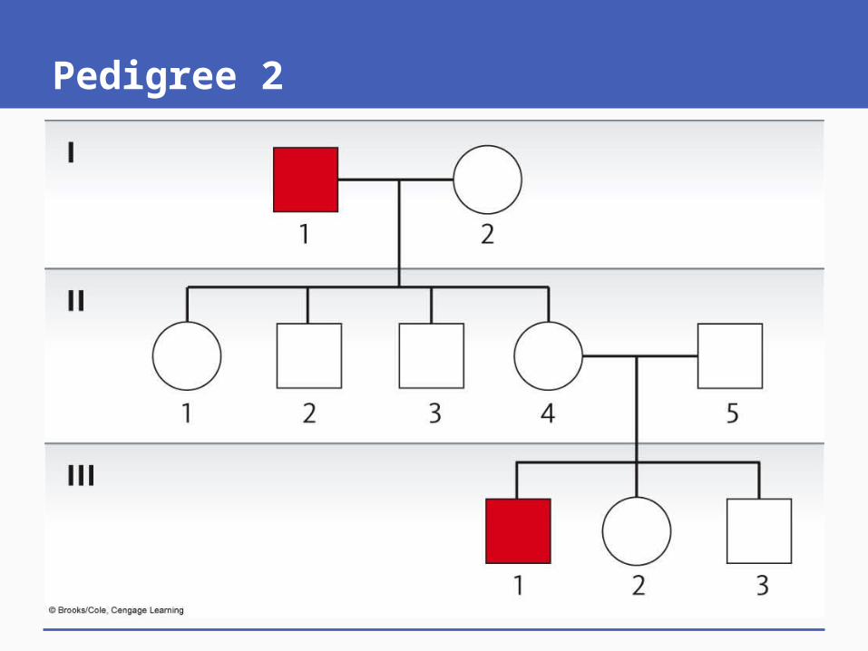
Pedigree 2

Pedigree Symbols

p. 62
Male
Female
Mating
Mating between relatives (consanguinous)
I Parents and children. Roman numerals symbolize generations. Arabic numbers symbolize birth order within generation (boy, girl, boy)II
1 2 3
I, II, III, etc. = each generation 1, 2, 3, etc. = individuals within a generation

p. 62
or Unaffected individual
or Affected individual
or Known heterozygotes
or Proband; a person in family who is the focus of the pedigree
PP
I, II, III, etc. = each generation 1, 2, 3, etc. = individuals within a generation

Pedigree Symbols

Proband
Person who is the focus of the pedigree
Indicated by an arrow and the letter P

4.2 Examining Human Pedigrees
Determine trait has dominant or recessive inheritance pattern
Predict genetic risk for:• Pregnancy outcome• Adult-onset disorder• In future offspring

Three Possible Patterns of Inheritance
Autosomal recessive
Autosomal dominant
X-linked recessive
Autosomal on chromosomes 1–22
X-linked traits on the X chromosome

Autosomal Recessive
Unaffected parents can have affected children
All children of affected parents are affected
Both parents Aa, risk of affected child is 25%
~Equal affected male and female
Both parents must transmit the gene for a child to be affected

Autosomal Recessive Pedigree

Autosomal Recessive Genetic Disorders

Albinism A = normal coloring; a = albinism
Group of genetic conditions, lack of pigmentation (melanin) in the skin, hair, and/or eyes
Normally, melanin in pigment granules inside melanocytes
In albinism, melanocytes present but cannot make melanin
Oculocutaneous albinism type I (OCA1)

Cystic Fibrosis (CF)
C = normal; c = cystic fibrosis
CF affects glands that produce mucus and digestive enzyme
CF causes production of thick mucus in lungs blocks airways
Develop obstructive lung diseases and infections
Identified CF gene and protein (CFTR)

Sickle Cell Anemia (SCA)
S = normal red blood cells; s = sickle
High frequency in areas of West Africa, Mediterranean Sea, India
Abnormal hemoglobin molecules aggregate to form rods
Red blood cells, crescent- or sickle-shaped, fragile and break open

Normal and Sickled Cells

Autosomal Dominant (1)
Requires one copy of the allele (Aa) rarely present in a homozygous condition (AA)
aa: Unaffected individuals
Affected individual has at least one affected parent
Aa X aa: Each child has 50% chance of being affected

Autosomal Dominant (2)
~Equal numbers of affected males and females
Two affected individuals may have unaffected children
Generally, AA more severely affected, often die before birth or in childhood
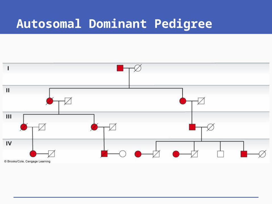
Autosomal Dominant Pedigree

Autosomal Dominant Genetic Disorders

Animation: Chromosomes and Human Inheritance (autosomal-dominant inheritance)

Animation: Chromosomes and Human Inheritance (autosomal-recessive inheritance)

Neurofibromatosis (NF)
N = Neurofibromatosis 1; n = normal
Many different phenotypes
Café-au-lait spots, or noncancerous tumors in the nervous system can be large and press on nerves
Deformities of the face or other body parts (rarely)
NF gene has a very high mutation rate

Neurofibromatosis

Huntington Disease (HD) H = Huntington disease; h = normal
Causes damage in brain from accumulation of huntingtin protein
Symptoms begin slowly (30–50 years old)
Affected individuals may have already had children (50% chance with one Hh parent)
Progressive neurological signs, no treatment, die within 10–25 years after symptoms

Adult-Onset Disorders
Expressed later in life
Present problems in pedigree analysis, genetic testing may be required
Examples:• Huntington disease (HD)• Adult polycystic kidney disease (ADPKD)
Both examples are autosomal dominant

4.3 X-Linked Recessive Traits
Genes on X chromosome: X-linked
Genes on Y chromosome: Y-linked
For X-linked traits:• Females XX, XX*, or X*X*• Males XY or X*Y• Males cannot be homozygous or heterozygous,
they are hemizygous for genes on X• Distinctive pattern of inheritance

X-Linked Recessive Inheritance
Mother gives one X chromosome to offspring
Father gives X to daughters and Y to sons
Sons carry X from mother
For recessive traits, X*X* and X*Y affected
More males affected

Pedigrees: X-Linked Inheritance

X-Linked Recessive Genetic Disorders

Inheritance of X-Linked Disorder

Animation: Chromosomes and Human Inheritance (X-linked inheritance)

Duchenne Muscular Dystrophy (DMD) (1) XM = normal; Xm = muscular dystrophy
Most common form, affects ~1/3,500 males
Infants appear healthy, symptoms age ~1–6 years
Rapid, progressive muscle weakness
Usually must use a wheelchair by age 12
Death, age ~20 from respiratory infection or cardiac failure

Duchenne Muscular Dystrophy (DMD) (2)
DMD gene on the end of X chromosome
Encodes protein dystrophin that supports plasma membrane during contraction
If dystrophin absent or defective, cells are torn apart
Two forms: DMD, and less-serious Becker muscular dystrophy (BMD)

Cells of a Person with MD

Hemophilia
XH = normal; Xh = hemophilia
Lack of clotting: factor VIII in blood
Affected individuals hemorrhage, often require hospitalization to treat bleeding
Hemophilia A most common form of X-linked hemophilia
Females affected if XhXh, both parents must carry the trait

Factor VIII
1980s, half of all people with hemophilia became infected with HIV
Recombinant DNA technology now used to make clotting factors free from contamination


