+ All Categories
Home > Documents > How I Treat Atypical Chronic Myeloid...

How I Treat Atypical Chronic Myeloid...

Date post: 11-Apr-2018
Category:
Upload: hanguyet
View: 219 times
Download: 3 times
Share this document with a friend
33
How I Treat Atypical Chronic Myeloid Leukemia Jason Gotlib 1 1 Division of Hematology, Stanford University School of Medicine / Stanford Cancer Institute, Stanford, CA Keywords: Atypical CML; MDS/MPN; BCR-ABL1 negative; SETBP1 Corresponding Author Jason Gotlib, MD, MS Professor of Medicine (Hematology) Stanford Cancer Institute 875 Blake Wilbur Drive, Room 2324 Stanford, CA 94305-5821 TEL: 650-736-1253 FAX: 650-724-5203 Email: [email protected] Word Count: Abstract: 140 Text: 4336 Tables: 1 Figures: 2 Blood First Edition Paper, prepublished online November 29, 2016; DOI 10.1182/blood-2016-08-693630 Copyright © 2016 American Society of Hematology For personal use only. on May 31, 2018. by guest www.bloodjournal.org From
Transcript

How I Treat Atypical Chronic Myeloid Leukemia

Jason Gotlib1

1Division of Hematology, Stanford University School of Medicine / Stanford Cancer Institute, Stanford, CA Keywords: Atypical CML; MDS/MPN; BCR-ABL1 negative; SETBP1 Corresponding Author Jason Gotlib, MD, MS Professor of Medicine (Hematology) Stanford Cancer Institute 875 Blake Wilbur Drive, Room 2324 Stanford, CA 94305-5821 TEL: 650-736-1253 FAX: 650-724-5203 Email: [email protected] Word Count: Abstract: 140 Text: 4336 Tables: 1 Figures: 2

Blood First Edition Paper, prepublished online November 29, 2016; DOI 10.1182/blood-2016-08-693630

Copyright © 2016 American Society of Hematology

For personal use only.on May 31, 2018. by guest www.bloodjournal.orgFrom

Abstract Atypical chronic myeloid leukemia, BCR-ABL1-negative (aCML) is a rare

myelodysplastic syndrome/myeloproliferative neoplasm for which no current standard of

care exists. The challenges of atypical CML relate to its heterogeneous clinical and

genetic features, high rate of transformation to acute myeloid leukemia, and historically

poor survival. Therefore, allogeneic hematopoietic stem cell transplantation should

always be an initial consideration for eligible patients with a suitable donor. Non-

transplant approaches for treating aCML have otherwise largely relied on adopting

treatment strategies used for MDS and MPN. However, such therapies, including

hypomethylating agents, are based on a paucity of data. With an eye toward making a

more meaningful impact on response rates and modification of the natural history of the

disease, progress will rely on enrollment of patients into clinical trials and molecular

profiling of individuals so that opportunities for targeted therapy can be exploited.

For personal use only.on May 31, 2018. by guest www.bloodjournal.orgFrom

Introduction

The current diagnostic criteria that comprise the World Health Organization (WHO)

entity ‘atypical chronic myeloid leukemia, BCR-ABL1 negative’ (Table 1) represents a

decades-long evolution of classifying diseases which exhibited morphologic similarity to

chronic myeloid leukemia (CML), but lacked both the Philadelphia (Ph) chromosome by

standard cytogenetics and BCR-ABL1 rearrangement by polymerase chain reaction

(PCR).1,2 The differential diagnosis of these BCR-ABL1-negative hematologic neoplasms

not only includes atypical CML, but also chronic myelomonocytic leukemia (CMML),

chronic neutrophilic leukemia (CNL), and MDS/MPN, unclassifiable (MDS/MPN, U).1-6

The knowledge gleaned from next generation sequencing has complemented

morphologic and laboratory WHO criteria for myeloid neoplasms, and can often provide

greater specificity in distinguishing atypical CML from alternative MDS/MPN or

MPNs.1,3-7 Invariably, how I pursue the diagnosis and treatment atypical CML requires

attention to the results of standard cytogenetic analysis and myeloid mutation testing

since druggable targets may be unmasked.

-----------------------------------------------------------------------------------------------------------

Case 1

LJ is a 62-year-old woman with a past medical history of hyperlipidemia and a left total

hip replacement. In the last 3 months, during two episodes of diverticulitis with

associated gastrointestinal bleeding requiring red blood cell transfusion support, a new

leukocytosis of 15-20 x 109/L was identified. The platelet count was normal and a

manual differential revealed 46% neutrophils, 12% band forms, 12% metamyelocytes,

6% monocytes, 6% myelocytes, 2% promyelocytes, 2% eosinophils, and 14%

For personal use only.on May 31, 2018. by guest www.bloodjournal.orgFrom

lymphocytes. The increased white blood cell count and left-shifted white blood cell

differential was felt to be reactive due to her acute medical condition. The patient was

seen in consultation in hematology clinic two months after hospital discharge because of

persistent blood count abnormalities despite resolution of her gastrointestinal issues. No

hepatosplenomegaly was noted on examination. A complete blood count (CBC) showed

persistent elevation of the WBC count to 24.2 x 10⁹/L, hemoglobin of 11.2 g/dL, platelet

count 160 x 109/Lwith a similar spectrum of myeloid immaturity. A bone marrow

aspirate and biopsy revealed hypercellularity for age, left-shifted myeloid hyperplasia

without increased blasts, and hypogranular granulocytes with abnormal nuclear

segmentation. Dyserythropoiesis and dysplastic megakaryocytes, including hypolobated

forms, were noted. Cytogenetics were normal and PCR for BCR-ABL1 was negative. A

diagnosis of atypical CML was made. The patient was referred for evaluation of

management options.

Case 2

CK is a 76 year-old man with a history of coronary artery disease and hepatitis C that had

been treated with pegylated-interferon-alpha-2a (PEG-IFN-alpha-2a) until 1.5 years ago

when it was discontinued because of progressive depressive symptoms and cytopenias.

After 4 months off therapy, the patient reported increasing fatigue and new night sweats.

A spleen tip was palpated on examination. The WBC count increased from 2.5 x 10⁹/L to

26.2 x 10⁹/L after PEG-IFN-alpha-2a was stopped; the hemoglobin was 10.6 g/dL, and

the platelet count was 133 x 109/L. Although an automatic differential showed 89%

neutrophils, a manual differential revealed 17% neutrophils, 27% band forms, 6%

For personal use only.on May 31, 2018. by guest www.bloodjournal.orgFrom

metamyelocytes, 8% monocytes, 15% myelocytes, 14% promyelocytes, 1% blasts, and

11% lymphocytes. Review of the peripheral blood and bone marrow aspirate revealed an

increased number of left-shifted leukocytes with hypolobation and pseudo Pelger-Huët

morphologies. A bone marrow aspirate was hypercellular without increased blasts; there

was subtle dyserythropoiesis, and dysmegakaryopoiesis primarily consisting of

hypolobated megakaryocytes with separate nuclear lobes. The bone marrow biopsy was

hypercellular (95%) with a M:E ratio of 5:1. Cytogenetics showed trisomy 8, and no Ph

chromosome. The patient was referred for a second opinion. Pathology was confirmed,

and next generation sequencing revealed CSF3R T618I (43% mutant allele frequency)

and U2AF1 Q157T (48% mutant allele frequency) mutations. Treatment options were

reviewed.

-----------------------------------------------------------------------------------------------------------

Some cases of atypical CML have been given the historical moniker ‘CML-like

syndrome’, because both diseases exhibit bone marrows with hyperplastic myeloid

hyperplasia and peripheral blood leukocytosis characterized by a spectrum of myeloid

immaturity. However, on morphologic grounds, this is where the similarity ends. Unlike

BCR-ABL1-positive CML, atypical CML is characterized by prominent dysplastic

granulopoiesis, (e.g. the acquired Pelger-Huët anomaly; nuclear abnormalities including

hypersegmentation, nuclear projections, and abnormally clumped nuclear chromatin; and

abnormalities of cytoplasmic granules, such as hypogranularity), and in some cases

multlineage dysplasia may be observed.1,2 The finding of >10% immature myeloid cells

(promyelocytes, myelocytes, and metamyelocytes) in the peripheral blood and/or

dysplasia are useful criteria in distinguishing aCML from CNL, which lacks these

For personal use only.on May 31, 2018. by guest www.bloodjournal.orgFrom

features.1,2 Additional features of aCML include absent or minimally present basophilia

(<2% of leukocytes) and monocytosis (<10% of leukocytes) which are additional

morphologic findings that help distinguish atypical CML from BCR-ABL1-positive CML

and CMML, respectively.1,2

Natural History and Prognostic Factors

In an Italian cohort of 55 WHO-defined aCML cases,8 the overall median survival was 25

months compared to survivals of 14-30 months derived from three smaller studies.9-11 A

recent U.S. multicenter study applied WHO 2008 criteria to compare aCML (n=65) and

MDS/MPN, U (n=69) cases, and found that the former exhibited a more aggressive

clinical course, with respective median overall survivals of 12.4 and 21.8 months.6 In the

U.S. and Italian studies,7,8 transformation to acute myeloid leukemia (AML) occurred in

37% and 40% of the patients, with a median time to transformation of 11.2 and 18

months, respectively. Increased WBC count (e.g. cutoffs of > 40 or 50 x 109/L),

increased percentage of peripheral blood immature precursors, female sex, and older age

have been shown to be adverse prognostic factors for overall survival or leukemia-free

survival in multivariate analyses.7,8

Molecular and Cytogenetic Features

In cases where subtle dysplasia or borderline levels of myeloid immaturity or

monocytosis are present, morphologic distinction between aCML, BCR-ABL1-positive

CML, CMML, CNL, or MDS/MPN-U can be challenging. In addition to

histopathologic analysis of the peripheral blood and bone marrow, modern evaluation of

For personal use only.on May 31, 2018. by guest www.bloodjournal.orgFrom

atypical CML should include next generation sequencing vis à vis myeloid mutation

panel testing in addition to standard karyotyping.1-7,12 Diagnosis of aCML first requires

testing for the Philadelphia chromosome and/or the BCR-ABL1 fusion gene to exclude

CML.1,2 Standard karyotyping, fluorescent in-situ hybridization, and myeloid mutation

testing not only complement morphologic analyses, but may also identify opportunities

for targeted therapy (Figure 1).

In contrast to BCR-ABL1, which operationally defines CML, no single genetic lesion

characterizes aCML. The mutations identified in aCML are commonly found in other

myeloid neoplasms.3-7, 12-15 The variability that exists in the reported frequency of specific

mutations in aCML may partly reflect the stringency to which the WHO definition of

aCML was applied in different publications.3 However, some basic observations can be

made about the molecular landscape of aCML: higher frequency mutations (e.g. >20%)

include SETBP1, ASXL1, N/K-RAS, SRSF2, and TET2, and lower frequency mutations

(<10%) include CBL, CSF3R, JAK2, and ETNK1.3-7,12-15

Recurrent SETBP1 mutations have been identified in ~ 25-33% of aCML patients and

represent one of the mostly frequently mutated genes in this disease.13-15 Set binding

protein (SETBP1) interacts with SET, a negative regulator of the tumor suppressor

protein phosphatase 2A (PP2A).16 SETBP1 protects SET from protease cleavage,

resulting in an increased amount of SET available to repress PP2A activity.17

Most SETBP1 mutations are located within a 14-amino acid stretch (codons 858-871)

which is also mutated in Schinzel-Giedion syndrome, a rare genetic disease characterized

For personal use only.on May 31, 2018. by guest www.bloodjournal.orgFrom

by congenital malformations, mental retardation, and frequent epithelial tumors.18

Normally, phosphorylation of this region leads to binding by E3 ubiquitin ligase subunit

b-TrCP1, resulting in ubiquitination and subsequent degradation of SETBP1.13 SETBP1

mutants disrupt this consensus b-TrCP motif, leading to increased SETBP1 and SET

expression, which decreases PP2A activity and increases cellular proliferation.13

SETBP1 mutations are associated with a higher leukocyte count, lower hemoglobin and

platelet counts, and worse overall survival.13,14 In one study, SETPB1 mutations were

associated with the presence of -7 and isochromosome i(17)(q10) cytogenetic

abnormalities as well as ASXL1 and CBL mutations, but were mutually exclusive of

mutations in the TET2 and JAK2 genes.14 However, SETBP1 is a ubiquitous molecular

abnormality among myeloid neoplasms, including CNL and CMML, and may be found

in tandem with other mutations, such as with CSF3R in cases of CNL.3,6,14,15,19

Although originally reported at a higher frequency in aCML,20 subsequent reports

indicate that the activating CSF3R T618I mutation is present in < 10% of cases. 1,3-7,12,15

Identification of CSF3R T618I in the context of neutrophilic leukocytosis strongly favors

a diagnosis of CNL where it is present in approximately 80% of patients.1,3,20,21

Although T618I is the most common activating mutation in CSF3R, an alternative

proximal membrane mutation, T640N, has been described in a case of MDS that

exhibited transformation to a secondary aCML-like picture.22 Rarely, nonsense and

frameshift mutations that result in truncation of the cytoplasmic tail of CSF3R have been

found in cases of aCML, and are similar to those identified in patients with severe

For personal use only.on May 31, 2018. by guest www.bloodjournal.orgFrom

congenital neutropenia who have been administered G-CSF therapy.3,6,20 Evolution of a

case of MPN,U to a phenotype of aCML was associated with new subclones of both

CSF3R proximal membrane (T618I, 35% mutant allele frequency) and truncation

(Q739*, 30% mutant allele frequency) mutants.23

JAK2 V617F is an uncommon mutation (3-7%) in aCML and its identification (as well as

similar Ph- MPN-associated mutations in CALR and MPL), tends to favor an alternative

diagnosis such as PV, ET, or MF in the appropriate clinicopathologic context.1,4-7,12,15

More recently, mutations in the ethanolamine kinase 1 (ETNK1) gene were found in 9%

of aCML cases,24 and were also enriched in patients with CMML and systemic

mastocytosis with associated eosinophilia.25 KRAS/NRAS mutations were identified in

7/20 (35%) aCML patients in the aforementioned U.S. multicenter analysis.7 Although

SETBP1 and ETNK1 mutations are not yet druggable targets, mutated CSF3R, JAK2, and

RAS are important to identify since clinical trial or off-label opportunities for targeted

therapy against these lesions may be available (see below; and Figures 1 and 2).

Non-specific karyotypic abnormalities have been reported in a moderate proportion of

aCML patients. These include single or double abnormalities, or complex cytogenetics,

including trisomy 8, del(20q), -7/7q- or isochromosome 17q [i17[q)].2,7,8,10,11 Notably,

the literature includes patients with rearrangement of PDGFRA, PDGFRB, FGFR1, or

PCM1-JAK2 who have been given the diagnosis of aCML.1,2,26-30 While in some of these

cases the term ‘atypical CML’ may have been loosely applied to indicate a CML-like

disease in the absence of BCR-ABL1, other cases may truly fulfill morphologic criteria

For personal use only.on May 31, 2018. by guest www.bloodjournal.orgFrom

for aCML. However, according to the WHO classification, the presence of any of these

genetic rearrangements re-assigns such cases to the major category of ‘Myeloid/lymphoid

neoplasms associated with eosinophilia and rearrangement of PDGFRA, PDGFRB,

FGFR1 or PCM1-JAK2’.1,2 Although not formally included in this category, cases with

rearranged FLT3 may rarely morphologically mimic aCML.31 Recognizing the relevant

breakpoints for reciprocal translocations that infer involvement of PDGFRA (4q12;

excluding the most common FIP1L1-PDGFRA rearrangement which is not visible by

standard karyotyping), PDGFRB (5q31~33), FGFR1 (8p11), and JAK2 (9p24) is also

critical for recognizing instances where use of tyrosine kinase inhibitors such as imatinib

should be considered (e.g. rearranged PDGFRA/B with confirmation by FISH or

sequencing), or where poor disease prognosis disease mandates high-intensity approaches

such as induction chemotherapy and/or allogeneic hematopoietic stem cell transplantation

(HSCT) (e.g. for patients with rearranged FGFR1).

Treatment

No standard of care exists for the treatment of atypical CML. In addition, no consensus

recommendations or risk-based treatment algorithms exist to help guide a watch-and-wait

approach versus initiation of therapy. However, progressive leukocytosis, anemia and/or

thrombocytopenia, or emergence of symptomatic splenomegaly or disease-related

constitutional symptoms should prompt treatment.

Given its unfavorable prognosis, my treatment algorithm (Figure 2) is first to consider

HSCT for eligible patients with a suitable donor without initially relying on the results of

For personal use only.on May 31, 2018. by guest www.bloodjournal.orgFrom

myeloid mutation testing. Although the best timing of transplantation, e.g. earlier in the

course of disease, or at the time of disease progression, remains an unresolved question,

the otherwise poor outcomes of aCML patients should encourage evaluation of the

feasibility of this treatment modality after the diagnosis is made. If a donor is not

immediately available and/or disease cytoreduction is recommended, I consider the

results of myeloid mutation testing to evaluate clinical trial options (preferred) or off-

label opportunities with targeted therapy. Current examples include ruxolitinib for

CSF3R or JAK2-mutated patients, or MEK inhibition in RAS-mutated patients (see

below, future prospects). Regardless of the results of mutation testing, hypomethylating

therapy may be considered in such individuals since the prognostic relevance of these

mutations to treatment response is unknown. I similarly use the results of myeloid

mutation panel testing for patients who are not transplant candidates to evaluate

opportunities for trials of targeted therapy. If no such option exists, my approach is to

consider hypomethylating therapy, or clinical trials of novel therapies not based on an

actionable mutation. Additionally, I co-opt treatment strategies used for either MDS or

MPN and apply them on a case-by-case basis to address a patient’s major clinical issues

(e.g. leukocytosis, anemia, constitutional symptoms, splenomegaly, and potential for

progression to AML). These second-line or adjunctive options may include pegylated-

interferon-α, hydroxyurea, and/or erythropoiesis stimulating agents.

Hematopoietic stem cell transplantation (HSCT)

A limited number of HSCT procedures for aCML have been published. Most are

included in series of patients with heterogeneous MDS/MPN where long-term disease

For personal use only.on May 31, 2018. by guest www.bloodjournal.orgFrom

free-survival of 40-50% has been recorded.3,32,33 Koldehoff and colleagues’ retrospective

analysis of 9 individuals with aCML represents the largest transplantation series focused

solely on this disease.34 In this series, allogeneic donor types consisted of an HLA-

identical sibling (N=4), or unrelated matched donor (n=4); 1 patient underwent syngeneic

transplantation from a twin sibling. Conditioning regimens included cyclophosphamide

with either TBI (n=5), busulfan (n=2), or bulsulfan and alemtuzumab (n=1); and in one

older patient, a reduced intensity conditioning (RIC) regimen consisting of busulfan,

fludarabine and anti-thymocyte globulin was used. All patients achieved a complete

remission; the patient who received bone marrow from his brother relapsed 19 months

after transplant but was successfully re-transplanted with peripheral blood stem cells

from this donor. Chronic graft-versus-host disease (GVHD) was observed in all

allografted patients, and grade II-IV acute GVHD occurred in 5 of 8 patients (63%). The

patient who received alemtuzumab developed cerebral toxoplasmosis and died of sepsis

273 days post transplant. A follow-up report by this group indicated that 21 patients with

aCML had been transplanted, with 17 of 21 patients alive at 5 years after transplantation

with a median survival of 47 months.35 These analyses compare more favorably to

another study of allogeneic transplantation that included 7 patients with Ph chromosome-

negative/BCR-AB1-negative CML.36 One patient suffered relapse at 9 months, and 5 of

the patients had died by 3-26 months of follow-up.

A more recent retrospective study evaluating allogeneic HSCT in 10 MDS/MPN patients

included 2 patients with aCML who received busulfan/ cyclophosphamide conditioning

with bone marrow allografts from matched sibling donors.33 Both patients remained alive

For personal use only.on May 31, 2018. by guest www.bloodjournal.orgFrom

with no evidence of disease after 96-99 months of follow-up. Notably, relapse was only

observed in the 5 of 10 MDS/MPN patients who received RIC compared to none of the

patients who received myeloablative conditioning. Because many patients with aCML or

other MDS/MPN are elderly and may only be eligible for RIC, novel strategies are

needed to reduce relapse in such individuals.

Molecular profiling to identify poor-risk mutations such as SETBP1 and ASXL1 may

prompt earlier consideration of HSCT for eligible aCML patients. It is currently

unknown whether HSCT can modify the adverse prognosis related to these mutations in

the context of aCML. However, among 36 MDS/MPN patients with DNA available for

serial molecular analysis, survival after HSCT was not influenced by ASXL1, CBL,

NRAS, or TET2 mutations (SETBP1 was not assessed).32 Detection of these pre-transplant

molecular markers, as well as CSF3R T618I may be useful for serial monitoring of

minimal residual disease after HSCT, as their detection has been associated with overt

relapse.37

Hypomethylating agents

The use of hypomethylating agents in aCML is a rational application of their established

activity in MDS and CMML. Among CMML patients treated in phase II studies of

hypomethylating agents, overall response rates range from 25-70% (average 30-40%),

with overall survival ranging from 12-37 months (reviewed in 38). It is challenging to

extrapolate from the limited data on proliferative CMML patients to broadly inform how

patients with aCML may respond. However, in one analysis, a WBC count > 13 x 109/L

For personal use only.on May 31, 2018. by guest www.bloodjournal.orgFrom

and bone marrow blasts > 10% were adverse prognostic factors for response to

azacitidine.39

I consider the use of azacitidine or decitabine in the following two scenarios: 1) as a

bridging therapy for those who are eligible for HSCT; and 2) as stand-alone treatment for

patients without a HSCT or clinical trial option. However, the experience with

hypomethylating agents in aCML is limited and cannot be considered a standard of care.

Decitabine (20 mg/m2 daily intravenously x 5 days IV) produced a complete hematologic

remission in 7 of the 8 patients described in four separate reports.40-43 Patients received a

total number of 1 to 6 cycles; 4 patients achieved a complete hematologic remission

(CHR) after 1 course of decitabine, and 3 patients achieved a CHR after 4 cycles.

Response duration and length of follow-up was variably described in these publications;

in one report, two patients were alive at 9 to 15 months after initiation of therapy,41 and 1

patient who maintained a response for 20 months expired at 26 months of follow-up.

Two patients treated with decitabine were successfully bridged to transplant, with one

succumbing to GVHD, BK viremia, and multiorgan failure on day +49.42 The small

number of patients treated thus far precludes a determination of which clinical,

laboratory, or molecular markers predict for response.

Ruxolitinib

Although uncommon, the identification of CSF3R T618I or JAK2 V617F in cases of

aCML provides an opportunity to consider JAK inhibitor therapy since both of these

For personal use only.on May 31, 2018. by guest www.bloodjournal.orgFrom

mutations result in JAK-STAT pathway activation. Pre-clinical studies indicate that

ruxolitinib potently inhibits CSF3R T618I-driven malignant cell growth and can reduce

leukocytosis and spleen size in a lethal myeloproliferative disease in mice driven by the

mutation.44 However, such data do not supplant my recommendation to first consider

HSCT for all eligible aCML patients, including individuals with either of these druggable

mutations. JAK inhibition may useful to consider as a bridge to allogeneic HSCT; in the

context of myelofibrosis, clinical improvement (vs. no clinical improvement) with JAK

inhibitors (defined by International Working Group-Myeloproliferative Neoplasms

Research and Treatment response criteria) was associated with improved overall survival

after transplant in a multivariate analysis45 and may relate to improvement of

performance status and reduction of splenomegaly.

Ruxolitinib is the only JAK inhibitor currently approved by the FDA (for patients with

intermediate or high risk myelofibrosis, or for patients with polycythemia vera

demonstrating intolerance or resistance to hydroxyurea). I recommend that patients be

treated with this agent in the context of a clinical trial. Currently, a multicenter study

(NCT02092324) is evaluating the safety and efficacy of ruxolitinib in patients with CNL

and aCML, regardless of mutation status. However, if clinical trial enrollment is not

feasible, I would consider off-label use of ruxolitinib in CSF3R T618I- or JAK2 V617F-

mutated patients.

The potential benefit of ruxolitinib in CSF3R T618I-mutated disease was first

demonstrated in a patient with CNL with CSF3R T618I who achieved a marked reduction

For personal use only.on May 31, 2018. by guest www.bloodjournal.orgFrom

in neutrophilic leukocytosis, and improvement of anemia and thrombocytopenia.20

Subsequently, a patient with hydroxyurea-refractory aCML dosed with ruxolitinib 10-20

mg twice daily resulted in similar hematologic improvements.46 Clinical benefits

experienced by the patient included reduction of peripheral blood myeloid immaturity,

marrow granulocytic hyperplasia and dysplastic megakaryocytes. In addition, ruxolitinib

decreased in spleen volume by 75% after 3 months of therapy, reverted weight loss, and

improved symptom scores. However, no change in CSF3R T618I mutant allele

frequency was observed.

In the pediatric setting, ruxolitinib (50 mg/m2) has been used in an eleven year-old girl

with aCML.47 Ruxolitinib decreased the leukocyte count from 101 to 7.9 x 10⁹/L after

one week, ultimately permitting the patient to be bridged to a successful allogeneic HSCT

Although not well studied, the presence of additional mutations besides CSF3R T618I,

such as SETBP1, may reduce responsiveness to JAK inhibitor therapy in aCML.48 Given

its similar oncogenicity to CSF3R T618I in cellular transformation assays and in an in

vivo murine transplantation model, aCML patients with the rarely described CSF3R

T640N mutation would also be predicted to respond to JAK inhibition. As previously

noted, patients with morphologic presentations consistent with aCML may exhibit

rearrangements of JAK2, most notably PCM1-JAK2, which are sensitive to JAK

inhibition, but with variable response duration.49-51 In contrast to CSF3R membrane

proximal mutations, CSF3R truncation mutants preferentially activate the downstream

SRC family kinases and TNK2. Although in vitro assays showed that dasatinib could

inhibit colony formation from bone marrow cells transduced with truncation mutant-

For personal use only.on May 31, 2018. by guest www.bloodjournal.orgFrom

CSF3R-containing retroviruses,20 the efficacy of dasatinib in patients with these

mutations has not yet been reported.

Other Medical Therapies

Complete and partial hematologic remissions have been reported with hydroxyurea in Ph

chromosome/BCR-ABL1-negative CML patients, but the remissions are usually short-

lived.9,10,52-54 Similar to its role in MPNs, I use hydroxyurea as a supportive care

measure either alone, or as an adjunct to other therapies to control leukocytosis or

progressive, symptomatic splenomegaly. Older studies evaluating standard interferon-

alpha noted partial or complete hematologic remitting activity with variable durability of

response. 9,10,52-54 In a phase II study of PEG-interferon-a-2b (starting dose of 3

mcg/kg/week), 2 of 5 BCR-ABL1 negative CML patients achieved complete remission

after 3 months of therapy.55 Median duration of therapy was 36 and 38 months at which

time both patients were discontinued due to toxicity. Given the more favorable toxicity

profile of pegylated interferons, these extended formulations merit further investigation

in aCML. Because treatment of anemia remains an unmet need, it may also be fruitful to

explore whether factors predicting response to erythropoiesis stimulating agents in

MDS54 also have potential applicability to aCML.

Splenectomy and splenic irradiation have limited roles in the management of aCML.9,10,52

In rare circumstances, either therapeutic modality may be useful for disease palliation

when other options have failed. However, the use of either modality must be weighed

For personal use only.on May 31, 2018. by guest www.bloodjournal.orgFrom

against morbid complications such as bleeding, thrombosis, infection, and potential for

acceleration of leukocytosis and hepatomegaly.

------------------------------------------------------------------------------------------------------------

Return to the Cases

Based on the limited data available for aCML therapy, and the diagnostic and treatment

algorithms I have outlined, we now return to the disposition of the two cases.

Case 1

We obtained a myeloid mutation panel on patient LJ which revealed three pathogenic

variants: SETBP1 G870S (mutant allele frequency 45%), SRSF2 P95H (mutant allele

frequency 50%), and ASXL1 P808fs*10 (1 base pair deletion with frame shift; mutant

allele frequency 45%). Although the patient’s white blood cell count was only mildly

elevated and the hemoglobin and platelets were well preserved, we discussed our concern

about aCML-associated survival survival as well as her molecular profile-- specifically

the unfavorable prognosis associated with SETBP1, and the generally poor-risk related to

ASXL1 and SRSF2 in the context of other myeloid neoplasms. The patient underwent

consultation for a reduced-intensity conditioning HSCT and for HLA typing. Because

her 2 siblings were not matches, an unrelated donor search was initiated. Over the next 2

months, her WBC count increased to 48 x 109/L, and progressive cytopenias developed

(Hb 9.5 g/dL; platelets 85 x 109/L). A repeat bone marrow revealed increased blasts

(8%) without clonal cytogenetic evolution. We recommended decitabine therapy (20

mg/m2 IV x 5 days on 28-day cycles). After 3 cycles, she achieved a complete

hematologic remission and a repeat bone marrow showed 3% blasts with persistent

For personal use only.on May 31, 2018. by guest www.bloodjournal.orgFrom

trilineage dysplasia. A 10/10 unrelated donor was identified and the patient proceeded to

a RIC HSCT. She remains in a hematologic and molecular remission 15 months after

transplant with mild-moderate chronic GVHD.

Case 2

This patient’s aCML was likely masked by treatment of his hepatitis C with PEG-IFN-

alpha-2a. With stoppage of treatment, laboratory features of aCML emerged. Due to

CK’s age and performance status, a mutual decision was made by the patient and

physician to seek alternative treatment besides HSCT. Because he carried the CSF3R

T618I mutation (albeit uncommon in aCML), he decided to pursue a clinical trial with

ruxolitinib. In order to be eligible, he first underwent treatment with ledipasvir/sofobuvir

which eradicated his hepatitis C. Before trial initiation, his CBC revealed a WBC count

of 32.7 x 10⁹/L, Hb 9.5 g/dL, and platelet count of 57 x 109/L, and the spleen had

increased to 7 cm below the left costal margin. No significant changes in the bone

marrow were observed except a borderline increase in blasts (5%). Treatment with 4

months of ruxolitinib in the range of 5-10 mg twice daily eliminated splenomegaly and

markedly improved his symptom burden. In addition, the WBC count decreased to 11.9

x 109/L, and the platelet count improved to 130 x 109/L; however, the patient became red

blood cell transfusion-dependent. After 2 additional months of therapy, the WBC and

platelet count began to worsen again. A repeat marrow demonstrated a further increase

in blasts to 10%. The patient has recently been switched to decitabine therapy.

------------------------------------------------------------------------------------------------------------

For personal use only.on May 31, 2018. by guest www.bloodjournal.orgFrom

Future Prospects

In this era of precision medicine, it is incumbent on physicians evaluating aCML patients

to employ myeloid mutation panels to uncover potentially druggable targets. A recent

example comes from investigators from Oregon Health and Sciences University who

identified an NRAS G12D mutation at 47% mutant allele frequency in an aCML patient.57

The mitogen-activated protein kinase kinase 1 (MEK1)/MEK2 inhibitor tramenitinib,

approved for malignant melanoma, also exhibits activity in RAS-driven leukemias in

vitro and in vivo.58,59 Treatment with trametinib 2 mg daily produced a durable near-

complete hematologic response, with the WBC count decreasing from 256 x 109/L to the

10 to 15 x 109/L range and the platelet count improving from 66 to 168 x 109/L.57

Pharmacologic reactivation of the tumor suppressor PP2A, which is functionally

suppressed due to SETPB1 mutations, is a promising therapeutic approach to pursue with

drugs such as fingolimod (FTY720).60,61 Spliceosome modulators are a novel class of

therapeutics entering clinical trials for myeloid neoplasms and may have a role in aCML

patients with mutations in SRSF2 or other genes that comprise the spliceosome

machinery.62 Combination strategies employing therapies targeting disease-associated

mutations in conjunction with either 1) hypomethylating agents, or 2) as an adjunct to

HSCT (either as a bridge to transplant or in the post-transplant setting to reduce relapse)

should be evaluated. Lastly, I encourage use of the new international MDS/MPN

response criteria so that treatment responses between regimens can be more accurately

compared.63

For personal use only.on May 31, 2018. by guest www.bloodjournal.orgFrom

Acknowledgements: J.G. is grateful for the support of the Charles and Ann Johnson

Foundation. He also wishes to thank Drs. Kim-Hien Dao, Julia Maxson, and Jeffrey

Tyner for their research collaboration on the molecular pathogenesis of CNL and aCML,

as well as Drs. Tracy George and Dan Arber for their input regarding the WHO

classification of MDS/MPN.

Disclosure of Conflicts of Interest: J.G. receives research funding as a sub-investigator

of an Incyte-trial sponsored of ruxolitinib in patients with chronic neutrophilic leukemia

and atypical CML. He also receives honoraria and has participated on advisory boards for

Incyte, Inc.

For personal use only.on May 31, 2018. by guest www.bloodjournal.orgFrom

References

1. Arber DA, Orazi A, Hasserjian R, Thiele J, et al. The 2016 revision to the World Health Organization classification of myeloid neoplasms and acute leukemia. Blood. 2016;127(20):2391-2405.

2. Vardiman JW, Bennett JM, Bain BJ, Brunning RD, Thiele J. Atypical chronic

myeloid leukaemia, BCR-ABL1 negative. In: Swerdlow S, Harris NL, Stein H, Jaffe ES, Theile J, Vardiman JW, editors. World Health Organization Classification of Tumours. Pathology and Genetics of Tumours of Haematopoietic and Lymphoid Tissues. Lyon, France: IARC Press, 2008. pp. 80–81.

3. Dao KH, Tyner JW. What’s different about atypical CML and chronic

neutrophilic leukemia? Hematology Am Soc Hematol Educ Program. 2015;2015:264-71

4. Mughal TI, Cross NCP, Padron E, et al. An International MDS/MPN Working

Group's perspective and recommendations on molecular pathogenesis, diagnosis and clinical characterization of myelodysplastic/myeloproliferative neoplasms. Haematologica. 2015;100(9):1117-1130.

5. Tiu RV, Sekeres MA. Making sense of the myelodysplastic/

myeloproliferative neoplasms overlap syndromes. Curr Opin Hematol. 2014;21(2):131-140.

6. Gotlib J, Maxson JE, George TI, Tyner JW. The new genetics of chronic

neutrophilic leukemia and atypical CML: implications for diagnosis and treatment. Blood. 2013;122(10):1707-1711.

7. Wang SA, Hasserjian RP, Fox PS, et al. Atypical chronic myeloid leukemia is

clinically distinct from unclassifiable myelodysplastic/myeloproliferative neoplasms. Blood. 2014;123(17):2645-2651.

8. Breccia M, Biondo F, Latagliata R, Carmosino I, Mandelli F, Alimena G.

Identification of risk factors in atypical chronic myeloid leukemia. Haematologica. 2006;91(11):1566-1568.

9. Kurzrock R, Bueso-Ramos CE, Kantarjian H, et al. BCR rearrangement-negative chronic myelogenous leukemia revisited. J Clin Oncol. 2001;19(11):2915-2926.

10. Martiat P, Michaux JL, Rodhain J. Philadelphia- negative (Ph-) chronic myeloid leukemia (CML): comparison with Ph1 CML and chronic myelomonocytic leukemia. The Groupe Français de Cytogénétique Hématologique. Blood. 1991;78(1):205-211.

For personal use only.on May 31, 2018. by guest www.bloodjournal.orgFrom

11. Hernández JM, del Cañizo MC, Cuneo A, et al. Clinical, hematological and cytogenetic characteristics of atypical chronic myeloid leukemia. Ann Oncol. 2000;11(4):441-444.

12. Zoi K, Cross NCP. Molecular pathogenesis of atypical CML, CMML and MDS/MPN-unclassifiable. Int J Hematol. 2015;101(3):229-242.

13. Piazza R, Valletta S, Winkelmann N, et al. Recurrent SETBP1 mutations in

atypical chronic myeloid leukemia. Nat Genet. 2013;45(1):18-24. 14. Meggendorfer M, Bacher U, Alpermann T, et al. SETBP1 mutations occur in

9% of MDS/MPN and in 4% of MPN cases and are strongly associated with atypical CML, monosomy 7, isochromosome i(17)(q10), ASXL1 and CBL mutations. Leukemia. 2013;27(9):1852-1860.

15. Meggendorfer M, Haferlach T, Alpermann T, et al. Specific molecular

mutation patterns delineate chronic neutrophilic leukemia, atypical chronic myeloid leukemia, and chronic myelomonocytic leukemia. Haematologica. 2014;99(12): e244-246.

16. Minakuchi M, Kakazu N, Gorrin-Rivas MJ, et al. Identification and characterization of SEB, a novel protein that binds to the acute undifferentiated leukemia- associated protein SET. Eur J Biochem. 2001; 268(5):1340-1351.

17. Cristóbal I, Blanco FJ, Garcia-Orti L, et al. SETBP1 overexpression is a novel leukemogenic mechanism that predicts adverse outcome in elderly patients with acute myeloid leukemia. Blood. 2010;115(3):615-625.

18. Hoischen A, van Bon BW, Gilissen C, et al.�De novo mutations of SETBP1 cause Schinzel- Giedion syndrome. Nat Genet. 2010;42(6):483-485.

19. Makishima H, Yoshida K, Nguyen N, et al. Somatic SETBP1 mutations in myeloid malignancies. Nat Genet. 2013;45(8):942-946.

20. Maxson JE, Gotlib J, Pollyea DA, et al. Oncogenic CSF3R mutations in

chronic neutrophilic leukemia and atypical CML. N Engl J Med. 2013. 368(19):1781-1790.

21. Pardanani A, Lasho TL, Laborde RR, et al. CSF3R T618I is a highly prevalent and specific mutation in chronic neutrophilic leukemia. Leukemia. 2013; 27(9):1870-1873.

22. Maxson JE, Luty SB, MacManiman JD, et al. The colony-stimulating factor 3 receptor T640N mutation Is oncogenic, sensitive to JAK inhibition, and mimics T618I. Clin Cancer Res. 2016;22(3):757-764.

For personal use only.on May 31, 2018. by guest www.bloodjournal.orgFrom

23. Bartels S, Lehmann U, Busche G, et al. De novo CSF3R mutation associated with transformation of myeloproliferative neoplasm to atypical CML. Ann Hematol. 2015;94(7):1255-1256.

24. Gambacorti-Passerini CB, Donadoni C, Parmiani A, et al. Recurrent ETNK1 mutations in atypical chronic myeloid leukemia. Blood. 2015;125(3):499-503.

25. Lasho TL, Finke CM, Zblewski D, et al. Novel recurrent mutations in ethanolamine kinase 1 (ETNK1) gene in systemic mastocytosis with eosinophilia and chronic myelomonocytic leukemia. Blood Cancer J. 2015;5:e275.

26. Bousquet M, Quelen C, De Mas V, et al. The t(8;9)(p22;p24) translocation in atypical chronic myeloid leukaemia yields a new PCM1-JAK2 fusion gene. Oncogene. 2005;24(48):7248-7252.

27. Reiter A, Walz C, Watmore A, et al. The t(8;9)(p22;p24) is a recurrent abnormality in chronic and acute leukemia that fuses PCM1 to JAK2. Cancer Res. 2005;65(7):2662-2667.

28. Baxter EJ, Hochhaus A, Bolufer P, et al. The t(4;22)(q12;q11) in atypical chronic myeloid leukaemia fuses BCR to PDGFRA. Hum Mol Genet. 2002;11(12):1391-1397.

29. Safley AM, Sebastian S, Collins TS, et al. Molecular and cytogenetic characterization of a novel translocation t(4;22) involving the breakpoint cluster regiona and platelet-derived growth factor receptor-alpha genes in a patient with atypical chronic myeloid leukemia. Genes Chromosomes Cancer. 2004;40(1):44-50.

30. Wittman B, Horan J, Goldberg J, et al. A 2-year-old with atypical CML with a t(5;12)(q33;p13) treated successfully with imatinib mesylate. Leuk Res. 2004;28 (Suppl1):S65-S69.

For personal use only.on May 31, 2018. by guest www.bloodjournal.orgFrom

31. Grand FH, Iqbal S, Zhang L, et al. A constitutively active SPTBN1-FLT3 fusion in atypical chronic myeloid leukemia is sensitive to tyrosine kinase inhibitors and immunotherapy. Exp Hematol. 2007;35(11):1723-1727.

32. Fu Y, Schroeder T, Zabelina T, et al. Postallogeneic monitoring with molecular markers detected by pretransplant next-generation or Sanger sequencing predicts clinical relapse in patients with myelodysplastic/ Mmyeloproliferative neoplasms. Eur J Haematol. 2014;92(3):189-194. �

33. Lim SN, Lee JH, Lee JH, et al. Allogeneic hematopoietic cell transplantation in adult patients with myelodysplastic/myeloproliferative neoplasms. Blood Res. 2013;48(3):178-184. �

34. Koldehoff M, Beelen DW, Trenschel R, et al. Outcome of hematopoietic stem

cell transplantation in patients with atypical chronic myeloid leukemia. Bone Marrow Transplant. 2004;34(12):1047-1050.

35. Koldehoff M, Steckel NK, Hegerfeldt Y, et al. Clinical course and molecular features in 21 patients with atypical chronic myeloid leukemia. Int J Lab Hematol. 2012;34(1):e3-5.

36. Mittal P, Saliba RM, Giralt SA, et al. Allogeneic transplantation: a therapeutic option for myelofibrosis, chronic myelomonocytic leukemia and Philadelphia-negative/BCR-ABL-negative chronic myelogenous leukemia. Bone Marrow Transplant. 2004;33(10):1005-1009.

37. Langabeer SE, McCarron SL, Haslam K, O'Donovan MT, Conneally E. The CSF3R T618I mutation as a disease-specific marker of atypical CML post allo-SCT. Bone Marrow Transplant. 2014;49(6):843-844.

38. Patnaik MM, Tefferi A. Chronic myelomonocytic leukemia: 2016 update on diagnosis, risk stratification, and management. Am J Hematol. 2016;91:631-642.

39. Ades L, Sekeres MA, Wolfromm A, et al. Predictive factors of response and survival among chronic myelomonocytic leukemia patients treated with azacitidine. Leuk Res. 2013;37:609-613.

For personal use only.on May 31, 2018. by guest www.bloodjournal.orgFrom

40. Mao L, You L, Yang M, et al. The first case of decitabine successfully in treatment of atypical chronic myeloid leukemia with CEPBPA double mutation. Chemotherapy. 2013;2:114

41. Tong, Li J, Zhou Z, et al. Efficacy and side-effects of decitabine in treatment of atypical chronic myeloid leukemia. Leuk Lymphoma. 2015;56(6):1911-1913.

42. Hausmann H, Bhatt VR, Yuan J, Maness LJ, Ganti AK. Activity of single-agent decitabine in atypical chronic myeloid leukemia. J Oncol Pharm Pract. 2015 Sep 15 pii: 1078155215605662. [Epub ahead of print]

43. Jiang H, Wu Z, Ren LI, Tao D, Tong H. Decitabine for the treatment of atypical chronic myeloid leukemia: A report of two cases. Oncol Lett. 2016;11(1):689-692.

44. Fleischman AG, Maxson JE, Luty SB, The CSF3R T618I mutation causes a lethal neutrophilic neoplasia in mice that is responsive to therapeutic JAK inhibition. Blood. 2013;122(22):3628-3631.

45. Shanavas M, Popat U, Michaelis LC, et al. Outcomes of allogeneic hematopoietic cell transplantation in patients with myelofibrosis with prior exposure to Janus kinase 1/2 inhibitors. Biol Blood Marrow Transplant. 2016;22(3):432-440.

46. Dao KH, Solti MB, Maxson JE, et al. Significant clinical response to JAK1/2 inhibition in a patient with CSF3R-T618I-positive atypical chronic myeloid leukemia. Leuk Res Rep. 2014;3(2):67-69.

47. Freedman JL, Desai AV, Bailey LC, et al. Atypical Chronic Myeloid Leukemia in Two Pediatric Patients. Pediatr Blood Cancer. 2016;63(1):156-159.

48. Ammatuna E, Eefting M, van Lom K, et al. Atypical chronic myeloid leukemia with concomitant CSF3R T618I and SETBP1 mutations unresponsive to JAK inhibitor ruxolitinib. Ann Hematol. 2015;94:879-880.

For personal use only.on May 31, 2018. by guest www.bloodjournal.orgFrom

49. Lierman E, Selleslag D, Smits S, Billiet J, Vandenberghe P. Ruxolitinib inhibits transforming JAK2 fusion proteins in vitro and induces complete cytogenetic remission in t(8;9)(p22;p24)/PCM1-JAK2-positive chronic eosinophilic leukemia. Blood. 2012;120(7):1529-31

50. Schwaab J, Knut M, Haferlach C, et al. Limited duration of complete remission on ruxolitinib in myeloid neoplasms with PCM1-JAK2 and BCR-JAK2 fusion genes. Ann Hematol. 2015;94(2):233-238.

51. Rumi E, Milosevic JD, Selleslag D, et al. Efficacy of ruxolitinib in myeloid neoplasms with PCM1-JAK2 fusion gene. Ann Hematol. 2015;94(11):1927-1928.

52. Kurzrock R, Kantarjian H, Shtalrid M, Gutterman JU, Talpaz M. Philadelphia chromosome-negative chronic myelogenous leukemia without breakpoint cluster region rearrangement: a chronic myeloid leukemia with a distinct clinical course. Blood. 1990;75(2):445-452.

53. Montefusco E, Alimena G, Lo Coco F, et al. Ph-negative and bcr-negative atypical chronic myelogenous leukemia: biological features and clinical outcome. Ann Hematol. 1992;65(1):17-21.

54. Costello R, Lafage M, Toiron Y, et al. Philadelphia chromosome-negative chronic myeloid leukaemia: a report of 14 new cases. Br J Haematol. 1995;90(2):346-352.

55. Jabbour E, Kantarjian H, Cortes J. PEG-IFN-alpha-2b therapy in BCR-ABL-negative myeloproliferative disorders: final result of a phase 2 study. Cancer. 2007;110(9):2012-2018.

56. Park S, Grabar S, Kelaidi C, et al. Predictive factors of response and survival in myelodysplastic syndrome treated with erythropoietin and G-CSF: the GFM experience. Blood. 2008;111(2):574-582.

For personal use only.on May 31, 2018. by guest www.bloodjournal.orgFrom

57. Khanna V, Pierce ST, Dao KH, et al. Durable Disease Control with MEK Inhibition in a Patient with NRAS-mutated Atypical Chronic Myeloid Leukemia. Cureus. 2015;7(12):e414.

58. Borthakur G, Popplewell L, Boyiadzis M, et al. Activity of the oral mitogen-activated protein kinase kinase inhibitor trametinib in RAS-mutant relapsed or refractory myeloid malignancies. Cancer. 2016;122(12):1871-1879

59. Burgess MR, Hwang E, Firestone AJ, et al. Preclinical efficacy of MEK inhibition in Nras-mutant AML. Blood. 2014;124(26):3947-3955.

60. Cristóbal I, Garcia-Orti L, Cirauqui C, et al. PP2A impaired activity is a common event in acute myeloid leukemia and its activation by forksolin has a potent anti-leukemic effect. Leukemia. 2011;24(4):606-614.

61. Perrotti D, Neviani P. Protein phosphatase 2A: a target for anticancer therapy. Lancet Oncol. 2013;14(6):e229-238.

62. Lee SC, Dvinge H, Kim E, et al. Modulation of splicing catalysis for therapeutic targeting of leukemia with mutations in genes encoding spliceosomal proteins. Nat Med. 2016;22(6):672-678.

63. Savona MR, Malcovati L, Komrokji R, et al. MDS/MPN International Working Group. An international consortium proposal of uniform response criteria for myelodysplastic/myeloproliferative neoplasms (MDS/MPN) in adults. Blood. 2015;125(12):1857-1865.

For personal use only.on May 31, 2018. by guest www.bloodjournal.orgFrom

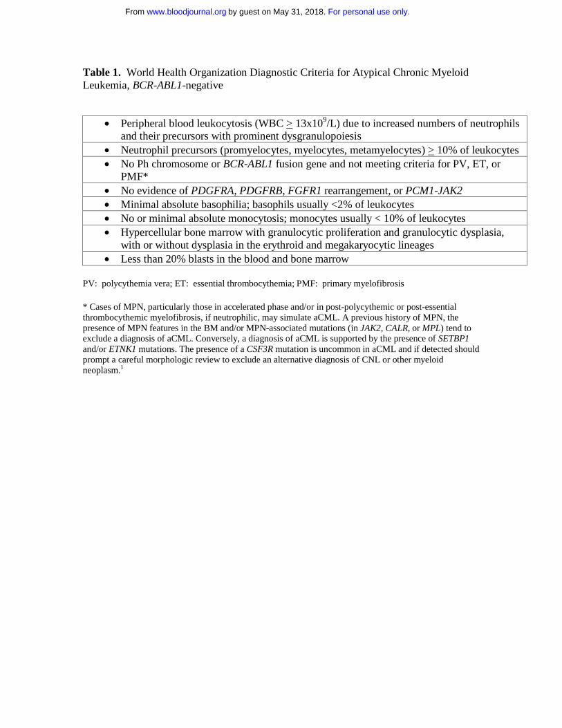
Table 1. World Health Organization Diagnostic Criteria for Atypical Chronic Myeloid Leukemia, BCR-ABL1-negative

PV: polycythemia vera; ET: essential thrombocythemia; PMF: primary myelofibrosis * Cases of MPN, particularly those in accelerated phase and/or in post-polycythemic or post-essential thrombocythemic myelofibrosis, if neutrophilic, may simulate aCML. A previous history of MPN, the presence of MPN features in the BM and/or MPN-associated mutations (in JAK2, CALR, or MPL) tend to exclude a diagnosis of aCML. Conversely, a diagnosis of aCML is supported by the presence of SETBP1 and/or ETNK1 mutations. The presence of a CSF3R mutation is uncommon in aCML and if detected should prompt a careful morphologic review to exclude an alternative diagnosis of CNL or other myeloid neoplasm.1

• Peripheral blood leukocytosis (WBC > 13x109/L) due to increased numbers of neutrophils and their precursors with prominent dysgranulopoiesis

• Neutrophil precursors (promyelocytes, myelocytes, metamyelocytes) > 10% of leukocytes • No Ph chromosome or BCR-ABL1 fusion gene and not meeting criteria for PV, ET, or

PMF* • No evidence of PDGFRA, PDGFRB, FGFR1 rearrangement, or PCM1-JAK2 • Minimal absolute basophilia; basophils usually <2% of leukocytes • No or minimal absolute monocytosis; monocytes usually < 10% of leukocytes • Hypercellular bone marrow with granulocytic proliferation and granulocytic dysplasia,

with or without dysplasia in the erythroid and megakaryocytic lineages • Less than 20% blasts in the blood and bone marrow

For personal use only.on May 31, 2018. by guest www.bloodjournal.orgFrom

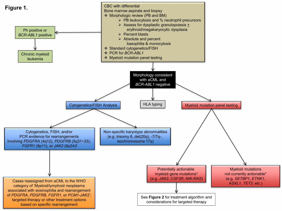
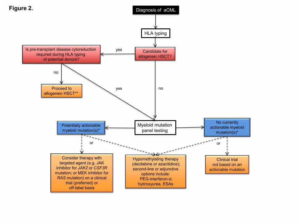
Figure Legends Figure 1. Diagnostic evaluation for atypical CML and identification of opportunities for targeted therapy. If a morphologic diagnosis of aCML is rendered, cytogenetic, FISH, and myeloid mutation panel testing are critical as they can unmask karyotypic or molecular abnormalities that have potential implications for use of targeted therapy approaches. *The ability to target certain genes is expected to change over time as new therapeutics are developed § Additional JAK2 rearrangements besides the PCM1-JAK2 fusion may present with morphologic features of aCML Figure 2. Treatment algorithm for atypical CML. Please refer to the text section ‘Treatment’ for a discussion of this treatment scheme for aCML. This algorithm is based on several decision nodes, including: 1) potential candidacy for allogeneic hematopoietic stem cell transplantation (HSCT); 2) the results of myeloid mutation panel testing; 3) eligibility for enrollment in clinical trials; and 4) opportunities to adopt strategies used for MDS or MPN (e.g. hypomethylating agents or second line/adjunctive therapies). HSCT: hematopoietic stem cell transplantation; ESAs: erythropoiesis-stimulating agents. *The ability to target certain genes is expected to change over time as new therapeutics are developed ** Myeloid mutation panel testing may also be performed prior to patients proceeding directing to allogeneic HSCT who do not require pre-transplant disease cytoreduction.

For personal use only.on May 31, 2018. by guest www.bloodjournal.orgFrom

CBC with differentialBone marrow aspirate and biopsy Morphologic review (PB and BM)

PB leukocytosis and % neutrophil precursors Assess for dysplastic granulopoiesis +

erythroid/megakaryocytic dysplasia Percent blasts Absolute and percent

basophilia & monocytosis Standard cytogenetics/FISH PCR for BCR-ABL1 Myeloid mutation panel testing

Ph positive orBCR-ABL1 positive

Chronic myeloidleukemia

Morphology consistentwith aCML and

BCR-ABL1 negative

Cytogenetics, FISH, and/or PCR evidence for rearrangements

involving PDGFRA (4q12), PDGFRB (5q31~33), FGFR1 (8p11), or JAK2 (9p24)§

Cases reassigned from aCML to the WHOcategory of ‘Myeloid/lymphoid neoplasms

associated with eosinophilia and rearrangementof PDGFRA, PDGFRB, FGFR1, or PCM1-JAK2’;

targeted therapy or other treatment optionsbased on specific rearrangement

Myeloid mutation panel testing

Potentially actionable myeloid gene mutations*

(e.g. JAK2, CSF3R, N/K-RAS)

Myeloid mutations not currently actionable* (e.g. SETBP1, ETNK1,

ASXL1, TET2, etc.)

Cytogenetics/FISH Analysis

Non-specific karyotype abnormalities(e.g. trisomy 8, del(20q), -7/7q-,

isochromosome 17q)

See Figure 2 for treatment algorithm andconsiderations for targeted therapy

Figure 1.

HLA typing

For personal use only.on May 31, 2018. by guest www.bloodjournal.orgFrom

Diagnosis of aCML

HLA typing

Candidate forallogeneic HSCT?

Myeloid mutation panel testing

Potentially actionable myeloid mutation(s)*

No currently actionable myeloid

mutation(s)*

Consider therapy with targeted agent (e.g. JAK

inhibitor for JAK2 or CSF3Rmutation; or MEK inhibitor for RAS mutation) on a clinical

trial (preferred) or off-label basis

or

Hypomethylating therapy(decitabine or azacitidine); second-line or adjunctive

options include:PEG-interferon-α;

hydroxyurea, ESAs

or

Clinical trialnot based on an

actionable mutation

Is pre-transplant disease cytoreductionrequired during HLA typing

of potential donors?

no

yes

Proceed toallogeneic HSCT**

yes

no

Figure 2.

For personal use only.on May 31, 2018. by guest www.bloodjournal.orgFrom

doi:10.1182/blood-2016-08-693630Prepublished online November 29, 2016;   

Jason Gotlib How I treat atypical chronic myeloid leukemia 

http://www.bloodjournal.org/site/misc/rights.xhtml#repub_requestsInformation about reproducing this article in parts or in its entirety may be found online at:

http://www.bloodjournal.org/site/misc/rights.xhtml#reprintsInformation about ordering reprints may be found online at:

http://www.bloodjournal.org/site/subscriptions/index.xhtmlInformation about subscriptions and ASH membership may be found online at:

    digital object identifier (DOIs) and date of initial publication. indexed by PubMed from initial publication. Citations to Advance online articles must include final publication). Advance online articles are citable and establish publication priority; they areappeared in the paper journal (edited, typeset versions may be posted when available prior to Advance online articles have been peer reviewed and accepted for publication but have not yet

  Copyright 2011 by The American Society of Hematology; all rights reserved.Hematology, 2021 L St, NW, Suite 900, Washington DC 20036.Blood (print ISSN 0006-4971, online ISSN 1528-0020), is published weekly by the American Society of

For personal use only.on May 31, 2018. by guest www.bloodjournal.orgFrom


Recommended