92 Paraskevas KI, et al. Stroke and Vascular Neurology 2018;3:e000129. doi:10.1136/svn-2017-000129 Open access How to identify which patients with asymptomatic carotid stenosis could benefit from endarterectomy or stenting Kosmas I Paraskevas, 1 Frank J Veith, 2,3 J David Spence 4 1 Department of Vascular and Endovascular Surgery, Royal Free Hospital, London, UK 2 Department of Vascular Surgery, New York University Langone Medical Center, New York, USA 3 Department of Vascular Surgery, Cleveland Clinic, Cleveland, Ohio, USA 4 Stroke Prevention & Atherosclerosis Research Centre, Robarts Research Institute, Western University, London, Canada Correspondence to Professor J David Spence; [email protected]To cite: Paraskevas KI, Veith FJ, Spence JD. How to identify which patients with asymptomatic carotid stenosis could benefit from endarterectomy or stenting. Stroke and Vascular Neurology 2018;3: e000129. doi:10.1136/ svn-2017-000129 Received 8 December 2017 Revised 6 February 2018 Accepted 7 February 2018 Published Online First 24 February 2018 ► http://dx.doi.org/10.1136/ svn-2018-000171 Review ABSTRACT Offering routine carotid endarterectomy (CEA) or carotid artery stenting (CAS) to patients with asymptomatic carotid artery stenosis (ACS) is no longer considered as the optimal management of these patients. Equally suboptimal, however, is the policy of offering only best medical treatment (BMT) to all patients with ACS and not considering any of them for prophylactic CEA. In the last few years, there have been many studies aiming to identify reliable predictors of future cerebrovascular events that would allow the identification of patients with high-risk ACS and offer a prophylactic carotid intervention only to these patients to prevent them from becoming symptomatic. All patients with ACS should receive BMT. The present article will summarise the evidence suggesting ways to identify these high-risk asymptomatic individuals, namely: (1) microemboli detection on transcranial Doppler, (2) plaque echolucency on Duplex ultrasound, (3) progression in the severity of ACS, (4) silent embolic infarcts on brain CT/MRI, (5) reduced cerebrovascular reserve, (6) increased size of juxtaluminal hypoechoic area, (7) identification of intraplaque haemorrhage using MRI and (8) carotid ulceration. The evidence suggests that approximately 10%–15% of patents with asymptomatic stenosis might benefit from intervention; this will become more clear after publication of ongoing studies comparing stenting or endarterectomy with best medical therapy. In the meantime, no patient should be offered intervention unless there is evidence of high risk of ipsilateral stroke, from modalities such as those discussed here. INTRODUCTION There are almost 800 000 strokes each year in USA, causing about 140 000 deaths annu- ally. 1 About 610 000 of these are first strokes. 1 Similarly, in UK, there are more than 100 000 strokes/year. 2 In 2015 alone, over 40 000 people died of stroke in UK. 2 Stroke causes twice as many deaths/year in women than breast cancer and twice as many deaths/ year in men than prostate and testicular cancer combined. 2 Stroke is the second most common cause of death in the world, causing around 6.7 million deaths each year (or one death every 5 s). 2 About 85% of all strokes are ischaemic and 15% are haemorrhagic. 2 Thromboemboli originating from an ipsilat- eral asymptomatic carotid stenosis (ACS) are the cause of a substantial proportion of first- ever ischaemic strokes. As a result of three landmark randomised controlled trials showing that carotid endar- terectomy (CEA) conferred a 50% relative risk (RR) reduction in the 5-year stroke risk compared with best medical treatment (BMT) alone, 3–5 offering CEA routinely to patients with ACS was considered as the treatment-of-choice in the 1980s and 1990s. In the early and mid-2000s, however, this began to change. Due to improvements in BMT (eg, smoking cessation strategies, implementation of statins and so on), the number of cerebrovascular events/year (ie, the annual stroke rate) among patients with ACS declined significantly. 6 7 It therefore became apparent that offering CEA routinely to patients with ACS was no longer optimal management. At the same time, however, the opposite theory supporting BMT alone as the treatment-of-choice for all patients with ACS and condemning prophylactic CEA for any patient with ACS 8 9 is equally suboptimal and misleading. This theory is not based on Level I Evidence; it is an extrapolation from the improved results achieved with current BMT in various observational studies and meta-analyses. In the last few years, several methods have been proposed as reliable predictors for the identification of ACS individuals at high risk of stroke. For some of these predictors, the evidence is adequate and robust, whereas for others it is weaker. The current article will outline methods to identify which asymp- tomatic carotid patients could benefit from a prophylactic carotid intervention. MICROEMBOLI DETECTION ON TRANSCRANIAL DOPPLER (TCD) The predictive value of microemboli detec- tion on TCD for the identification of patients with ACS at high risk for future stroke is well-established. Spence et al first reported that patients with ACS with >2 microemboli/ on February 6, 2020 by guest. Protected by copyright. http://svn.bmj.com/ Stroke Vasc Neurol: first published as 10.1136/svn-2017-000129 on 24 February 2018. Downloaded from
Transcript
92 Paraskevas KI, et al. Stroke and Vascular Neurology 2018;3:e000129. doi:10.1136/svn-2017-000129
Open access
How to identify which patients with asymptomatic carotid stenosis could benefit from endarterectomy or stentingKosmas I Paraskevas,1 Frank J Veith,2,3 J David Spence4
1Department of Vascular and Endovascular Surgery, Royal Free Hospital, London, UK2Department of Vascular Surgery, New York University Langone Medical Center, New York, USA3Department of Vascular Surgery, Cleveland Clinic, Cleveland, Ohio, USA4Stroke Prevention & Atherosclerosis Research Centre, Robarts Research Institute, Western University, London, Canada
Correspondence toProfessor J David Spence; dspence@ robarts. ca
To cite: Paraskevas KI, Veith FJ, Spence JD. How to identify which patients with asymptomatic carotid stenosis could benefit from endarterectomy or stenting. Stroke and Vascular Neurology 2018;3: e000129. doi:10.1136/svn-2017-000129
Received 8 December 2017Revised 6 February 2018Accepted 7 February 2018Published Online First 24 February 2018
AbsTrACTOffering routine carotid endarterectomy (CEA) or carotid artery stenting (CAS) to patients with asymptomatic carotid artery stenosis (ACS) is no longer considered as the optimal management of these patients. Equally suboptimal, however, is the policy of offering only best medical treatment (BMT) to all patients with ACS and not considering any of them for prophylactic CEA. In the last few years, there have been many studies aiming to identify reliable predictors of future cerebrovascular events that would allow the identification of patients with high-risk ACS and offer a prophylactic carotid intervention only to these patients to prevent them from becoming symptomatic. All patients with ACS should receive BMT. The present article will summarise the evidence suggesting ways to identify these high-risk asymptomatic individuals, namely: (1) microemboli detection on transcranial Doppler, (2) plaque echolucency on Duplex ultrasound, (3) progression in the severity of ACS, (4) silent embolic infarcts on brain CT/MRI, (5) reduced cerebrovascular reserve, (6) increased size of juxtaluminal hypoechoic area, (7) identification of intraplaque haemorrhage using MRI and (8) carotid ulceration. The evidence suggests that approximately 10%–15% of patents with asymptomatic stenosis might benefit from intervention; this will become more clear after publication of ongoing studies comparing stenting or endarterectomy with best medical therapy. In the meantime, no patient should be offered intervention unless there is evidence of high risk of ipsilateral stroke, from modalities such as those discussed here.
InTroduCTIonThere are almost 800 000 strokes each year in USA, causing about 140 000 deaths annu-ally.1 About 610 000 of these are first strokes.1 Similarly, in UK, there are more than 100 000 strokes/year.2 In 2015 alone, over 40 000 people died of stroke in UK.2 Stroke causes twice as many deaths/year in women than breast cancer and twice as many deaths/year in men than prostate and testicular cancer combined.2 Stroke is the second most common cause of death in the world, causing around 6.7 million deaths each year (or one death every 5 s).2 About 85% of all strokes are ischaemic and 15% are haemorrhagic.2 Thromboemboli originating from an ipsilat-eral asymptomatic carotid stenosis (ACS) are
the cause of a substantial proportion of first-ever ischaemic strokes.
As a result of three landmark randomised controlled trials showing that carotid endar-terectomy (CEA) conferred a 50% relative risk (RR) reduction in the 5-year stroke risk compared with best medical treatment (BMT) alone,3–5 offering CEA routinely to patients with ACS was considered as the treatment-of-choice in the 1980s and 1990s. In the early and mid-2000s, however, this began to change. Due to improvements in BMT (eg, smoking cessation strategies, implementation of statins and so on), the number of cerebrovascular events/year (ie, the annual stroke rate) among patients with ACS declined significantly.6 7 It therefore became apparent that offering CEA routinely to patients with ACS was no longer optimal management. At the same time, however, the opposite theory supporting BMT alone as the treatment-of-choice for all patients with ACS and condemning prophylactic CEA for any patient with ACS8 9 is equally suboptimal and misleading. This theory is not based on Level I Evidence; it is an extrapolation from the improved results achieved with current BMT in various observational studies and meta-analyses.
In the last few years, several methods have been proposed as reliable predictors for the identification of ACS individuals at high risk of stroke. For some of these predictors, the evidence is adequate and robust, whereas for others it is weaker. The current article will outline methods to identify which asymp-tomatic carotid patients could benefit from a prophylactic carotid intervention.
MICroeMbolI deTeCTIon on TrAnsCrAnIAl doppler (TCd)The predictive value of microemboli detec-tion on TCD for the identification of patients with ACS at high risk for future stroke is well-established. Spence et al first reported that patients with ACS with >2 microemboli/
on February 6, 2020 by guest. P
rotected by copyright.http://svn.bm
j.com/
Stroke V
asc Neurol: first published as 10.1136/svn-2017-000129 on 24 F
93Paraskevas KI, et al. Stroke and Vascular Neurology 2018;3:e000129. doi:10.1136/svn-2017-000129
Open access
hour on TCD had a >1500% increased risk of 1-year ipsi-lateral ischaemic stroke compared with patients with ACS without TCD-detected microemboli (15.6% vs 1.0%, respectively; P<0.0001).6 Figure 1 shows a microembolus. In 2010, the same group reported that as a result of improvements in BMT, there was a marked reduction in TCD-detected microemboli (12.6% before 2003 vs 3.7% after 2003; P<0.001) and in cardiovascular events (17.6% before 2003 vs 5.2% after 2003; P<0.001) in 468 patients with ACS.6 A few months later, these results were verified in an independent multicentre international study on 467 patients with ACS, the Asymptomatic Carotid Emboli Study (ACES).10 As in the study by Spence et al,6 patients participating in ACES also had two 1-hour TCD record-ings 1 week apart.10 Patients with one or more TCD emboli had a >550% higher risk of 1 year ipsilateral stroke compared with patients without emboli (HR: 5.57; 95% CI 1.61 to 19.32; P=0.007).10
Contradictory results had been earlier reported by a small prospective, observational, cohort study.11 This early study showed a trend to higher stroke risk in patients with ACS with microemboli, but was underpow-ered, with only 202 patients.11 Besides the small sample size, another likely reason for the negative results of this study was that it accepted one microembolus as positive, and the test was repeated at 6-monthly intervals.11 There is compelling evidence that at least two embolic signals detected in a recording lasting 1 hour identifies patients with ACS at very high risk of stroke,12 13 suggesting a
high-risk, unstable asymptomatic plaque or a plaque with a thrombus on its surface.13
TCD embolus detection is currently the best validated method for the identification of high-risk patients with ACS.14 This is also supported by a meta-analysis including five prospective studies (n=677 patients).15 In this meta-analysis, the presence of TCD-detected embolic signals in patients with ACS was a significant predictor of ipsilateral stroke (OR: 7.46; 95% CI 2.24 to 24.89; P=0.001).15 The 2017 European Society for Vascular Surgery carotid guidelines recommend intervention based on TCD microemboli.16
plAque eCholuCenCy on duplex ulTrAsoundEarly studies from the 1990s demonstrated that carotid plaque echolucency corresponds to lipid-rich necrotic core or intraplaque haemorrhage, more commonly found in patients with symptomatic rather than ACS.17 18 Several studies evaluated whether or not carotid plaque echolucency was associated with increased risk of future stroke in patients with ACS (table 1).19–26
The majority of studies independently reported a strong association between plaque echolucency and increased risk of stroke in patients with ACS.19–21 23–26 The only exception was a study from Denmark which demon-strated that carotid plaque echolucency was positively associated with risk of future stroke in patients with symp-tomatic, but not with ACS.22
Figure 1 Transcranial Doppler embolus detection. Microembolus in a patient with asymptomatic carotid stenosis. The upper channel is an M-mode image of an embolus in the middle cerebral artery; the lower panel shows the high-intensity transit signal in the Doppler channel. Besides the visual appearance of the microembolus, a characteristic clicking sound is heard. (Reproduced by permission of the Society for Vascular Ultrasound from: Spence JD. Transcranial Doppler: uses in stroke prevention. The Journal for Vascular Ultrasound 2015;39:183–7.)
on February 6, 2020 by guest. P
rotected by copyright.http://svn.bm
j.com/
Stroke V
asc Neurol: first published as 10.1136/svn-2017-000129 on 24 F
94 Paraskevas KI, et al. Stroke and Vascular Neurology 2018;3:e000129. doi:10.1136/svn-2017-000129
Open access
A recent meta-analysis (7557 patients; mean follow-up: 37.2 months) demonstrated a positive association between plaque echolucency and the risk of future ipsi-lateral stroke (RR: 2.31; 95% CI 1.58 to 3.39; P<0.001).27 Of the total study sample, 1741 patients (23.0%) had a positive ultrasound test for echolucency, whereas 5816 (77.0%) had a negative test for echolucency. A total of 100 ipsilateral strokes occurred in the echolucent-positive test group, while 141 ipsilateral strokes occurred in the echolucency-negative test group (cumulative incidence of ipsilateral stroke: 5.7% vs 2.4%, respectively). In patients with ≥50% carotid stenosis, the stroke risk was higher (RR: 2.61; 95% CI 1.47 to 4.63; P=0.001).27 The associa-tion between carotid plaque echolucency and increased risk of future ipsilateral stroke was verified in an indepen-dent meta-analysis.28
The predictive value of echolucent plaque morphology on carotid ultrasound increases even further if it is combined with TCD-detected emboli.24 In ACES,24 carotid plaque echolucency was associated with a >600% increased risk of ipsilateral risk (HR: 6.43; 95% CI 1.36 to 30.44; P=0.019), while the combination of plaque echo-lucency with TCD-detected emboli was associated with a >1000% increased risk of ipsilateral stroke (HR: 10.61; 95% CI 2.98 to 37.82; P=0.0003).24
progressIon of The severITy of sTenosIsMost would agree that the progression of the severity of ACS in successive ultrasound examinations despite the implementation of BMT is not a good sign. Several reasons may account for ACS progression despite BMT. Up to half of patients with ACS may have ‘resistant ather-osclerosis’.29
The largest prospective study on patients with ACS undergoing medical intervention alone, the Asymp-tomatic Carotid Stenosis and Risk of Stroke (ACSRS) study,30 demonstrated quite clearly that progression in the severity of ACS was a predictor of future stroke. As shown in ACSRS,30 the 8-year cumulative ipsilateral isch-aemic stroke rate was 0% in patients with regression of stenosis, 9% if the stenosis was unchanged and 16% if there was progression of stenosis. In the subgroup with unchanged stenosis, the 8-year cumulative ipsilateral cerebral ischaemic stroke rates for patients with baseline stenosis of 50%–69%, 70%–89% and 90%–99% were 4%, 8% and 13%, respectively.30 In contrast, in the presence of progression, the stroke rate was 8%, 15% and 25%, respectively.30
An independent study from Boston, Massachusetts, verified that progression of ACS despite BMT is not a good prognostic factor.31 In this study, 794 patients (900 carotid arteries) with moderate (50%–69%) ACS had a mean follow-up of 3.6 (range: 0.3–6.7) years. Stenosis progression occurred in 262 of 900 (29.1%) carotid arteries despite BMT and 36 (13.7%) of these developed symptoms. The symptomatic conversion rate in patients with stenosis progression was considerably higher than of those without stenosis progression (13.7% vs 8.5%, respectively; P=0.02). Overall, BMT failed to prevent carotid disease progression or the development of ipsilat-eral neurologic symptoms in a significant proportion of the patient cohort.31
These results once again verify the results of an earlier study on 1065 patients with ACS followed up with carotid ultrasound.32 During the initial study period of a median 7.5 (range: 6–9) months, progression of carotid lesions was demonstrated in 93 of 1065 patients (9%). During a median follow-up of 3.2 (IQR: 2.9–3.5) years, 495 major adverse cardiovascular events (a composite of myocar-dial infarction (MI), percutaneous coronary interven-tion, coronary artery bypass graft, stroke, peripheral percutaneous angioplasty, peripheral vascular surgery, amputation due to critical limb ischaemia and all-cause mortality) were recorded in 421 patients (40%). Patients with progressive ACS had a 200% higher risk for composite major adverse cardiovascular events compared with patients with non-progressive disease (adjusted HR: 2.01; 95% CI 1.48 to 2.67; P<0.001), consisting of a >200% higher risk of MI (HR: 2.38; 95% CI 1.07 to 5.35; P=0.044), a 200% higher risk for stroke (adjusted HR: 2.0; 95% CI 1.02 to 4.11; P=0.035) and a 175% higher risk for cardiovascular death (adjusted HR: 1.75; 95% CI 1.03 to 2.97, P=0.039).32
Progression of carotid plaque burden may be a better predictor of cardiovascular outcomes than carotid intima-media thickness (cIMT). A study from Canada compared the progression/regression of cIMT, total plaque area and total plaque volume as predictors of cardiovascular outcomes in 349 patients attending stroke prevention clinics.33 After a median follow-up of 3.17 (range: 0.07–5.0) years, there were 50 first events: 20
Table 1 Studies evaluating the association between carotid plaque echolucency and risk of ipsilateral stroke
Study (year)Number of patients
Mean follow-up (months)
Ipsilateral strokeRR (95% CI)
O’Holleran et al19 (1987)
293 46 5.12 (2.01 to 13.04)
Polak et al20 (1998) 4886 39.6 1.96 (1.25 to 2.90)
Mathiesen et al21 (2001)
177 36 3.85 (0.46 to 32.28)
Grønholdt et al22 (2001)
111 52.8 0.87 (0.34 to 2.23)
Nicolaides et al23 (2005)
1092 37.1 2.23 (1.28 to 3.87)
Topakian et al24 (2011)
435 21.8 6.61 (1.42 to 30.75)
Silvestrini et al25 (2013)
621 27* 2.37 (1.14 to 4.92)
Huibers et al26 (2016)
814 60 2.52 (1.20 to 5.25)
*Median follow-up.RR, relative risk.
on February 6, 2020 by guest. P
rotected by copyright.http://svn.bm
j.com/
Stroke V
asc Neurol: first published as 10.1136/svn-2017-000129 on 24 F
95Paraskevas KI, et al. Stroke and Vascular Neurology 2018;3:e000129. doi:10.1136/svn-2017-000129
Open access
vascular deaths, 11 strokes, 13 transient ischaemic attacks (TIAs) and 6 MIs. Progression of total plaque volume predicted stroke, death or TIA (P=0.001), stroke, death or MI (P=0.008) and stroke, death, TIA or MI (P=0.001). Progression of total plaque area weakly predicted stroke, TIA or death (P=0.097), but not stroke, death or MI (P=0.59) or TIA, stroke, death or MI (P=0.143). Simi-larly, change in cIMT did not predict stroke, death or MI (P=0.13) or TIA, stroke, death or MI (P=0.455). By Cox regression analysis using a backward stepwise Wald approach, total plaque volume progression remained a significant predictor of events after adjustment for coronary risk factors (P=0.001). This study showed that measurement of total plaque volume is a superior predictor of cardiovascular events that either total plaque area or cIMT.33 In patients with ACS, plaque burden, but not per cent stenosis, predicted the risk of stroke.34 In the High Risk Plaque BioImage study,35 plaque burden was strongly correlated with coronary calcium whereas IMT was not, and plaque burden was as predictive of events as coronary calcium.36
sIlenT eMbolIC InfArCTs on brAIn CT or MrIBoth the Cardiovascular Health Study37 and the Rotterdam Scan Study38 demonstrated that the presence of silent embolic infarcts on brain CT or MRI scans is associated with an increased risk of stroke in the general population. Two studies (ACSRS39 and an independent study from Japan)40 showed that silent embolic infarcts on brain CT or MRI are an independent predictor of stroke. In ACSRS, patients with 60%–99% ACS having silent embolic infarcts on brain CT scans had a 300% higher risk of future ipsilateral stroke compared with patients without silent embolic infarcts (annual stroke rate: 3.6% vs 1.0%, respectively; HR: 3.0; 95% CI 1.46 to 6.29; P=0.002).39
These results suggest that the presence of silent infarcts on brain CT or MRI in patients with ACS reliably identi-fies patients with ACS at high risk for a future ipsilateral cerebrovascular event. However, one of the limitations of brain CT scans is that they may miss up to 40% of brain infarcts in patients with ACS.41
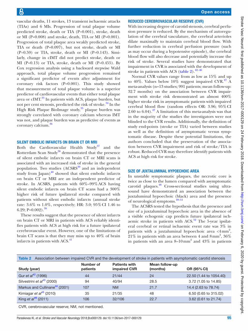
reduCed CerebrovAsCulAr reserve (Cvr)With increasing degree of carotid stenosis, cerebral perfu-sion pressure is reduced. By the mechanism of autoregu-lation of the cerebral vasculature, the cerebral arterioles dilate maximally to maintain cerebral blood flow. With further reduction in cerebral perfusion pressure (such as may occur during a hypotensive episode), the cerebral blood flow will also decrease and potentially increase the risk of stroke. Several studies have demonstrated that impairment in CVR is associated with the development of stroke in patients with ACS (table 2).42–46
Normal CVR values range from as low as 15% and up to 40%. Values below 10% suggest impaired CVR.47 A meta-analysis (n=13 studies; 991 patients; mean follow-up: 32.7 months) on the association between CVR impair-ment with stroke risk demonstrated an almost 400% higher stroke risk in asymptomatic patients with impaired cerebral blood flow (random effects OR: 3.96; 95% CI 2.60 to 6.04).47 A limitation of this meta-analysis was that in the majority of the studies the investigators were not blinded to the CVR results. Additionally, the definition of study end-points (stroke or TIA) varied between studies as well as the definition of asymptomatic versus symp-tomatic disease. Despite these potential limitations, the authors concluded that the preservation of the associa-tion between CVR impairment and risk of stroke/TIA is robust. Reduced CVR may therefore identify patients with ACS at high risk for stroke.
sIze of juxTAluMInAl hypoeChoIC AreAIn unstable symptomatic plaques, the necrotic core is twice as close to the lumen compared with asymptomatic carotid plaques.48 Cross-sectional studies using ultra-sound have demonstrated an association between the juxtaluminal hypoechoic (black) area and the presence of neurological symptoms.49–51
The ACSRS tested the hypothesis that the presence and size of a juxtaluminal hypoechoic area in the absence of a visible echogenic cap predicts future ipsilateral isch-aemic stroke in patients with ACS.52 The 5-year ipsilat-eral cerebral or retinal ischaemic event rate was 3% in patients with a juxtaluminal hypoechoic area <4 mm2, 21% in patients with an area between 4 and 8 mm2, 36% in patients with an area 8–10 mm2 and 43% in patients
Table 2 Association between impaired CVR and the development of stroke in patients with asymptomatic carotid stenosis
Study (year)Number of patients
Patients with impaired CVR
Mean follow-up (months) OR (95% CI)
Gur et al42 (1996) 44 21/44 24 22.50 (1.44 to 1054.40)
Silvestrini et al43 (2000) 94 40/94 28.5 3.72 (1.05 to 14.85)
Markus and Cullinane44 (2001) 107 NM 21.7 14.4 (2.63 to 78.74)
Kimiagar et al45 (2010) 35 21/35 48 6.50 (0.65 to 315.02)
King et al46 (2011) 106 32/106 22.7 3.62 (0.61 to 21.74)
CVR, cerebrovascular reserve; NM, not mentioned.
on February 6, 2020 by guest. P
rotected by copyright.http://svn.bm
j.com/
Stroke V
asc Neurol: first published as 10.1136/svn-2017-000129 on 24 F
96 Paraskevas KI, et al. Stroke and Vascular Neurology 2018;3:e000129. doi:10.1136/svn-2017-000129
Open access
with a juxtaluminal black area>10 mm2 (average annual rates: 0.6%, 4.2%, 7.2% and 8.6%, respectively).52 These results support the theory that the size of juxtaluminal hypoechoic area may be a predictor of future ipsilateral ischaemic stroke.
IdenTIfICATIon of InTrAplAque hAeMorrhAge usIng MrISeveral studies have evaluated whether or not MRI assess-ment of specific components of the carotid plaque can be used to predict stroke in patients with ACS (table 3).
In some of these studies, the authors described blinding of MRI results to researchers who assessed ischaemic outcomes53 54 55 whereas such blinding was not reported in others.56–58 The majority of the studies showed that carotid plaques with intraplaque haemorrhage, lipid-rich necrotic core or thinning/rupture of the fibrous cap are significantly more likely to result in ipsilateral ischaemic events with this increased risk present across a wide range of stenosis severity.
The ability of carotid intraplaque haemorrhage, lipid-rich necrotic core and thinning/rupture of the fibrous cap to predict future ipsilateral ischaemic stroke was verified in a meta-analysis (n=9 studies; 779 patients).59 The HRs (95% CI) for intraplaque haemorrhage, lipid-rich necrotic core and thinning/rupture of the fibrous
cap as predictors of subsequent stroke/TIA were 4.59 (2.91–7.24), 3.00 (1.51–5.95) and 5.93 (2.65–13.20), respectively. This meta-analysis concluded that MRI characterisation of these specific plaque elements
Table 3 Studies evaluating the risk of stroke in patients with ACS using carotid plaque MRI
Study (year)Number of patients
Mean follow-up (months)
HR (95% CI) for stroke
Takaya et al56 (2006)
154 38.2 IPH: 5.2 (1.6 to 7.3)TRFC: 2.2 to 132.0LRNC: 0.6 to 33.7
Singh et al53 (2009)
98 24.9 IPH: 2.48 to 4.71
Sadat et al57 (2010)
61 16.9 IPH: 1.27 to 26.77TRFC: 7.39 (1.61 to 33.82)LRNC: 1.75 (0.55 to 5.54)
Mono et al54 (2012)
65 18.9 IPH: 0.03 (0.00 to 86.62)TRFC: 1.103 (0.11 to 10.70)LRNC: 7.2 (1.12 to 46.28)
Kwee et al55 (2013)
126 12.0 IPH: 3.5 (1.06 to 11.96)TRFC: 5.8 (1.91 to 17.32)LRNC: 3.2 (1.08 to 9.50)
Hosseini et al58 (2013)
179 17.5 IPH: 12 (4.8 to 30.1)
ACS, asymptomatic carotid stenosis; IPH, intraplaque haemorrhage; LRNC, lipid-rich necrotic core; TRFC, thinning/rupture of the fibrous cap.
Figure 2 Carotid ulcer volume as a predictor of risk. (A) Measurement of ulcer volume and ulcer depth. Contours of ulcers were traced and depth of ulcers measured in cross-sectional views. Each slice had a thickness of 1 mm; total ulcer volume (TUV) was computed from the sum of the volumes of all slices in which ulceration was traced. (B) Kaplan–Meier survival analysis curves for participants with TUV≥5.00 mm3 and those with no ulcerations or TUV<5 mm3. Time is shown in days until the first occurrence of any of the following events: stroke, TIA or cardiovascular death during the duration of follow-up; log-rank P=0.009. TIA, transient ischaemic attack; TUV, total ulcer volume. (Reproduced by permission of Wolters Kluwer Health. from: Kuk M, Wannarong T, Beletsky V, Parraga G, Fenster A, Spence JD. Volume of Carotid Artery Ulceration as a predictor of Cardiovascular Events. Stroke 2014;45:1437–41.)
on February 6, 2020 by guest. P
rotected by copyright.http://svn.bm
j.com/
Stroke V
asc Neurol: first published as 10.1136/svn-2017-000129 on 24 F
97Paraskevas KI, et al. Stroke and Vascular Neurology 2018;3:e000129. doi:10.1136/svn-2017-000129
Open access
can provide additional measures of stroke risk not provided by simple measurement of luminal stenosis. These results suggest that carotid plaque MRI may be used to select high-risk groups that may benefit from revascularisation.
CAroTId ulCerATIonThe North American Symptomatic Carotid Endar-terectomy Trial showed that ulceration on angiog-raphy was associated with up to a 350% higher RR of stroke.60 A study from Canada showed that, in ACS, compared with no ulcers, the presence of three or more ulcers (the sum of both carotids) predicted the 3-year risk of stroke or death (18.2% vs 1.7%, p = 0.03 respectively) to a similar degree as microemboli (20% vs 2.0%, p = 0.003).61 Ulcer volume also predicts risk among patients attending a stroke prevention clinic (figure 2). Contrast-enhanced ultrasound is superior to colour Doppler ultrasound for the detection of ulcer-ated plaques.62 Contrast-enhanced ultrasound can be used to identify the ‘vulnerable carotid plaque’ associ-ated with high embolic potential.63 Three-dimensional carotid ultrasound-based texture analysis is another way to evaluate both the composition of the carotid plaque and to predict vascular events.64 65
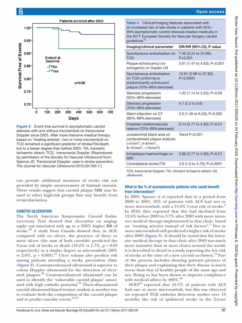
What is the % of asymptomatic patients who could benefit from intervention?In 2005, Spence et al reported that in a period from 2000 to 2005, 10% of patients with ACS had two or more microemboli, with a 15.6% 1-year risk of stroke.6 In 2010, they reported that this had declined from 12.6% before 2003 to 3.7% after 2003 with more inten-sive medical therapy implemented in that clinic, based on ‘treating arteries instead of risk factors’.7 Two or more microemboli still predicted a higher risk of stroke after 2003 (figure 3). It should be noted that the inten-sive medical therapy in that clinic after 2003 was much more intensive than in most clinics around the world; it is described in detail in a study reporting the low risk of stroke at the time of a new carotid occlusion.34 Part of the process includes showing patients pictures of their plaque and explaining that their disease is much worse than that of healthy people of the same age and sex. Doing so has been shown to improve compliance with medical advice by 400%.66
ACES10 reported that 16.5% of patients with ACS had one or more microemboli, but this was observed on repeated TCD embolus detection studies over 18 months; the risk of ipsilateral stroke in the 2 years
Table 4 Clinical/imaging features associated with an increased risk of late stroke in patients with 50%–99% asymptomatic carotid stenosis treated medically in the 2017 European Society for Vascular Surgery carotid guidelines16
Imaging/clinical parameter OR/HR (95% CI); P value
Spontaneous embolisation on TCD
7.46 (2.24 to 24.89); P=0.001
Plaque echolucency (vs echogenic) on Duplex US
2.61 (1.47 to 4.63); P=0.001
Spontaneous embolisation on TCD+uniformly or predominantly echolucent plaque (70%–99% stenoses)
Figure 3 Event-free survival in asymptomatic carotid stenosis with and without microemboli on transcranial Doppler since 2003. After more intensive medical therapy based on ‘treating arteries’, two or more microemboli on TCD remained a significant predictor of stroke/TIA/death, but to a lesser degree than before 2003. TIA, transient ischaemic attack; TCD, transcranial Doppler. (Reproduced by permission of the Society for Vascular Ultrasound from: Spence JD. Transcranial Doppler: uses in stroke prevention. The Journal for Vascular Ultrasound 2015;39:183–7.)
on February 6, 2020 by guest. P
rotected by copyright.http://svn.bm
j.com/
Stroke V
asc Neurol: first published as 10.1136/svn-2017-000129 on 24 F
98 Paraskevas KI, et al. Stroke and Vascular Neurology 2018;3:e000129. doi:10.1136/svn-2017-000129
Open access
following baseline microembolus detection was 3.62% in patients with embolic signals versus 0.70% without.10 In the study of 3D ultrasound detection of carotid ulceration described above,6110% of patients had either three or more ulcers or microemboli, with comparable risks and, surprisingly, these did not overlap by much: only 1.2% of patients with ACS had both two or more microemboli and three or more ulcers. However, some of these patients were studied before implementation of intensive medical therapy in 2003. In 2009, Singh et al reported intraplaque haem-orrhage in 36.7% of 98 carotid arteries with moderate asymptomatic stenosis.53 In a larger pathological study of endarterectomy specimens, intraplaque haemor-rhage was observed in 69.9% of plaques from patients who had been asymptomatic prior to surgery.67 It is likely that many of the pathologically observed haem-orrhages may have been too small for detection on MRI or ultrasound.
It seems reasonable to assume that some of the other features that predict risk of stroke, such as reduced CBF reserve, intraplaque haemorrhage and plaque inflammation, will also not overlap with microemboli or ulceration, so approximately 10%–15% of patients with ACS could benefit from CEA or carotid artery stenting (CAS) despite intensive medical therapy.16 No patient with asymptomatic stenosis should be offered intervention in the absence of such evidence. In most cases, particularly in the elderly, CEA is associated with
a lower risk of stroke compared to CAS. Another paper in this issue of the journal will review that topic.
ConClusIonCurrent evidence suggests that certain patients with ACS (eg, those with TCD-detected microemboli, silent embolic infarcts on brain CT/MRI scans, reduced CVR, ACS severity progression despite BMT, size of black juxtaluminal plaque area≥8 mm2 without a visible echogenic cap and intraplaque haemorrhage on MRI) are at increased stroke risk and should be considered for prophylactic CEA or CAS.16 68 The 2017 guide-lines by the European Society for Vascular Surgery recommend that in average surgical risk patients with a 60%–99% ACS, CEA (Class IIa; Level of Evidence: B) or CAS (Class IIb; Level of Evidence: B) should be considered for intervention in the presence of one or more imaging characteristics that may be associ-ated with an increased risk of late ipsilateral stroke (table 4), provided documented perioperative stroke/death rates are <3% and the patient’s life expectancy exceeds 5 years.16
This is a new recommendation taking into account the increased stroke risk of these patients with ACS managed with BMT alone. Further research on the above (and other) possible predictors of ischaemic stroke in patients with ACS such as plaque texture,65 plaque neovascularity69 70 and plaque inflammation or
Figure 4 Imaging of active calcification by PET/CT with [18]F Sodium Fluoride. NaF PET/CT imaging of left and right internal carotid arteries of active calcification in a 72-year-old symptomatic patient evaluated at the University of Ottawa Heart Institute. Upper row: evidence of NaF uptake with a small foci of calcification on CT in the left internal carotid symptomatic culprit vessel. There is a mismatch between the region of NaF uptake and calcification on CT. Lower row: evidence of calcium nodules with matched NaF uptake at the right internal carotid artery. PET, positron emission tomography. (Reproduced by permission of the Journal of Nuclear Cardiology from: Cocker MS, Mc Ardle B, Spence JD, et al. Imaging atherosclerosis with hybrid [(18)F]fluorodeoxyglucose positron emission tomography/CT imaging: What Leonardo da Vinci could not see. J Nucl Cardiol 2012;19:1211–25.)
on February 6, 2020 by guest. P
rotected by copyright.http://svn.bm
j.com/
Stroke V
asc Neurol: first published as 10.1136/svn-2017-000129 on 24 F
99Paraskevas KI, et al. Stroke and Vascular Neurology 2018;3:e000129. doi:10.1136/svn-2017-000129
Open access
active calcification (figure 4) on PET/CT71 is essential in order to appropriately select for carotid revascu-larisation procedures the few patients with ACS who could benefit from these procedures. Two ongoing trials comparing BMT with CEA or CAS are under way; when those studies have been completed, we will have better evidence about the role of intervention versus medical therapy in ACS.
Contributors KIP wrote the first draft. FJV and JDS made revisions.
funding This research received no specific grant from any funding agency in the public, commercial or not-for-profit sectors.
Competing interests None declared.provenance and peer review Commissioned; externally peer reviewed.data sharing statement No additional data are available.
guest chief editor J David Spence
open access This is an open access article distributed in accordance with the Creative Commons Attribution Non Commercial (CC BY-NC 4.0) license, which permits others to distribute, remix, adapt, build upon this work non-commercially, and license their derivative works on different terms, provided the original work is properly cited and the use is non-commercial. See: http:// creativecommons. org/ licenses/ by- nc/ 4. 0/
2. Stroke. org. uk. State of the nation. Stroke statistics, 2017. https://www. stroke. org. uk/ sites/ default/ files/ state_ of_ the_ nation_ 2017_ final_ 1. pdf. (accessed on 4 Nov 2017).
3. Hobson RW, Fields WS, Fields WS, et al. Efficacy of carotid endarterectomy for asymptomatic carotid stenosis. The Veterans Affairs Cooperative Study Group. N Engl J Med 1993;328:221–7.
4. Executive Committee for the Asymptomatic Carotid Atherosclerosis Study. Endarterectomy for asymptomatic carotid artery stenosis. JAMA 1995;273:1421–8.
5. Halliday A, Mansfield A, Marro J, et al. MRC Asymptomatic Carotid Surgery Trial (ACST) Collaborative Group. Prevention of disabling and fatal strokes by successful carotid endarterectomy in patients without recent neurological symptoms: randomised controlled trial. Lancet 2004;363:1491–502.
6. Spence JD, Tamayo A, Lownie SP, et al. Absence of microemboli on transcranial Doppler identifies low-risk patients with asymptomatic carotid stenosis. Stroke 2005;36:2373–8.
7. Spence JD, Coates V, Li H, et al. Effects of intensive medical therapy on microemboli and cardiovascular risk in asymptomatic carotid stenosis. Arch Neurol 2010;67:180–6.
8. Abbott AL. Medical (nonsurgical) intervention alone is now best for prevention of stroke associated with asymptomatic severe carotid stenosis: results of a systematic review and analysis. Stroke 2009;40:e573–e583.
9. Abbott A. Asymptomatic carotid artery stenosis--it's time to stop operating. Nat Clin Pract Neurol 2008;4:4–5.
10. Markus HS, King A, Shipley M, et al. Asymptomatic embolisation for prediction of stroke in the Asymptomatic Carotid Emboli Study (ACES): a prospective observational study. Lancet Neurol 2010;9:663–71.
11. Abbott AL, Chambers BR, Stork JL, et al. Embolic signals and prediction of ipsilateral stroke or transient ischemic attack in asymptomatic carotid stenosis: a multicenter prospective cohort study. Stroke 2005;36:1128–33.
12. King A, Shipley M, Markus H. ACES Investigators. Optimizing protocols for risk prediction in asymptomatic carotid stenosis using embolic signal detection: the Asymptomatic Carotid Emboli Study. Stroke 2011;42:2819–24.
13. Bogiatzi C, Cocker MS, Beanlands R, et al. Identifying high-risk asymptomatic carotid stenosis. Expert Opin Med Diagn 2012;6:139–51.
14. Spence JD. Transcranial Doppler monitoring for microemboli: a marker of a high-risk carotid plaque. Semin Vasc Surg 2017;30:62–6.
15. King A, Markus HS. Doppler embolic signals in cerebrovascular disease and prediction of stroke risk: a systematic review and meta-analysis. Stroke 2009;40:3711–7.
16. Naylor AR, Ricco JB, de Borst GJ, et al. Management of Atherosclerotic Carotid and Vertebral Artery Disease: 2017 Clinical Practice Guidelines of the European Society for Vascular Surgery (ESVS). Eur J Vasc Endovasc Surg 2017. doi: 10.1016/j.ejvs.2017.06.021. [Epub ahead of print 26 Aug 2017].
18. Grønholdt ML, Nordestgaard BG, Wiebe BM, et al. Echo-lucency of computerized ultrasound images of carotid atherosclerotic plaques are associated with increased levels of triglyceride-rich lipoproteins as well as increased plaque lipid content. Circulation 1998;97:34–40.
19. O'Holleran LW, Kennelly MM, McClurken M, et al. Natural history of asymptomatic carotid plaque. Five year follow-up study. Am J Surg 1987;154:659–62.
20. Polak JF, Shemanski L, O'Leary DH, et al. Hypoechoic plaque at US of the carotid artery: an independent risk factor for incident stroke in adults aged 65 years or older. Cardiovascular Health Study. Radiology 1998;208:649–54.
21. Mathiesen EB, Bønaa KH, Joakimsen O. Echolucent plaques are associated with high risk of ischemic cerebrovascular events in carotid stenosis: the tromsø study. Circulation 2001;103:2171–5.
23. Nicolaides AN, Kakkos SK, Griffin M, et al. Asymptomatic Carotid Stenosis and Risk of Stroke (ACSRS) Study Group. Effect of image normalization on carotid plaque classification and the risk of ipsilateral hemispheric ischemic events: results from the asymptomatic carotid stenosis and risk of stroke study. Vascular 2005;13:211–21.
24. Topakian R, King A, Kwon SU, et al. Ultrasonic plaque echolucency and emboli signals predict stroke in asymptomatic carotid stenosis. Neurology 2011;77:751–8.
25. Silvestrini M, Altamura C, Cerqua R, et al. Ultrasonographic markers of vascular risk in patients with asymptomatic carotid stenosis. J Cereb Blood Flow Metab 2013;33:619–24.
26. Huibers A, de Borst GJ, Bulbulia R, et al. Plaque Echolucency and the Risk of Ischaemic Stroke in Patients with Asymptomatic Carotid Stenosis Within the First Asymptomatic Carotid Surgery Trial (ACST-1). Eur J Vasc Endovasc Surg 2016;51:616–21.
27. Gupta A, Kesavabhotla K, Baradaran H, et al. Plaque echolucency and stroke risk in asymptomatic carotid stenosis: a systematic review and meta-analysis. Stroke 2015;46:91–7.
28. Jashari F, Ibrahimi P, Bajraktari G, et al. Carotid plaque echogenicity predicts cerebrovascular symptoms: a systematic review and meta-analysis. Eur J Neurol 2016;23:1241–7.
29. Spence JD, Solo K. Resistant Atherosclerosis: The Need for Monitoring of Plaque Burden. Stroke 2017;48:1624–9.
30. Kakkos SK, Nicolaides AN, Charalambous I, et al. Asymptomatic Carotid Stenosis and Risk of Stroke (ACSRS) Study Group. Predictors and clinical significance of progression or regression of asymptomatic carotid stenosis. J Vasc Surg 2014;59:956–67.
31. Conrad MF, Boulom V, Baloum V, et al. Progression of asymptomatic carotid stenosis despite optimal medical therapy. J Vasc Surg 2013;58:128–35.
32. Sabeti S, Schlager O, Exner M, et al. Progression of carotid stenosis detected by duplex ultrasonography predicts adverse outcomes in cardiovascular high-risk patients. Stroke 2007;38:2887–94.
33. Wannarong T, Parraga G, Buchanan D, et al. Progression of carotid plaque volume predicts cardiovascular events. Stroke 2013;44:1859–65.
34. Yang C, Bogiatzi C, Spence JD. Risk of Stroke at the Time of Carotid Occlusion. JAMA Neurol 2015;72:1261–7.
35. Sillesen H, Muntendam P, Adourian A, et al. Carotid plaque burden as a measure of subclinical atherosclerosis: comparison with other tests for subclinical arterial disease in the High Risk Plaque BioImage study. JACC Cardiovasc Imaging 2012;5:681–9.
36. Baber U, Mehran R, Sartori S, et al. Prevalence, impact, and predictive value of detecting subclinical coronary and carotid atherosclerosis in asymptomatic adults: the BioImage study. J Am Coll Cardiol 2015;65:1065–74.
37. Bernick C, Kuller L, Dulberg C, et al. Cardiovascular Health Study Collaborative Research Group. Silent MRI infarcts and the risk of future stroke: the cardiovascular health study. Neurology 2001;57:1222–9.
on February 6, 2020 by guest. P
rotected by copyright.http://svn.bm
j.com/
Stroke V
asc Neurol: first published as 10.1136/svn-2017-000129 on 24 F
100 Paraskevas KI, et al. Stroke and Vascular Neurology 2018;3:e000129. doi:10.1136/svn-2017-000129
Open access
38. Vermeer SE, Hollander M, van Dijk EJ, et al. Silent brain infarcts and white matter lesions increase stroke risk in the general population: the Rotterdam Scan Study. Stroke 2003;34:1126–9.
39. Kakkos SK, Sabetai M, Tegos T, et al. Asymptomatic Carotid Stenosis and Risk of Stroke (ACSRS) Study Group. Silent embolic infarcts on computed tomography brain scans and risk of ipsilateral hemispheric events in patients with asymptomatic internal carotid artery stenosis. J Vasc Surg 2009;49:902–9.
40. Miwa K, Hoshi T, Hougaku H, et al. Silent cerebral infarction is associated with incident stroke and TIA independent of carotid intima-media thickness. Intern Med 2010;49:817–22.
41. Hougaku H, Matsumoto M, Handa N, et al. Asymptomatic carotid lesions and silent cerebral infarction. Stroke 1994;25:566–70.
42. Gur AY, Bova I, Bornstein NM. Is impaired cerebral vasomotor reactivity a predictive factor of stroke in asymptomatic patients? Stroke 1996;27:2188–90.
43. Silvestrini M, Vernieri F, Pasqualetti P, et al. Impaired cerebral vasoreactivity and risk of stroke in patients with asymptomatic carotid artery stenosis. JAMA 2000;283:2122–7.
44. Markus H, Cullinane M. Severely impaired cerebrovascular reactivity predicts stroke and TIA risk in patients with carotid artery stenosis and occlusion. Brain 2001;124:457–67.
45. Kimiagar I, Bass A, Rabey JM, et al. Long-term follow-up of patients with asymptomatic occlusion of the internal carotid artery with good and impaired cerebral vasomotor reactivity. Eur J Neurol 2010;17:1285–90.
46. King A, Serena J, Bornstein NM, et al. Does impaired cerebrovascular reactivity predict stroke risk in asymptomatic carotid stenosis? A prospective substudy of the asymptomatic carotid emboli study. Stroke 2011;42:1550–5.
47. Gupta A, Chazen JL, Hartman M, et al. Cerebrovascular reserve and stroke risk in patients with carotid stenosis or occlusion: a systematic review and meta-analysis. Stroke 2012;43:2884–91.
48. Bassiouny HS, Sakaguchi Y, Mikucki SA, et al. Juxtalumenal location of plaque necrosis and neoformation in symptomatic carotid stenosis. J Vasc Surg 1997;26:585–94.
49. Sztajzel R, Momjian-Mayor I, Comelli M, et al. Correlation of cerebrovascular symptoms and microembolic signals with the stratified gray-scale median analysis and color mapping of the carotid plaque. Stroke 2006;37:824–9.
50. Griffin MB, Kyriacou E, Pattichis C, et al. Juxtaluminal hypoechoic area in ultrasonic images of carotid plaques and hemispheric symptoms. J Vasc Surg 2010;52:69–76.
51. Pedro LM, Fernandes e Fernandes J, Pedro MM, et al. Ultrasonographic risk score of carotid plaques. Eur J Vasc Endovasc Surg 2002;24:492–8.
52. Kakkos SK, Griffin MB, Nicolaides AN, et al. Asymptomatic Carotid Stenosis and Risk of Stroke (ACSRS) Study. The size of juxtaluminal hypoechoic area in ultrasound images of asymptomatic carotid plaques predicts the occurrence of stroke. J Vasc Surg 2013;57:609–18.
53. Singh N, Moody AR, Gladstone DJ, et al. Moderate carotid artery stenosis: MR imaging-depicted intraplaque hemorrhage predicts risk of cerebrovascular ischemic events in asymptomatic men. Radiology 2009;252:502–8.
54. Mono ML, Karameshev A, Slotboom J, et al. Plaque characteristics of asymptomatic carotid stenosis and risk of stroke. Cerebrovasc Dis 2012;34:343–50.
55. Kwee RM, van Oostenbrugge RJ, Mess WH, et al. MRI of carotid atherosclerosis to identify TIA and stroke patients who are at risk of a recurrence. J Magn Reson Imaging 2013;37:1189–94.
56. Takaya N, Yuan C, Chu B, et al. Association between carotid plaque characteristics and subsequent ischemic cerebrovascular events: a prospective assessment with MRI--initial results. Stroke 2006;37:818–23.
57. Sadat U, Teng Z, Young VE, et al. Association between biomechanical structural stresses of atherosclerotic carotid plaques and subsequent ischaemic cerebrovascular events--a longitudinal in vivo magnetic resonance imaging-based finite element study. Eur J Vasc Endovasc Surg 2010;40:485–91.
58. Hosseini AA, Kandiyil N, Macsweeney ST, et al. Carotid plaque hemorrhage on magnetic resonance imaging strongly predicts recurrent ischemia and stroke. Ann Neurol 2013;73:774–84.
59. Gupta A, Baradaran H, Schweitzer AD, et al. Carotid plaque MRI and stroke risk: a systematic review and meta-analysis. Stroke 2013;44:3071–7.
60. Eliasziw M, Streifler JY, Fox AJ, et al. Significance of plaque ulceration in symptomatic patients with high-grade carotid stenosis. North American Symptomatic Carotid Endarterectomy Trial. Stroke 1994;25:304–8.
61. Madani A, Beletsky V, Tamayo A, et al. High-risk asymptomatic carotid stenosis: ulceration on 3D ultrasound vs TCD microemboli. Neurology 2011;77:744–50.
62. Alonso A, Artemis D, Hennerici MG. Molecular imaging of carotid plaque vulnerability. Cerebrovasc Dis 2015;39:5–12.
63. Faggioli GL, Pini R, Mauro R, et al. Identification of carotid 'vulnerable plaque' by contrast-enhanced ultrasonography: correlation with plaque histology, symptoms and cerebral computed tomography. Eur J Vasc Endovasc Surg 2011;41:238–48.
64. Awad J, Krasinski A, Parraga G, et al. Texture analysis of carotid artery atherosclerosis from three-dimensional ultrasound images. Med Phys 2010;37:1382–91.
65. van Engelen A, Wannarong T, Parraga G, et al. Three-dimensional carotid ultrasound plaque texture predicts vascular events. Stroke 2014;45:2695–701.
66. Korcarz CE, DeCara JM, Hirsch AT, et al. Ultrasound detection of increased carotid intima-media thickness and carotid plaque in an office practice setting: does it affect physician behavior or patient motivation? J Am Soc Echocardiogr 2008;21:1156–62.
67. Hellings WE, Peeters W, Moll FL, et al. Composition of carotid atherosclerotic plaque is associated with cardiovascular outcome: a prognostic study. Circulation 2010;121:1941–50.
68. Paraskevas KI, Spence JD, Veith FJ, et al. Identifying which patients with asymptomatic carotid stenosis could benefit from intervention. Stroke 2014;45:3720–4.
69. Saito K, Nagatsuka K, Ishibashi-Ueda H, et al. Contrast-enhanced ultrasound for the evaluation of neovascularization in atherosclerotic carotid artery plaques. Stroke 2014;45:3073–5.
70. Virmani R, Narula J, Farb A. When neoangiogenesis ricochets. Am Heart J 1998;136:937–9.
71. Beanlands R, Cocker M, Spence JD, et al. [18F]-Fluorodeoxyglucose PET/CT Imaging as a Marker of Carotid Plaque Inflammation: Comparison to Immunohistology and relationship to acuity of events. Int J Cardiol 2017. In press. on F
ebruary 6, 2020 by guest. Protected by copyright.
http://svn.bmj.com
/S
troke Vasc N
eurol: first published as 10.1136/svn-2017-000129 on 24 February 2018. D