Attachments Lilavati Hospital Mumbai, India Fortis / Seven Hills / BSESMG / Shushrusha Hospital, Mumbai Mobile - 9821046391, Appt. – 9773580791 Email - [email protected]Dear Doctor This communication is a follow up to my previous letter of personal introduction to you. From this and future communications through “HPB news” I intend to give information on common problems of liver, pancreas & biliary tract useful in day to day practice for non HPB practitioners and how to handle them till the patient can be sent to a HPB specialist unit. These problems include obstructive jaundice, a liver mass (benign or cancer), a pancreatic mass (usually cancer), a gall bladder mass (usually cancer), complex biliary stones, patient with hepatitis B or C or cirrhosis of liver with complications & inquiring about liver transplantation, bile duct injury, liver or pancreatic cyst, acute or chronic pancreatitis, recurent acute pancreatitis, liver metastasis, liver & pancreatic trauma etc & many more. The 1st issue is on “approach to a patient with liver mass”. I sincerely hope it will help you in your clinical practice at some point. Regards Dr. D. R. Kulkarni is a Consultant Surgeon at renowned hospitals in Mumbai ( Lilavati Hospital, BSESMG, Fortis, Seven Hills & Shushrusha Hospital). He did MS from Mumbai University and advanced training in HPB. Surgery and Liver Transplantation in Hong Kong and Paris, France. In HK he worked at the Queen Mary Hospital & trained in liver resection techniques, Hepatocellular cancer treatment and Living Donor Liver Transplantation. In Paris he worked at Paul. Brousse Hospital and gathered experience in segmental liver resection, cadaveric liver transplantation and surgery for metastatic liver cancer. Now Dr. D.R.Kulkarni with his multidisciplinary colleagues(hepatologist, intensivist, endoscopist, interventional radiologist & oncologist) provides comprehensive care for disorders of HPB tract like. (Dr. D. R. Kulkarni) Approach to patient with liver mass From the desk of Dr. D.R. Kulkarni M.S (Mumbai), Fellowship in HepatoPancreatoBiliary Surgery (HongKong & France) HepatoPancreatoBiliary (HPB) Surgeon (Mumbai) HPB NEWS – ISSUE 1 Liver Ÿ Primary liver cancers like Hepatocellular cancer &Cholangiocarcinoma . Ÿ Metastatic liver tumors especially from colorectal cancer & neuroendocrine tumors Ÿ Benign tumors like hemangioma, adenoma &FNH Ÿ Cirrhosis & chronic liver diseases requiring liver transplantation. Ÿ Liver Cysts of various types, liver trauma Pancreas Ÿ Cancer of pancreas, Neuroendocrine tumors, Cystic tumors of pancreas Ÿ Severe acute pancreatitis / necrotizing pancreatitis Ÿ Chronic pancreatitis / Pancreatic pseudocyst / stones / strictures / fistula Ÿ Pancreatic trauma Ÿ Hepatic resection Ÿ Whipple’s operation ( Pancreatoduodenectomy), Distal pancreatic resection, Ÿ Procedures for chronic pancreatitis, Pancreatic necrosectomy. Ÿ Repair of biliary injury & stricture Ÿ Liver transplantation Ÿ RadioFrequency Ablation ( RFA) of liver tumors Laparoscopic HPB surgery Gall Bladder stones & Cancer of gall bladder Bile duct stones, Cancer of bile duct, Bile duct injury, Benign strictures, Choledocal cyst The supramajor surgeries conducted by Dr. Kulkarni for the above disorders Dr. Kulkarni is a member of the International HPB Association. He is faculty in international and national conferences. He is actively involved in teaching of doctors registered for DNB in GI surgery at Lilavati Hospital. The supportive procedures performed by the team are ERCP & stone extraction, biliary stenting, Variceal endotherapy, Percutaneous Transhepatic Biliary Drainage (PTBD), stenting & stricture. Dilatation, Transjugular Intrahepatic PortoSystemic Stenting ( TIPSS), Portal Vein Embolisation (PVE), TransArterialChemoEmbolisation ( TACE ) & RadioEmbolisation ( TARE) and Endoscopic UltraSound ( EUS )
This communication is a follow up to my previous letter of personal introduction to you. From this and future communications through “HPB news”
I intend to give information on common problems of liver, pancreas & biliary tract useful in day to day practice for non HPB practitioners and
how to handle them till the patient can be sent to a HPB specialist unit. These problems include obstructive jaundice, a liver mass (benign or cancer),
a pancreatic mass (usually cancer), a gall bladder mass (usually cancer), complex biliary stones, patient with hepatitis B or C or cirrhosis of liver with
complications & inquiring about liver transplantation, bile duct injury, liver or pancreatic cyst, acute or chronic pancreatitis, recurent acute pancreatitis,
liver metastasis, liver & pancreatic trauma etc & many more. The 1st issue is on “approach to a patient with liver mass”.
I sincerely hope it will help you in your clinical practice at some point.
Regards
Dr. D. R. Kulkarni is a Consultant Surgeon at renowned hospitals in Mumbai ( Lilavati Hospital, BSESMG, Fortis, Seven Hills & Shushrusha Hospital). He did MS
from Mumbai University and advanced training in HPB. Surgery and Liver Transplantation in Hong Kong and Paris, France. In HK he worked at the Queen Mary
Hospital & trained in liver resection techniques, Hepatocellular cancer treatment and Living Donor Liver Transplantation. In Paris he worked at Paul. Brousse
Hospital and gathered experience in segmental liver resection, cadaveric liver transplantation and surgery for metastatic liver cancer. Now Dr. D.R.Kulkarni with his
multidisciplinary colleagues(hepatologist, intensivist, endoscopist, interventional radiologist & oncologist) provides comprehensive care for disorders of HPB tract
like.
(Dr. D. R. Kulkarni)
Approach to patient with liver mass
From the desk of
Dr. D.R. Kulkarni
M.S (Mumbai),
Fellowship in HepatoPancreatoBiliary Surgery
(HongKong & France)
HepatoPancreatoBiliary (HPB) Surgeon (Mumbai)
HPB NEWS – ISSUE 1
Liver
Ÿ Primary l iver cancers l ike Hepatocel lu lar cancer
&Cholangiocarcinoma .
Ÿ Metastatic liver tumors especially from colorectal cancer &
Ÿ Procedures for chronic pancreatitis, Pancreatic necrosectomy.
Ÿ Repair of biliary injury & stricture
Ÿ Liver transplantation
Ÿ RadioFrequency Ablation ( RFA) of liver tumors Laparoscopic HPB
surgery
Gall Bladder stones & Cancer of gall bladder
Bile duct stones, Cancer of bile duct, Bile duct injury, Benign strictures, Choledocal cyst
The supramajor surgeries conducted by Dr. Kulkarni for the above disorders
Dr. Kulkarni is a member of the International HPB Association. He is faculty in international and national conferences. He is actively involved in teaching of
doctors registered for DNB in GI surgery at Lilavati Hospital.
The supportive procedures performed by the team are ERCP & stone extraction, biliary stenting, Variceal endotherapy, Percutaneous Transhepatic Biliary
TransArterialChemoEmbolisation ( TACE ) & RadioEmbolisation ( TARE) and Endoscopic UltraSound ( EUS )
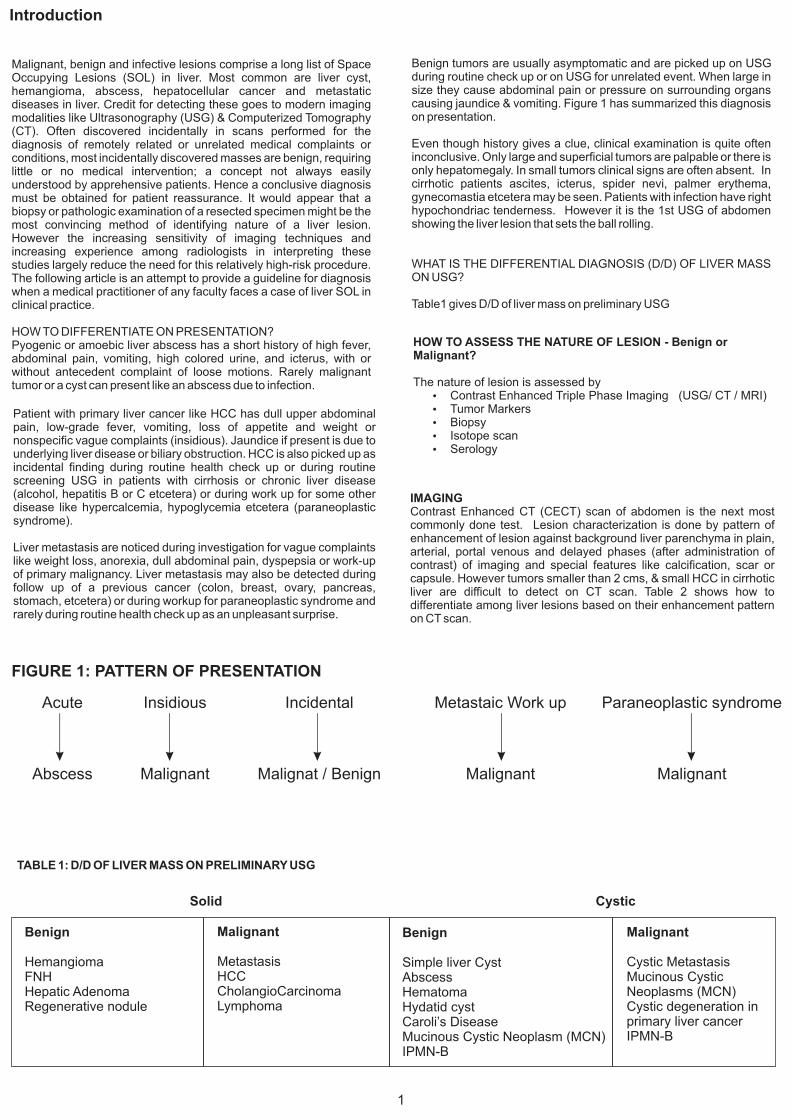
Malignant, benign and infective lesions comprise a long list of Space Occupying Lesions (SOL) in liver. Most common are liver cyst, hemangioma, abscess, hepatocellular cancer and metastatic diseases in liver. Credit for detecting these goes to modern imaging modalities like Ultrasonography (USG) & Computerized Tomography (CT). Often discovered incidentally in scans performed for the diagnosis of remotely related or unrelated medical complaints or conditions, most incidentally discovered masses are benign, requiring little or no medical intervention; a concept not always easily understood by apprehensive patients. Hence a conclusive diagnosis must be obtained for patient reassurance. It would appear that a biopsy or pathologic examination of a resected specimen might be the most convincing method of identifying nature of a liver lesion. However the increasing sensitivity of imaging techniques and increasing experience among radiologists in interpreting these studies largely reduce the need for this relatively high-risk procedure. The following article is an attempt to provide a guideline for diagnosis when a medical practitioner of any faculty faces a case of liver SOL in clinical practice.
HOW TO DIFFERENTIATE ON PRESENTATION?Pyogenic or amoebic liver abscess has a short history of high fever, abdominal pain, vomiting, high colored urine, and icterus, with or without antecedent complaint of loose motions. Rarely malignant tumor or a cyst can present like an abscess due to infection.
FIGURE 1: PATTERN OF PRESENTATION
Acute Insidious Incidental Metastaic Work up Paraneoplastic syndrome
Cystic MetastasisMucinous Cystic Neoplasms (MCN)Cystic degeneration in primary liver cancerIPMN-B
Patient with primary liver cancer like HCC has dull upper abdominal pain, low-grade fever, vomiting, loss of appetite and weight or nonspecific vague complaints (insidious). Jaundice if present is due to underlying liver disease or biliary obstruction. HCC is also picked up as incidental finding during routine health check up or during routine screening USG in patients with cirrhosis or chronic liver disease (alcohol, hepatitis B or C etcetera) or during work up for some other disease like hypercalcemia, hypoglycemia etcetera (paraneoplastic syndrome).
Liver metastasis are noticed during investigation for vague complaints like weight loss, anorexia, dull abdominal pain, dyspepsia or work-up of primary malignancy. Liver metastasis may also be detected during follow up of a previous cancer (colon, breast, ovary, pancreas, stomach, etcetera) or during workup for paraneoplastic syndrome and rarely during routine health check up as an unpleasant surprise.
IMAGINGContrast Enhanced CT (CECT) scan of abdomen is the next most commonly done test. Lesion characterization is done by pattern of enhancement of lesion against background liver parenchyma in plain, arterial, portal venous and delayed phases (after administration of contrast) of imaging and special features like calcification, scar or capsule. However tumors smaller than 2 cms, & small HCC in cirrhotic liver are difficult to detect on CT scan. Table 2 shows how to differentiate among liver lesions based on their enhancement pattern on CT scan.
Benign tumors are usually asymptomatic and are picked up on USG during routine check up or on USG for unrelated event. When large in size they cause abdominal pain or pressure on surrounding organs causing jaundice & vomiting. Figure 1 has summarized this diagnosis on presentation.
Even though history gives a clue, clinical examination is quite often inconclusive. Only large and superficial tumors are palpable or there is only hepatomegaly. In small tumors clinical signs are often absent. In cirrhotic patients ascites, icterus, spider nevi, palmer erythema, gynecomastia etcetera may be seen. Patients with infection have right hypochondriac tenderness. However it is the 1st USG of abdomen showing the liver lesion that sets the ball rolling.
WHAT IS THE DIFFERENTIAL DIAGNOSIS (D/D) OF LIVER MASS ON USG?
Table1 gives D/D of liver mass on preliminary USG
1
HOW TO ASSESS THE NATURE OF LESION - Benign or Malignant?
The nature of lesion is assessed by Ÿ Contrast Enhanced Triple Phase Imaging (USG/ CT / MRI)Ÿ Tumor MarkersŸ BiopsyŸ Isotope scanŸ Serology
Introduction
A contrast enhanced triple phase Magnetic Resonance Imaging (MRI) does better lesion detection & characterization than CT and is more useful than CT for small HCC, cirrhosis, chronic hepatitis and as such whenever in doubt after a CT. (PIC 5). However MRI is not available easily and difficult to interpret than CT. Contrast enhanced ultrasound (CEUS) is a technique of using ultrasonography for detecting contrast enhancement pattern of tumor. In spite of CT scan, MRI & CEUS solid lesions like benign cirrhotic nodules (regenerative nodule or dysplastic nodules) often cannot be detected or distinguished from HCC. Similarly atypical imaging features of benign tumors like hemangioma, adenoma or FNH cannot be differentiated from HCC. In these situations tumor markers may give a clue.
confirmed the diagnosis with characteristic enhancement pattern
in arterial, portal and delayed phase (E, F, G, H)
Hyperdense – bright / white compared to liver parenchyma, Hypodense – dark / grey to black. Compared to liver parenchyma, Isodense – lesion density similar to liver parenchyma
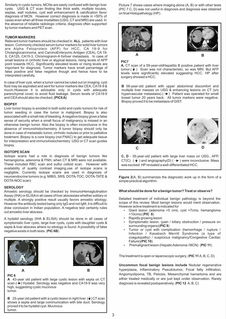
PIC 6 A 43-year old patient with large cystic lesion with septa on CT scan.( ) Hydatid. Serology was negative and CA19-9 was very high, suggesting cystic mucinous tumor.
B 25-year old patient with a cystic lesion in right liver. ( ) CT scan shows a septa and large communication with bile duct. Serology proved it to be hydatid cyst. Mucinous tumor.
A B
TUMOR MARKERSRelevant tumor markers should be checked in ALL patients with liver lesion. Commonly checked serum tumor markers for solid liver tumors are Alpha Fetoprotein (AFP) for HCC, CA 19-9 for Cholangiocarcinoma, and CarcinoEmbryonic Antigen (CEA), CA 19-9, CA125, CA15-3, Chromogranin-A forliver metastasis. In case of small lesions in cirrhotic liver or atypical lesions, rising levels of AFP point towards HCC. Significantly elevated levels or rising levels are important for diagnosis. Tumor markers have small percentage of false positive and false negative though and hence have to be interpreted carefully.
B 58-year-old patient with upper abdominal discomfort and multiple liver masses on USG & enhancing lesions on CT (s/o hypervascular metastasis).( ) Patient was operated for small bowel tumor 20 years back. All tumor markers were negative. Biopsy proved it to be metastasis of GIST.
PIC7A. CT scan of a 59-year-oldHepatitis B positive patient with liver tumor.( ) Scan was not characteristic, so was MRI. But AFP levels were significantly elevated suggesting HCC. HP after surgery showed a HCC.
C, D - 35-year-old patient with large liver mass on USG., AFP,
CT(C) ( ) and angiography(D) ( ) were inconclusive. Mass
was excised. HP revealed a well-differentiated HCC.
C D
Figure 2(A, B) summarizes the diagnostic work up in the form of a simple practical algorithm.
What should be done for a benign tumor? Treat or observe?
Detailed treatment of individual benign pathology is beyond the scope of this review. Most benign lesions would merit observation. However active treatment is indicated for
Similarly in cystic tumors, MCNs are easily confused with benign liver cysts. USG & CT scan finding like thick walls, multiple locules, septae, wall nodules, cyst wall enhancement & calcification favor diagnosis of MCN. However correct diagnosis is made in <50% of cases even when all three modalities (USG, CT and MRI) are used. In the absence of reliable radiologic criteria, diagnosis often supported by tumor markers and PET scan.
In case of liver cyst, when a tumor cannot be ruled out on imaging, cyst fluid may be aspirated and sent for tumor markers like CEA, CA19-9 & mucin.However it is advisable only in cysts with adequate parenchymal cover, to avoid fluid leakage. Serum levels of CA19-9 and CEA should also be checked. (PIC6 A)
BIOPSYLiver tumor biopsy is avoided in both solid and cystic tumors for risk of tumor seeding in case the tumor is malignant. Biopsy is also associated with a small risk of bleeding. A negative biopsy gives a false sense of security when a small focus of malignancy is missed in an otherwise benign tumor. Also the biopsy is often inconclusive in the absence of immunohistochemistry. A tumor biopsy should only be done in case of metastatic tumor, cirrhotic nodules or prior to palliative treatment. Biopsy is a core biopsy (not FNAC) to get adequate tissue for interpretation and immunohistochemistry. USG or CT scan guides biopsy.
ISOTOPE SCANIsotope scans had a role in diagnosis of benign tumors like hemangioma, adenoma & FNH, when CT & MRI were not available. These included RBC scan and sulfur colloid scan. However with availability of quality contrast imaging,use of isotope scans is negligible. Currently isotope scans are used in diagnosis of neuroendocrine tumors (e.g. MIBG, SRS, DOTA-TOC, DOTA-TATE & DOTA-NOC scan)
SEROLOGYAmoebic serology should be checked by ImmunoHemaglutination Assay (IHA) or ELISA in all cases of liver abscesses whether solitary or multiple. A strongly positive result usually favors amoebic etiology. However the antibody tested being only IgG and not IgM, it is difficult to draw conclusion in Indian population. A negative test certainly rules out amoebic liver abscess.
A hydatid serology (IHA & ELISA) should be done in all cases of symptomatic liver cysts, large liver cysts, cysts with daughter cysts & septa & liver abscess where no etiology is found. A possibility of false negative exists in both tests. (PIC 6B)
The treatment is open or laparoscopic surgery. (PIC 11 A, B, C, D)
Uncommon focal benign lesions include Nodular regenerative
Angiomyolipoma, TB, Peliosis, Mesenchymal hamartoma and are
either treated medically or are just kept under observation. Rarely
diagnosis is revealed postoperatively. (PIC 12 A, B, C)
Picture 7 shows cases where imaging alone (A, B) or with other tests (PIC 7 C, D) was not useful in diagnosis and diagnosis was obtained on final Histopathology (HP).
3
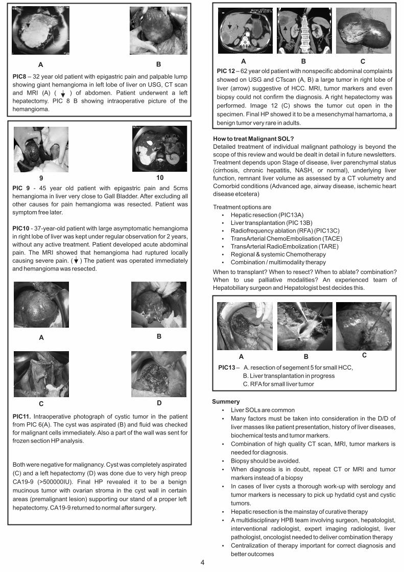
PIC 9 - 45 year old patient with epigastric pain and 5cms
hemangioma in liver very close to Gall Bladder. After excluding all
other causes for pain hemangioma was resected. Patient was
symptom free later.
PIC10 - 37-year-old patient with large asymptomatic hemangioma
in right lobe of liver was kept under regular observation for 2 years,
without any active treatment. Patient developed acute abdominal
pain. The MRI showed that hemangioma had ruptured locally
causing severe pain. ( ) The patient was operated immediately
and hemangioma was resected.
9 10
Both were negative for malignancy. Cyst was completely aspirated
(C) and a left hepatectomy (D) was done due to very high preop
CA19-9 (>500000IU). Final HP revealed it to be a benign
mucinous tumor with ovarian stroma in the cyst wall in certain
areas (premalignant lesion) supporting our stand of a proper left
hepatectomy. CA19-9 returned to normal after surgery.
A B
C D
4
PIC13 – A. resection of segement 5 for small HCC,
B. Liver transplantation in progress
C. RFA for small liver tumor
Summery
Ÿ Liver SOLs are common
Ÿ Many factors must be taken into consideration in the D/D of
liver masses like patient presentation, history of liver diseases,
biochemical tests and tumor markers.
Ÿ Combination of high quality CT scan, MRI, tumor markers is
needed for diagnosis.
Ÿ Biopsy should be avoided.
Ÿ When diagnosis is in doubt, repeat CT or MRI and tumor
markers instead of a biopsy
Ÿ In cases of liver cysts a thorough work-up with serology and
tumor markers is necessary to pick up hydatid cyst and cystic
tumors.
Ÿ Hepatic resection is the mainstay of curative therapy
Ÿ A multidisciplinary HPB team involving surgeon, hepatologist,
Figure 2 -- Management algorithm for a patient with solid(A) or cystic (B) liver mass
Disclaimer: The views expressed in this article solely belong to the author. The information provided here is for educational and informational purposes only.