16
HPI • 48 yo F comes to the clinic complaining of left knee pain What questions would you like to ask?
| Date post: | 30-Dec-2015 |
| Category: |
Documents |
| Upload: | bertha-cannon |
| View: | 217 times |
| Download: | 1 times |

HPI• 48 yo F comes to the clinic complaining
of left knee pain
What questions would you like to ask?

HPI• 2 month history of L knee pain, worse with ROM
• Reports noticeable L knee “stiffness”
• Recently noticed a “bump” behind the left knee
• - history of recent falls or trauma to the knee
• Denies other joint pain/discomfort

PMH: HTN, DM
PSH: cholecystectomy
Family Hx: colon cancer
Social: social drinker, ½ pack/day smoker, denies illicits
Meds: HCTZ, lisinopril
Allergies: none
Differential Diagnosis?

Differential Diagnosis
• Osteoarthritis• Baker’s cyst• Rheumatoid arthritis• Ligamentous, meniscal injury• Soft tissue/ bone tumor

PE
• Vitals: AVSS• CV, Pulm, Abd: WNL• Musc: Left knee exam: • Fixed solid mass palpated over the posterior knee.
No warmth, erythema appreciated. No joint line tenderness. Tender to palpation over posterior aspect of the knee. Decreased ROM (0-90 degrees). Negative anterior/posterior drawer. Negative Mcmurray’s. Neurovascularly intact distal to R knee.
Labs:WNL

Xray (AP)

Xray (Lateral)

MRI (T1)Coronal

MRI Sagittal

Histology

Histology
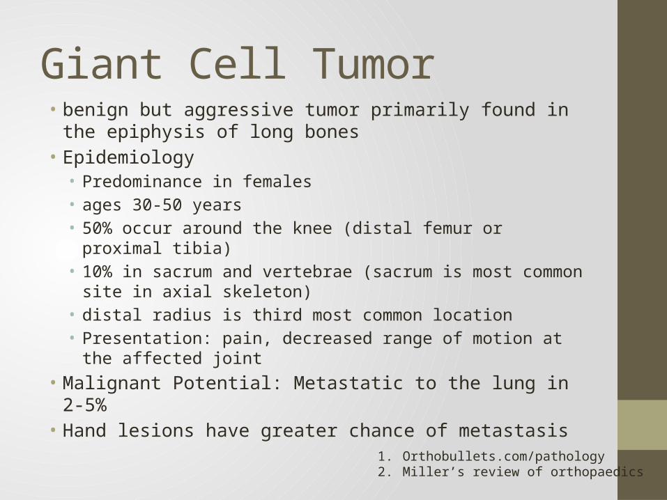
Giant Cell Tumor• benign but aggressive tumor primarily found in the epiphysis
of long bones • Epidemiology• Predominance in females • ages 30-50 years• 50% occur around the knee (distal femur or proximal tibia)• 10% in sacrum and vertebrae (sacrum is most common site in
axial skeleton) • distal radius is third most common location• Presentation: pain, decreased range of motion at the affected
joint• Malignant Potential: Metastatic to the lung in 2-5% • Hand lesions have greater chance of metastasis
1. Orthobullets.com/pathology2. Miller’s review of orthopaedics

Characteristic Features
• Xray• eccentric lytic epiphyseal/metaphyseal lesion that often extends
into the distal epiphysis and borders subchondral bone

Characteristic Features
• MRI • clear demarcation on T1 image between fatty marrow and tumor

• Bone scan is very “hot”
Characteristic Features

Characteristic Features
• Histology• Characteristic cell is the mononucleur stromal cell • hallmark giant cells are numerous • nuclei of giant cell appears same as stromal cells


