Vol. 49, No. 1 APPLIED AND ENVIRONMENTAL MICROBIOLOGY, Jan. 1985, p. 221-228 0099-2240/85/010221-08$02.00/0 Copyright C) 1985, American Society for Microbiology Ecology of Legionella pneumophila within Water Distribution Systems JANET E. STOUT, VICTOR L. YU,* AND MICHELE G. BEST Special Pathogen and Infectious Disease Sections, Veterans Administration Medical Center, and University of Pittsburgh School of Medicine, Pittsburgh, Pennsylvania 15240 Received 31 May 1984/Accepted 16 October 1984 The reservoir for hospital-acquired Legionnaires disease has been shown to be the potable water distribution system. We investigated the influence of the natural microbial population and sediment (scale and organic particulates) found in water systems as growth-promoting factors for Legionella pneumophila. Our in vitro experiments showed that: (i) water from a hot-water storage tank readily supported the survival of L. pneumophila, (ii) the concentration of sediment was directly related to the survival of L. pneumophila, (iii) the presence of environmental bacteria improved the survival of L. pneumophila via nutritional symbiosis, (iv) the combination of sediment and environmental bacteria acted synergistically to improve the survival of L. pneumophila, and (v) the role of sediment in this synergistic effect was determined to be nutritional. Sediment was found to stimulate the growth of environmental microflora, which in turn stimulated the growth of L. pneumophila. These findings confirm the empiric observations of the predilection of L. pneumophila for growth in hot-water tanks and its localization to sediment. L. pneumophila occupies an ecological niche within the potable water system, with interrelationships between microflora, sediment, and temperature. The prevalence of Legionella pneumophila, the causative agent of Legionnaires disease, in natural and manmade aquatic environments has been well documented (5, 8, 9, 19, 21, 22). Ongoing surveillance for L. pneumophila at the Pittsburgh Veterans Administration Medical Center has dem- onstrated that the presence of L. pneumophila in the hospital water distribution system is epidemiologically linked to the acquisition of Legionnaires disease in susceptible, hospital- ized patients (3, 19). Recent experience suggests that hospi- tal-acquired Legionnaires disease not only can be treated but can be prevented with control measures directed at the reservoir (2a, 3, 10, 14). The success of eradication measures directed at water distribution systems will be determined by information obtained from the analysis of the ecology of this organism within the habitat of the water distribution system. MATERIALS AND METHODS Media and test organisms. Buffered charcoal yeast extract agar (BCYE) was prepared as previously described (7, 13). L. pneumophila was recovered from mixed cultures by using a selective differential medium which is a modification of previously described media for the isolation of L. pneumo- phila (18). This medium, DGVP, is buffered yeast extract agar to which 0.001% bromocresol purple, 0.001% bromo- thymol blue, 0.3% glycine, 1 jig of vancomycin per ml, 50 U of polymyxin B per ml, and 1.5 g of charcoal are added. One lot of media was used in each experiment to control for sampling variation due to media composition. L. pneumo- phila, serogroup 1, which had been isolated from our hot- water storage tank was used in all experiments. Environ- mental microorganisms were recovered by plating a water sample from the hot- and cold-water storage tanks on BCYE plates and incubating at 25, 37, and 42°C (Fig. 1). A stereo- microscope was used to screen the plates to differentiate organisms by colony morphology. Single colonies were picked and subcultured to BCYE. Organisms were stocked by making a heavy suspension of 1 to 2 ml of 50% fetal * Corresponding author. bovine serum-50% (vol/vol) tryptic soy broth mixture and freezing at -20°C. Thirty-two different environmental organ- isms (16 isolated from cold-water-tank water and 16 from hot-water-tank water) were stocked. Standard methods were utilized to identify these organisms. These included API 20E for the differentiation of non-Enterobacteriaceae (Analytab Products) and the UNI-N/F system (Flow Laboratories, Inc.). One environmental organism, identified as a Pseudo- monas species by standard methods, was used in certain experiments as a representative environmental organism. This Pseudomonas species could not be further identified to the species level by standard morphological and biochemical criteria (G. Gilardi, personal communication). Sampling and characteristics of hot-water-tank water and sediment. A water sample was collected from the bottom of a hot-water storage tank which supplied our hospital and was divided into two fractions. One fraction was concen- trated by centrifugation at 5,000 rpm for 30 min. The supernatant (sediment absent) was removed, and the con- centrate (sediment present) was suspended in 1/10 of the original volume of supernatant. The unconcentrated fraction of tank water, sediment, and supernatant were further divided into sterile (microflora absent) and nonsterile (mi- croflora present) fractions. The sediment suspensions were designated 1.0 (undiluted sediment), 0.25 (1:4 dilution of sediment), 0.05 (1:16 dilution of sediment), and 0.0 (super- natant). The supernatant was used as the diluent. Steriliza- tion of the tank water and sediment was accomplished by boiling for 30 min, followed by incubation at 37°C for 24 h. This process was done to detect the presence of spore-form- ing bacteria and was repeated until sterility was achieved. Sterility was determined by direct plating of 0.1 ml of the suspension onto BCYE plates, followed by incubation at 37°C for 72 h. The supernatant was filter sterilized with a 0.2-p.m filter sterilization unit (Nalgene Labware Div., Nalge/Sybron Corp.). Determinations of total organic car- bon content, total suspended solids, and volatile residuals as well as atomic absorption spectrophotometric analysis for 221
Transcript
Vol. 49, No. 1APPLIED AND ENVIRONMENTAL MICROBIOLOGY, Jan. 1985, p. 221-2280099-2240/85/010221-08$02.00/0Copyright C) 1985, American Society for Microbiology
Ecology of Legionella pneumophila within WaterDistribution Systems
JANET E. STOUT, VICTOR L. YU,* AND MICHELE G. BEST
Special Pathogen and Infectious Disease Sections, Veterans Administration Medical Center, and University of PittsburghSchool of Medicine, Pittsburgh, Pennsylvania 15240
Received 31 May 1984/Accepted 16 October 1984
The reservoir for hospital-acquired Legionnaires disease has been shown to be the potable water distributionsystem. We investigated the influence of the natural microbial population and sediment (scale and organicparticulates) found in water systems as growth-promoting factors for Legionella pneumophila. Our in vitroexperiments showed that: (i) water from a hot-water storage tank readily supported the survival of L.pneumophila, (ii) the concentration of sediment was directly related to the survival of L. pneumophila, (iii) thepresence of environmental bacteria improved the survival of L. pneumophila via nutritional symbiosis, (iv) thecombination of sediment and environmental bacteria acted synergistically to improve the survival of L.pneumophila, and (v) the role of sediment in this synergistic effect was determined to be nutritional. Sedimentwas found to stimulate the growth of environmental microflora, which in turn stimulated the growth of L.pneumophila. These findings confirm the empiric observations of the predilection of L. pneumophila for growthin hot-water tanks and its localization to sediment. L. pneumophila occupies an ecological niche within thepotable water system, with interrelationships between microflora, sediment, and temperature.
The prevalence of Legionella pneumophila, the causativeagent of Legionnaires disease, in natural and manmadeaquatic environments has been well documented (5, 8, 9, 19,21, 22). Ongoing surveillance for L. pneumophila at thePittsburgh Veterans Administration Medical Center has dem-onstrated that the presence of L. pneumophila in the hospitalwater distribution system is epidemiologically linked to theacquisition of Legionnaires disease in susceptible, hospital-ized patients (3, 19). Recent experience suggests that hospi-tal-acquired Legionnaires disease not only can be treated butcan be prevented with control measures directed at thereservoir (2a, 3, 10, 14). The success of eradication measuresdirected at water distribution systems will be determined byinformation obtained from the analysis of the ecology of thisorganism within the habitat of the water distribution system.
MATERIALS AND METHODSMedia and test organisms. Buffered charcoal yeast extract
agar (BCYE) was prepared as previously described (7, 13).L. pneumophila was recovered from mixed cultures by usinga selective differential medium which is a modification ofpreviously described media for the isolation of L. pneumo-phila (18). This medium, DGVP, is buffered yeast extractagar to which 0.001% bromocresol purple, 0.001% bromo-thymol blue, 0.3% glycine, 1 jig of vancomycin per ml, 50 Uof polymyxin B per ml, and 1.5 g of charcoal are added. Onelot of media was used in each experiment to control forsampling variation due to media composition. L. pneumo-phila, serogroup 1, which had been isolated from our hot-water storage tank was used in all experiments. Environ-mental microorganisms were recovered by plating a watersample from the hot- and cold-water storage tanks on BCYEplates and incubating at 25, 37, and 42°C (Fig. 1). A stereo-microscope was used to screen the plates to differentiateorganisms by colony morphology. Single colonies werepicked and subcultured to BCYE. Organisms were stockedby making a heavy suspension of 1 to 2 ml of 50% fetal
* Corresponding author.
bovine serum-50% (vol/vol) tryptic soy broth mixture andfreezing at -20°C. Thirty-two different environmental organ-isms (16 isolated from cold-water-tank water and 16 fromhot-water-tank water) were stocked. Standard methods wereutilized to identify these organisms. These included API 20Efor the differentiation of non-Enterobacteriaceae (AnalytabProducts) and the UNI-N/F system (Flow Laboratories,Inc.).One environmental organism, identified as a Pseudo-
monas species by standard methods, was used in certainexperiments as a representative environmental organism.This Pseudomonas species could not be further identified tothe species level by standard morphological and biochemicalcriteria (G. Gilardi, personal communication).
Sampling and characteristics of hot-water-tank water andsediment. A water sample was collected from the bottom ofa hot-water storage tank which supplied our hospital andwas divided into two fractions. One fraction was concen-trated by centrifugation at 5,000 rpm for 30 min. Thesupernatant (sediment absent) was removed, and the con-centrate (sediment present) was suspended in 1/10 of theoriginal volume of supernatant. The unconcentrated fractionof tank water, sediment, and supernatant were furtherdivided into sterile (microflora absent) and nonsterile (mi-croflora present) fractions. The sediment suspensions weredesignated 1.0 (undiluted sediment), 0.25 (1:4 dilution ofsediment), 0.05 (1:16 dilution of sediment), and 0.0 (super-natant). The supernatant was used as the diluent. Steriliza-tion of the tank water and sediment was accomplished byboiling for 30 min, followed by incubation at 37°C for 24 h.This process was done to detect the presence of spore-form-ing bacteria and was repeated until sterility was achieved.Sterility was determined by direct plating of 0.1 ml of thesuspension onto BCYE plates, followed by incubation at37°C for 72 h. The supernatant was filter sterilized with a0.2-p.m filter sterilization unit (Nalgene Labware Div.,Nalge/Sybron Corp.). Determinations of total organic car-bon content, total suspended solids, and volatile residuals aswell as atomic absorption spectrophotometric analysis for
221
222 STOUT, YU, AND BEST
H WT 10-1
CWT 10-1
VIU. 1. Isolation of environmental microorganisms from water samples obtained from the hot-water tank (HWT) and cold-water tank(CWT). Plates were incubated at 25, 37, and 42°C. Different microbial populations inhabit each water tank due to differences in their optimalgrowth temperature.
metals were performed by the standard methods for theexamination of water and wastewater (1). Both the nonster-ile tank water and sediment were plated on selective mediato obtain the number of L. pneumophila bacteria present inthe sample before incubation or inoculation.
Survival and growth curve studies. Several hypotheseswere tested by generating survival and growth curve data forL. pneumophila when incubated in suspensions which var-ied in biotic and abiotic composition. These experimentswere conducted as follows. A 10-,ul loopful of a freezer stocksuspension of L. pneumophila was used to inoculate aBCYE plate. After incubation at 37°C for 72 h, a portion ofthe actively growing culture was used to inoculate a secondBCYE plate. This plate was incubated at 37°C for only 24 hto obtain a culture composed of viable L. pneumophila. Thisculture was used to prepare a suspension on L. pneumophilain sterile water which contained ca. 105 CFU/ml. Thissuspension (0.5 ml) was used to inoculate 4.5 ml of a testsuspension. When experiments required the addition of anenvironmental organism to the test suspension, 50 ,ul of anorganism suspension (containing ca. 109 CFU/ml) was added.The final growth curve experiment utilized nutrient broth(Difco Laboratories) as the test suspension. The nutrientbroth was buffered to pH 6.9 with ACES buffer [N-(2-acetamido)-2-aminoethane-sulfonic acid] and filter sterilizedthrough a Nalgene 0.2-,um sterilization unit. Two steriletubes were filled with 4.5 ml of broth and inoculated with 0.5ml of a suspension of L. pneumophila which had a concen-tration of ca. 105 CFU/ml. A suspension (50 p.l) of an
environmental pseudomonad with a concentration of ca. 106CFU/ml was added to one tube. Broth (5.0 ml) was added toanother sterile tube and inoculated only with the pseu-domonad.
Nutrient broth was chosen over other possible nutrientsources because it will not inherently stimulate the growth of
L. pneumophila. All suspensions were incubated at 37°C andsampled at various intervals. The viability of L. pneumo-phila and nonlegionella bacteria in a test suspension wasdetermined by plating 0.1 ml of a given dilution onto DGVPand BCYE plates. All plates were incubated at 37°C for 5days. All suspensions were vortexed daily and before platingto provide aeration, disperse bacterial aggregates, and sus-pend sedimented materials. Each experiment was carriedout for 28 days.
Demonstration of syntrophy between L. pneumophila andother environmental microorganisms (satellitism). Pour platesof L. pneumophila (109 CFU/ml) were prepared with BCYElacking cysteine, an essential nutrient for the growth of L.pneumophila. A Steers replicator was used to inoculate 32environmental organisms by placing 0.001 ml of a suspen-sion of each organism containing ca. 108 CFU/ml onto eachpour plate (17). Plates were incubated at 25, 37, and 42°C for48 to 72 h. The appearance of satellite colonies of L.pneumophila around the peripheral edge of an environmen-tal organism indicated nutritional symbiosis.
Since cysteine is an essential nutrient for the growth of L.pneumophila, this test system would determine the ability ofother environmental bacteria to provide this essential aminoacid (or a metabolic substitute) and thus demonstrate thepotential for a symbiotic relationship between these organ-isms.
RESULTSSampling and characteristics of hot-water-tank water and
sediment. Quantitative characteristics of the water sampleobtained from the hot-water tank and its two fractions,supernatant and sediment, involved determinations of totalsolids, total organic carbon content, and volatile residuals aswell as atomic absorption spectrophotometric analysis (Ta-ble 1) and the measurement of viable plate counts for L.
250c 370c 420c
II
Ii.
APPL. ENVIRON. MICROBIOL.
L. PNEUMOPHILA IN WATER SYSTEMS 223
TABLE 1. Characteristics of hot-water-tank water, supernatant, and sediment
Total Total organic Atomic absorption spectrophotometrySample suspended Volatile residuals carbon content Mg (mg/
solids (mg/liter) (mg/iter) (mg/liter) Pb (mg/dl) Cu (mg/dl) Ca (mg/dl) liter) Fe (mg/dl) Zn (.ld/ml)
pneumophila. These tests indicate the relative availability oforganic and inorganic compounds for utilization as sub-strates and energy sources. Total suspended solids andvolatile residuals indicate the amount of solid particulatematter in the sample and the portion which is organic innature, respectively. The sediment has a higher concentra-tion of both total suspended solids and volatile residual whencompared with either supernatant or hot-water-tank water(Table 1). The total organic content of the sediment wasdetermined to be 128 mg/liter. Atomic absorption analysis ofhot-water-tank water, supernatant, and sediment showedsignificant differences in the concentration of copper andiron only. The concentration of L. pneumophila in hot-wa-ter-tank water, supernatant, and sediment was 1.6 x 10⁴,<1.0 x 101, and 1.8 x 10⁵ CFU/ml, respectively.
Survival and growth curve studies. Hypothesis I. Hot-wa-ter-tank water supports the growth and survival of L.pneumophila. Figure 2 demonstrates the response of L.pneumophila incubated in nonsterile (microflora and sedi-ment present) hot-water-tank water, sterile supernatant (mi-croflora and sediment absent), and sterile high-pressureliquid chromatography water (microflora, sediment, anddissolved organic nutrients absent). Colony counts of L.pneumophila determined on selective agar plates showed L.pneumophila to persist at a level equal to or above the initialconcentration only in hot-water-tank water. After 21 days ofincubation, the concentration of L. pneumophila increasedin the hot-water-tank water from an initial concentration of2.0 x 104 to 6.0 x 10⁴ CFU/ml. It is also apparent that thegrowth-promoting effect of dissolved organic nutrients wasminimal, as evidenced by the precipitous decline in theconcentration of L. pneumophila in both sterile supernatant(containing dissolved organic nutrients) and sterile high-pres-sure liquid chromatography water (devoid of dissolved or-ganic nutrients) (Fig. 2). To control for the effect of filtrationon the dissolved organic nutrient content of the supernatant,sterilization by UV light was also done. The results werevirtually identical (data not shown). Thus, L. pneumophilacannot multiply in a low-nutrient, aqueous environment.
Hypothesis II. Sediment contributes to the survival of L.pneumophila. There was a direct correlation between theconcentration of sediment and growth or survival of L.pneumophila (Fig. 3). L. pneumophila increased most dra-matically when incubated with concentrated nonsterile (mi-croflora present) sediment (Fig. 3A). One-fourth the concen-tration of sediment had a lesser effect on the growth of L.pneumophila, and both the nonsterile supernatant and thesuspension of lowest sediment concentration were not capa-ble of supporting the survival of L. pneumophila. These datareflect a dose-response phenomenon; as the concentration ofnonsterile sediment was increased, the survival of L. pneu-mophila also increased. L. pneumophila incubated in sterilesediment, regardless of sediment concentration, did not
increase in concentration, but its survival rate correlatedwith sediment concentration (Fig. 3B). However, the de-crease in the concentration of L. pneumophila was greaterwith successive dilutions of sediment. Therefore, sterilesediment had a lesser effect on the survival and growth of L.pneumophila than did nonsterile sediment which containedits natural complement of living microflora.
This set of experiments was also performed with a watersample which had been collected from a different hot-watertank. This was done to determine whether the phenomenawhich we had observed were intrinsic to this particularwater sample and whether these observations were repro-ducible. This set of experiments also demonstrated that asthe concentration of nonsterile sediment increased, thesurvival of L. pneumophila improved. The initial concentra-tion of L. pneumophila in each suspension was ca. 104CFU/ml. After 28 days of incubation in concentrated sedi-ment (relative sediment concentration, 1.0 Sed), the concen-tration of L. pneumophila was 1.6 x 10⁶ CFU/ml; at one-fourth the sediment concentration (0.25 Sed), 1.6 x 10⁵ CFUof L. pneumophila per ml was detectable; in the lowestsediment concentration (0.05 Sed), the concentration of L.
6-
5
:f~ _11~> L+Env'sa 4 - +HWT
Q) 3
bj 2o-\ .
0 3 7 10 14 21 28
Time (days)FIG. 2. Hot-water-tank water support of the growth and survival
of L. pneumophila. Nonsterile hot-water-tank water in which mi-croflora and sediment were present (0) supported the survival of L.pneumophila. The growth-promoting effect of dissolved organicnutrients was minimal as evidenced by the decline in viable L.pneumophila in both sterile supernatant (O), which containeddissolved organic nutrients, and sterile high-pressure liquid chroma-tography water (A), which did not contain dissolved organicnutrients.
VOL. 49, 1985
224 STOUT, YU, AND BEST
6
-I
A Lp+EnvI1-
\ ~~~~~~~~~Lp+Env'
_ _ _ _ _ ~~~~~~LpEn\
O 3 7 14 21 28
Time (days)6-
U-C5-
-i
CZI 4 -
0
x3-Q)Q 3.-
b 2a
B
12
Time (days)FIG. 3. Sediment contribution to the survival of L.
(A) Correlation between the concentration of nonsterilwhich environmental microflora was present and the s
pneumophila. Symbols (in Sed units): *, 1.0; 0, 0.2'0.05. (B) Correlation between the concentration of stein which living microflora was absent and the supneumophila. Symbols are as in (A).
pneumophila was 2.4 x 10³ CFU/ml; and in asuspension totally devoid of sediment, the concL. pneumophila at 28 days had fallen to 1.0 x 1
Hypothesis III. Environmental microflora wit)water tank promotes the growth and survival of:phila. The first experiment demonstrated syntropL. pneumophila and other environmental micr(satellitism). Altogether, 32 environmental orgatested for their ability to stimulate the productiorcolonies of L. pneumophila in pour plates of BCcysteine. Subsurface satellite colonies were obs(peripheral edge in 16 of 32 environmental organ2, Fig. 4). Although conventional attempts to id(organisms to the species level were often urorganisms were identified to the genus levelFlavobacterium, Pseudomonas, Alcaligenes,Disease Control group II, and Acinetobacter.A larger percentage of environmental organ
produced satellitism were isolated from the hotrather than the cold-water tank (Table 1). These dthe observed predilection of L. pneiumophila fosystems.
The second experiment produced survival curves of L.pneumophila in sediment-free suspensions containing a
s+l.OSed mixed population of environmental microflora. L. pneumo-
phila decreased more precipitously when incubated in asterile supernatant suspension devoid of environmental mi-croflora than in a nonsterile suspension which containedenvironmental microflora (Fig. 5A). This experimental ob-servation indicated that microflora alone can contribute to
r's+0.25Sed the improved survival of L. pneiumophila.The third experiment was an extension of the second
experiment and demonstrated the effect of a single symbioticorganism on the survival curve of L. pneumophila in asediment-free suspension. Incubation of L. pneumophila in
v's+O.OSed the presence of a symbiotic pseudomonad improved theLp + Env's + survival of L. pneuinophila when compared with an identical
0.05 Sed suspension without the symbiote (Fig. SB). This was ob-served in a sediment-free suspension (supernatant) andreiterates the observation made in the second experimentthat under more controlled conditions, the presence of asymbiotic organism (microflora) can contribute to the im-proved survival of L. pneumophila. Replicate experimentswere performed with several different initial concentrationsof the pseudomonad, ranging from 102 to 107 CFU/ml.Again, we observed the same pattern; the viability of L.pneumophila decreased more precipitously in the suspen-sions which contained the lower initial concentrations of the
Lp+ 1.0 Sed pseudomonad. Growth of L. pneumophila was never ob-served.
Lp+0.05 Sed Hypothesis IV. Combined effect of sediment and environ-Lp+0.25 Sed mental microflora on the survival of L. pneumophila is
greater than either factor alone. Having already establishedthat neither sediment alone (Fig. 3B) nor microorganisms
LP+ 0.0 Sed alone (Fig. 5) was as growth stimulating as hot-water-tank3 water (Fig. 1), we hypothesized that the combination of
sediment and microorganisms would provide the optimalenvironment for L. pneumophila.
mneumophila. The first experiment provided survival curves of L. pneu-e sediment in mophila and was a comparison of the effect of the combina-survival of L. tion of sediment and a syntrophic microbial population5; 0.0 ; *, versus the effect of sediment or the microbial populationrile sediment alone. Throughout the 28-day period, the concentration of L.irvival of L. pneumophila in the suspension which contained both sedi-
ment and the syntrophic pseudomonad maintained at ahigher level than in the suspensions which represented
supernatant sediment or microflora alone (Fig. 6). In sterile supernatantentration of and sterile supernatant plus the pseudomonad, the concen-02 CFU/ml. tration of L. pneumophila dropped below detectable levelshin the hot- after 21 and 28 days, respectively. This experiment indicatedL. pneumo- that the combination of sediment and microflora had ahy between greater effect on the survival of L. pneumophila than didroorganisms sediment or microflora (pseudomonad) alone. This experi-inisms were ment was repeated with several different initial concentra-n of satellite tions of the pseudomonad, ranging from 102 to 107 CFU/ml."YE lackingerved at therisms (Table TABLE 2. Nutritional symbiosis (satellitism) between L.entify these pneumophila and other environmental microorganismsisuccessful,
1, includingCenters for
isms which,-water tanklata supportor hot-water
Source of environmental No. of Satellitism"organisms strains
tested 25°C 37°C 42°C
Cold-water-tank water 16 0 2 1Hot-water-tank water 16 0 13 3")
" Number of environmental organisms found to stimulate production ofsatellite colonies of L. ptCeluitnopila when tested at three temperatures.
h Of the 13 organisms which produced satellitism at 37°C. 3 also demon-strated satellitism at 42°C.
APPL. ENVIRON. MICROBIOL.
d
L. PNEUMOPHILA IN WATER SYSTEMS 225
FIG. 4. Nutritional symbiosis of L. pneumophila with other environmental microorganisms. Subsurface satellite colonies of L.pneumophila (arrows) were observed at the peripheral edge in 16 of 32 environmental organisms.
In each case, L. pneumophila maintained a higher concen-tration in the suspension which contained both sediment andmicroflora (pseudomonad).Hypothesis V. Postulated mechanism for the "synergis-
tic" effect of sediment plus environmental microorganismsin promoting the growth of L. pneumophila derivesfrom thenutritive properties of sediment. The first experiment dem-onstrated the growth of environmental microflora in non-sterile sediment. The total microbial population in nonsterile(microflora present) concentrated sediment was ca. 104CFU/ml on day zero of this experiment. After incubation at37°C for 3 days, the concentration of environmental bacteriahad increased to 4.0 x 10⁵ CFU/ml and maintained at thislevel throughout the 28-day experiment. These data indicatethat sediment can support the growth and multiplication ofhigh concentrations of indigenous microbial flora.The second experiment was a comparison of the growth
curves of an environmental pseudomonad in suspensions ofvarious sediment concentrations. The growth rate of thesymbiotic pseudomonad was directly related to the concen-tration of sterile sediment (Fig. 7A). We also performed thisexperiment with initial concentrations of the pseudomonadof 102, 104, and 107 CFU/ml. The concentration of thepsueodmonad always increased with increasing sedimentconcentration (data not shown). This experiment confirmsthe growth-promoting properties of sediment, using a singlesymbiotic pseudomonad.The third experimnent demonstrated the indirect effect of
sediment in promoting the growth and survival of L. pneu-mophila by substituting nutrient broth for sediment. Theincubation of L. pneumophila with the pseudomonad innutrient broth enabled L. pneumophila to survive and even
multiply in a liquid medium that normally does not supportits growth (Fig. 7B). After 24 h of incubation, the concen-tration of L. pneumophila in both the mixed culture and thesuspension of L. pneumophila alone had decreased. Overthe next 72 h, the concentrations of L. pneumophila in-creased nearly 1 log in the mixed culture but continued todrop in the suspension of L. pneumophila alone.
DISCUSSIONFliermans et al. (9) were able to detect L. pneumophila in
natural aquatic habitats by using centrifugation and directfluorescent-antibody staining techniques, although it wasnoted that the population densities of L. pneumophila wereextremely low, with serogroups 1 through 4 accounting forless than 1% of the total bacterial population. L. pneumo-phila has also been readily detectable in man-made aqueousenvironments (5, 19, 21, 22) and shown to be capable ofmultiplication in water (16, 23, 24). In our institution, theconcentration of L. pneumophila (as determined by directculture techniques without the need for concentration bycentrifugation or filtration) at numerous sites throughout thewater distribution system was alarmingly high, >3.0 x 10³CFU/ml (19). Obviously, the conditions which exist inhot-water systems tip the ecological balance in favor of L.pneumophila. In the present study, we delineate some of thefactors and conditions within a water distribution systemwhich lead to colonization with L. pneumophila and, there-fore, potential acquisition of Legionnaires disease in ex-posed susceptible individuals.Our data demonstrate that the water obtained from a
hot-water storage tank provides an environment in which L.pneumophila can maintain concentrations exceeding 104
VOL. 49, 1985
226 STOUT, YU, AND BEST
E
-i
:9
C0-
Q)
b4
0
-
C
-a
6
5-
4
3
2
6-
A
1 3 7 10 14 21 28
Time (days)
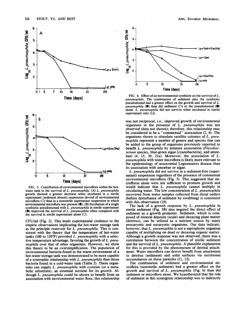
Time (days)FIG. 5. Contribution of environmental microflora within the hot-
water tank to the survival of L. pneumophila. (A) L. pneumophilagrowth showed a greater decrease when incubated in a sterilesupernatant (sediment absent) suspension devoid of environmentalmicroflora (0) than in a nonsterile supernatant suspension in whichenvironmental microflora was present (0). (B) Incubation of a singlesymbiotic pseudomonad with L. pneumophila in sterile supernatant(0) improved the survival of L. pneumophila when compared withthe survival in sterile supematant alone (0).
CFU/ml (Fig. 2). This lends experimental credence to theempiric observations implicating the hot-water storage tankas the principle reservoir for L. pneumophila. This is con-sistent with the theory that the temperature of hot-watertanks (100 to 120°F) provided L. pneumophila with a selec-tive temperature advantage, favoring the growth of L. pneu-mophila over that of other organisms. However, we showthis theory to be an oversimplification. The population ofenvironmental bacteria found in the warm environment of a
hot-water storage tank was demonstrated to be more capableof a syntrophic relationship with L. pneumophila than thosebacteria found in a cold-water tank (Table 2). These organ-isms can supply L. pneumophila with cysteine (or a meta-bolic substitute), an essential nutrient for its growth. Al-though L. pneumophila could be shown to benefit from an
association with environmental water flora, this relationship
E
c-a 5
Lp+Sed+Env(Ps)
Lp+Env(Ps)Lp only
O 5 12 21 28
Time (days)FIG. 6. Effect of an environmental symbiote on the survival ofL.
pneumophila. The combination of sediment plus the symbioticpseudomonad had a greater effect on the growth and survival of L.pneumophila (0) than did sediment (0) or the pseudomonad (-)alone. L. pneumophila did not survive when incubated in sterilesupernatant only (E).
was not reciprocal, i.e., improved growth of environmentalorganisms in the presence of L. pneumophila was notobserved (data not shown); therefore, this relationship maybe considered to be a "commensal" association (2, 6). Theorganisms shown to stimulate satellite colonies of L. pneu-mophila represent a number of genera and species that canbe added to the group of organisms previously reported tobenefit L. pneumophila by intimate association (Flavobac-terium species, blue-green algae [cyanobacteria], and amoe-bae) (4, 15, 20, 21a). Moreover, the association of L.pneumophila with water microflora is likely more relevant tothe epidemiology of nosocomial Legionnaires disease thanits association with amoebae or algae.
L. pneumophila did not survive in a sediment-free (super-natant) suspension regardless of the presence of commensalenvironmental microflora (Fig. 5). This suggested that mi-crofloras alone were not sufficient to promote growth andwould indicate that L. pneumophila cannot multiply incirculating water. The low concentration of L. pneumophilarecovered from water samples collected from distal outlets(before disturbance of sediment by swabbing) is consistentwith this observation (19).The lack of a growth response by L. pneumophila in
sterile sediment (Fig. 3B) also negated the direct effect ofsediment as a growth promoter. Sediment, which is com-posed of mineral deposits (scale) and decaying plant matter(detritus), can be utilized as a nutrient source by manyprocaryotic and eucaryotic organisms. Our data indicate,however, that L. pneumophila is not a saprophytic organismcapable of multiplying on dead or decaying organic matter.Although a growth response was not observed, there was acorrelation between the concentration of sterile sedimentand the survival of L. pneumophila. A plausible explanationfor this is provided by the phenomenon of detrital attach-ment. Water microflora can derive benefit from attachmentto detritus (sediment) and solid surfaces via nutritionalaccumulation on these particles (11, 12).The combination of sediment and environmental mi-
croflora (nonsterile sediment) had a greater effect on thegrowth and survival of L. pneumophila (Fig. 6) than didsediment or microflora alone. We hypothesized that the roleof sediment in this synergistic relationship was to indirectly
APPL. ENVIRON. MICROBIOL.
L. PNEUMOPHILA IN WATER SYSTEMS 227
8-
7
6-
5
4
A
Env(Ps)+ 1.0 Sed
,,' \ _ E~~~~nv(Ps)+0.25Sed
}' " ~~~~~~~~Env(Ps)+0.0 SedI,~~ ~ ~ ~ ~ I
'o-/_____"O, Env(Ps)+0.05Sed
) 5 12
Time (days)21 28
2 3
Time Idays)FIG. 7. Mechanism for the synergistic effect of sediment plus
environmental microorganisms is dependent upon the nutritiveproperties of sediment. (A) Growth curves of an environmentalpseudomonad in suspensions of various sediment concentrations.The growth rate of a symbiotic pseudomonad was directly related tothe concentration of sediment. Symbols (in Sed units): 0, 1.0; 0,
0.25; 0, 0.0;*, 0.05. (B) Substitution of nutrient broth for sedimentdemonstrating the indirect role of sediment in promoting growth ofL. pneumophila. Nutrient broth was used to replace sediment sinceit will stimulate growth of symbiotic microflora; however, nutrientbroth alone (0) did not stimulate growth of L. pneumophila. Thecombination of nutrient broth plus a symbiotic population (A) had a
greater effect on the growth and survival of L. pneumophila.
promote the growth of L. pneumophila by stimulating thegrowth of environmental microorganisms (Fig. 7A). Theirmetabolic by-products would, in turn, stimulate the growthof L. pneumophila via nutritional symbiosis (Fig. 4, Table 2).To test this hypothesis, we provided a syntrophic populationwith a different nutrient source (nutrient broth), therebyduplicating the nutritional role of sediment (Fig. 7B). Theresults indicated that our hypothesis was valid; growth of L.pneumophila was observed when incubated in the presenceof an actively growing symbiote (regardless of the presence
of sediment). In addition, the growth of L. pneumophilaoccurred only after the symbiotic population had reachedthe exponential growth phase.
With these findings, the ubiquity of L. pneumophila withinthe confines of a hot-water system now becomes less enig-matic. Although the growth requirements of L. pneumophilaare correctly considered to be fastidious, these requirementsare readily fulfilled by the conditions which exist in ahot-water system. Our data suggest that L. pneumophilatends to multiply in areas of stagnation in which sedimentwould accumulate. We emphasize that eradication attemptswill likely fail if the presence of L. pneumophila in thesediment of distal outlets is not addressed by the eradicationprotocol. In retrospect, it is not surprising that during ourheat eradication protocol, distal water sites remained posi-tive for L. pneumophila unless water outlets were system-atically flushed with hot water (3). And, finally, it may be amisnomer to refer to water systems as being contaminatedwith L. pneumophila when this organism merely representsone of hundreds of microorganisms which occupy an eco-logical niche in this environment.
ACKNOWLEDGMENTSWe thank Richard M. Vickers for suggesting the satellitism method
for determining nutritional stimulation, Aphia Abdou (Medical Me-dia) for assistance with the figures, Robert R. Muder for review ofthe manuscript, and Dorothy Zadinski and Shirley Brinker for sec-retarial assistance.
LITERATURE CITED1. American Public Health Association. 1976. Standard methods for
the examination of water and wastewater, 14th ed. AmericanPublic Health Association, Inc., Washington, D.C.
2. Atlas, R. M., and R. Bartha. 1981. Interactions between micro-organisms, p. 249-283. In Microbial ecology. Addison-Wesley,Reading. Mass.
2a.Beam, T. R., Jr., D. Moreton, T. A. Raab, W. Heaslip, M.Montes, J. Hanrahan, M. Best, and V. Yu. 1984. Epidemiologyand control of Legionellaceae in state developmental centers, p.236-237. In C. Thornsberry, A. Balows, J. C. Feeley, and W.Jakubowski (ed.), Legionella: Proceedings of the 2nd Interna-tional Symposium. American Society for Microbiology, Wash-ington, D.C.
3. Best, M., V. L. Yu, J. Stout, A. Goetz, R. R. Muder, and F.Taylor. 1983. Legionellaceae in the hospital water supply-epidemiological link with disease and evaluation of a method forcontrol of nosocomial Legionnaires' disease and Pittsburghpneumonia. Lancet ii:307-310.
4. Bohach, G. A., and I. S. Snyder. 1982. Cyanobacterial stimula-tion of growth and oxygen uptake by Legionella pneumophila.Appl. Environ. Microbiol. 46:528-531.
5. Dennis, P. J., J. A. Taylor, R. Fitzgeorge, C. Bartlett, andG. Barrow. 1982. Legionella pneumophila in water plumbingsystems. Lancet ii:949-951.
6. Doetsch, R. N., and T. M. Cook. 1973. Bacterial interactions, p.188-190. In Introduction to bacteria and their ecobiology.University Park Press, Baltimore.
7. Feeley, J. C., R. J. Gibson, G. W. Gorman, N. C. Langford,J. K. Rasheed, D. C. Mackel, and W. B. Baine. 1979. Charcoal-yeast extract agar: primary isolation medium for Legionellapneumophila. J. Clin. Microbiol. 10:437-441.
8. Fliermans, C. B., W. B. Cherry, L. H. Orrison, S. J. Smith,D. L. Tison, and D. H. Pope. 1981. Ecological distribution ofLegionella pneumophila. Appl. Environ. Microbiol. 41:6-9.
9. Fliermans, C. B., W. B. Cherry, L. H. Orrison, and L. Thacker.1979. Isolation of Legionella pneumophila from nonepidemic-related aquatic habitats. Appl. Environ. Microbiol. 37:1239-1242.
10. Helms, C. M., R. M. Massanari, R. Zeiter, S. Streed, M. J. R.
a.-2(0
(b
Q)
Q.-I.
E
aC.3
Q0
-Z
_-1E
VOL. 49, 1985
228 STOUT, YU, AND BEST
Gilchrist, N. Hall, W. J. Hansler, J. Sywassink, W. Johnson, L.Wintermeyer, and W. J. Hierholzer. 1983. Legionnaires' diseaseassociated with a hospital water system: a cluster of 24 noso-comial cases. Ann. Intern. Med. 99:172-178.
11. Jannasch, H. W. 1978. Microbial ecology of aquatic low nutrienthabitats, p. 243-260. In M. Shilo (ed.), Strategies of microbiallife in extreme environments: report of the Dahlem workshop onstrategy of life in extreme environments. Verlag Chemie, Berlin.
12. Marshall, K. C. 1979. Growth at interfaces, p. 281-290. In M.Shilo (ed.), Strategies of microbial life in extreme environments:report of the Dahlem workshop on strategy of life in extremeenvironments. Verlag Chemie, Berlin.
13. Pasculle, A. W., J. C. Feeley, R. J. Gibson, L. G. Cordes, R. L.Myerowitz, C. M. Patton, G. W. Gorman, C. L. Carmack, J. W.Ezzell, and J. N. Dowling. 1980. Pittsburgh pneumonia agent:direct isolation from human lung tissue. J. Infect. Dis. 141:727-732.
14. Plouffe, J. F., L. R. Webster, and IB. Hackman. 1983. Relation-ship between colonization of hospital buildings with Legionellapneumophila and hot water temperatures. Appl. Environ. Mi-crobiol. 46:769-770.
15. Rowbotham, T. J. 1980. Preliminary report on the pathogenicityof Legionella pneumophila for freshwater and soil amoebae. J.Clin. Pathol. 33:1179-1183.
16. Skaliy, P., and H. V. McEachern. 1979. Survival of the Legion-naires' disease bacterium in water. Ann. Intern. Med.90:662-663.
17. Steers, E., E. L. Foltz and S. B. Graves. 1959. An inoculareplicating apparatus for routine testing of bacterial susceptibil-
ity to antibiotics. Antibiot. Chemother. 9:307-311.18. Stout, J., V. L. Yu, R. M. Vickers, and J. Shonnard. 1982.
Potable water supply as the hospital reservoir for Pittsburghpneumonia agent. Lancet i:471-472.
19. Stout, J., V. L. Yu, R. M. Vickers, J. Zuravleff, M. Best, A.Brown, R. B. Yee, and R. Wadowsky. 1982. Ubiquitousness ofLegionella pneumophila in the water supply of a hospital withendemic Legionnaires' disease. N. Engl. J. Med. 306:466-468.
20. Tison, D. L., D. H. Pope, W. B. Cherry, and C. B. Fliermans.1980. Growth of Legionella pneumophila in association withblue-green algae (cyanobacteria). Appl. Environ. Microbiol.39:456459.
21. Tobin, J. O., C. L. R. Bartlett, S. Watkins, G. Barrow, A. D.Macrae, A. Taylor, R. J. Fallon, and F. Lynch. 1981. Legion-naires' disease: further evidence to implicate water storage anddistribution systems as sources. Br. Med. J. 282:573.
21a.Wadowsky, R. M., and R. B. Yee. 1983. Satellite growth ofLegionella pneumophila with an environmental isolate ofFlavobacterium breve. Appl. Environ. Microbiol. 46:1447-1449.
22. Wadowsky, R. M., R. B. Yee, L. Mezmar, E. J. Wing, and J. N.Dowling. 1982. Hot water systems as sources of Legionellapneumophila in hospital and non-hospital plumbing fixtures.Appl. Environ. Microbiol. 43:1104-1110.
23. Wang, S. L. L., M. J. Blaser, J. Cravens, and M. A. Johnson.1979. Growth, survival, and resistance of Legionnaires' diseasebacterium. Ann. Intern. Med. 90:614-618.
24. Yee, R. B., and R. M. Wadowsky. 1982. Multiplication ofLegionella pneumophila in unsterilized tap water. Appl. Envi-ron. Microbiol. 43:1330-1334.