Human �1-Adrenergic Receptor Is Subject to Constitutiveand Regulated N-terminal Cleavage*
Received for publication, May 31, 2010 Published, JBC Papers in Press, June 29, 2010, DOI 10.1074/jbc.M110.149989
Anna E. Hakalahti1, Miia M. Vierimaa, Minna K. Lilja, Esa-Pekka Kumpula, Jussi T. Tuusa, and Ulla E. Petaja-Repo2
From the Department of Anatomy and Cell Biology, Institute of Biomedicine, University of Oulu, FI-90014 Oulu, Finland
The �1-adrenergic receptor (�1AR) is the predominant �ARin the heart, mediating the catecholamine-stimulated increasein cardiac rate and force of contraction. Regulation of thisimportant G protein-coupled receptor is nevertheless poorlyunderstood. We describe here the biosynthetic profile of thehuman �1AR and reveal novel features relevant to its regulationusing an inducible heterologous expression system in HEK293icells. Metabolic pulse-chase labeling and cell surface biotinyla-tion assays showed that the synthesized receptors are efficientlyand rapidly transported to the cell surface. The N terminus ofthemature receptor is extensivelymodified by sialylatedmucin-type O-glycosylation in addition to one N-glycan attached toAsn15. Furthermore, the N terminus was found to be subject tolimited proteolysis, resulting in twomembrane-boundC-termi-nal fragments. N-terminal sequencing of the fragments identi-fied two cleavage sites between Arg31 and Leu32 and Pro52 andLeu53, which were confirmed by cleavage site and truncationmutants. Metalloproteinase inhibitors were able to inhibit thecleavage, suggesting that it is mediated by a matrix metallopro-teinase or a disintegrin and metalloproteinase (ADAM) familymember. Most importantly, the N-terminal cleavage was foundto occur not only in vitro but also in vivo. Receptor activationmediated by the �AR agonist isoproterenol enhanced the cleav-age in a concentration- and time-dependent manner, and it wasalso enhanced by direct stimulation of protein kinase C andadenylyl cyclase. Mutation of the Arg31–Leu32 cleavage site sta-bilized the mature receptor. We hypothesize that the N-termi-nal cleavage represents a novel regulatory mechanism of cellsurface �1ARs.
The �1-adrenergic receptor (�1AR)3 is one of the three �ARsubtypes that are activated by the endogenous catecholaminesadrenaline andnoradrenaline (1). These receptors belong to theG protein-coupled receptor (GPCR) family, one of the largestmembrane protein families involved in cellular signaling (2, 3).
The �1AR is the predominant �AR subtype in the heart, medi-ating the increase in cardiac rate and force of contraction (4, 5).This makes it the most important target receptor for the �-blockers that are used to treat common cardiac diseases such aschronic heart failure, coronary artery disease, hypertension,and arrhythmias. The mechanisms that regulate human �1AR(h�1AR) are therefore of considerable interest.The h�2AR is one of themost extensively studiedGPCRs, but
much less is known about h�1AR. The suggestion that itmay bemore resistant to agonist-mediated desensitization (6), inter-nalization (7–12), and down-regulation (7, 10, 13, 14) couldindicate that the two receptors are regulated by distinct mech-anisms. Their ligand-binding sites are well conserved, but theoverall homology of the two �ARs is only 54% (15). The mostdiverse regions are the intervening loops that connect thetransmembrane domains, the extracellular N terminus and theintracellular C terminus. The third intracellular loops andthe C-terminal tails of �1AR and �2AR have been implicated inthe mediation of interactions with distinct intracellular pro-teins, including a number of PSD-95/Discs-large/ZO-1-homol-ogy (PDZ) domain-containing proteins that have divergentroles in receptor signaling and trafficking (reviewed in Refs. 16,17). In contrast, the extracellular domains of the two�ARs havearoused only limited interest and have been thought to have anegligible role in receptor activation and regulation. This isprobably related to the relatively short length of the N terminiand the absence of noticeable functional domains with con-served motifs as opposed to GPCRs, e.g. in the adhesion recep-tor family (2, 3). Recent evidence nevertheless suggests that theextracellular �AR domains may have a more important rolethan had been anticipated. For example, the crystal structure ofturkey �1AR revealed that the second extracellular loop partic-ipates in ligand binding, defining the entrance to the ligandbinding pocket within the 7-transmembrane domain bundle(18). On the other hand, ligand binding was found to lead toconformational changes in the �2AR N terminus and extracel-lular loops, aswas revealed by conformation-specific antibodiesand NMR spectroscopy, respectively (19, 20).To initiate investigations into the mechanisms that regulate
h�1AR, we set out to characterize the specific steps that takeplace during the biogenesis of thisGPCR.Wedemonstrate herethat the receptor N terminus is extensively modified by mucin-type O-glycosylation during the rapid transport to the cell sur-face in addition to one N-glycan. Furthermore, the N terminusof the mature receptor with fully processed oligosaccharideswas found to be subject to proteolytic cleavage by metallopro-teinases, a cleavage that occurred both in vitro and in vivo. Thiscleavagewas enhanced by agonist-mediated receptor activation
* This work was supported in part by the Sigrid Juselius Foundation andGrants 107922 and 127199 from the Academy of Finland.
1 Supported by the Finnish Foundation for Cardiovascular Research and theFinnish Medical Society Duodecim.
2 To whom correspondence should be addressed. Tel.: 358-8-537-5193; Fax:358-8-537-5172; E-mail: [email protected].
3 The abbreviations used are: �1AR, �1-adrenergic receptor; h, human; ADAM,a disintegrin and metalloproteinase; CGP-20712, 1-[2-((3-carbamoyl-4-hydroxy)phenoxy)ethylamino]-3-[4-(1-methyl-4-trifluoromethyl-2-imida-zolyl)phenoxy]-2-propanol dihydrochloride; DDM, n-dodecyl-�-D-malto-side; Endo H, endo-�-N-acetylglucosaminidase H; ER, endoplasmicreticulum; GPCR, G protein-coupled receptor; ISO, isoproterenol; NHS,N-hydroxysuccinimide; PMA, phorbol 12-myristate 13-acetate; PNGase F,peptide-N-glycosidase F; BFA, brefeldin A.
and by direct activation of protein kinase C and adenylylcyclase. On the other hand, mutation of the primary cleavagesite was found to lead to stabilization of mature receptors.These results allowus to hypothesize that theN-terminal cleav-age represents a novel way of controlling the number of func-tionally active cell surface �1ARs.
EXPERIMENTAL PROCEDURES
Materials—Endo-�-N-acetylglucosaminidase H (Endo H),peptide-N-glycosidase F (PNGase F),O-glycosidase, and neura-minidase were obtained from Roche Applied Science. Anti-FLAGM2 and anti-HAmonoclonal antibodies, anti-FLAGM2and anti-c-Myc antibody affinity resins, tunicamycin, phorbol12-myristate 13-acetate (PMA), and forskolin were productsfrom Sigma. The anti-h�1AR polyclonal antibody and anti-c-Myc (9E10) monoclonal antibody were from Santa CruzBiotechnology. EZ-linked sulfo-N-hydroxysuccinimide (NHS)biotin and immobilized streptavidin were from Pierce. FLAGand c-Myc peptides were obtained from Sigma or were synthe-sized at the Biocenter Oulu Protein Analysis Core Facility. Theligands propranolol, isoproterenol (ISO), dobutamine, and1-[2-((3-carbamoyl-4-hydroxy)phenoxy)ethylamino]-3-[4-(1-methyl-4-trifluoromethyl-2-imidazolyl)phenoxy]-2-propanol dihydrochloride (CGP-20712) were purchased fromTocris, and cell culture reagentswere fromBioWhittaker, Invitro-gen, or Sigma. TAPI-1 (tumor necrosis factor-� protease inhibi-tor-1: N-(R)-[2-(hydroxyaminocarbonyl)methyl]-4-methyl-pentanoyl-L-naphthylalanyl-L-alanine,2-aminoethyl amide),GM6001, and its inactive form (negative control) were obtainedfrom Calbiochem, and bisindolylmaleimide I was from Alexis.All the other reagents were of analytical grade and purchasedfrom various commercial suppliers.DNA Constructs—A DNA construct encoding the h�1AR
with a cleavable influenza hemagglutinin signal peptide (KTI-IALSYIFCLVFA), N-terminal Myc tag (EQKLISEEDL), andC-terminal FLAG tag (DYKDDDDK) was created. Briefly,cDNA for the h�1AR (GenBankTM accession number P08588)(a generous gift from Professor M. Bouvier, Montreal, Canada)was amplified by PCR using the oligonucleotides 5�-TCGCCCG-CTAGCATGGGCGCGGGGGTGCTC-3� and 5�-CGCCGGC-CTAGGCACCTTGGATTCCGAGGC-3�, digested with NheIand AvrII (New England Biolabs), ligated into the pFT-SMMFvector, and transformed into Escherichia coli JM109. The h�1ARconstruct with a mutated cleavage site (R31H,L32A) was createdby site-directed mutagenesis using the QuikChange mutagenesiskit (Stratagene) and 5�-GGCCACCGCGGCGCATGCGCTGG-TGCCCGC-3� and the complementary oligonucleotide. TheN-terminally truncated h�1AR constructs were cloned by PCRamplification from cDNAs encoding Met32–447 and Met53–447,using the primer pairs 5�-GTCGAAGCTTATGCTGCTGGTG-CCCGCG-3�/5�-GTCGAAGCTTATGCTGTCTCAGCAGTG-GACAG-3�, and 5�-CGCCGGCCTAGGCACCTTGGATTCC-GAGGC-3�, followed by digestion with HindIII and AvrII (NewEnglandBiolabs), and ligation into thepFT-SMMFvectordigestedwith the same enzymes. The pFT-SMMF vector was modifiedfrom the pcDNA5/FRT/TO vector (Invitrogen) as described pre-viously (21). The DNA construct for the N-terminally HA-tagged
h�1AR in pcDNA3 (Invitrogen) was obtained from Professor M.Bouvier and has been described elsewhere (22).Cell Culture and Transfections—Cells were cultured at 37 °C
in a humidified atmosphere of 5% CO2. Dulbecco’s modifiedEagle’s medium (DMEM), supplemented with 10% (w/w) fetalbovine serum, 100 units/ml penicillin, 0.1 mg/ml streptomycin(complete DMEM), and the appropriate selection antibiotics,was used for the human embryonic kidney (HEK)293-derivedcell lines. The medium was supplemented with Zeocin (100�g/ml, Invitrogen or InvivoGen) and blasticidin S (4 �g/ml,InvivoGen) for the maintenance of the HEK293i cells thatexpress the Tet repressor (23), and with Zeocin (100 �g/ml) forthe Flp-In-293 cells (Invitrogen). Flp-In-CHO (Chinese ham-ster ovary) cells (Invitrogen)were cultured inHam’s F-12 nutri-entmixture (Sigma), supplementedwith 10% (w/w) fetal bovineserum, 2 mM glutamine, 100 units/ml penicillin, 0.1 mg/mlstreptomycin, and 100 �g/ml Zeocin. Stable cell lines withinducible h�1AR expression were established by co-transfect-ing the receptor construct and pOG44 plasmid (Invitrogen)into HEK293i cells with the Lipofectamine 2000 transfectionreagent (Invitrogen) under blasticidin S (4 �g/ml) and hygro-mycin (400 �g/ml) selection. For maintenance of the isolatedclones, the hygromycin concentration was lowered to 100�g/ml. The selected clones were sensitive to Zeocin, lacked�-galactosidase activity, and showed very low basal but highlyinducible h�1AR expression (see Fig. 1A). After a 24-h induc-tion, themaximal binding capacity (Bmax) for the clone that wasroutinely used for the experiments reached 160 pmol/mgmem-brane protein, as determined by saturation binding assays with[3H]alprenolol. The calculated binding affinity for [3H]alpreno-lol (kd) was 2.9 nM. An independent clone with lower receptorexpression was also isolated and was used to verify the keyfindings.The cells for the experiments were plated onto culture flasks/
plates (4.5 � 106 cells/75-cm2 flask or 100-mm plate or 1.5 �106 cells/25-cm2 flask) and cultured for 3 days. Receptorexpression was induced by adding tetracycline (0.5 �g/mlunless otherwise indicated) (Invitrogen) into the medium fordifferent periods of time, as specified in the figures.Metallopro-teinase inhibitors were added to the culture medium 60 minbefore tetracycline, except in the metabolic labeling experi-ments (see below). Transient transfections were performed asdescribed earlier (24). Cells were harvested on ice in cold phos-phate-buffered saline, quick frozen in liquid nitrogen, andstored thereafter at �70 °C.Radioligand Binding Assays—Cellular membranes were
prepared, and saturation ligand binding assays were performedas described previously (25), using 1–4 �g of membrane pro-tein and increasing concentrations of [3H]dihydroalprenolol(PerkinElmer Life Sciences, 117.8 Ci/mmol; 0.1–15 nM). Thenonspecific binding was determined in the presence of 10 �M
propranolol.Metabolic Labeling with [35S]Methionine/Cysteine—Prior to
pulse-chase labeling, the stably transfected cells were treatedwith 0.5 �g/ml tetracycline for 16 h and then incubated inmethionine- and cysteine-free DMEM for 60min (depletion)before labeling in fresh medium containing 75 �Ci/ml[35S]methionine/cysteine (EasyTagTM Express 35S-protein
�1-Adrenergic Receptor N-terminal Cleavage
SEPTEMBER 10, 2010 • VOLUME 285 • NUMBER 37 JOURNAL OF BIOLOGICAL CHEMISTRY 28851
labeling mix, 1175 Ci/mmol; PerkinElmer Life Sciences) for 15min. After washing twice with the chase medium (completeDMEM supplemented with 5 mM methionine), the cells werechased for various periods of time before harvesting, as speci-fied in the figures. The transiently transfected cells were labeledin a similar manner 20 h after transfection. In the case of thebrefeldin A (BFA, Alexis)-treated cells, the drug was added tothe depletion medium to a final concentration of 5 �g/ml andwas maintained thereafter. The �AR agonists ISO (0.001–10�M) and dobutamine (10 �M), the protein kinase C activatorPMA (0.5 �M), and the adenylyl cyclase activator forskolin (15�M) were added to the chase medium 45 min after the pulse,and the �AR antagonist CGP-20712 (10 �M), the metallopro-teinase inhibitor GM6001 (10 �M), and the protein kinase Cinhibitor bisindolylmaleimide I (10 �M) 30 min after the pulse.The induction time for the tunicamycin-treated cells was 14 h,and the drug was added simultaneously with tetracycline to aconcentration of 5 �g/ml, after which it was increased to 25�g/ml during depletion, which was extended to 150 min. Thechase was performed in the absence of the drug.Preparation and Solubilization of Membranes and Whole
Cell Extracts—Membranes and whole cell extracts were pre-pared and solubilized as described before (25). The homogeni-zation buffers used for membrane preparation (25 mM Tris-HCl, pH 7.4, 20 mM N-ethylmaleimide) contained the proteaseinhibitors EDTA (2 mM), 1,10-phenanthroline (2 mM), aproti-nin (2 �g/ml), phenylmethylsulfonyl fluoride (0.5 mM), leupep-tin (5 �g/ml), soybean trypsin inhibitor (5 �g/ml), and benza-midine (10 �g/ml), although for some experiments themetalloproteinase inhibitors EDTA and 1,10-phenanthrolinewere omitted or replaced with inactive or active GM6001 (10�M) or TAPI-1 (10 �M), as indicated in the figures. The bufferfor membrane solubilization and for preparation of cellularlysates was supplemented with 0.5% (w/v) n-dodecyl �-D-mal-toside (DDM) (Alexis) and 140 mM NaCl, without N-ethylma-leimide. Alternatively, themembranes were solubilized directlyin SDS sample buffer (62.5 mMTris-HCl, pH 6.8, 2% (w/v) SDS,10% (v/v) glycerol, 0.001% (w/v) bromphenol blue).Immunoprecipitation of Solubilized Membranes—Solubi-
lized membranes were supplemented with 0.1% (w/v) bovineserum albumin, subjected to immunoprecipitation using eitherimmobilized anti-c-Myc or anti-FLAG M2 antibodies, andeluted with 200 �g/ml c-Myc or FLAG peptide, respectively, asdescribed previously (26, 27).Deglycosylation of Immunoprecipitated Receptors—Enzy-
matic deglycosylation was performed as described previously(23, 28). Prior to the digestions, the immunoprecipitates werediluted either 7.5- (EndoH andPNGase F) or 5-fold (neuramin-idase andO-glycosidase) in the corresponding digestion bufferscontaining 1% (v/v) �-mercaptoethanol.Cell Surface Biotinylation—Cell surface biotinylation was
performed as described previously (26), and the receptors weresubjected to one-step purification with the anti-c-Myc anti-body or immobilized streptavidin (Fig. 2C) or two-step purifi-cation using either immobilized streptavidin followed by theanti-FLAGM2 antibody or two consecutive steps with the anti-body (Fig. 7C).
SDS-PAGE, Western Blotting, and Fluorography—Sampleswere reduced by heating at 95 °C for 2min in the presence of 50mM dithiothreitol, and SDS-PAGE was run on a Bio-RadMini-PROTEAN 3 cell apparatus (10% SDS-polyacrylamide gels)using reagents from Bio-Rad or Amresco (NextGel system).Broad Range molecular weight standards from Bio-Rad wereused as markers and stained with Ponceau S (Sigma) after blot-ting. Proteins were electroblotted onto Immobilon P (Milli-pore) or ProBlott (Applied Biosystems) membranes using aBio-Rad mini trans-blot cell apparatus at 35–50 V for 16 h or100V for 60min at 4 °C. The blots were probedwith anti-FLAGM2 (0.5 �g/ml), anti-c-Myc 9E10 (1:10,000), anti-h�1AR(1:100), or anti-HA (1:1,000) antibodies, followed by horserad-ish peroxidase-conjugated goat anti-mouse (1:15,000, Invitro-gen), donkey anti-mouse F(ab)2, or donkey anti-rabbit F(ab)2antibodies (Jackson Immunochemicals; 1:15,000 for ECL and1:100,000 for ECL Plus, respectively). ECL or ECL Plus detec-tion reagents (GE Healthcare) were used to reveal the blots.Gels containing radiolabeled samples were treated for fluoro-graphy as described previously (21). Exposed films werescanned with the Umax PowerLook 1120 color scanner andImage Master 2D Platinum 6.0 software, and the data werequantified and analyzed as described previously (21).N-terminal Sequencing of Cleaved Receptor Fragments—Pu-
rified receptor fragments on ProBlott membranes were stainedwith Coomassie Blue as instructed by the manufacturer. Thefragments were excised and subjected to automated Edmandegradation on the protein sequencer ProciseTM 492 (AppliedBiosystems). The amino acid residues sequentially removedfrom theN terminus were identified by reverse-phase high per-formance liquid chromatography.Sequence Alignment—The sequence alignment was per-
formed as described previously (24).Data Analysis—The data were analyzed using the GraphPad
Prism 4.01 software.
RESULTS
h�1AR Is Subject to Metalloproteinase-mediated N-terminalCleavage in Vitro—The h�1AR was expressed in stably trans-fected tetracycline-inducible HEK293i cells with N-terminalMyc and C-terminal FLAG epitope tags. Western blot analysisof solubilizedmembranes from24-h induced cells revealed thatonly one major specific band with an apparent molecularweight of 69,000 was detected with the anti-c-Myc antibody(Fig. 1A, lane 2). This species was also recognized with the anti-FLAG M2 antibody (Fig. 1A, lane 4), indicating that it repre-sents the full-length receptor. Interestingly, the latter antibodyalso detected smaller specific species of Mr 54,000 and 47,000(Fig. 1A, lane 4), the intensity of which varied from one exper-iment to another. The relative abundance of the three receptorforms remained unaltered in cells that were induced for variousperiods of time (Fig. 1B), indicating that their appearance wasnot dependent on the receptor expression level. Thus, thesmaller molecular weight species are likely to represent proteo-lytic products of the full-length receptor, missing a part of theirN terminus. This conclusion is consistent with previous reportson N-terminal proteolysis in the turkey �1AR (29).
�1-Adrenergic Receptor N-terminal Cleavage
28852 JOURNAL OF BIOLOGICAL CHEMISTRY VOLUME 285 • NUMBER 37 • SEPTEMBER 10, 2010
Because various protease inhibitors, including those thataffect metalloproteinases, have been shown to inhibit the pro-teolysis of �ARs in vitro (29–31), we tested this possibility onthe h�1AR by omitting EDTA and 1,10-phenanthroline rou-tinely used for membrane preparation from the homogeniza-
tion buffer. As seen in Fig. 1C, the cleaved receptor species weresubstantially less abundant in the presence of the two inhibitorsthan in their absence (lanes 5 and 1, respectively). The cleavagewas also inhibited by GM6001, a broad spectrum hydroxamateinhibitor of metalloproteinases (Fig. 1C, lane 3), but not by itsinactive form (Fig. 1C, lane 2), and likewise by TAPI-1, anotherhydroxamate inhibitor showing some specificity for tumornecrosis factor-�-converting enzyme (TACE; also known as adisintegrin and metalloproteinase (ADAM)-17)) (Fig. 1C, lane4). These results indicate that the h�1AR is very susceptible toin vitro proteolysis, resulting in two cleaved fragments. Thecleavage is mediated by a matrix metalloproteinase or a prote-ase belonging to the ADAM family of metalloproteinases.To verify that the observed susceptibility of the h�1AR to
N-terminal cleavage was not dependent on the Myc and FLAGepitope tags added to the receptor N and C termini, an N-ter-minally HA-tagged receptor construct was transiently trans-fected into Flp-In-293 cells, and the receptor species expressedwere identified by Western blotting using either HA antibodyor an antibody directed against the receptor C terminus. Asexpected, the former antibody recognized only one receptorspecies, whereas the C-terminal antibody identified two spe-cific receptor forms (Fig. 1D, lanes 2 and 6, respectively). Sim-ilar results were obtained with transiently transfected Flp-In-CHO cells (Fig. 1D, lane 4 and 8, respectively), confirming thatthe N-terminal cleavage of the receptor is not a cell-specificphenomenon.h�1AR Is Expressed as Two Full-length Receptor Forms—The
predicted molecular weight of the h�1AR as calculated fromthe amino acid sequence (477 amino acids) is 51,323 (15).Withthe added Myc and FLAG epitope tags, the calculated molecu-lar weight for the construct used for preparing the stable cellline was 53,815. Thus, the apparentMr 69,000 detected for thefull-length receptor byWestern blotting suggests that this spe-cies represents a post-translationally modified cell surfacereceptor rather than an intracellular biosynthetic intermediate.To characterize the Mr 69,000 h�1AR species further and tofind out whether a biosynthetic intermediate can be identified,the receptors were immunoprecipitated with either anti-c-Mycor anti-FLAGM2 antibodies and subjected toWestern blottingwith the former antibody. This technique visualizes only thefull-length receptor. As expected, the Mr 69,000 receptor spe-cies was detected in both immunoprecipitates (Fig. 2A, lanes 2and 4), but in addition, one smaller band became apparent(Fig. 2A, lanes 2 and 4). This barely detectable Mr 54,100species migrated slightly more slowly in SDS-PAGE than themore abundant Mr 54,000 N-terminally cleaved receptorform that was detected in the anti-FLAG M2 antibodyimmunoprecipitates probed with the same antibody (see Fig.1B; see also Fig. 3B).The identities of the two full-length Mr 54,100 and 69,000
receptor species were studied by enzymatic deglycosylation. Asthe former species was sensitive to Endo H (Fig. 2B, lane 2), itmust represent a receptor carryingmannose-typeN-glycans, asis typical for intracellular biosynthetic intermediates. The shiftin its electrophoretic mobility was small (�3,000), yet detecta-ble, correlating with the removal of one N-linked glycanattached to Asn15, the only putativeN-glycosylation site on the
FIGURE 1. h�1AR is susceptible to N-terminal cleavage. A, receptor speciesexpressed after long term induction. HEK293i cells stably transfected withthe N-terminally Myc-tagged and C-terminally FLAG-tagged h�1AR wereinduced with 0.5 �g/ml tetracycline for 24 h (lanes 2 and 4) or not (lanes 1 and3). Isolated membranes were solubilized in SDS-sample buffer and subjectedto Western blotting, using either anti-c-Myc (lanes 1 and 2) or anti-FLAG M2(lanes 3 and 4) antibodies (Ab). B, time-dependent induction of receptorexpression. Stably transfected HEK293i cells were induced for 0, 4, 6, 10, or24 h, and the receptors were solubilized in DDM buffer, subjected to anti-FLAG M2 antibody immunoprecipitation, and analyzed by Western blottingwith the same antibody. C, in vitro proteolysis. Stably transfected HEK293icells were induced for 24 h, and cellular membranes were isolated using 25mM Tris-HCl, pH 7.4, containing 2 �g/ml aprotinin, 0.5 mM phenylmethylsul-fonyl fluoride, 5 �g/ml leupeptin, 5 �g/ml trypsin inhibitor, and 10 �g/mlbenzamidine (lane 1). The buffer was supplemented with 10 �M inactiveGM6001 (lane 2), 10 �M GM6001 (lane 3), 10 �M TAPI-1 (lane 4), or 2 mM EDTAand 2 mM 1,10-phenanthroline (lane 5). The DDM-solubilized receptors weresubjected to anti-FLAG M2 antibody immunoprecipitation in the presence ofthe respective reagents, and the purified samples were analyzed by Westernblotting with the same antibody. D, receptor expression in transiently trans-fected cells. The N-terminally HA-tagged h�1AR was transiently transfectedinto Flp-In-293 (lanes 2 and 6) or Flp-In-CHO (lanes 4 and 8) cells. Control cells(lanes 1, 3, 5, and 7) were transfected with vector DNA. The receptors weresolubilized in DDM buffer and subjected to Western blotting, using either theanti-HA antibody (lanes 1– 4) or the anti-h�1AR antibody (lanes 5– 8), which isdirected against the C terminus of the receptor. The full-length receptor isindicated with a closed circle and the proteolytic fragments with a closedsquare and a triangle. Molecular weight markers are indicated. IP, immuno-precipitation; WB, Western blotting.
�1-Adrenergic Receptor N-terminal Cleavage
SEPTEMBER 10, 2010 • VOLUME 285 • NUMBER 37 JOURNAL OF BIOLOGICAL CHEMISTRY 28853
h�1AR (15). In contrast, the Mr 69,000 species was sensitiveonly to PNGase F and was digested to a species of Mr 67,000(Fig. 2B, lane 4). This indicates that the N-glycan of this recep-tor formhas been processed in theGolgi to a hybrid or complextype. Its electrophoretic mobility was not increased any furtherwhen the PNGase F concentration was increased (data notshown), indicating that the enzyme reaction was complete.Thus the larger full-length receptor form apparently carriesother post-translational modifications in addition to the oneN-glycan.The cellular localization of theMr 69,000 and 54,100 recep-
tor forms was then investigated by cell surface biotinylation.Induced cells were treated withmembrane-impermeable sulfo-NHS-biotin, and the receptors, which were purified withimmobilized anti-c-Myc antibody or streptavidin, were identi-fied by Western blotting using the anti-c-Myc antibody. Asexpected, both receptor forms were purified by the antibody,but only the Mr 69,000 form was purified by streptavidin (Fig.2C, lanes 1 and 2, respectively). These results confirm that theMr 69,000 receptor form represents themature cell surface spe-cies and theMr 54,100 one the intracellular precursor.Maturation of the h�1AR Is Efficient and Displays Relatively
Fast Kinetics—The low relative amount of h�1AR precursors inHEK293i cells suggests that synthesized receptors mature effi-ciently, a finding that is in contrast to those obtained in thesame cellular background for two other GPCRs in the rhodop-sin family, namely the human �-opioid (24, 26) and the rat-luteinizing hormone receptors (21). Thus, to investigate h�1AR
processing and maturation in moredetail, cells were subjected to meta-bolic labeling. Induced cells werepulse-labeled with [35S]methio-nine/cysteine for 15 min, chased for0, 15, 30, 60, 120, or 240 min, andsubjected to immunoprecipitationwith the anti-c-Myc antibody. Asseen in Fig. 3A, the major receptorformdetected at the end of the pulsewas the Mr 54,100 precursor (lane1). This had already disappearedafter the 60-min chase, however,and its half-life was calculated to beabout 23 min (Fig. 3C). In line withthe fast disappearance of the pre-cursor, thematureMr 69,000 recep-tor form was already detected at theend of the 15-min pulse (Fig. 3A,lane 1), and the calculated half-timefor its maturation was only 26 min(Fig. 3C). Thus, as predicted fromthe high mature receptor to precur-sor ratio seen in Fig. 2A, h�1ARmaturation appears to be both effi-cient and rapid.The labeled samples were then
subjected to purification with theanti-FLAGM2 antibody to examinethe appearance of the cleaved recep-
Induction-- ++
1 2 3 4
FLAGAbcMyc AbIP:
-
IP: cMyc Ab
1 2 3
Endo H- ---
+-
-+ PNGase F
4
Purification
1 2
--+ cMyc Ab+ Streptavidin
66 6645 45
45 45
66
11697
11697
66
FIGURE 2. Identification of full-length precursor and mature h�1AR spe-cies. A, identification of the two full-length receptor species. Stably trans-fected HEK293i cells were treated with (lanes 2 and 4) or without (lanes 1 and3) tetracycline for 6 h, and DDM-solubilized receptors were subjected toimmunoprecipitation (IP) with anti-c-Myc (lanes 1 and 2) or anti-FLAG M2(lanes 3 and 4) antibodies (Ab) and to Western blotting (WB) with the formerantibody. B, deglycosylation of purified receptors. Stably transfected HEK293icells were induced for 6 h, and anti-c-Myc antibody-immunoprecipitatedreceptors were digested with Endo H (50 milliunits/ml; lane 2) or PNGase F (50units/ml; lane 4) for 16 h at 30 °C. The control samples (lanes 1 and 3) con-tained buffer only. The samples were analyzed by Western blotting using theanti-c-Myc antibody. C, cell surface biotinylation. Stably transfected HEK293icells were induced for 6 h, and the cell surface proteins were labeled withsulfo-NHS-biotin (0.5 mg/ml) for 30 min on ice. DDM-solubilized receptorswere purified with the anti-c-Myc antibody (lane 1) or immobilized streptavi-din (lane 2) and subjected to Western blotting with the anti-c-Myc antibody.Symbols are as in Fig. 1.
FIGURE 3. Pulse-chase analysis of h�1AR synthesis and processing. Stably transfected HEK293i cells weretreated with tetracycline for 16 h, labeled with 75 �Ci/ml [35S]methionine/cysteine for 15 min, and chased forthe times indicated. Cellular membranes were isolated, and the DDM-solubilized extracts were divided into 2equal aliquots and subjected to immunoprecipitation (IP) with immobilized anti-c-Myc (A) or anti-FLAG M2antibodies (Ab) (B). The latter samples were further subjected to Endo H digestion (50 milliunits/ml, 16 h, 30 °C),or not, as indicated (D). Aliquots were analyzed by SDS-PAGE and fluorography. C shows the time course of theappearance and disappearance of mature and precursor receptor forms, respectively, when purified with theanti-c-Myc antibody. Symbols refer to those used to identify the different receptor forms. The intensities oflabeled receptor species were obtained by densitometric scanning, the values being normalized to the label-ing of the receptor precursors at the end of the pulse. The values given are means � S.E. from seven independ-ent experiments. Symbols are as in Fig. 1.
�1-Adrenergic Receptor N-terminal Cleavage
28854 JOURNAL OF BIOLOGICAL CHEMISTRY VOLUME 285 • NUMBER 37 • SEPTEMBER 10, 2010
tor forms. As seen in Fig. 3B, the fragments were clearly observ-able after the 60-min chase (lane 4), at a timewhen the receptorprecursors had almost disappeared (compare Fig. 3A, lane 4).This suggests that only mature receptors are susceptible tocleavage. To confirm this notion, the purified samples weredigested with Endo H. As expected (Fig. 3D), only the precur-sors were sensitive to the enzyme. The more slowly migratingEndo H-resistant receptor fragment first became detectableafter the 15-min chase (Fig. 3D, lanes 3–4), was the pre-
dominant form after 60 min, andremained relatively stable until theend of the chase (lanes 9–12).h�1AR Is O-Glycosylated—Anal-
ysis of the amino acid sequence ofthe h�1AR N-terminal domainrevealed that it contains numerousSer and Thr residues that may beO-glycosylation sites (Fig. 4). Thispost-translational modification wassuggested by the observation thatprecursor and mature receptorforms showed a considerable differ-ence in migration in the enzymaticN-deglycosylation experiments (seeFig. 2B). To investigateO-glycosyla-tion of the h�1AR, N-glycosylationwas first inhibited with tunicamy-cin, and the effectiveness of the drugtreatment was verified in pulse-chase labeling experiments. As seen
in Fig. 5A, lane 1, a single receptor species of Mr 51,000 waspurified from the treated cells with the anti-c-Myc antibody atthe end of the pulse. This species was resistant to Endo H andco-migrated with Endo H-digested precursors from the non-treated control cells (Fig. 5C, compare lanes 2 and 4). Duringthe chase, the non-N-glycosylated precursor was converted to aspecies ofMr 67,000 (Fig. 5A). This confirms that the receptoracquires additionalmodifications after leaving the endoplasmicreticulum (ER). Interestingly, when chase samples from thetunicamycin-treated cells were subjected to anti-FLAG M2antibody purification, a smaller receptor fragment was identi-fied, which co-migrated with the Mr 54,000 fragment of thenontreated control cells (Fig. 5B). This indicates that theN-gly-can at Asn15 does not appear to have any effect on receptorcleavage.To find out whether theMr 67,000 receptor form in the tuni-
camycin-treated cells carriesO-glycans, samples that had beenchased for 2 h were subjected to enzymatic deglycosylation.The Mr 67,000 species was resistant to PNGase F, as wasexpected, but was sensitive to both neuraminidase and O-gly-cosidase, resulting in a species ofMr 59,000 (Fig. 5D). Thus, thereceptor carries sialylated O-glycans. In view of the specificityof O-glycosidase (32), it can be concluded that the O-glycansare of themucin-type and consist of the disaccharide galactose-N-acetylgalactosamine attached to Ser/Thr.Identification of N-terminal Cleavage Sites of the h�1AR—
The finding of a cleaved receptor fragment ofMr 54,000 in bothtunicamycin-treated and nontreated control cells (Fig. 5B) sug-gests that the cleavage site resulting in the formation of thisspecies lies in the C-terminal direction relative to Asn15. Toconfirm this finding, we subjected anti-FLAG M2 antibodyimmunoprecipitated samples of nontreated control cells todeglycosylation with PNGase F and analyzed the samples byWestern blotting. EDTA and 1,10-phenanthroline were omit-ted from the homogenization buffer tomaximize the amount ofthe cleaved receptor species. PNGase F reduced the molecularweight of the full-length receptor to about Mr 67,000 in a
FIGURE 4. Sequence analysis of the N terminus of vertebrate �1ARs. The sequence alignment was per-formed as described under “Experimental Procedures.” The last N-terminal amino acid preceding the firsttransmembrane domain was predicted according to the crystal structure of the turkey �1AR (18). The O-gly-cosylated Ser/Thr residues predicted by the NetOGlyc 3.1 Server (52) are indicated in boldface. The conservedconsensus sequence for N-glycosylation is framed, and the h�1AR proteolytic cleavage sites identified areindicated with arrows. Dashes show gaps introduced in the sequence to optimize the alignment. The accessionnumbers are as follows: P08588 (Homo sapiens), P47899 (Macaca mulatta), Q9TT96 (Bos taurus), P79148 (Canisfamiliaris), Q9TST6 (Felis silvestris catus), Q28998 (Sus scrofa), B0FL73 (Cavia porcellus), P34971 (Mus musculus),P18090 (Rattus norvegicus), Q28927 (Ovis aries), P07700 (Meleagris gallopavo), O42574 (Xenopus laevis), andB3DHM6 (Danio rerio).
FIGURE 5. h�1AR carries O-linked glycans. Stably transfected HEK293i cellswere labeled with [35S]methionine/cysteine as described in the legend to Fig.3, in the presence or absence of 25 �g/ml tunicamycin (TM), as indicated.DDM-solubilized receptors were subjected to immunoprecipitation (IP) withanti-c-Myc (A, C, and D) or anti-FLAG M2 (B) antibodies (Ab) and analyzed bySDS-PAGE and fluorography. Some samples were subjected to enzymaticdeglycosylation for 16 h at 30 °C or not before SDS-PAGE, as indicated (C andD). C, lanes 1 and 3, untreated control. Lanes 2 and 4, Endo H (50 milliunits/ml).D, lane 1, untreated control. Lane 2, PNGase F (50 units/ml). Lane 3, neuramin-idase (NEU; 50 milliunits/ml). Lane 4, neuraminidase (50 milliunits/ml) andO-glycosidase (O-glyc; 100 milliunits/ml). Symbols are as in Fig. 1.
�1-Adrenergic Receptor N-terminal Cleavage
SEPTEMBER 10, 2010 • VOLUME 285 • NUMBER 37 JOURNAL OF BIOLOGICAL CHEMISTRY 28855
concentration-dependent manner, but it had no effect on theelectrophoreticmobility of theMr 54,000 or 47,000 species (Fig.6A, lanes 1–5). Thus, the h�1AR appears to be cleaved in the Nterminus at sites that reside in a C-terminal direction relative toAsn15. This is consistent with findings obtained for the turkey�1AR, which has been shown to be cleaved between aminoacids 15 and 28, after the consensus site forN-linked glycosyla-tion at Asn14 (29, 33).
The h�1AR N-terminal domain carries several Ser/Thr resi-dues that are located C-terminally relative to Asn15 (Fig. 4).Wetherefore testedwhether the proteolytic fragments carryO-gly-cans by subjecting the immunoprecipitated receptors to diges-tion with neuraminidase andO-glycosidase. As seen in Fig. 6A,lanes 6 and 7, the two enzymes were able to reduce the molec-ular weight of the full-length and the cleavedMr 54,000 recep-tor down to aboutMr 61,000 and 48,000, respectively.However,the enzymes were not able to digest the cleavedMr 47,000 spe-cies. These results are consistent with the notion that the cleav-age site resulting in the formation of the larger fragment liesC-terminally relative to at least one of the O-glycosylated Ser/Thr residues that are located after the N-glycosylated Asn15.On the other hand, the second cleavage site resulting in the
formation of the smaller receptor fragment lies C-terminallyrelative to all the O-glycosylation sites (Fig. 4).To identify the cleavage sites more specifically, a larger scale
receptor purification was performed using cellular membranesthat were prepared in a buffer devoid of EDTA and 1,10-phe-nanthroline. After a two-step immunoprecipitation with theanti-FLAGM2antibody, the purified sampleswere subjected toSDS-PAGE and blotted onto PVDF membrane, and the Coo-massie Blue-stained bands of Mr 54,000 and 47,000 wereexcised and subjected to N-terminal sequencing. The sequenceobtained for the larger cleaved fragment, 32LLVPA36,was foundto resideC-terminally relative toAsn15 and one of the predictedO-glycosylation sites, Thr28 (Fig. 4). Thus, the first cleavage sitewas identified as being betweenArg31 andLeu32. TheMr 47,000fragment gave the sequence 53LSQQXT58 (Trp at position 57was not identified), which is located close to the first transmem-brane domain of the receptor, showing the second cleavage siteto lie between Pro52 and Leu53 (Fig. 4).To verify the cleavage sites determined by N-terminal
sequencing, alternative strategies were employed. First, thereceptor constructs h�1AR�2–31 and h�1AR�2–52 were pre-pared, which were truncated at the first and second proposedcleavage sites, respectively. These truncated receptors lackingthe N-terminal Myc tag were then expressed in transientlytransfected Flp-In-293 cells and analyzed by Western blottingusing the anti-FLAGM2 antibody. As seen in Fig. 6B, the largertruncated receptor, h�1AR�2–31, was expressed as two speciesthat co-migrated with the wild-type receptor fragments of Mr54,000 and 47,000, whereas the second truncated receptor,h�1AR�2–52, was expressed as a single species that co-mi-grated with the smaller cleaved form.As another way of confirming the cleavage sites, a mutant
receptor construct was prepared in which the Arg31 and Leu32that flank the first proposed cleavage site were replaced by Hisand Ala, respectively. Analysis of the expressed N-terminallyMyc-tagged and C-terminally FLAG-tagged protein in tran-siently transfected Flp-In-293 cells (Fig. 6C) revealed that thesemutations inhibited the appearance of the larger receptor frag-ment but not that of the smaller fragment. Taken together,these results confirm the two cleavage sites identified at thereceptor N terminus and suggest further that the two cleavagesteps do not occur sequentially but are independent events.Furthermore, the cleavage at Arg312Leu32 appears to be highlysequence-specific.N-terminal Cleavage of the h�1AR Also Occurs in Vivo—As
only the mature h�1AR carrying fully processed N-glycansappears to be subject to proteolytic cleavage, and as the pulse-chase labeling experiments showed that the cleaved receptorfragments appeared with somewhat slower kinetics than thefull-length receptor, cleavage of the h�1AR is not likely to beentirely an in vitro event. We thus tested whether the cleavagecan be inhibited in vivo. For this purpose GM6001 and TAPI-1were added to the culture medium 60 min after tetracycline,and receptor expression was induced for 5 h. As seen byWest-ern blotting (Fig. 7A), both inhibitors, but not the inactiveGM6001, were able to reduce the amount of the Mr 54,000cleaved product, in line with the view that at least the cleavagebetween Arg31 and Leu32 takes place in vivo.
FIGURE 6. Identification of h�1AR cleavage sites. A, localization of cleavagesites in relation to N- and O-glycans. Stably transfected HEK293i cells wereinduced for 24 h, and DDM-solubilized receptors were purified with the anti-FLAG M2 antibody (Ab). EDTA and 1,10-phenanthroline were omitted fromthe homogenization buffer. Aliquots of the eluates were subjected to diges-tion with 0.1, 1, 10, or 50 units/ml PNGase F (lanes 2–5, respectively) or, alter-natively, with 50 milliunits/ml neuraminidase (NEU; lane 6), or 50 milliunits/mlneuraminidase and 100 milliunits/ml O-glycosidase (O-glyc; lane 7) for 16 h at30 °C. The control samples (lanes 1 and 8) contained buffer only. B, identifica-tion of the cleavage sites by means of receptor truncation mutants. C, site-directed mutagenesis of the major cleavage site at Arg31. Constructs for thewild-type h�1AR, the truncation mutants h�1AR�2–31 and h�1AR�2–52, andthe R31H,L32A mutant were transiently expressed for 24 h in Flp-In-293 cell,the control cells being transfected with vector DNA. Cells were homogenizedin the absence or presence of EDTA and 1,10-phenanthroline, as indicated,and DDM-solubilized receptors were subjected to Western blotting (WB)using the anti-FLAG M2 antibody. The truncation mutants were expressed ata substantially lower level than the wild-type receptor (C), most likely becausethey lacked the HA signal sequence. WT, wild type; IP, immunoprecipitation.Symbols are as in Fig. 1.
�1-Adrenergic Receptor N-terminal Cleavage
28856 JOURNAL OF BIOLOGICAL CHEMISTRY VOLUME 285 • NUMBER 37 • SEPTEMBER 10, 2010
To find out whether the cleavage requires receptor deliveryto the cell surface, the appearance of receptor fragments wasstudied in metabolically labeled cells treated with the transportblocker BFA, which causes disassembly of the Golgi and intra-cellular accumulation of newly synthesized proteins (25, 34).Purification of labeled receptors from BFA-treated cells withthe anti-FLAG M2 antibody revealed that the h�1AR precur-sors were converted to the Mr 66,000 species (Fig. 7B), andnotably, no smaller receptor specieswere identified. Thiswas incontrast to the situation in the nontreated controls (comparelane 6 in Fig. 7B with Fig. 3B) or the tunicamycin-treated cells(Fig. 5B). TheMr 66,000 receptor remained intracellular, as wasverified by cell surface biotinylation (Fig. 7C), and carried bothN- and O-glycans (Fig. 7D). This was expected, because Golgienzymes are retrotranslocated to the ER in BFA-treated cells,causing partial processing of N- and O-glycans in the ER-re-tained proteins (26). Taken together, these results indicate thatthe N-terminal cleavage of the receptor occurs in vivo after thenewly synthesized receptors have been transported through thetrans-Golgi network to the plasma membrane.
We also tested whether the cleaved h�1AR N terminus isextracted into the culture medium and made several attemptsto immunoprecipitate the cleaved fragment from the mediumwith the anti-c-Myc antibody. The attempts were unsuccessful,however. This negative finding suggests that the fragment (orthe N-terminal epitope) is either degraded soon after cleavageor that the fragment somehow remains attached to cellularmembranes despite the cleavage. Alternative explanationswould be that the Myc epitope of the cleaved fragment ismasked as a consequence of a conformational change in thepeptide structure or that the cleavage occurs in endosomesafter internalization.In Vivo N-terminal Cleavage of the h�1AR Is a Regulated
Event—We next wanted to find out whether there is anyregulation of h�1AR N-terminal cleavage. For this purpose,cells were subjected to metabolic labeling, and the appear-ance of cleaved receptor fragments was studied during thechase in the presence of the �AR agonist ISO. When theligand was added to the medium 45 min after the beginningof the chase and cells were harvested 195min later, there wasa significant concentration-dependent increase in the pro-portion of the Mr 54,000 species relative to the full-lengthreceptor (Fig. 8, A and B). At the same time, the total amountof receptor decreased, but only slightly (Fig. 8, A and C). Thechanges in the relative amounts of the full-length andcleaved receptor fragments were already apparent after 30min of incubation and were enhanced with time (Fig. 8D),but they could be inhibited by adding 10 �M GM6001 to themedium 15 min before ISO (Fig 8A, compare lane 5 with 7).Another �AR agonist, dobutamine, was also able to enhancereceptor cleavage (Fig. 8E), whereas the enhanced cleavageinduced by 1 �M ISO was inhibited with 10 �M CGP-20712,a �1AR-specific antagonist (Fig. 8A, compare lane 5 with 8).These results are in line with the notion that the agonist-induced �1AR cleavage most likely results from direct acti-vation of the receptor.As the N-terminal cleavage of the h�1AR appears to be a
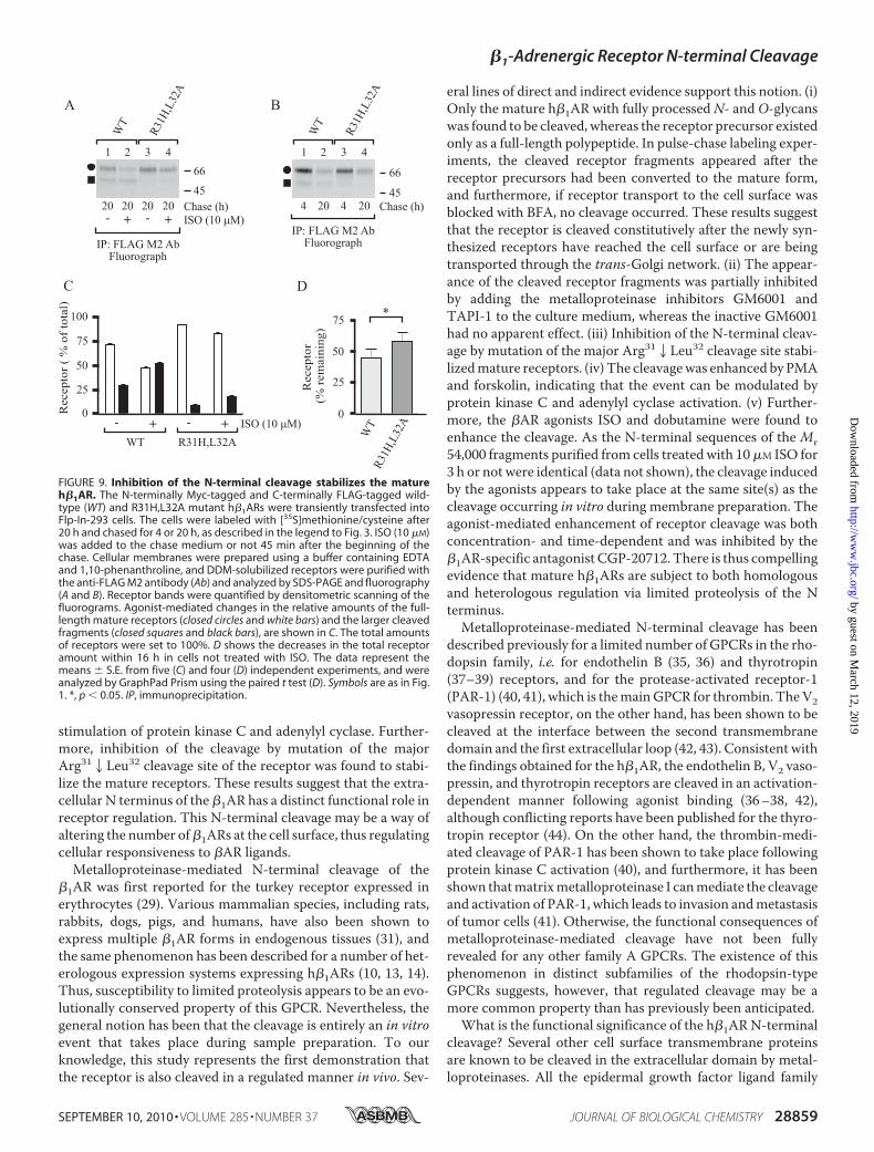
regulated event involving agonist-mediated activation, it can bespeculated that it may have a role in receptor down-regulation.To test this possibility, the wild-type receptor and theR31H,L32A cleavage site mutant were transiently transfectedinto Flp-In-293 cells, and ISO-mediated long term changes inreceptor numbers were monitored in the pulse-chase labelingassay. A 20-h treatment with ISO enhanced cleavage of thewild-type receptor (Fig. 9, A and C), and the amount of totalreceptors was decreased by 26 � 6% when compared with thenontreated control cells. The cleavage of the mutant receptorwas significantly attenuated, as expected, but was enhanced inISO-treated cells (Fig. 9, A and C). The agonist-mediateddecrease in the amount of total mutant receptors was similarthan in cells expressing the wild-type receptor (26 � 8%). Ago-nist-induced h�1AR down-regulation thus does not appear tobe dependent on receptor cleavage at the Arg312Leu32 cleav-age site. It can be argued that the residual cleavage that occursat, or very close to, the mutated site was adequate enough tolead to similar long term down-regulation of the mutantreceptor. Interestingly, the total amount of the mutant re-ceptor was consistently higher than that of the wild-type
FIGURE 7. h�1AR N-terminal cleavage takes place after receptors leavethe ER. A, inhibition of in vivo cleavage by metalloproteinase inhibitors. Sta-bly transfected HEK293i cells were induced to express h�1ARs for 6 h in theabsence or presence of various protease inhibitors, as indicated. DDM-solu-bilized receptors were analyzed by Western blotting (WB) with the anti-FLAGM2 antibody (Ab). B, pulse-chase labeling of BFA-treated cells. Stably trans-fected HEK293i cells were induced, labeled with [35S]methionine/cysteine,and chased as described in the legend to Fig. 3 in the presence of 5 �g/mlBFA, which was added to the culture medium 60 min before labeling. DDM-solubilized receptors were purified with the anti-FLAG M2 antibody and ana-lyzed by SDS-PAGE and fluorography. C, cell surface biotinylation. Stablytransfected control and BFA-treated HEK293i cells were labeled as describedfor B and chased for 240 min. Cell surface proteins were biotinylated withsulfo-NHS biotin before harvesting. DDM-solubilized receptors were sub-jected to a two-step purification with the anti-FLAG M2 antibody (lanes 1 and2; one-fifth of the samples) or immobilized streptavidin, followed by immu-noprecipitation (IP) with the anti-FLAG M2 antibody (lanes 3 and 4; four-fifthsof the samples). The samples were analyzed by SDS-PAGE and fluorography.D, enzymatic deglycosylation. Stably transfected BFA-treated HEK293i cellswere labeled as described for B and chased for 240 min. DDM-solubilizedreceptors were purified with the anti-FLAG M2 antibody and digested withglycosidases for 16 h at 30 °C or not before analysis by SDS-PAGE and fluorog-raphy. Lanes 1 and 5, untreated controls. Lane 2, PNGase F (50 units/ml). Lane3, neuraminidase (NEU; 50 milliunits/ml). Lane 4, neuraminidase (50 milliunits/ml) and O-glycosidase (O-glyc; 100 milliunits/ml). The receptor precursor inthe BFA-treated cells is indicated with an open circle and the receptor speciescarrying partially processed oligosaccharides with a closed diamond. Symbolsare as in Fig. 1.
�1-Adrenergic Receptor N-terminal Cleavage
SEPTEMBER 10, 2010 • VOLUME 285 • NUMBER 37 JOURNAL OF BIOLOGICAL CHEMISTRY 28857
receptor in the absence of ISO (Fig. 9A, compare lane 1 with3), suggesting that the N-terminal cleavage might have a rolein the general turnover of mature receptors. Consistent withthis idea, the total amount of wild-type and R31H,L32Amutant receptors decreased by 55 and 42% within 16 h,respectively (Fig. 9, B and D).Finally, we wanted to assess whether the h�1AR N-terminal
cleavage might be regulated in a heterologous manner. As seenin Fig. 8F, addition of PMA to the cell culture medium in thepulse-chase labeling assay increased the proportion of the Mr54,000 receptor species, whereas the enhanced cleavage wasinhibited with 10 �M bisindolylmaleimide I, a protein kinase Cinhibitor (lanes 2 and 3). The cleavage was also enhanced byforskolin (Fig. 8F, lane 4). These results suggest that h�1ARcleavage maybe under heterologous regulation, involving pro-tein kinase C as well as adenylyl cyclase activation.
DISCUSSION
The present description of the biosynthetic profile of theh�1AR in a heterologous expression system reveals several dis-tinct features of the behavior of the receptor that are relevant toits regulation. We show that the h�1AR is a GPCR that is effi-ciently expressed at the cell surface and is extensively glycosy-lated during transport to the plasma membrane. OneN-glycanis added to the conservedAsn at position 15, and severalmucin-type sialylated O-glycans are added to the N-terminal Ser/Thrresidues. However, despite thesemodifications, theN terminusappears to be very susceptible to cleavage by a metalloprotein-ase of either the matrix metalloproteinase or the ADAM fami-lies, resulting in the formation of at least two smaller mem-brane-bound C-terminal receptor fragments. The cleavage wasfound to take place constitutively in vivo and, importantly, wasenhanced after agonist-mediated receptor activation or direct
FIGURE 8. h�1AR N-terminal cleavage is a regulated process. A, receptoractivation by increasing concentrations of the �AR-agonist ISO. Stably trans-fected HEK293i cells were induced, labeled with [35S]methionine/cysteine,and chased for 240 min, as described in the legend to Fig, 3. ISO at the
concentrations indicated was added to the chase medium 45 min after thebeginning of the chase. GM6001 (10 �M) and CGP-20712 (10 �M) were added15 min prior to ISO. Cellular membranes were prepared using a buffer con-taining EDTA and 1,10-phenanthroline, and DDM-solubilized receptors werepurified with the anti-FLAG M2 antibody (Ab) and analyzed by SDS-PAGE andfluorography. Changes in the relative amounts of the full-length maturereceptor (a closed circle) and the larger cleaved fragment (a closed square), asrevealed by densitometric scanning of fluorograms, are shown in B. The dataare expressed as (Fl/(Fl � Fr)) � 100 and (Fr/(Fl � Fr)) � 100 for the full-lengthreceptor and cleaved fragment, respectively, where Fl � full-length receptor,and Fr � receptor fragment. C shows relative changes in the total amount ofreceptor. The values were normalized to that in the nontreated cells, whichwas set to 100%. The data shown represent the means � S.E. from threeindependent experiments and were analyzed by GraphPad Prism using therepeated measures two-way analysis of variance followed by Bonferroni’spost-test to compare the ISO-treated samples with the nontreated controls.The post-test did not reveal any significant differences for the data in C,although the analysis of variance performed before normalization revealedsignificant differences between the samples (p � 0.0081). D, time-dependentreceptor activation by ISO. E, receptor activation by dobutamine. F, activationof protein kinase C and adenylyl cyclase by PMA and forskolin, respectively.Stably transfected HEK293i cells were induced and labeled with [35S]methi-onine/cysteine as described for A. ISO (10 �M) was added to the chasemedium 45 min after the beginning of the chase or not, and the cells wereharvested 30, 60, 120, or 240 min later, as indicated (D). Dobutamine (DOB) (10�M) was added 45 min after the beginning of the chase, and the cells wereharvested 195 min later (E), Bisindolylmaleimide I (Bis I; 10 �M) was added 30min after the beginning of the chase, and PMA (0.5 �M) and forskolin (15 �M)15 min later, and the cells were harvested after a further 195 min (F). DDM-solubilized receptors were analyzed as described for A. Symbols are as in Fig. 1.***, p 0.001; *, p 0.05. IP, immunoprecipitation.
�1-Adrenergic Receptor N-terminal Cleavage
28858 JOURNAL OF BIOLOGICAL CHEMISTRY VOLUME 285 • NUMBER 37 • SEPTEMBER 10, 2010
stimulation of protein kinase C and adenylyl cyclase. Further-more, inhibition of the cleavage by mutation of the majorArg312Leu32 cleavage site of the receptor was found to stabi-lize the mature receptors. These results suggest that the extra-cellular N terminus of the �1AR has a distinct functional role inreceptor regulation. This N-terminal cleavage may be a way ofaltering the number of�1ARs at the cell surface, thus regulatingcellular responsiveness to �AR ligands.
Metalloproteinase-mediated N-terminal cleavage of the�1AR was first reported for the turkey receptor expressed inerythrocytes (29). Various mammalian species, including rats,rabbits, dogs, pigs, and humans, have also been shown toexpress multiple �1AR forms in endogenous tissues (31), andthe same phenomenon has been described for a number of het-erologous expression systems expressing h�1ARs (10, 13, 14).Thus, susceptibility to limited proteolysis appears to be an evo-lutionally conserved property of this GPCR. Nevertheless, thegeneral notion has been that the cleavage is entirely an in vitroevent that takes place during sample preparation. To ourknowledge, this study represents the first demonstration thatthe receptor is also cleaved in a regulated manner in vivo. Sev-
eral lines of direct and indirect evidence support this notion. (i)Only the mature h�1AR with fully processedN- andO-glycanswas found to be cleaved, whereas the receptor precursor existedonly as a full-length polypeptide. In pulse-chase labeling exper-iments, the cleaved receptor fragments appeared after thereceptor precursors had been converted to the mature form,and furthermore, if receptor transport to the cell surface wasblocked with BFA, no cleavage occurred. These results suggestthat the receptor is cleaved constitutively after the newly syn-thesized receptors have reached the cell surface or are beingtransported through the trans-Golgi network. (ii) The appear-ance of the cleaved receptor fragments was partially inhibitedby adding the metalloproteinase inhibitors GM6001 andTAPI-1 to the culture medium, whereas the inactive GM6001had no apparent effect. (iii) Inhibition of the N-terminal cleav-age by mutation of the major Arg312Leu32 cleavage site stabi-lizedmature receptors. (iv) The cleavagewas enhanced by PMAand forskolin, indicating that the event can be modulated byprotein kinase C and adenylyl cyclase activation. (v) Further-more, the �AR agonists ISO and dobutamine were found toenhance the cleavage. As the N-terminal sequences of the Mr54,000 fragments purified from cells treatedwith 10�M ISO for3 h or not were identical (data not shown), the cleavage inducedby the agonists appears to take place at the same site(s) as thecleavage occurring in vitro during membrane preparation. Theagonist-mediated enhancement of receptor cleavage was bothconcentration- and time-dependent and was inhibited by the�1AR-specific antagonist CGP-20712. There is thus compellingevidence that mature h�1ARs are subject to both homologousand heterologous regulation via limited proteolysis of the Nterminus.Metalloproteinase-mediated N-terminal cleavage has been
described previously for a limited number of GPCRs in the rho-dopsin family, i.e. for endothelin B (35, 36) and thyrotropin(37–39) receptors, and for the protease-activated receptor-1(PAR-1) (40, 41), which is themainGPCR for thrombin. TheV2vasopressin receptor, on the other hand, has been shown to becleaved at the interface between the second transmembranedomain and the first extracellular loop (42, 43). Consistent withthe findings obtained for the h�1AR, the endothelin B, V2 vaso-pressin, and thyrotropin receptors are cleaved in an activation-dependent manner following agonist binding (36–38, 42),although conflicting reports have been published for the thyro-tropin receptor (44). On the other hand, the thrombin-medi-ated cleavage of PAR-1 has been shown to take place followingprotein kinase C activation (40), and furthermore, it has beenshown thatmatrixmetalloproteinase I canmediate the cleavageand activation of PAR-1, which leads to invasion andmetastasisof tumor cells (41). Otherwise, the functional consequences ofmetalloproteinase-mediated cleavage have not been fullyrevealed for any other family A GPCRs. The existence of thisphenomenon in distinct subfamilies of the rhodopsin-typeGPCRs suggests, however, that regulated cleavage may be amore common property than has previously been anticipated.What is the functional significance of the h�1AR N-terminal
cleavage? Several other cell surface transmembrane proteinsare known to be cleaved in the extracellular domain by metal-loproteinases. All the epidermal growth factor ligand family
FIGURE 9. Inhibition of the N-terminal cleavage stabilizes the matureh�1AR. The N-terminally Myc-tagged and C-terminally FLAG-tagged wild-type (WT) and R31H,L32A mutant h�1ARs were transiently transfected intoFlp-In-293 cells. The cells were labeled with [35S]methionine/cysteine after20 h and chased for 4 or 20 h, as described in the legend to Fig. 3. ISO (10 �M)was added to the chase medium or not 45 min after the beginning of thechase. Cellular membranes were prepared using a buffer containing EDTAand 1,10-phenanthroline, and DDM-solubilized receptors were purified withthe anti-FLAG M2 antibody (Ab) and analyzed by SDS-PAGE and fluorography(A and B). Receptor bands were quantified by densitometric scanning of thefluorograms. Agonist-mediated changes in the relative amounts of the full-length mature receptors (closed circles and white bars) and the larger cleavedfragments (closed squares and black bars), are shown in C. The total amountsof receptors were set to 100%. D shows the decreases in the total receptoramount within 16 h in cells not treated with ISO. The data represent themeans � S.E. from five (C) and four (D) independent experiments, and wereanalyzed by GraphPad Prism using the paired t test (D). Symbols are as in Fig.1. *, p 0.05. IP, immunoprecipitation.
�1-Adrenergic Receptor N-terminal Cleavage
SEPTEMBER 10, 2010 • VOLUME 285 • NUMBER 37 JOURNAL OF BIOLOGICAL CHEMISTRY 28859
members, for example, are synthesized as inactive transmem-brane precursors that undergo ectodomain proteolytic cleav-age. The growth factors released this way then act in an endo-crine, paracrine, or autocrine manner via binding to theircognate receptors (45). A similar functional role for the cleavedh�1AR N terminus is very unlikely, however, as it lacks anyapparent functional protein domains. It is more likely that thecleavage has a role in attenuating receptor activity and activa-tion-mediated signaling in cells. This notion was supported bythe observation that the inhibition of receptor cleavage bymutation of the Arg312Leu32 cleavage site stabilized maturereceptors, suggesting that the proteolytic cleavage may have arole in the turnover of cell surface receptors. Removal of thereceptor N terminus may represent the first step in a pathwaythat directs the receptor protein toward degradation. It remainsto be demonstrated in future studies whether cleavage of thereceptor requires internalization or whether it occurs at the cellsurface. Internalization and targeting to lysosomal degradationmay be the primary way of disposing of cell surfaceGPCRs (46),but it has also been suggested, e.g. for the �2AR (47), that recep-tor degradation may occur without internalization at the cellsurface.The �1AR ligand-binding site resides within the 7-trans-
membrane helix bundle and involves amino acids in transmem-brane helices 3 and 5–7 and in the second extracellular loop(18). Thus, it is not immediately obvious why agonist bindingand receptor activation should lead to enhanced cleavage of thereceptor in the N-terminal domain. It can be hypothesized,however, that activation-induced conformational changes inthe transmembrane �-helices may be reflected in the N termi-nus. Thismay in turn increase susceptibility to enzymatic cleav-age. This possibility is in line with previous findings that havesuggested thatmetalloproteinase-mediated cleavage is not nec-essarily dependent on the primary structure of the substrateprotein but rather on conformational attributes (48). Interest-ingly, recent studies by Gupta et al. (19, 49) have demonstratedthat activation-induced conformational alterations do indeedoccur in the N terminus of several GPCRs in the rhodopsinfamily, including the �2AR. Furthermore, it was also found thatan N-terminally directed antibody of the �-opioid receptor wasable to recognize post-activation-mediated changes in thereceptor C terminus, an ability that was impaired after muta-tions at the C-terminal phosphorylation sites (49). If changes inthe phosphorylation of �1AR intracellular domains are con-verted to conformational changes in the receptorN terminus, itcan be hypothesized that the observed increase in the cleavageof the h�1AR by PMA and forskolin may be mediated by thesame mechanism as binding of the agonist to the receptor. Analternative hypothesis is that the enhanced cleavage of thereceptor results from increased functional activity of the cleav-ing enzyme itself. This could occur via phosphorylation of themetalloprotease through a protein kinase that is activated bythe second messengers. Extracellular signal-regulated kinase(ERK), for example, is known to phosphorylate ADAM-17 inresponse to PMAatThr735 (50), amodification that is indispen-sable for maturation of the protein and its inducible traffickingto the cell surface (51). These highly speculative mechanisms
that may be responsible for regulated h�1AR cleavage need tobe verified in future studies.The h�1AR was found to be modified by both N- and O-gly-
cosylation. The latter modification was first suggested by thefact that removal of the single N-glycan at Asn15 in the maturereceptor resulted in a receptor form that migrated more slowlythan the corresponding de-N-glycosylated receptor precursor.Treatment of the mature receptor with neuraminidase andO-glycosidase revealed that it carries mucin-type sialylatedO-glycans. Because of a lack of the consensus sequence forO-glycosylation, we used the NetOGlyc 3.1 Server (52) to predictthe mucin-type O-glycosylation sites in the receptor N termi-nus. This program predicts five potential O-glycosylation sitesfor the h�1AR, namely Thr28, Ser37, Ser41, Ser47, and Ser49,which are well conserved in mammalian �1ARs (Fig. 4).Apart from the h�1AR, an increasing number of other
GPCRs in the rhodopsin family have been reported to be mod-ified by O-linked carbohydrates. These include the human�-opioid (26, 28), �-opioid (53), V2 vasopressin (54), CCR5 che-mokine (55, 56), and bradykinin B2 (57) receptors, as well asoctopus rhodopsin and chicken iodopsin (58). The functionalsignificance of themodification has nevertheless remained elu-sive. Bannert et al. (56) found that O-glycosylation contributesto the binding of chemokines to the CCR5 chemokine receptorby providing an array of negative charges that allow electro-static interactions. On the other hand, Sadeghi and Birnbaumer(54) showed that neither the level of expression of the V2 vaso-pressin receptor nor its function is altered by eliminating theputatively O-glycosylated Ser/Thr residues. We cannot at themoment deduce the functional role of the O-glycosylation ofthe h�1AR, and further studies will have to be performed beforeany definite conclusions are drawn.One intriguing possibility isthat the extensive O-glycosylation might modulate the suscep-tibility of the N-terminal domain to metalloproteinases, possi-bly by restricting the conformational flexibility of the N termi-nus or by introducing negative charges. By comparison,O-glycans of several other cell surface proteins, including thetransferrin receptor, have been shown to protect the proteinfrom excessive proteolytic cleavage (59). The clustered Ser res-idues in the h�1AR N terminus (Ser37, Ser41, Ser47, and Ser49)are flanked by two proteolytic cleavage sites identified here,Arg312Leu32 and Pro522Leu53 (Fig. 4), which speaks for thepossibility that O-glycans may indeed have a protective role.
Acknowledgments—We thank Dr. Hongmin Tu (Biocenter Oulu Pro-tein Sequencing andAmino Acid Analysis Core Service) for assistancewith the N-terminal sequencing and Paula Salmela for technicalassistance. We are grateful to other members of the GPCR team fordiscussions during this work and to Prof. HannuRajaniemi for fruitfulideas.We also thank Prof. Michel Bouvier for the cDNAs encoding theuntagged and HA-tagged h�1AR constructs.
REFERENCES1. Bylund, D. B., Eikenberg, D. C., Hieble, J. P., Langer, S. Z., Lefkowitz, R. J.,
Minneman, K. P., Molinoff, P. B., Ruffolo, R. R., Jr., and Trendelenburg, U.(1994) Pharmacol. Rev. 46, 121–136
2. Fredriksson, R., Lagerstrom,M.C., Lundin, L. G., and Schioth,H. B. (2003)Mol. Pharmacol. 63, 1256–1272
�1-Adrenergic Receptor N-terminal Cleavage
28860 JOURNAL OF BIOLOGICAL CHEMISTRY VOLUME 285 • NUMBER 37 • SEPTEMBER 10, 2010
3. Lagerstrom, M. C., and Schioth, H. B. (2008) Nat. Rev. Drug Discov. 7,339–357
4. Rockman, H. A., Koch, W. J., and Lefkowitz, R. J. (2002) Nature 415,206–212
5. Brodde, O. E., Bruck, H., and Leineweber, K. (2006) J. Pharmacol. Sci. 100,323–337
6. Rousseau, G., Nantel, F., and Bouvier, M. (1996) Mol. Pharmacol. 49,752–760
7. Suzuki, T., Nguyen, C. T., Nantel, F., Bonin, H., Valiquette, M., Frielle, T.,and Bouvier, M. (1992)Mol. Pharmacol. 41, 542–548
8. Green, S. A., and Liggett, S. B. (1994) J. Biol. Chem. 269, 26215–262199. Shiina, T., Kawasaki, A., Nagao, T., and Kurose, H. (2000) J. Biol. Chem.
275, 29082–2909010. McLean, A. J., and Milligan, G. (2000) Br. J. Pharmacol. 130, 1825–183211. Xiang, Y., Devic, E., and Kobilka, B. (2002) J. Biol. Chem. 277,
33783–3379012. Liang, W., Curran, P. K., Hoang, Q., Moreland, R. T., and Fishman, P. H.
(2004) J. Cell Sci. 117, 723–73413. Dunigan, C. D., Hoang, Q., Curran, P. K., and Fishman, P. H. (2002) Bio-
chemistry 41, 8019–803014. Liang, W., Austin, S., Hoang, Q., and Fishman, P. H. (2003) J. Biol. Chem.
278, 39773–3978115. Frielle, T., Collins, S., Daniel, K. W., Caron, M. G., Lefkowitz, R. J., and
Kobilka, B. K. (1987) Proc. Natl. Acad. Sci. U.S.A. 84, 7920–792416. Hall, R. A. (2004) Semin. Cell Dev. Biol. 15, 281–28817. Marchese, A., Paing, M. M., Temple, B. R., and Trejo, J. (2008) Annu. Rev.
Pharmacol. Toxicol. 48, 601–62918. Warne, T., Serrano-Vega, M. J., Baker, J. G., Moukhametzianov, R., Ed-
wards, P. C., Henderson, R., Leslie, A. G., Tate, C. G., and Schertler, G. F.(2008) Nature 454, 486–491
19. Gupta, A., Decaillot, F. M., Gomes, I., Tkalych, O., Heimann, A. S., Ferro,E. S., and Devi, L. A. (2007) J. Biol. Chem. 282, 5116–5124
20. Bokoch, M. P., Zou, Y., Rasmussen, S. G., Liu, C. W., Nygaard, R., Rosen-baum, D.M., Fung, J. J., Choi, H. J., Thian, F. S., Kobilka, T. S., Puglisi, J. D.,Weis, W. I., Pardo, L., Prosser, R. S., Mueller, L., and Kobilka, B. K. (2010)Nature 463, 108–112
21. Pietila, E. M., Tuusa, J. T., Apaja, P. M., Aatsinki, J. T., Hakalahti, A. E.,Rajaniemi, H. J., and Petaja-Repo, U. E. (2005) J. Biol. Chem. 280,26622–26629
22. Lavoie, C., Mercier, J. F., Salahpour, A., Umapathy, D., Breit, A., Ville-neuve, L. R., Zhu,W. Z., Xiao, R. P., Lakatta, E. G., Bouvier,M., andHebert,T. E. (2002) J. Biol. Chem. 277, 35402–35410
23. Apaja, P. M., Tuusa, J. T., Pietila, E. M., Rajaniemi, H. J., and Petaja-Repo,U. E. (2006)Mol. Biol. Cell 17, 2243–2255
24. Leskela, T. T., Markkanen, P. M., Alahuhta, I. A., Tuusa, J. T., and Petaja-Repo, U. E. (2009) Traffic 10, 116–129
25. Leskela, T. T., Markkanen, P. M., Pietila, E. M., Tuusa, J. T., and Petaja-Repo, U. E. (2007) J. Biol. Chem. 282, 23171–23183
26. Petaja-Repo, U. E., Hogue, M., Laperriere, A., Walker, P., and Bouvier, M.(2000) J. Biol. Chem. 275, 13727–13736
27. Petaja-Repo, U. E., Hogue, M., Laperriere, A., Bhalla, S., Walker, P., andBouvier, M. (2001) J. Biol. Chem. 276, 4416–4423
28. Markkanen, P. M., and Petaja-Repo, U. E. (2008) J. Biol. Chem. 283,29086–29098
29. Jurss, R., Hekman, M., and Helmreich, E. J. (1985) Biochemistry 24,3349–3354
30. Benovic, J. L., Stiles, G. L., Lefkowitz, R. J., and Caron, M. G. (1983) Bio-chem. Biophys. Res. Commun. 110, 504–511
31. Stiles, G. L., Strasser, R. H., Lavin, T. N., Jones, L. R., Caron, M. G., andLefkowitz, R. J. (1983) J. Biol. Chem. 258, 8443–8449
32. Umemoto, J., Bhavanandan, V. P., andDavidson, E. A. (1977) J. Biol. Chem.252, 8609–8614
33. Yarden, Y., Rodriguez, H., Wong, S. K., Brandt, D. R., May, D. C., Burnier,J., Harkins, R.N., Chen, E. Y., Ramachandran, J., Ullrich,A., andRoss, E.M.(1986) Proc. Natl. Acad. Sci. U.S.A. 83, 6795–6799
34. Petaja-Repo, U. E., Hogue, M., Bhalla, S., Laperriere, A., Morello, J. P., andBouvier, M. (2002) EMBO J. 21, 1628–1637
35. Kozuka, M., Ito, T., Hirose, S., Lodhi, K. M., and Hagiwara, H. (1991)J. Biol. Chem. 266, 16892–16896
36. Grantcharova, E., Furkert, J., Reusch, H. P., Krell, H. W., Papsdorf, G.,Beyermann, M., Schulein, R., Rosenthal, W., and Oksche, A. (2002) J. Biol.Chem. 277, 43933–43941
37. Couet, J., Sar, S., Jolivet, A., Hai,M. T.,Milgrom, E., andMisrahi,M. (1996)J. Biol. Chem. 271, 4545–4552
38. Ando, T., Latif, R., Pritsker, A., Moran, T., Nagayama, Y., and Davies, T. F.(2002) J. Clin. Invest. 110, 1667–1674
39. Kaczur, V., Puskas, L. G., Nagy, Z. U., Miled, N., Rebai, A., Juhasz, F.,Kupihar, Z., Zvara, A., Hackler, L., Jr., and Farid, N. R. (2007) J. Mol.Recognit. 20, 392–404
40. Ludeman, M. J., Zheng, Y. W., Ishii, K., and Coughlin, S. R. (2004) J. Biol.Chem. 279, 18592–18599
41. Boire, A., Covic, L., Agarwal, A., Jacques, S., Sherifi, S., and Kuliopulos, A.(2005) Cell 120, 303–313
42. Kojro, E., and Fahrenholz, F. (1995) J. Biol. Chem. 270, 6476–648143. Kojro, E., Postina, R., Gilbert, S., Bender, F., Krause, G., and Fahrenholz, F.
(1999) Eur. J. Biochem. 266, 538–54844. Chazenbalk, G. D., Chen, C. R., McLachlan, S.M., and Rapoport, B. (2004)
Endocrinology 145, 4–1045. Blobel, C. P. (2005) Nat. Rev. Mol. Cell Biol. 6, 32–4346. Hanyaloglu, A. C., and von Zastrow, M. (2008) Annu. Rev. Pharmacol.
Toxicol. 48, 537–56847. Jockers, R., Angers, S., Da Silva, A., Benaroch, P., Strosberg, A. D., Bouvier,
M., and Marullo, S. (1999) J. Biol. Chem. 274, 28900–2890848. Huovila, A. P., Turner, A. J., Pelto-Huikko, M., Karkkainen, I., and Ortiz,
R. M. (2005) Trends Biochem. Sci. 30, 413–42249. Gupta, A., Rozenfeld, R., Gomes, I., Raehal, K. M., Decaillot, F. M., Bohn,
L. M., and Devi, L. A. (2008) J. Biol. Chem. 283, 10735–1074450. Díaz-Rodríguez, E., Montero, J. C., Esparís-Ogando, A., Yuste, L., and
Pandiella, A. (2002)Mol. Biol. Cell 13, 2031–204451. Soond, S. M., Everson, B., Riches, D.W., andMurphy, G. (2005) J. Cell Sci.
118, 2371–238052. Julenius, K., Mølgaard, A., Gupta, R., and Brunak, S. (2005) Glycobiology
15, 153–16453. Li, J. G., Chen, C., and Liu-Chen, L. Y. (2007) Biochemistry 46,
10960–1097054. Sadeghi, H., and Birnbaumer, M. (1999) Glycobiology 9, 731–73755. Farzan, M., Mirzabekov, T., Kolchinsky, P., Wyatt, R., Cayabyab, M., Ge-
rard, N. P., Gerard, C., Sodroski, J., and Choe, H. (1999) Cell 96, 667–67656. Bannert, N., Craig, S., Farzan, M., Sogah, D., Santo, N. V., Choe, H., and
Sodroski, J. (2001) J. Exp. Med. 194, 1661–167357. Michineau, S., Alhenc-Gelas, F., and Rajerison, R. M. (2006) Biochemistry
45, 2699–270758. Nakagawa, M., Miyamoto, T., Kusakabe, R., Takasaki, S., Takao, T.,
Shichida, Y., and Tsuda, M. (2001) FEBS Lett. 496, 19–2459. Rutledge, E. A., Root, B. J., Lucas, J. J., and Enns, C. A. (1994) Blood 83,
580–586
�1-Adrenergic Receptor N-terminal Cleavage
SEPTEMBER 10, 2010 • VOLUME 285 • NUMBER 37 JOURNAL OF BIOLOGICAL CHEMISTRY 28861


![Odoribacter splanchnicus type strain (1651/6T)...Strain 1651/6T (= DSM 20712 = ATCC 29572 = JCM 15291) is the type strain of Odoribacter splan-chnicus [1,2]. Currently, there are three](https://static.documents.pub/doc/80x56/611ce1fbeb7fcc56bc32d083/odoribacter-splanchnicus-type-strain-16516t-strain-16516t-dsm-20712-.jpg)

