Nucleic Acids Research, Vol. 18, No. 21 6231 Human DNA polymerase a. catalytic polypeptide binds ConA and RCA and contains a specific labile site in the N-terminus Kuo-Liang Hsi, William C.Copeland and Teresa S.-F.Wang* Laboratory of Experimental Oncology, Department of Pathology, Stanford University School of Medicine, Stanford, CA 94305, USA Received August 9, 1990; Revised and Accepted October 2, 1990 ABSTRACT The catalytic polypeptide of DNA polymerase a is often observed In vitro as a family of phosphopolypeptides predominantly of 180 and 165 kDa derived from a single primary structure. The estimated Mr of this polypeptide deduced from the full-length cDNA is 165 kDa. Immunoblot analysis with polyclonal antibodies against peptldes of the N- and C-terminl of the deduced primary sequence indicates that the observed family of polypetldes from 180 kDa to lower molecular weight results from proteolytic cleavage from the N-termlnus. Antibodies against the N-termlnal peptide detect only the 180 kDa species suggesting that this higher molecular weight polypeptide may be the result of posttranslational modification of the 165 kDa primary translation product. The catalytic polypeptide is not only phosphorylated but is also found to react with lectins ConA and RCA. N-terminal sequencing of the isolated catalytic polypeptide from human cells and of the recombinant fusion proteins indicates that the often observed 165 kDa polypeptide is the In vitro proteolytic cleavage product of the modified 180 kDa protein at the specific site between lys 123 and lys 124 within the sequence -RNVKKLAVTKPNN-. INTRODUCTION DNA polymerase a purified from species as distinct as unicellular fungi and mammals displays a remarkably similar set of constituent polypeptides (1—7). Each contains a cluster of large polypeptides predominantly 165 to 180 kDa with catalytic function, a 70 kDa protein of unknown function and two polypeptides, 55 and 49 kDa, reported to contain the primase activity (5 — 10). Peptide mapping indicates that the catalytic polypeptides, 180 and 165 kDa have identical peptide maps (6). This finding suggests that these high molecular weight polypeptides are derivatives of a single primary translation product. The full length cDNA from human cells and a genomic clone from 5. cerevisiae encoding the catalytic polypeptide have been isolated (11-13). The primary sequence deduced from human or from yeast genetic sequences predicts a molecular mass of 165 kDa (11,13). In this report, we have used polyclonal antisera against two synthetic peptides corresponding to the N- and C-termini of the catalytic polypeptide, lectin binding assays, and amino acid sequence analysis of the N-terminal portion of the protein to resolve the discrepency of the observed and the predicted molecular mass of DNA polymerase a catalytic polypeptide. MATERIALS AND METHODS Production of antibodies against synthetic peptides Two amino acid sequences of 20 residues corresponding to the N- and C-termini of human DNA polymerase a catalytic polypeptide were chosen as peptide antigens based on the criteria described (14). The peptides were synthesized with an Applied Biosystems Automated Peptide Synthesizer (ABI, 430) with an additional cysteine at the C-terminus and then conjugated to myoglobin or ovalbumin according to the method of (15). Two hundred fifty micrograms of each conjugated peptide were used to immunize New Zealand white rabbits according to the described protocol (16). Antisera titer were screened by ELJSA assay and further characterized by immunoblotting with human DNA polymerase a purified by monoclonal antibody immunoaffinity chromatography (5,6,17). Antiserum against the N-terminal peptide is designated DPN; while antiserum against the C-terminus is designated DPC. Detection of polymerase a catalytic polypeptide with antisera and lectins Immunoaffinity purified DNA polymerase a polypeptides were subjected to gel electrophoresis and transferred to Problott membrane (Applied Biosy stems, Inc.) as described (18). Detection of catalytic polypeptide with antisera DPN or DPC was performed according to (16) with goat anti-rabbit IgG conjugated with horseradish peroxidase (Bio-Rad Laboratories) as secondary antibody followed by developing in 3,3'-diaminobenzidine and peroxide as decribed (16). For lectin binding, the membranes were incubated with 10 /tg/ml of biotinylated lectins (Vector Laboratories, CA) for 1 to 2 hours and developed with precomplexed avidin-biotin-peroxidase • To whom correspondence should be addressed Downloaded from https://academic.oup.com/nar/article-abstract/18/21/6231/2388468 by guest on 03 February 2018
Transcript
Nucleic Acids Research, Vol. 18, No. 21 6231
Human DNA polymerase a. catalytic polypeptide bindsConA and RCA and contains a specific labile site in theN-terminus
Kuo-Liang Hsi, William C.Copeland and Teresa S.-F.Wang*Laboratory of Experimental Oncology, Department of Pathology, Stanford University School ofMedicine, Stanford, CA 94305, USA
Received August 9, 1990; Revised and Accepted October 2, 1990
ABSTRACT
The catalytic polypeptide of DNA polymerase a is oftenobserved In vitro as a family of phosphopolypeptidespredominantly of 180 and 165 kDa derived from a singleprimary structure. The estimated Mr of this polypeptidededuced from the full-length cDNA is 165 kDa.Immunoblot analysis with polyclonal antibodies againstpeptldes of the N- and C-terminl of the deduced primarysequence indicates that the observed family ofpolypetldes from 180 kDa to lower molecular weightresults from proteolytic cleavage from the N-termlnus.Antibodies against the N-termlnal peptide detect onlythe 180 kDa species suggesting that this highermolecular weight polypeptide may be the result ofposttranslational modification of the 165 kDa primarytranslation product. The catalytic polypeptide is notonly phosphorylated but is also found to react withlectins ConA and RCA. N-terminal sequencing of theisolated catalytic polypeptide from human cells and ofthe recombinant fusion proteins indicates that the oftenobserved 165 kDa polypeptide is the In vitro proteolyticcleavage product of the modified 180 kDa protein at thespecific site between lys123 and lys124 within thesequence -RNVKKLAVTKPNN-.
INTRODUCTION
DNA polymerase a purified from species as distinct as unicellularfungi and mammals displays a remarkably similar set ofconstituent polypeptides (1—7). Each contains a cluster of largepolypeptides predominantly 165 to 180 kDa with catalyticfunction, a 70 kDa protein of unknown function and twopolypeptides, 55 and 49 kDa, reported to contain the primaseactivity (5 — 10). Peptide mapping indicates that the catalyticpolypeptides, 180 and 165 kDa have identical peptide maps (6).This finding suggests that these high molecular weightpolypeptides are derivatives of a single primary translationproduct. The full length cDNA from human cells and a genomicclone from 5. cerevisiae encoding the catalytic polypeptide havebeen isolated (11-13). The primary sequence deduced fromhuman or from yeast genetic sequences predicts a molecular mass
of 165 kDa (11,13). In this report, we have used polyclonalantisera against two synthetic peptides corresponding to the N-and C-termini of the catalytic polypeptide, lectin binding assays,and amino acid sequence analysis of the N-terminal portion ofthe protein to resolve the discrepency of the observed and thepredicted molecular mass of DNA polymerase a catalyticpolypeptide.
MATERIALS AND METHODSProduction of antibodies against synthetic peptidesTwo amino acid sequences of 20 residues corresponding to theN- and C-termini of human DNA polymerase a catalyticpolypeptide were chosen as peptide antigens based on the criteriadescribed (14). The peptides were synthesized with an AppliedBiosystems Automated Peptide Synthesizer (ABI, 430) with anadditional cysteine at the C-terminus and then conjugated tomyoglobin or ovalbumin according to the method of (15). Twohundred fifty micrograms of each conjugated peptide were usedto immunize New Zealand white rabbits according to thedescribed protocol (16). Antisera titer were screened by ELJSAassay and further characterized by immunoblotting with humanDNA polymerase a purified by monoclonal antibodyimmunoaffinity chromatography (5,6,17). Antiserum against theN-terminal peptide is designated DPN; while antiserum againstthe C-terminus is designated DPC.
Detection of polymerase a catalytic polypeptide with antiseraand lectinsImmunoaffinity purified DNA polymerase a polypeptides weresubjected to gel electrophoresis and transferred to Problottmembrane (Applied Biosy stems, Inc.) as described (18).Detection of catalytic polypeptide with antisera DPN or DPCwas performed according to (16) with goat anti-rabbit IgGconjugated with horseradish peroxidase (Bio-Rad Laboratories)as secondary antibody followed by developing in3,3'-diaminobenzidine and peroxide as decribed (16). For lectinbinding, the membranes were incubated with 10 /tg/ml ofbiotinylated lectins (Vector Laboratories, CA) for 1 to 2 hoursand developed with precomplexed avidin-biotin-peroxidase
• To whom correspondence should be addressed
Downloaded from https://academic.oup.com/nar/article-abstract/18/21/6231/2388468by gueston 03 February 2018
6232 Nucleic Acids Research, Vol. 18, No. 21
according to the manufacturer's instruction (Vector Laboratories,CA). For ConA and LCA bindings, 1 mM Ca+2 and Mn+2
were added to each incubation, respectively.
Production of recombinant human DNA polymerase a asfusion proteinsTwo plasmids, pBC5O9 and pBC943, that express the N-terminalportion of human DNA polymerase a fused with /3-galactosidaseprotein under control of the T7 RNA polymerase promoter wereconstructed. Both plasmids were constructed by linker ligatingthe 5' Ncol site of El - 1 9 cDNA clone (11) with EcoRI linkersfollowed by subsequent digestion with EcoRI and either Sau3AIor Bell. The 509 bp EcoRI-Sau3AI and the 943 bp EcoRI-BclIrestriction fragments from El - 1 9 were separately ligated to the6.2 Kb BamHI-Sall restriction fragment from pMC874 (19) andinserted into the EcoRI-Sall digested pT7-7 vector (20).Induction of the fusion proteins in E. coli was accomplished bya modified procedure of Tabor and Richardson (20) and purifiedfrom the cleared cell lysate using p-aminophenyl /3-D-thiogalactopyranoside-agarose (APTG, Sigma) as described (21).Purified fusion proteins were subjected to gel electrophoresis and
transferred to Problott membrane for either amino acid sequenceanalysis or immunoblotting with DPN as described above.
N-terminal amino acid sequence analysisHuman DNA polymerase a protein from KB cells (6) and DNApolymerase a-//3-galactosidase fusion protein were subjected toelectrophoresis and transferred onto Problott membrane. Proteinbands of interest were excised and subjected to automated Edmandegradation performed on an Applied Biosystems Model 477Agas phase sequencer with an on-line PTH-amino acid analyserModel 120A, using the NORMAL-1 program (22).
Other methodsThe amount of immunoaffinity purified DNA polymerase aprotein applied to each gel electrophoresis is estimated in termsof units of polymerase a activity. One unit of polymerase aactivity is defined as the amount of enzyme that catalyzes theincorporation of 1 nmole of labeled dNMP into acid-insolubleproduct in 1 hour at 37 °C under standard assay conditions (26).By densitometric scanning of Coomassie stained gels loaded withknown units of DNA polymerase a, it was previously estimatedthat 1 unit of polymerase a activity is equivalent to 5 ng of protein
DPN
SRABRFXKSKKGROEA1.EKC
DPC
EKLTTDHEKDICLKKOnTPKC
NH. COOH
KOa
200 — 200 — — 200p 180
p 165
97 _ — 97
68 - — 68
68 —-p70
43 —
— IgCKH)
43 —
DPNSilverstain
DPC
Figure 1. A. Schematic diagram of position of peptide antigens in the predicted primary protein sequence of human DNA polymerase a for antisera, DPN andDPC. DPN was raised against a peptide of 20 amino acids beginning at 19 amino acids from the translation start she. DPC was raised against a 20 residues peptide37 amino acids upstream from the C-terminus stop codon. The line represents the entire 1462 amino acid sequence derived from the full-length cDNA, the solidboxes depict the six conserved regions (11, 23). B. Immunoblot of DNA polymerase a by antisera DPN and DPC. Sixty units of immunopurified human DNApolymerase a were immunoblotted by antisera DPN or DPC, and used in silver stain for comparison as described in Materials and Methods. Protein markers of200, 97, 68 and 43 kDa are myosin, phosphorylase b, bovine serum albumin and ovalbumin, respectively.
Downloaded from https://academic.oup.com/nar/article-abstract/18/21/6231/2388468by gueston 03 February 2018
Nucleic Acids Research, Vol. 18, No. 21 6233
containing the four subunits of the polymerase a/primasecomplex.
a-D-mannosidase, a-D-glycosidase and /3-D-galactosidase arefrom Boehringher-Mannhein, Biochemicals and used accordingto the manufacturer's instruction.
RESULTSDNA polymerase a is degraded from the N-terminusDNA polymerase a purified either by conventional biochemicalmethods or by an immunoaffinity protocol with monoclonalantibody contains: a family of high molecular weight polypeptidesof 165 to 180 kDa with catalytic function, a 70 kDa polypeptideof unknown function and two polypetides of 55 and 49 kDareported to be associated with DNA primase activity, as shownin Figure IB (4—6,11). Previous studies have demonstrated thatthe two predominant high molecular weight members of thecatalytic polypeptide family have identical peptide maps (6). Themolecular mass difference between these two catalyticpolypeptides (pl80 and pl65) can be explained either bydifferential modification or N- or C-terminal degradation of asingle primary translation product. The full length cDNA ofhuman DNA polymerase a catalytic polypeptide encodes a 165kDa protein (11,23; Pearson, Nasheuer and Wang, submitted).Genomic sequence of the yeast Poll, a homolog of polymerasea, also predicts a protein of 167 kDa (13).
To resolve the difference between the observed and thepredicted molecular mass of DNA polymerase a catalyticpolypeptide, purified human DNA polymerase a immobilizedon membrane was incubated with polyclonal antisera raisedagainst 20 amino acid residue synthetic peptides correspondingto sequences at the N- or C-termini, Figure 1A. The antiseraagainst the N-terminus, DPN, recognized epitopes on a singlehigh molecular weight polypeptide of 180 kDa (pi80), whileantisera against the C-terminus, DPC, recognized epitopes onboth 180 and 165 kDa polypeptides (pl80 and pl65), Figure IB.Two smaller polypeptides of this catalytic polypeptide cluster withapparent molecular masses of 140 and 125 kDa which areoccasionally observed were also found to contain the DPCepitopes but not DPN epitopes (Fig. IB). These results indicatethat the pi80 contains the N-terminal sequence recognized byDPN antisera, whereas pl65, pl40 and pl25 proteins lack thisN-terminal epitope. This result in addition to the observation thatfull-length recombinant human polymerase a when expressed inE. coli yielded a 165 kDa polypeptide (Copeland and Wang,unpublished observation), strongly suggest that the pi80 is amodified form of the predicted 165 kDa primary translationproduct of DNA polymerase a transcript, while pl65, as wellas pl40 and pl25 polypeptide, are N-terminally degradedproducts of the pi80.
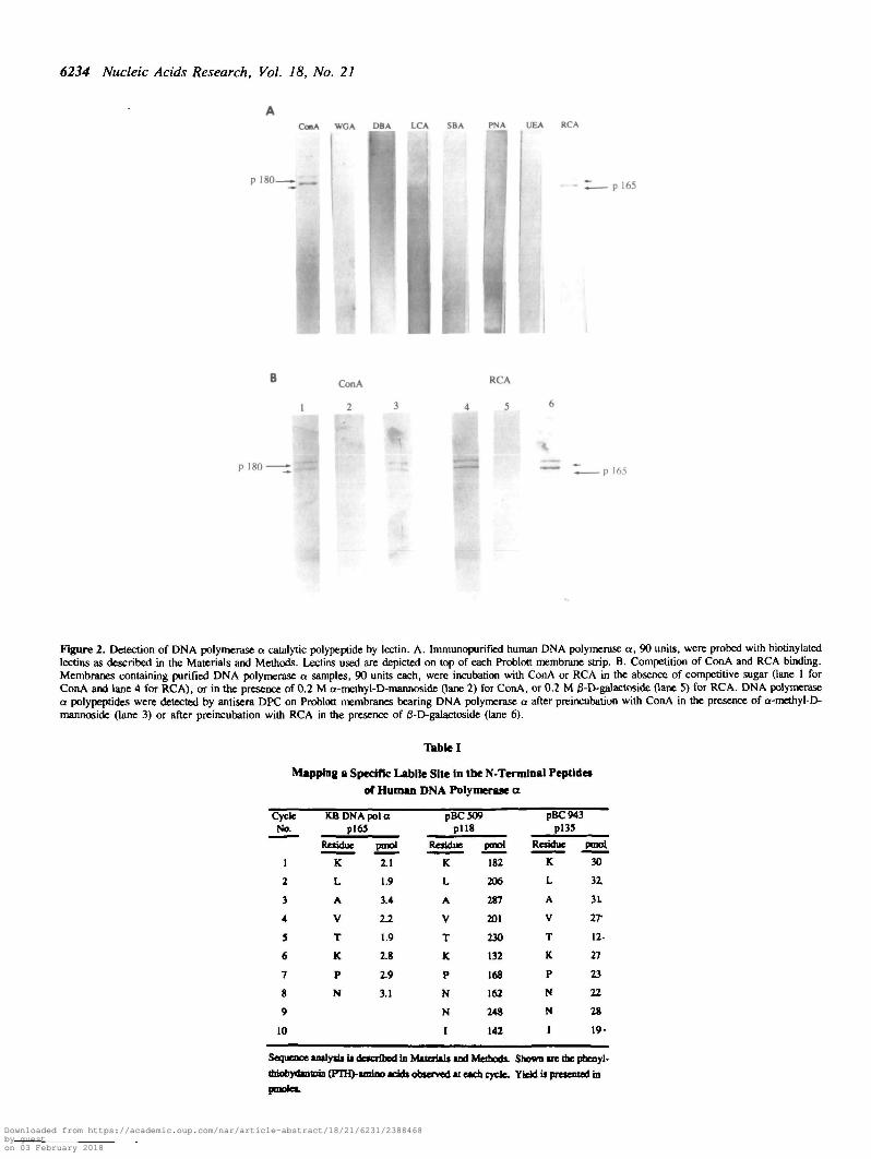
DNA polymerase a catalytic polypeptide is specifically boundby the lectins ConA and RCAIt is possible that the observed higher molecular mass catalyticpolypeptide is due to posttranslational modification. We havepreviously reported that pl80/pl65 and p70 of DNA polymerasea enzyme preparations are phosphoproteins (6). The recentfindings that some nuclear proteins are glycoproteins (24)prompted us to test whether the catalytic polypeptide can bindlectins. Membranes bearing immunoaffinity purified human DNApolymerase a proteins were treated with eight biotinylated lectinderivatives: concanavalin A (ConA), which primarily recognizesa-D-mannoside and a-D-glucoside residues; wheat germ
agglutinin (WGA), which recognizes N-acetylglucosamineresidues; dolchos biflorus agglutinin (DBA), specific for a-linkedN-acetylgalactosamine; lens culinaris agglutinin (LCA), specificfor a-linked fucose residue attached to the N-acetylchitobioseportion of the core oligosaccharide; soybean agglutinin (SBA)which binds to oligosaccharide structures with terminal a- or /3-linked N-acetylglucosamine or galactose residues; peanutagglutinin (PNA) which recognizes galactosyl (/3-l,3) N-acetylgalactosamine; ricinus communis agglutinin I (RCA) whichpreferentially binds to oligosaccharides ending in galactose andalso binds to N-acetylgalactosamine; and Aulex europeaeusagglutinin (UEA), specific for glycoproteins containing a-linkedfucose residues (25). Binding of ConA and RCA to both pi80and pl65 was observed, Fig 2A. In the presence of 0.2 M a-D-mannoside, a competitive inhibitor of ConA binding, or 0.2 Ma-D-galactoside, a competitive inhibitor of RCA binding,reactivity with ConA or RCA, respectively, was completelyabolished, as shown in Fig. 2B, lane 2 and 5. ConA binding couldlikewise be competitively abolished by a-D-glucoside (data notshown). The inability of ConA or RCA to bind to thesepolypeptides in the presence of these inhibitors is not caused byremoval or absence of the polymerase a protein on themembrane, because these membranes did not lose the ability tobind antisera raised against the C-terminus, DPC, as demonstratedin Fig. 2B, lane 3 and 6. In addition, it is unlikely that the ConAreactivity is due to comigration of another glycoprotein of 180and 165 kDa sizes, since covalently linked ConA-Sepharose isable to deplete DNA polymerase a activity from a partiallypurified enzyme fraction (data not shown).
To further define the specificity of the ConA and RCAinteraction, purified polymerase a were incubated with threeglycosidases, a-mannosidase, a-glucosidase and jS-galactosidaseprior to ConA or RCA binding. Preincubation with a-glucosidasedid not abrogate the ConA binding, whereas preincubation witha-mannosidase completely abolished the binding of ConA (Fig.3A, lane 2 and 3, respectively). This result suggests but doesnot rigorously prove that the ConA binding moiety on thispolypeptide is a-D-mannoside. Treatment with /S-galactosidaseabolished binding by lectin RCA, as shown in Fig 3B, lane 2.As with the binding competition experiments, the inability to bindConA or RCA after a-mannosidase or j3-galactosidase treatmentwas not due to removal or absence of polymerase a proteins fromthe membrane, because either enzyme treatment did not diminshthe binding of DPC to pl80 or pl65 (Fig. 3A, lane 4 and Fig.3B, lane 3). It should be noted that in the polymerase a enrymefraction only the catalytic polypeptide reacts with ConA and RC-A. The 70 kDa subunit and the two primase related subunits didnot bind to any of the eight biotinylated lectins tested.
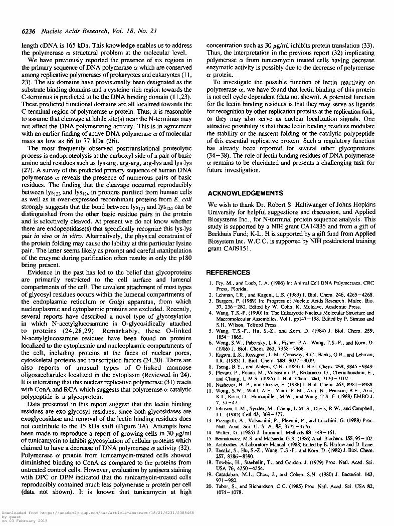
Proteolytic cleavage occurs at a specific labile site of the N-terminuspi65 is often reproducibly observed in DNA polymerase apreparations from a variety of eukaryotic cell sources. Resultsof immunoblots with antisera DPN and DPC strongly suggestthat pi65 is a proteolytic degradation product of pi80 (Figure1). To investigate this proteolytic event in detail, the N-terminusof pi65 isolated from KB cells by the immunoaffinity protocolwas sequenced and found to be KLAVTKPN-, Table 1. Twofusion proteins each containing overlapping 5' portions of thepolymerase a cDNA fused with the lacZ gene at the 3' end wereexpressed from the pT7-7 vector in E. coli, purified, andanalyzed on SDS gels. For each construct, more than one major
Downloaded from https://academic.oup.com/nar/article-abstract/18/21/6231/2388468by gueston 03 February 2018
6234 Nucleic Acids Research, Vol. 18, No. 21
ConA WGA DBA LCA SBA PNA UEA RCA
p 180— _ p 165
1 2 3
RCA
4 5 6
p!65
t
Figure 2. Detection of DNA polymerase a catalytic polypeptide by lectin. A. Immunopurified human DNA polymerase a, 90 units, were probed with biotinylatedlectins as described in the Materials and Methods. Lectins used are depicted on top of each Problott membrane strip. B. Competition of ConA and RCA binding.Membranes containing purified DNA polymerase a samples, 90 units each, were incubation with ConA or RCA in the absence of competitive sugar (lane 1 forConA and lane 4 for RCA), or in the presence of 0.2 M a-methyl-D-mannoside Oane 2) for ConA, or 0.2 M 0-D-galactoside Oane 5) for RCA. DNA polymerasea polypeptides were detected by antisera DPC on Problott membranes bearing DNA polymerase a after preincubation with ConA in the presence of a-methyl-D-mannoside Oane 3) or after preincubation with RCA in the presence of 0-D-galactoside Qane 6).
Table I
Mapping a Specific Labile Site In the N-Tertnlnal Peptldes
of Human DNA Polymerase a
CycleNo.
1
2
3
4
S
6
7
8
9
10
KB DNA pol apl6J
Residue
K
L
A
V
T
K
P
N
ptnol
2.1
1.9
3.4
2 J
1.9
2.8
2.9
3.1
pBC509pi 18
Residue
K
L
A
V
T
K
P
N
N
I
pmo)
182
206
287
201
230
132
168
162
248
142
pBC943pl35
Residue
K
L
A
V
T
K
P
N
N
1
pmot
30
3Z
31
rr12-
27
23
22
28
19-
Sequence imrysis U described in Material* md Methods. Shown n e the pbenyl-
dnohydnuoin (PTH>imlno adds observed at each cycle. Yield u presented in
pmolca.
Downloaded from https://academic.oup.com/nar/article-abstract/18/21/6231/2388468by gueston 03 February 2018
Nucleic Acids Research, Vol. 18, No. 21 6235
ConA RCAS i u 3 * Bell
Ncol Ncol
KDa
200 —
68 —
43 —
Figure 3. ConA and RCA binding specificity. A.Immunoaffinity purified DNApolymerase a, 120 units, was incubated with no glycosidase Gane 1), 2.5 unitsof ot-glucosidase (lane 2), or 5.0 units of a-mannosidase 0ane 3 and lane 4) for20 hours at 37 °C under the conditions recommended by Bochringer MannheimBiochemicals. These glycosidase treated DNA polymerase a samples were thensubjected to SDS gel electrophoresis, transferred to Problot membrane and ConAbinding as described in Figure 2. Lane 4 demonstrates the detection of DNApolymerase a polypeptide by antisera DPC after the sample was treated witha-mannosidase and stained with ConA as described in lane 3. The tower molecularweight ConA binding proteins in lane 2, 3 and 4 are a-glucosidase anda-mannosidase which are both glycoproteins and bind to ConA. Depending onthe batch of ConA used, minor ConA binding bands in the lower molecular weightrange were occasionally observed. B. RCA binding to purified DNA polymerasea (90 units) after incubation with no /3-galactosidase (lane 1) and with 2.2 unitsof |3-D-galactosidase (lane 2). Detection of DNA polymerase a polypeptide byantisera DPC after 0-galactosidase treatment (lane 3).
protein species was isolated by APTG-affinity column, as shownin Figure 4B. Expression of pBC509 yielded two protein bandsof 135 and 118 kDa (pl35 and pi 18) as shown in Fig 4B, lane2, while pBC943 yielded three protein bands of 151, 135 and118 kDa (pl51, pl35 and pi 18), as shown in Fig 4B, lane 3.Immunoblotting with DPN antisera revealed that only the largestproteins from each construct (i.e. pl35 from pBC509 and pl51from pBC943) contain the N-terminal epitopes. The smallerproteins, pi 18 from pBC5O9 or pi35 and pi 18 from pBC943,do not contain the epitopes recognized by DPN antisera, as shownin Fig. 4C, lane 2 and 3. Interestingly, the 15 kDa molecularmass decrement within each set of fusion proteins closely matchesthe difference between the observed polymerase a proteins (pi80and pi65) isolated from KB cells. Sequence analyses of fusionproteins pi35 of pBC934 and pi 18 of pBC509 revealed N-terminal amino acid sequence identical with that of pi65 isolatedfrom KB cells (Table 1). These sequence analyses stronglysuggest the presence of a specific labile site between l y s ^ andlys|24 within the N-terminal domain -RNVKKLAVTKPNN- ofhuman polymerase a catalytic polypeptide. Cleavage of pi80 atthis labile site generates the often observed pi65.
Taken together, the results of anti-peptide antibody studies,lectin binding and N-terminal sequence analyses support theconclusion that posttranslational modification of a 165 kDaprimary translation product results in a protein of apparentmolecular mass of 180 kDa, and that subsequent proteolyticcleavage of the modified pi80 species at a labile site within the
cDNA
Hvtdlll BimHI
- Poly A
T 7 SluJA/BwiHIpBC509 -mm^'
PBC943
B
200
9*3
1 2 3 1 2 3
200 —»
116—»
68—»
43
29—»
Figure 4. N-terminal peptides of human DNA polymerase a in recombinant fusionproteins. A. Schematic diagram of N-terminal peptide fusion protein constructs.The top diagram is the restriction map of the full-length cDNA of human DNApolymerase a. pBC509 and pBC943 were constructed as described in the Materialsand Methods. The heavy arrow is the T7 RNA polymerase promoter, while theN-terminal portion of polymerase a and the lacZ gene are illustrated in differentpatterns. Number 509 and 943 represent the cDNA nucleotide numbers whichencode the portions of N-terminal amino acids of DNA polymerase a in thesefusion constructs. B. Recombinant proteins produced from the fusion constructs.Lane 1 demonstrates 5 ng, of /3-galactosidase, lane 2 and 3 are 25 and 10 /igof affinity purified fusion proteins from pBC504 and pBC943, respectively. DNApolymerase a fusion proteins expressed from each construct are indicated by solidtriangles. C. Immunoblot of polymerase a N-terminal fusion proteins by DPN.Lane 1 is 0-galactosidase, lane 2 and 3 are 25 and 10 /jg of expressed fusionproteins from pBC509 and pBC943, respectively. Locations of the immunorcactiveproteins to DPN are indicated by solid triangles.
N-terminus gives rise to the protein with an apparent molecularmass of 165 kDa which is commonly observed in purified DNApolymerase a.
DISCUSSION
The reproducible observation of the catalytic polypeptide as -a.family of proteins, with apparent molecular mass differences ofapproximately 15 to 20 kDa has been a perplexing issue.Recently, we have isolated a genomic clone which spans the 5'end of this gene and 1.62 kb of sequence upstream from thetranslation start site (Pearson, Nasheuer and Wang, submitted).Characterization of the genomic clone has confirmed that thecDNA previously isolated is indeed full-length (11), and thus thatthe predicted molecular mass of the protein encoded by the full-
Downloaded from https://academic.oup.com/nar/article-abstract/18/21/6231/2388468by gueston 03 February 2018
6236 Nucleic Acids Research, Vol. 18, No. 21
length cDNA is 165 kDa. This knowledge enables us to addressthe polymerase a structural problem at the molecular level.
We have previously reported the presence of six regions inthe primary sequence of DNA polymerase a which are conservedamong replicative polymerases of prokaryotes and eukaryotes (11,23). The six domains have provisionally been designated as thesubstrate binding domains and a cysteine-rich region towards theC-terminus is predicted to be the DNA binding domain (11,23).These predicted functional domains are all localized towards theC-terminal region of polymerase a protein. Thus, it is reasonableto assume that cleavage at labile site(s) near the N-terminus maynot affect the DNA polymerizing activity. This is in agreementwith an earlier finding of active DNA polymerase a of molecularmass as low as 66 to 77 kDa (26).
The most frequently observed posttranslational proteolyticprocess is endoproteolysis at the carboxyl side of a pair of basicamino acid residues such as lys-arg, arg-arg, arg-lys and lys-lys(27). A survey of the predicted primary sequence of human DNApolymerase a reveals the presence of numerous pairs of basicresidues. The finding mat the cleavage occurred reproduciblybetween l y s ^ and lyS[24 in proteins purified from human cellsas well as in over-expressed recombinant proteins from E. colistrongly suggests that the bond between Iys123 and Iys124 can bedistinguished from the other basic residue pairs in the proteinand is selectively cleaved. At present we do not know whetherthere are endopeptidase(s) that specifically recognize this lys-lyspair in vivo or in vitro. Alternatively, the physical constraint ofthe protein folding may cause the lability at this particular lysinepair. The latter seems likely as prompt and careful manipulationof the enzyme during purification often results in only the pi80being present.
Evidence in the past has led to the belief that glycoproteinsare primarily restricted to the cell surface and lumenalcompartments of the cell. The covalent attachment of most typesof glycosyl residues occurs within die lumenal compartments ofthe endoplasmic reticulum or Golgi apparatus, from whichnucleoplasmic and cytoplasmic proteins are excluded. Recendy,several reports have described a novel type of glycosylationin which N-acetylglucosamine is O-glycosidically attachedto proteins (24,28,29). Remarkably, these O-linkedN-acetylglucosamine residues have been found on proteinslocalized to the cytoplasmic and nucleoplasmic compartments ofthe cell, including proteins at the faces of nuclear pores,cytoskeletal proteins and transcription factors (24,30). There arealso reports of unusual types of O-linked mannoseoligosaccharides localized in the cytoplasm (Reviewed in 24).It is interesting that this nuclear replicative polymerase (31) reactswith ConA and RCA which suggests that polymerase a catalyticpolypeptide is a glycoprotein.
Data presented in this report suggest that the lectin bindingresidues are exo-glycosyl residues, since both glycosidases areexoglycosidase and removal of the lectin binding residues doesnot contribute to the 15 kDa shift (Figure 3A). Attempts havebeen made to reproduce a report of growing cells in 30 /ig/mlof tunicamycin to inhibit glycosylation of cellular proteins whichclaimed to have a decrease of DNA polymerase a activity (32).Polymerase a protein from tunicamycin-treated cells showeddiminished binding to ConA as compared to the proteins fromuntreated control cells. However, evaluation by antisera stainingwith DPC or DPN indicated that the tunicamycin-treated cellsreproducibly contained much less polymerase a protein per cell(data not shown). It is known that tunicamycin at high
concentration such as 30 /tg/ml inhibits protein translation (33).Thus, the interpretation in the previous report (32) implicatingpolymerase a from tunicamycin treated cells having decreaseenzymatic activity is possibly due to the decrease of polymerasea protein.
To investigate the possible function of lectin reactivity onpolymerase a, we have found that lectin binding of this proteinis not cell cycle dependent (data not shown). A potential functionfor the lectin binding residues is that they may serve as ligandsfor recognition by other replication proteins at the replication fork,or they may also serve as nuclear localization signals. Oneattractive possibility is that these lectin binding residues modulatethe stability or the nascent folding of the catalytic polypeptideof this essential replicative protein. Such a regulatory functionhas already been reported for several other glycoproteins(34-38). The role of lectin binding residues of DNA polymerasea remains to be elucidated and presents a challenging task forfuture investigation.
ACKNOWLEDGEMENTS
We wish to thank Dr. Robert S. Haltiwanger of Johns HopkinsUniversity for helpful suggestions and discussion, and AppliedBiosystems Inc., for N-terminal protein sequence analysis. Thisstudy is supported by a NTH grant CA14835 and from a gift ofBeekhuis Fund; K-L. H is supported by a gift fund from AppliedBiosystem Inc. W.C.C. is supported by NTH postdoctoral traininggrant CA09151.
REFERENCES1. Fry, M., and Loeb, L.A. (1986) In: Animal Cell DNA Polymerases, CRC
Press, Florida.2. Lehman, I.R., and Kaguni, L.S. (1989) J. Biol. Chem. 246, 4265-4268.3. Burgers, P. (1989) In: Progress of Nucleic Acids Research. Molec. Bio.
37, 236-280. Edited by W. Conn, K. Moldave, Academic Press.4. Wang, T.S.-F. (1990) In: The Eukaryotic Nucleus Molecular Structure and
Macromolecular Assemblies. Vol I. ppl47-198. Edited by P. Strauss andS.H. Wilson, Telford Press.
5. Wang, T.S.-F., Hu, S.-Z., and Kom, D. (1984) J. Biol. Chem. 259,1854-1865.
6. Wong, S.W., Paborsky, L.R., Fisher, P.A., Wang, T.S.-F., and Kom, D.(1986) J. Biol. Chem. 261, 7958-7968.
7. Kaguni, L.S., Rossignol, J.-M., Conaway, R.C., Banks, G.R., and Lehman,I.R. (1983) J. Biol. Chem. 288, 9037-9039.
8. Tseng, B.Y., and Ahlem, C.N. (1983) J. Biol. Chem. 258, 9845-9849.9. Plevani, P., Foiani, M., Valsasnini, P., Bodaracco, G., Cheriathundam, E.,
and Chang, L.M.S. (1985) J. Biol. Chem. 260, 7120-7107.10. Nasheuer, H.-P., and Grossc, F. (1988) J. Biol. Chem. 263, 8981-8988.11. Wong, S.W., Wan], A.F., Yuan, P.-M., Arai, N., Pearson, B.E., Arai,
K-L, Kom, D., Hunkapiller, M.W., and Wang, T.S.-F. (1988) EMBO J.7, 37-47.
12. Johnson, L.M., Synder, M., Chang, L.M.-S., Davis, R.W., and Campbell,J.L. (1985) CeU 43, 369-377.
13. Pizzagalli, A., Valsasnini, P., Plevani, P., and Lucchini, G. (1988) Proc.Nad. Acad. Sci. U. S. A. 85, 3772-3776.
14. Walter, G. (1986) J. Immunol. Methods 88, 149-161.15. Bcmatowicz, M.S. and Matsueda, G.R. (1986) Anal. Biochem. 155, 95-102.16. Antibodies. A Laboratory Manual. (1988) Edited by E. Hariow and D. Lane.17. Tanaka, S., Hu, S.-Z., Wang, T.S.-F., and Kom, D. (1982) J. Biol. Chem.
257, 8386-8390.18. Towbin, H., Staehelin, T., and Gordon, J. (1979) Proc. Natl. Acad. Sci.
USA 76, 4350-4354.19. Casadaban, M.J., Chou, J., and Cohen, S.N. (1980) J. Bacteriol. 143,
971-980.20. Tabor, S., and Richardson, C.C. (1985) Proc. Natl. Acad. Sci. USA 82,
1074-1078.
Downloaded from https://academic.oup.com/nar/article-abstract/18/21/6231/2388468by gueston 03 February 2018
Nucleic Acids Research, Vol. 18, No. 21 6237
21. Ullmann, A. (1984) Gene 29, 27 -31 .22. HunkapUler, M.W., Hewick, R.M., Freyer, W.J., and Hood, L.E. (1983)
Methods of Enzymology. 91, 399-413.23. Wang, T.S.-F., Wong, S.W., and Korn, K. (1989) The FASEBJ. 3, 14-21.24. Hart, G.W., Haltiwanger, R.S., Holt, G.D., and Kelly, W.G. (1989) Ann.
Rev. Biochem. 58, 841-874.25. Goldstein, I. J. and Hayes, C. E. (1978) Adv. Carbohydr. Chem. Biochem.
35, 127-340.26. Fisher, P.A., and Kom.D. (1977) J. Biol. Chem. 252, 6528-6535.27. Douglass, J., Civelli, O., and Herbert, E. (1984) Ann. Rev. Biochem. 53,
655-715.28. Torres, C-R., and Hart, G.W. (1984) J. Biol. Chem. 259, 3308-3317.29. Holt, G.D., and Hart, G.W. (1986) J. Biol. Chem. 261, 8049-8057.30. Jackson, S.P., and Tjian, R. (1988) Cell 55, 126-133.31. Bensch, K.G., Tanaka, S., Hu, S.-Z., Wang, T.S.-F., and Korn.D. (1982)
J. Biol. Chem. 257, 8391-8396.32. Bhattacharya, P. andBasu, S. (1982) Proc. Natl. Acad. Sci. 79, 1488-1491.33. Elbein, A. (1987) Ann. Rev. Biochem. 56, 497-534.34. Davis, L.I., and Blobel, G. (1986) Cell 45, 699-709.35. Schindler, M., Hogan, M., Miller, R., and DeGaetano, D. (1987) J. Biol.
Chem. 262, 1254-1260.36. Nyame, K., Cummings, R.D., and Damian, R.T. (1987) J. Biol. Chem.
262, 7990-7995.37. Holt, G.D., Snow, CM. , Senin, A., Haltiwanger, R.S., Gexace, L., and
Hart, G.W. (1987) J. Cell Biol. 104, 1157-1164.38. Gallagher, P., Henneberry, J., Wilson, I., Sambrook, J., and Gething, M.J.
(1988) J. Cell. Biol. 107, 2059-2073.
Downloaded from https://academic.oup.com/nar/article-abstract/18/21/6231/2388468by gueston 03 February 2018


![ch02.ppt [Read-Only] - IJSbio.ijs.si/brigita/faculty/pdf/biokemija/Evolucija.pdf · Amino acid Growing polypeptide U.. 5'-... A tRNA carrying an amino acid binds to the RNA template.](https://static.documents.pub/doc/80x56/606c6ba80322546d8607d733/ch02ppt-read-only-amino-acid-growing-polypeptide-u-5-a-trna-carrying.jpg)


