33
HUZAIFA UMAR DEPARTMENT OF BIOENGINEERING STUDENT NO: 20142894 CYPRUS INTERNATIONAL UNIVERSITY 15 TH December, 2014 1
| Date post: | 16-Jul-2015 |
| Category: |
Science |
| Upload: | huzaifa-umar |
| View: | 696 times |
| Download: | 4 times |
HUZAIFA UMARDEPARTMENT OF BIOENGINEERING
STUDENT NO: 20142894CYPRUS INTERNATIONAL UNIVERSITY
15TH
December, 2014 1
Outlines
Introduction
General Application of Biomaterials
Cellular Measurements in Proteomics
Protein Immunostaining
Immunohistochemistry/Immunocytochemistry
Application of IHC
Flow Cytometry
Application of Immunostaining
Conclusion
2
Definition: Biomaterial
Any material of natural or of syntheticorigin that comes in contact with tissue,blood or biological fluids, and intendedfor use in prosthetic, diagnostic,therapeutic or storage applicationswithout adversely affecting the livingorganism and its components.
3
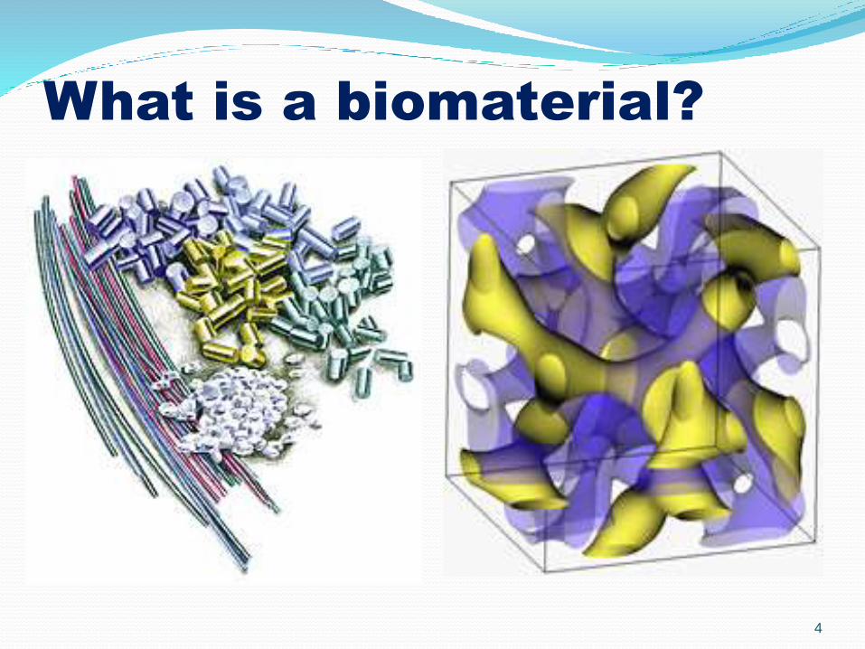
What is a biomaterial?
4
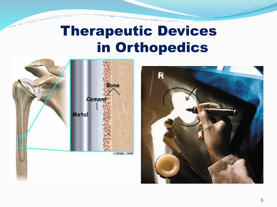
Therapeutic Devices
in Orthopedics
5
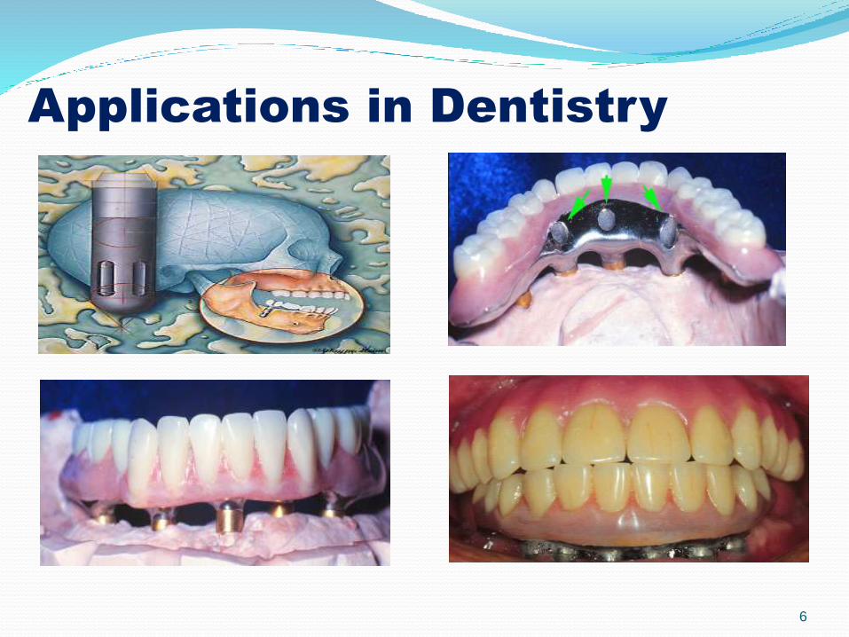
Applications in Dentistry
6
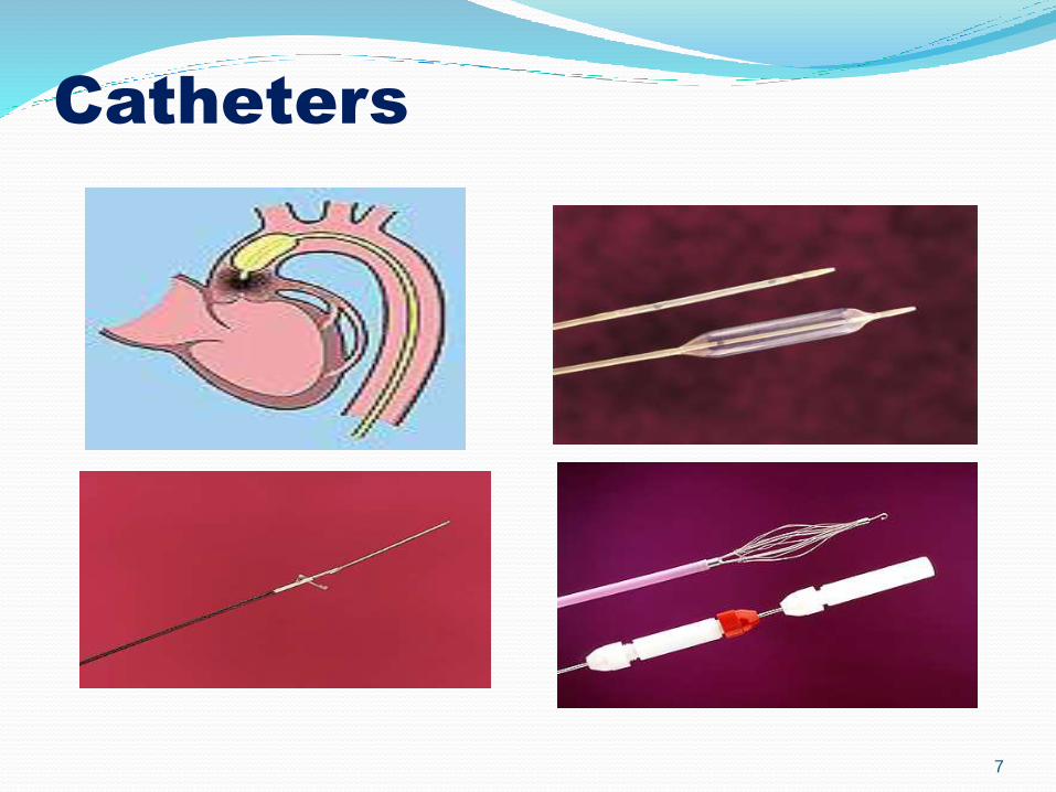
Catheters
7
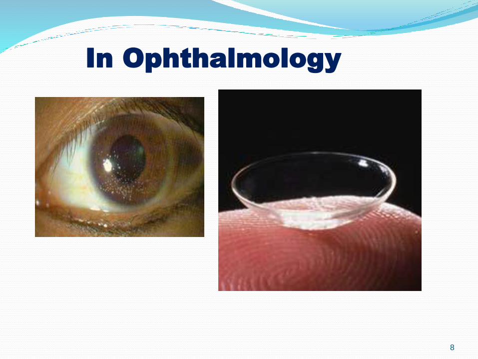
In Ophthalmology
8
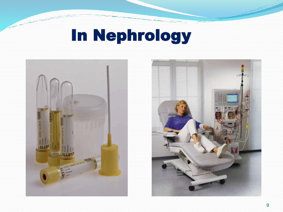
In Nephrology
9
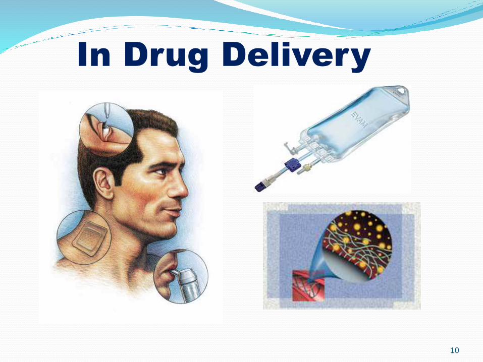
In Drug Delivery
10
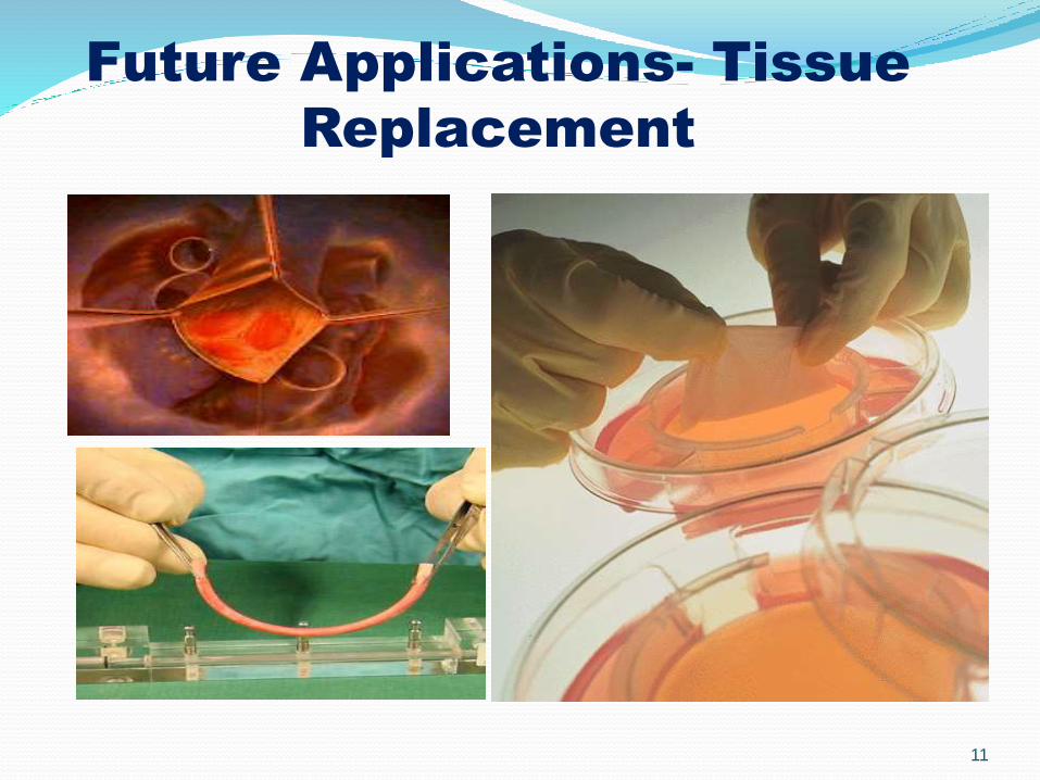
Future Applications- Tissue
Replacement
11
12/17/2014 12
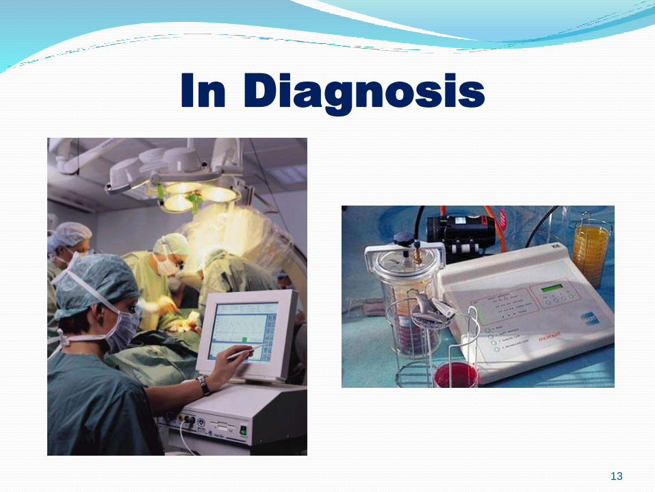
In Diagnosis
13
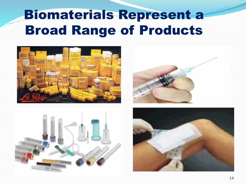
Biomaterials Represent a
Broad Range of Products
14
General Applications of
Biomaterials
Storage of fluids, tissues, and other
biological products
Diagnosis
Monitoring
Therapy
15
Biomaterials can be derived either fromnature or synthesized in the laboratory usinga variety of chemical approaches utilizingmetallic components, polymers, ceramics orcomposite materials.
They are often used and/or adapted for amedical application, and thus compriseswhole or part of a living structure orbiomedical device which performs,augments, or replaces a natural function.
Biomaterials are also used every day indental applications, surgery, and drugdelivery.
16
Cellular Measurements in Proteomics
Common methods of protein detection are:-
Immunohistology/Immunocytology
Flow Cytometry
Western Blotting
ELISA
Immunoprecipatation
17
Immunohistochemisty
IHC is a technique that uses antibodies (matchingmolecules) that can seek, identify and attachthemselves to these markers on cells. The antibodiesthemselves can be seen under microscope, whichhelps the technician to make a preciseidentification.
For example, several diseases or disease sub-typesmay look alike or appear to have similar size cellsunder a microscope but have different behavioursand necessary treatments. The best way todifferentiate them is to detect specific molecules onthese cells that act as markers.
18
Protein immunostaining
is a general term in biochemistry that applies toany use of antibody-based method to detect aspecific protein in a sample.
The term immunostaining was originally used torefer to the immunohistochemical staining oftissue sections, as first described by Albert Coonsin 1941. Now however, immunostainingencompasses a broad range of techniques used inhistology, cell biology, and molecular biology thatutilize antibody-based staining method
19
Immunochemistry - what’s
good about it?
Antibodies bind to antigen in specific manner
Gives a spatial location
Can be used to locate particular cells and proteins
Can be used to identify cellular events- e.g. apoptosis
And it can also be use in cancer diagnosis (Lymphomas).
20
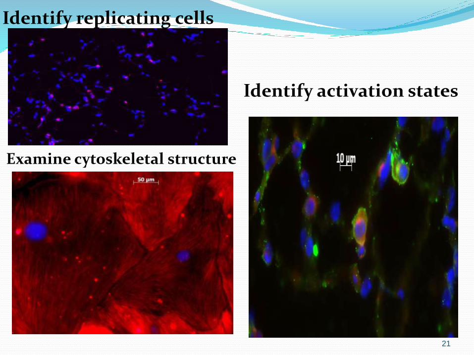
Identify replicating cells
Examine cytoskeletal structure
Identify activation states
21
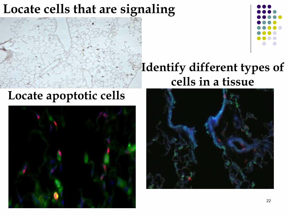
Locate cells that are signaling
Locate apoptotic cells
Identify different types of cells in a tissue
22
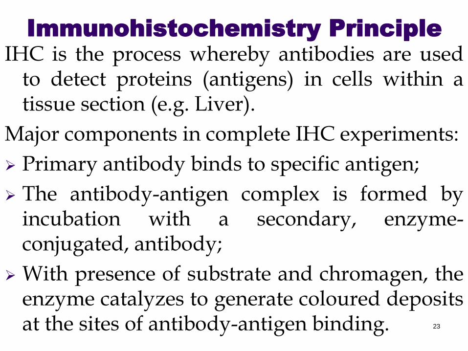
Immunohistochemistry Principle
IHC is the process whereby antibodies are usedto detect proteins (antigens) in cells within atissue section (e.g. Liver).
Major components in complete IHC experiments:
Primary antibody binds to specific antigen;
The antibody-antigen complex is formed byincubation with a secondary, enzyme-conjugated, antibody;
With presence of substrate and chromagen, theenzyme catalyzes to generate coloured depositsat the sites of antibody-antigen binding. 23
24
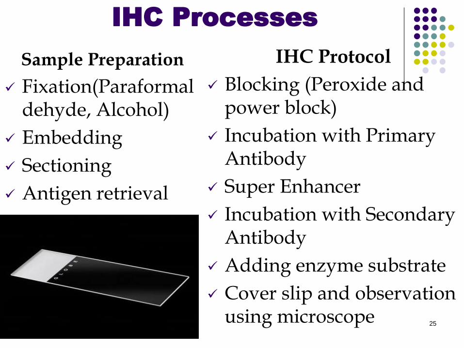
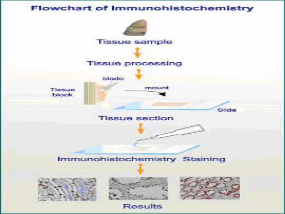
IHC Processes
Sample Preparation
Fixation(Paraformaldehyde, Alcohol)
Embedding
Sectioning
Antigen retrieval
IHC Protocol
Blocking (Peroxide and power block)
Incubation with Primary Antibody
Super Enhancer
Incubation with Secondary Antibody
Adding enzyme substrate
Cover slip and observation using microscope
25
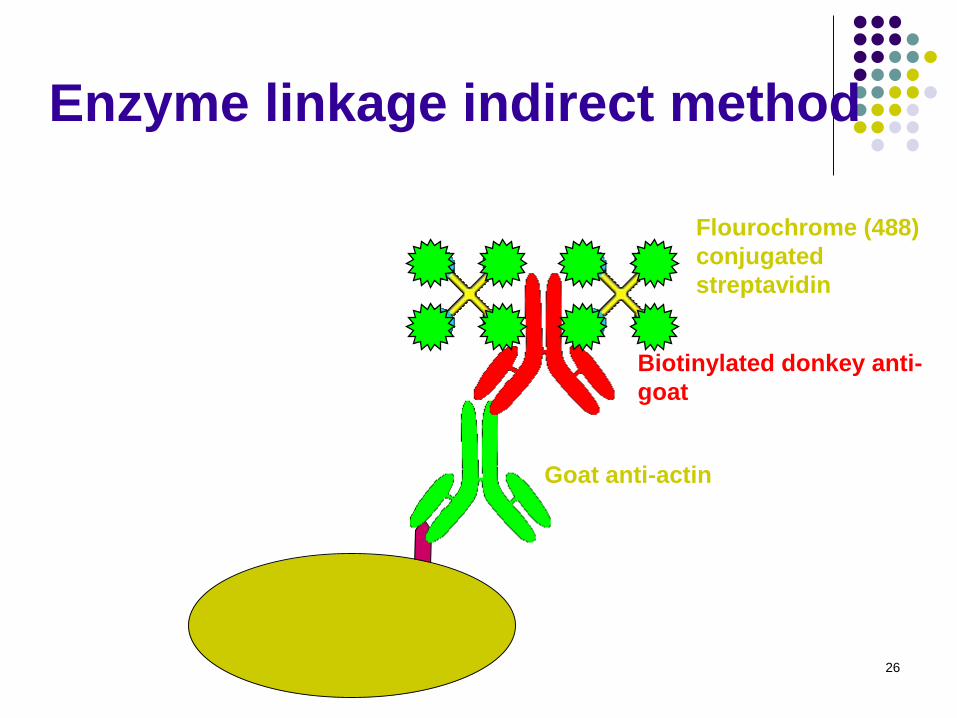
26
Goat anti-actin
Flourochrome (488)
conjugated
streptavidin
Biotinylated donkey anti-
goat
Enzyme linkage indirect method
Enzymatic detection methods
Brightfield microscope sufficient for analysis of specimens
Suitable for tissue analysis at low magnification
Resolution of subcellular structures not as good as with fluorescence methods, but can be combined with electron microscopy
Unimited shelf life of labelled specimens
Substrate reagents often toxic/carcinogenic
27
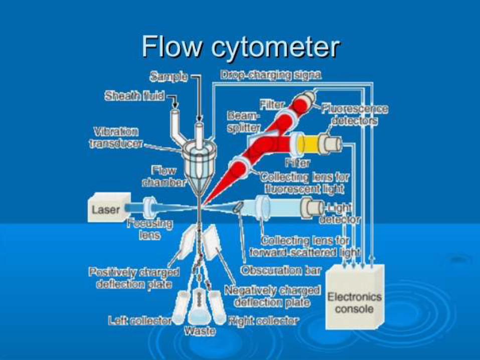
Flow cytometer is a technology that allows a single cell to be measured
for a variety of characterestics, determined by looking at how it flow in
liquid. Instruments used for this can gather information about cells by
measuring visible and flourescent light emissions, allowing cell sorting
based on physical, biochemical and antigenic traits.
It can be used for the direct analysis of cells expressing one or more
specific proteins, Cells are immunostained in solution using method
similar to the one used for immunofluorescence, and then analyzed by
flow cytometry.
Flow cytometry has several advantages over IHC including: the ability
to define distinct cell populations are defined by their size and
granularity; the capacity to get out dead cells; improved sensitivity; and
multi-colour analysis to measure several antigens simultenously.
However, flow cytometry can be less effective at detecting extremely
rare cell populations, and there is a loss of architectural relationships in
the absence of a tissue section.
Flow Cytometry
28
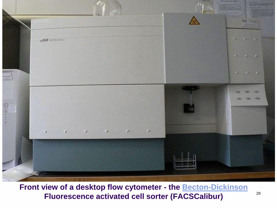
Front view of a desktop flow cytometer - the Becton-Dickinson
Fluorescence activated cell sorter (FACSCalibur)29
30
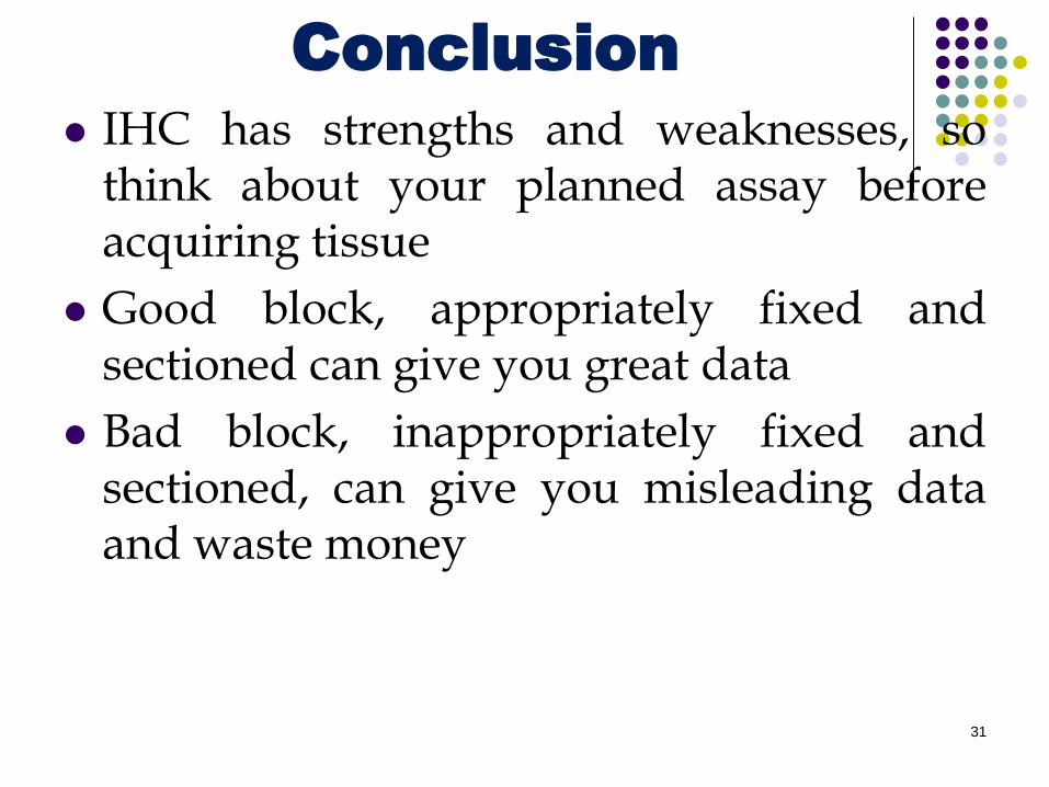
Conclusion
IHC has strengths and weaknesses, sothink about your planned assay beforeacquiring tissue
Good block, appropriately fixed andsectioned can give you great data
Bad block, inappropriately fixed andsectioned, can give you misleading dataand waste money
31
32

Questions?
Comments/Observations
33


