Abstract Water confi ned on nanometer-length scales is found in many physical and biological environments. Confi nement induces special dynamics in liquids, dif-ferent from that of their bulk counterparts. Reverse micelles, formed by the self-assembly of amphiphilic surfactants in nonpolar solvents, have emerged as an appropriate molecular assembly to monitor the property of water upon confi nement due to a number of reasons. A unique advantage of reverse micelles is that molecu-lar dynamics can be monitored with varying states of hydration that is diffi cult to achieve with assemblies, such as membranes. In this article, we focus on the change in confi ned hydration dynamics accompanied with increasing hydration, monitored by red edge excitation shift (REES). REES can be effectively used to directly moni-tor the environment and dynamics around a fl uorophore in a molecular assembly utilizing slow solvent relaxation around an excited state fl uorophore. It is apparent from the examples discussed that change in solvent relaxation with hydration is complicated and could depend on a number of factors, such as the location of the probe in the reverse micelle and the structure and compactness of the fl uorophore involved. We conclude that care should be exercised in interpreting such results.
S. Haldar • A. Chattopadhyay (*) Centre for Cellular and Molecular Biology, Council of Scientifi c and Industrial Research , Uppal Road , Hyderabad 500 007 , India e-mail: [email protected]
Hydration Dynamics of Probes and Peptides in Captivity
EGFP Enhanced green fl uorescent protein GFP Green fl uorescent protein NBD 7-Nitrobenz-2-oxa-1,3-diazol-4-yl NBD-cholesterol 25-[ N -[(7-nitrobenz-2-oxa-1,3-diazol-4-yl)-methyl]amino]-27-
norcholesterol NBD-PE N -(7-nitrobenz-2-oxa-1,3-diazol-4-yl)-1,2-dipalmitoyl- sn -
glycero-3-phosphoethanolamine REES Red edge excitation shift
1 Introduction
Although water is the most abundant liquid on the surface of the earth, it is often found in captivity [ 44 ] . There are numerous examples in geochemistry, tribology, nanofl uidics, and biology, where water is often found in confi nement. In fact, cel-lular water in biology has been referred to as tamed hydra [ 51 ] . Confi nement brings about interesting properties in bound water in terms of organization and dynamics that are very different from that experienced in bulk water [ 9, 28 ] .
Reverse micelles have emerged as an appropriate molecular assembly to monitor the property of water upon confi nement. Amphiphilic surfactants self-assemble to form reverse (or inverted) micelles in nonpolar solvents in which the polar head groups of the surfactant monomers cluster to form a micellar core and are directed toward the center of the assembly and the hydrophobic tails extend outward into the bulk organic phase [ 46, 47 ] . Reverse micelles can solubilize an appreciable amount of water to form a spherical pool in the center. They are optically transparent, nano-meter-sized water droplets of various size surrounded by a layer of surfactant mol-ecules dispersed in nonpolar solvents. Studies on reverse micellar organization and dynamics are relevant since the general principle underlying their formation (the hydrophobic effect) is common to other related organized assemblies, such as micelles, bilayers, liposomes, and biological membranes [ 37, 73– 75 ] . Reverse micelles provide an attractive model system for biomembranes since they mimic a number of important and essential features of biological membranes although lack-ing much of the complexity associated with them.
The water pools entrapped in reverse micelles have been extensively used as micromedia for chemical and biochemical reactions. The nature of water in reverse micelles, especially at low water content, has been studied extensively and is believed to be different from that of bulk water. Both experimental [ 36, 38, 79 ] and theoretical [ 25 ] approaches have shown that the crucial structural parameter of reverse micelles is [water]/[surfactant] molar ratio ( w
o ) that determines the micellar size as well as the
extent of deviation of the properties of the entrapped water from those of normal bulk water [ 53, 60 ] . Reverse micelles, therefore, represent a type of organized molecular assembly which offer the unique advantage of monitoring dynamics of molecules with varying degrees of hydration. A number of physicochemical proper-ties of the entrapped water, such as micropolarity, dielectric constant, microviscosity,
157Hydration Dynamics of Probes and Peptides in Captivity
water activity, freezing point, proton transfer effi ciency, and the hydrogen-bonding potential, can be experimentally varied with w
o , thereby providing a unique and ver-
satile reaction medium. The double-chain anionic surfactant AOT has been extensively used to form
reverse micelles in nonpolar solvents [ 21 ] . A major advantage with AOT is that reverse micelles formed by AOT can solubilize a large quantity of water in a nonpo-lar solvent without the use of any cosurfactant [ 47 ] . In addition, reverse micelles formed by AOT retain a spherical shape over a wide range of w
o . As a result of this,
the radius of the entrapped water pool can be linearly related to w o [ 24, 50 ] . Three
types of water populations (pools) have been shown to coexist in reverse micelles (see Fig. 1 ). These are bound water, trapped water, and free water [ 34, 36, 38 ] . The relative proportions of these three types of water pools are determined by w
o . The
properties of water in reverse micelles of AOT at low w o values are rather different
from those of bulk water [ 36, 38, 79 ] . Even at higher water content ( w o = 50), the
apparent microviscosity is 6–9 times greater than that of free aqueous solutions [ 5 ] . The trapped water pool is characterized by the fact that this population of water mol-ecules does not hydrogen bond to other water molecules [ 25, 38 ] . The various types
Fig. 1 ( a ) Chemical structure of AOT. ( b ) A schematic diagram of a reverse micelle showing highly structured yet heterogeneous water pools of graded dynamics: trapped water, bound water, and free water molecules. The crucial parameter is the [water]/[surfactant] molar ratio ( w
o ) which
determines the relative proportions of these three types of water pools and the micellar size. Adapted and modifi ed from Chattopadhyay [ 12 ]
158 S. Haldar and A. Chattopadhyay
of water pools in reverse micelles, characterized by graded dynamics, represent interesting models for water present in biological systems, such as membranes (see Fig. 1 ). The physical and chemical properties of the entrapped water are markedly different from the properties of bulk water but similar in several aspects to those of biological interfacial water as found in membrane or protein interfaces [ 10, 36, 38, 79 ] . For example, the dielectric constant of the aqueous phase in reverse micelles has been estimated to be ~60–70 [ 19 ] . This review is focused on the application of the wavelength-selective fl uorescence approach [ 12, 22, 23, 56, 65 ] as a powerful tool to monitor the organization and dynamics of probes and peptides inside reverse micelles.
2 Red Edge Excitation Shift
Fluorescence techniques have been widely used to characterize reverse micellar organization and dynamics due to suitable timescale, noninvasive nature, and intrinsic sensitivity [ 7, 45 ] . Reverse micelles offer certain inherent advantages in fl uorescence studies since they are small and optically transparent, have well-defi ned sizes, and are relatively scatter-free. They, therefore, represent model sys-tems appropriate for the study of probes, peptides, and proteins in membrane-mimetic, hydration-controlled environment [ 62, 72, 78 ] . Reverse micelles are highly coop-erative, organized molecular assemblies of amphiphilic surfactants and are dynamic in nature. A direct consequence of such organized systems is the restriction imposed on the dynamics and mobility of their constituent structural units. We have previ-ously shown that the microenvironment of molecules bound to such organized assemblies can be conveniently monitored using wavelength-selective fl uorescence as a novel tool [ 12 ] . Wavelength-selective fl uorescence comprises a set of approaches based on the red edge effect in fl uorescence spectroscopy, which can be used to directly monitor the environment and dynamics around a fl uorophore in a complex system [ 12, 22, 23, 56, 65, 68 ] . A shift in the wavelength of maximum fl uorescence emission toward higher wavelengths, caused by a shift in the excita-tion wavelength toward the red edge of absorption band, is termed red edge excita-tion shift (REES). This effect is mostly observed with polar fl uorophores in motionally restricted media, such as very viscous solutions or condensed phases, where the dipolar relaxation time for the solvent shell around a fl uorophore is com-parable to or longer than its fl uorescence lifetime. REES arises due to slow rates of solvent relaxation (reorientation) around an excited state fl uorophore, which depends on the motional restriction imposed on the solvent molecules [or the dipo-lar environment, as in green fl uorescent protein (GFP) ( [ 29 ] ; see later)] in the immediate vicinity of the fl uorophore. Utilizing this approach, it becomes possible to probe the mobility parameters of the environment itself (which is represented by the relaxing solvent molecules) using the fl uorophore merely as a reporter group.
159Hydration Dynamics of Probes and Peptides in Captivity
Furthermore, because the ubiquitous solvent for biological systems is water, the information obtained in such cases comes from the otherwise “optically silent” water molecules. The unique feature of REES is that while fl uorescence techniques, such as fl uorescence quenching, energy transfer, and anisotropy measurements, yield information about the fl uorophore itself (either intrinsic or extrinsic) REES provides information about the relative rates of solvent relaxation dynamics, not possible to obtain by other techniques. This makes REES in particular and the wavelength-selective fl uorescence approach in general extremely useful since hydration plays a crucial modulatory role in a large number of important cellular events, such as protein folding, lipid–protein interactions, and ion transport [ 33, 82 ] . As mentioned above, reverse micelles represent a type of organized molecular assembly which offers the unique advantage of monitoring dynamics of molecules with varying states of hydration. This feature, along with the confi nement imposed on reverse micellar water, makes reverse micelles a spatiotemporally suitable sys-tem for the application of the wavelength-selective fl uorescence approach.
3 Fluorescent Probes in Reverse Micelles: Depth-Dependent Solvent Relaxation
The applicability of the wavelength-selective fl uorescence approach to reverse micellar systems has been tested using the interfacial fl uorescence probe NBD-PE incorporated in AOT reverse micelles. The NBD group possesses some of the most desirable properties for serving as an excellent probe for both spectroscopic [ 57 ] as well as microscopic [ 61 ] applications. It is very weakly fl uorescent in water and upon transfer to hydrophobic media, it fl uoresces brightly in the visible range and shows a large degree of environmental sensitivity [ 14, 27, 43, 55 ] . This large degree of environmental sensitivity of NBD fl uorescence is very useful in monitoring vari-ous types of reverse micellar organizations formed under conditions of varying [water]/[surfactant] molar ratio. NBD-labeled lipids are widely used as fl uorescent analogues of native lipids in biological and model membranes and in membrane-mimetic assemblies to study a variety of processes [ 8, 11, 49 ] . In NBD-PE (see Figs. 2 and 3a ), the NBD group is covalently attached to the head group of a phos-phatidylethanolamine molecule. The NBD group in NBD-PE has earlier been shown to be localized in the interfacial region of the membrane ( [ 4, 13, 14, 52, 81 ] ; see Fig. 2 ) and its location in the reverse micellar environment is most likely to be inter-facial. The emission maximum of NBD-PE in AOT reverse micelles displays a pro-gressive red shift with increasing w
o , implying that the polarity inside reverse
micelles gradually increases with increasing w o . NBD-PE was found to exhibit
REES when incorporated in AOT reverse micelles [ 18 ] . Interestingly, the extent of REES was found to decrease with increasing w
o indicating that REES is sensitive to
changing hydration dynamics (see Fig. 3a ) and it is possible to detect differences in
160 S. Haldar and A. Chattopadhyay
water dynamics that is accompanied with increasing water content. The choice of a suitable probe is of considerable importance in wavelength-selective fl uorescence studies of organized molecular assemblies [ 15– 17 ] . NBD-PE is a suitable probe since the location of the NBD group in NBD-PE is expected to be interfacial, a region in the reverse micellar organization that is important from structural and functional aspects and is characterized by unique dynamics of water molecules. More importantly, we have earlier shown using solvatochromic and quantum chem-ical approaches that the dipole moment of the NBD group changes by ~4 D upon excitation [ 55 ] , an important criterion for a fl uorophore to exhibit REES effects [ 12 ] . These results show that REES is sensitive to the changing dynamic hydration profi le (Fig. 3a ) and can be conveniently used to probe dynamics of molecules in various states of hydration.
The ability of a fl uorophore incorporated in a reverse micellar assembly to exhibit red edge effects depends on a number of factors, such as its polarity, as well as the effective polarity of its immediate environment, and its fl uorescence characteristics (e.g., lifetime). Since all these properties of a probe are a function of its location in the reverse micelle, the extent of REES would depend on its location in the reverse micelle. In order to test this, we carried out REES experiments with NBD-cholesterol in which the NBD group is covalently attached to the fl exible acyl chain of the cho-lesterol molecule (see Figs. 2 and 3b ). The NBD group of this molecule has been found to be localized in the hydrocarbon region of the membrane [ 13, 14, 57, 67 ]
Fig. 2 A schematic representation of a leafl et of the membrane bilayer showing the localizations of the NBD groups of NBD-PE and NBD-cholesterol in phosphatidylcholine bilayer. The NBD group of NBD-PE localizes at the interfacial region while that of NBD-cholesterol resides at the nonpolar hydrocarbon region. These are time-averaged locations of the NBD group (in ns times-cale) and fl uctuations at shorter timescales are possible. The horizontal line at the bottom indicates the center of the bilayer. Adapted and modifi ed from Mukherjee et al. [ 58 ]
161Hydration Dynamics of Probes and Peptides in Captivity
and membrane-mimetic media, such as micelles [ 66 ] . Due to its deeper location, the NBD group of NBD-cholesterol is capable of reporting solvation dynamics in the deeper regions of the organized molecular assembly in which it is incorporated. This is important since unlike bulk solvents, compartmentalized molecular assem-blies are anisotropic in nature and therefore display differential solvent relaxation
Fig. 3 Probe depth-dependent REES in reverse micelles. Effect of increasing amounts of water on the magnitude of REES of ( a ) NBD-PE and ( b ) NBD-cholesterol in reverse micelles of AOT. The insets show the chemical structures of NBD-PE and NBD-cholesterol. Adapted and modifi ed from Chattopadhyay et al. [ 18 ] and Kelkar and Chattopadhyay [ 39 ]
162 S. Haldar and A. Chattopadhyay
rates in different regions of the assembly [ 16 ] . In our previous work, we utilized the spatial localization of NBD-cholesterol in the membrane bilayer to monitor water penetration in the deeper hydrocarbon region of the membrane [ 17 ] .
In contrast to what was observed in case of NBD-PE, the emission maximum of NBD-cholesterol in AOT reverse micelles does not exhibit any dependence on hydration ( w
o ), thereby confi rming its deeper localization in the reverse micellar
interior, a region characterized by reduced water penetration. Interestingly, REES of NBD-cholesterol incorporated in AOT reverse micelles exhibits increase with increasing water-to-surfactant ratio ( w
o ) (see Fig. 3b ) [ 39 ] . This difference in the
pattern of change of REES with w o for NBD-PE and NBD-cholesterol could possibly
be due to the deeper location of the NBD group in the reverse micellar assembly in the case of NBD-cholesterol. This implies that the rate of solvent relaxation (reori-entation) varies with probe location. We have earlier shown such depth-dependent solvent relaxation with NBD-labeled lipid probes [ 17 ] and anthroyloxy-labeled fatty acid probes [ 16 ] incorporated in membranes. The above results bring out the important point that increasing w
o in an organized anisotropic molecular assembly,
such as a reverse micelle, does not necessarily imply uniform polarity shifts in all regions of the reverse micelle. In other words, the change in polarity experienced in an anisotropic assembly is dependent on the position (location) of the incorporated probe.
To further explore depth-dependent polarity in reverse micelles, we designed experiments using the well-known series of anthroyloxy fatty acid probes. The anthroyloxy fatty acids in which an anthracene group is attached by an ester linkage to various positions of an alkyl chain have been extensively used as fl uorescent probes of micellar and bilayer structures [ 3, 16, 80 ] . The anthroyloxy fatty acids are well-designed to monitor such position-dependent effects since they locate at a graded series of depths in the bilayer (see Fig. 4a ). It has been shown that the depth of the anthroyloxy group in organized assemblies, such as a membrane bilayer, is almost linearly related to the number of carbon atoms between it and the carboxyl group [ 3, 80 ] . The fl uorescence emission maximum of the shallow interfacial probe 2-AS is found to be extremely sensitive to a change in w
o from 0 to 25 which is
accompanied by a red shift of the emission maximum by 15 nm (see Fig. 4b ). Interestingly, the sensitivity of the emission maximum of the anthroyloxy probes appears to reduce as the position (location) of the anthroyloxy group increases from the water pool. In support of this, the red shifts in emission maximum of 6- and 12-AS are reduced to 6 and 2 nm, respectively, upon changing w
o from 0 to 25.
The sensitivity of the emission maximum to hydration, therefore, appears to be dependent on the position of the fl uorophore in the reverse micelle and decreases with increasing distance of the fl uorophore from the water pool, that is, 2-AS > 6-AS > 12-AS.
163Hydration Dynamics of Probes and Peptides in Captivity
4 Peptides in Reverse Micelles: Effect of Depth Heterogeneity in Solvent Relaxation
Water has a crucial role in determining the folding, structure, dynamics, and, in turn, the function of proteins and peptides [ 20, 26, 48, 76, 83 ] . It is estimated that a threshold level of hydration (less than 0.4 grams of water per gram of protein) is required to fully activate the dynamics and function of globular proteins [ 6 ] . Knowledge of the dynamics of hydration at the molecular level is of considerable importance in understanding cellular structure and function since water plays a crucial role in the formation and maintenance of organized molecular assemblies,
Fig. 4 ( a ) A schematic diagram of a leafl et of the membrane bilayer showing the localizations of the anthroyloxy groups of 2-, 6-, and 12-AS in phosphatidylcholine bilayers. The horizontal line at the bottom indicates the center of the bilayer. Adapted and modifi ed from Chattopadhyay and Mukherjee [ 16 ] . ( b ) Effect of water on the wavelength of maximum emission of differentially localized anthroyloxy probes (2-, 6-, and 12-AS) in AOT reverse micelles. The blue bars represent emission maximum recorded at w
o = 0 and the maroon bars correspond to w
o = 25. The cyan bars
represent the difference in emission maximum ( D l max
) in these two cases. Adapted and modifi ed from Kelkar and Chattopadhyay [ 39 ]
164 S. Haldar and A. Chattopadhyay
such as proteins and membranes [ 51 ] . In particular, hydration has been shown to be a crucial parameter in protein folding and it has been suggested that water-mediated interactions could guide the folding process even before the formation of native contacts [ 59 ] . Interestingly, water has been shown to act as a catalyst for hydrogen bond exchange in protein folding, thereby acting as a “foldase” [ 83 ] . Reverse micelles represent popular assemblies for studying hydration effects on peptide and protein structure and dynamics [ 21, 46, 47 ] . Proteins trapped in reverse micelles are widely utilized in protein biotechnology [ 50 ] . As mentioned above, the entrapped water in reverse micelles has properties that are markedly different from the proper-ties of bulk water but similar in several aspects to those of biological interfacial water as found in membranes or protein interfaces [ 10, 36, 38, 79 ] . The interfacial water is crucial for the induction of secondary structure in peptides and proteins when bound to surfaces, such as membranes or micelles, as well as for variation of their local internal motion. Confi nement of a peptide chain within a restricted environ-ment has been reported to increase the relative stability of the folded state against unfolded states [ 2 ] . This observation has potential biological relevance since intrin-sically disordered proteins could be structured in their native cellular environment. In such a scenario, it becomes important to monitor the effects of hydration on the conformation and dynamics of proteins and peptides. Small peptides (such as melit-tin and gramicidin) are particularly suitable for such hydration studies due to high surface/volume ratio.
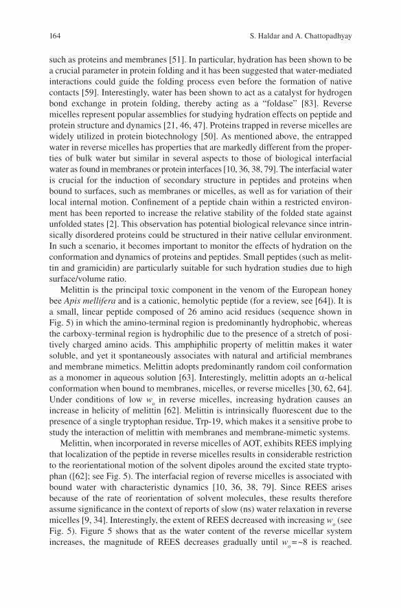
Melittin is the principal toxic component in the venom of the European honey bee Apis mellifera and is a cationic, hemolytic peptide (for a review, see [ 64 ] ). It is a small, linear peptide composed of 26 amino acid residues (sequence shown in Fig. 5 ) in which the amino-terminal region is predominantly hydrophobic, whereas the carboxy-terminal region is hydrophilic due to the presence of a stretch of posi-tively charged amino acids. This amphiphilic property of melittin makes it water soluble, and yet it spontaneously associates with natural and artifi cial membranes and membrane mimetics. Melittin adopts predominantly random coil conformation as a monomer in aqueous solution [ 63 ] . Interestingly, melittin adopts an a -helical conformation when bound to membranes, micelles, or reverse micelles [ 30, 62, 64 ] . Under conditions of low w
o in reverse micelles, increasing hydration causes an
increase in helicity of melittin [ 62 ] . Melittin is intrinsically fl uorescent due to the presence of a single tryptophan residue, Trp-19, which makes it a sensitive probe to study the interaction of melittin with membranes and membrane-mimetic systems.
Melittin, when incorporated in reverse micelles of AOT, exhibits REES implying that localization of the peptide in reverse micelles results in considerable restriction to the reorientational motion of the solvent dipoles around the excited state trypto-phan ( [ 62 ] ; see Fig. 5 ). The interfacial region of reverse micelles is associated with bound water with characteristic dynamics [ 10, 36, 38, 79 ] . Since REES arises because of the rate of reorientation of solvent molecules, these results therefore assume signifi cance in the context of reports of slow (ns) water relaxation in reverse micelles [ 9, 34 ] . Interestingly, the extent of REES decreased with increasing w
o (see
Fig. 5 ). Figure 5 shows that as the water content of the reverse micellar system increases, the magnitude of REES decreases gradually until w
o = ~8 is reached.
165Hydration Dynamics of Probes and Peptides in Captivity
At w o > 8, REES attains a more or less steady value and becomes less sensitive to
further addition of water into the system. This essentially means that there is a reor-ganization of water molecules in the reverse micellar assembly upon increasing w
o
from 0 to 8. This is in excellent agreement with earlier reports in which it was shown that water relaxation rates in reverse micelles become faster with an increase in w
o [ 69 ] . This suggests that the overall motional restriction experienced by the
reorienting solvent molecules is reduced as more water is added to the reverse micelles. Similar results have previously been obtained for amphiphilic probes, such as hemicyanine dye [ 35 ] or fl uorescent phospholipid ( [ 18 ] ; Fig. 3a ), incorpo-rated in reverse micelles.
The linear peptide gramicidin forms prototypical ion channels specifi c for mon-ovalent cations and has been extensively used to study the organization, dynamics, and function of membrane-spanning channels [ 41 ] . Gramicidin is a multitryptophan protein (Trp-9, 11, 13, and 15) which serves as an excellent model for transmem-brane channels due to its small size, ready availability, and the relative ease with which chemical modifi cations can be performed. This makes gramicidin unique among small membrane-active peptides and provides the basis for its use to explore the principles that govern the folding and function of membrane-spanning channels in particular and membrane proteins in general. The unique sequence of alternating l - and d -chirality (see Fig. 6 ) renders gramicidin sensitive to the environment in which it is placed. The head-to-head (amino terminal-to-amino terminal), single-stranded b 6.3 helical dimer form is the cation conducting channel conformation of gramicidin in membranes. In this conformation, the carboxy terminus is exposed to
Fig. 5 Hydration-dependent REES of a surface-active hemolytic peptide. Effect of increasing amounts of water on the magnitude of REES of melittin in reverse micelles of AOT. The inset shows the amino acid sequence of melittin (the sole tryptophan residue is highlighted). Adapted and modifi ed from Raghuraman and Chattopadhyay [ 62 ]
166 S. Haldar and A. Chattopadhyay
the membrane–water interface and the amino terminus is buried in the hydrophobic core of the membrane. This places the tryptophan residues clustered at the membrane–water interface at the entrance to the channel [ 42, 54 ] .
Gramicidin assumes single-stranded b 6.3 helical conformation in AOT reverse micelles and the tryptophan residues experience motional restriction and exhibit REES [ 40 ] . This implies that the tryptophans in the gramicidin single-stranded b 6.3 conformation, on the average, are localized in a motionally restricted region of the reverse micelle. Interestingly, the magnitude of REES is found to be more or less independent of w
o (see Fig. 6 ). Gramicidin is a multitryptophan peptide, and there-
fore the REES may be indicative of the average environment experienced by the tryptophans. The locations of these tryptophans would, therefore, be heterogeneous in the reverse micelle. The presence of tryptophans at various locations would con-tribute to spectral heterogeneity (and also gives rise to spectral broadening).
The overall invariance of REES with water content in the reverse micelle is sur-prising. This is because, as mentioned above, the extent of REES has generally been shown to decrease with increasing w
o for probes and peptides incorporated at the
reverse micellar interface ( [ 18, 35, 62 ] ; also see Figs. 3a and 5 ) and in the water pool [ 70 ] . This indicates that addition of water to the reverse micellar system in these cases leads to a reduction in the overall motional restriction experienced by the reorienting solvent molecules in the region of localization of the fl uorophore. However, this has been shown to be not true for probes localized in the deeper acyl
Fig. 6 Hydration-dependent REES of an ion-channel peptide. Effect of increasing amounts of water on the magnitude of REES of gramicidin in reverse micelles of AOT. The inset shows the amino acid sequence of gramicidin. Notice the unique alternating l - and d- chirality in gramicidin. Data taken from Kelkar and Chattopadhyay [ 40 ]
167Hydration Dynamics of Probes and Peptides in Captivity
chain regions of the reverse micellar assembly. Thus, in case of NBD-cholesterol in which the fl uorescent NBD moiety is positioned at a deeper acyl chain location in the reverse micellar assembly, the extent of REES increases with increasing w
o
( [ 39 ] ; see Fig. 3b ). This implies that the rate of solvent relaxation (reorientation) varies with probe location in the reverse micellar assembly. In the background of these results, the relative invariance of the magnitude of REES with increasing w
o in
case of gramicidin in AOT reverse micelles (Fig. 6 ) presents an interesting case. Gramicidin is a multitryptophan protein (Trp-9, 11, 13, and 15) and the location of these tryptophan residues would be heterogeneous in the reverse micelle. While the carboxy-terminal tryptophan (Trp-15) would occupy an interfacial position, the tryptophan residue at position 9 (Trp-9) would be placed in a relatively deep acyl chain region of the reverse micelle in the single-stranded b 6.3 conformation. The overall variation in the extent of REES with increasing w
o would then be dependent
on the average of the variations with individual tryptophans. This could explain the apparent insensitivity of the magnitude of REES to increasing w
o for gramicidin in
AOT reverse micelles.
5 “Solvent” Relaxation in Green Fluorescent Protein Incorporated in Reverse Micelles
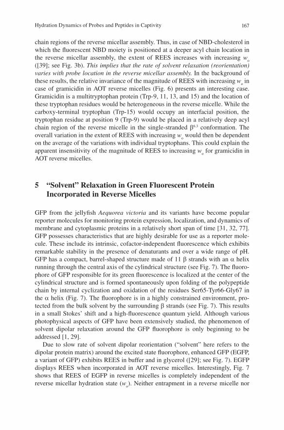
GFP from the jellyfi sh Aequorea victoria and its variants have become popular reporter molecules for monitoring protein expression, localization, and dynamics of membrane and cytoplasmic proteins in a relatively short span of time [ 31, 32, 77 ] . GFP possesses characteristics that are highly desirable for use as a reporter mole-cule. These include its intrinsic, cofactor-independent fl uorescence which exhibits remarkable stability in the presence of denaturants and over a wide range of pH. GFP has a compact, barrel-shaped structure made of 11 b strands with an a helix running through the central axis of the cylindrical structure (see Fig. 7 ). The fl uoro-phore of GFP responsible for its green fl uorescence is localized at the center of the cylindrical structure and is formed spontaneously upon folding of the polypeptide chain by internal cyclization and oxidation of the residues Ser65-Tyr66-Gly67 in the a helix (Fig. 7 ). The fl uorophore is in a highly constrained environment, pro-tected from the bulk solvent by the surrounding b strands (see Fig. 7 ). This results in a small Stokes’ shift and a high-fl uorescence quantum yield. Although various photophysical aspects of GFP have been extensively studied, the phenomenon of solvent dipolar relaxation around the GFP fl uorophore is only beginning to be addressed [ 1, 29 ] .
Due to slow rate of solvent dipolar reorientation (“solvent” here refers to the dipolar protein matrix) around the excited state fl uorophore, enhanced GFP (EGFP, a variant of GFP) exhibits REES in buffer and in glycerol ( [ 29 ] ; see Fig. 7 ). EGFP displays REES when incorporated in AOT reverse micelles. Interestingly, Fig. 7 shows that REES of EGFP in reverse micelles is completely independent of the reverse micellar hydration state ( w
o ). Neither entrapment in a reverse micelle nor
168 S. Haldar and A. Chattopadhyay
the hydration state of the reverse micelle appear to infl uence the magnitude of REES of EGFP (see Fig. 7 ). This implies that the extent of REES of EGFP is independent of the viscosity and hydration of the surrounding medium, implying that the dynam-ics of the protein matrix, rather than the dynamics of the surrounding medium, plays an important role. Interestingly, it has been previously shown by measurement of kinetics of proton transfer in EGFP that the dynamics in the interior of the protein is very weakly coupled to viscosity changes of the bulk medium [ 71 ] .
6 Conclusion
Water confi ned on nanometer-length scales is found in many physical and biologi-cal environments. As mentioned earlier, the dynamics of liquids in confi ned spaces is different from that of their bulk counterparts [ 10, 28, 44 ] , and this constitutes one of the main reasons for the popularity that reverse micelles enjoy as a model system in studies of water dynamics. The highly structured yet heterogeneous water mole-cules in reverse micelles represent interesting models for water molecules present in biological systems, such as membranes, which are more diffi cult to analyze experi-mentally. Moreover, the dimension, shape, and overall charge of reverse micelles
Fig. 7 Hydration-independent REES of a green fl uorescent protein (GFP). Effect of increasing amounts of water on the magnitude of REES of EGFP in reverse micelles of AOT. The inset shows the GFP fl uorophore ( p -hydroxybenzylideneimidozolidinone) and the b -barrel structure of GFP. It turns out that the observed REES of EGFP is due to the constrained environment experienced by the EGFP fl uorophore in the rigid protein matrix, rather than due to the dynamics of the host reverse micellar assembly. Adapted and modifi ed from Haldar and Chattopadhyay [ 29 ]
169Hydration Dynamics of Probes and Peptides in Captivity
can be conveniently modulated which make them particularly useful for monitoring dynamics of confi ned liquids. The interior dimensions of reverse micelles are believed to be similar to confi ned spaces found in cavities in biomolecules. These nanopockets of water are thought to be involved in folding and relaxation in pro-teins. In addition, reverse micellar structures are proposed to be formed during endosome formation [ 44 ] .
The focus of this article is the change in confi ned hydration dynamics with increasing hydration, monitored by REES. We discuss here that while solvent relax-ation for probes and peptides incorporated in reverse micelles is generally depen-dent on the extent of hydration ( w
o ), this effect is dependent on the position (location)
of the probe in the reverse micellar assembly. For molecules with multiple fl uoro-phores at various locations (such as gramicidin), positional heterogeneity could complicate the observed hydration effects. An interesting case is presented by EGFP, where the extent of REES appears to be independent of hydration, since the dynamics of the protein matrix is the important determinant in such a case. We con-clude that change in solvent relaxation with hydration could be context dependent and care should be exercised in interpreting such results.
Acknowledgments Work in A.C.’s laboratory was supported by the Council of Scientifi c and Industrial Research and Department of Science and Technology, Government of India. S.H. thanks the Council of Scientifi c and Industrial Research for the award of a Senior Research Fellowship. A.C. is an Adjunct Professor at the Special Centre for Molecular Medicine of Jawaharlal Nehru University (New Delhi, India) and Honorary Professor of the Jawaharlal Nehru Centre for Advanced Scientifi c Research (Bangalore, India). A.C. gratefully acknowledges J.C. Bose Fellowship (Department of Science and Technology, Government of India). Some of the work described in this article was carried out by former members of A.C.’s group whose contributions are gratefully acknowledged. We thank Arunima Chaudhuri for help with Fig. 1 and members of our laboratory for critically reading the manuscript.
References
1. Abbyad P, Childs W, Shi X, Boxer SG (2007) Dynamic Stokes shift in green fl uorescent protein variants. Proc Natl Acad Sci USA 104:20189–20194
2. Abel S, Waks M, Urbach W, Marchi M (2006) Structure, stability, and hydration of a polypeptide in AOT reverse micelles. J Am Chem Soc 128:382–383
3. Abrams FS, Chattopadhyay A, London E (1992) Determination of the location of fl uorescent probes attached to fatty acids using the parallax analysis of fl uorescence quenching: effect of carboxyl ionization state and environment on depth. Biochemistry 31:5322–5327
4. Abrams FS, London E (1993) Extension of the parallax analysis of membrane penetration depth to the polar region of model membranes: use of fl uorescence quenching by a spin-label attached to the phospholipid polar headgroup. Biochemistry 32:10826–10831
5. Andrade SM, Costa SM, Pansu R (2000) The infl uence of water on the photophysical and photochemical properties of Piroxicam in AOT/iso-octane/water reversed micelles. Photochem Photobiol 71:405–412
6. Bizzarri AR, Cannistraro S (2002) Molecular dynamics of water at the protein–solvent inter-face. J Phys Chem B 106:6617–6633
170 S. Haldar and A. Chattopadhyay
7. Behera GB, Mishra BK, Behera PK, Panda M (1999) Fluorescent probes for structural and distance effect studies in micelles, reversed micelles and microemulsions. Adv Colloid Interface Sci 82:1–42
8. Bertorelle F, Dondon R, Fery-Forgues S (2002) Compared behavior of hydrophobic fl uores-cent NBD probes in micelles and in cyclodextrins. J Fluoresc 12:205–207
9. Bhattacharyya K, Bagchi B (2000) Slow dynamics of constrained water in complex geome-tries. J Phys Chem A 104:10603–10613
10. Brubach J-B, Mermet A, Filabozzi A, Gerschel A, Lairez D, Krafft MP, Roy P (2001) Dependence of water dynamics upon confi nement size. J Phys Chem B 105:430–435
11. Chattopadhyay A (1990) Chemistry and biology of N-(7-nitrobenz-2-oxa-1,3-diazol-4-yl)-labeled lipids: fl uorescent probes of biological and model membranes. Chem Phys Lipids 53:1–15
12. Chattopadhyay A (2003) Exploring membrane organization and dynamics by the wavelength-selective fl uorescence approach. Chem Phys Lipids 122:3–17
13. Chattopadhyay A, London E (1987) Parallax method for direct measurement of membrane penetration depth utilizing fl uorescence quenching by spin-labeled phospholipids. Biochemistry 26:39–45
14. Chattopadhyay A, London E (1988) Spectroscopic and ionization properties of N -(7-nitrobenz-2-oxa-1,3-diazol-4-yl)-labeled lipids in model membranes. Biochim Biophys Acta 938: 24–34
15. Chattopadhyay A, Mukherjee S (1993) Fluorophore environments in membrane-bound probes: a red edge excitation shift study. Biochemistry 32:3804–3811
16. Chattopadhyay A, Mukherjee S (1999) Depth-dependent solvent relaxation in membranes: wavelength-selective fl uorescence as a membrane dipstick. Langmuir 15:2142–2148
17. Chattopadhyay A, Mukherjee S (1999) Red edge excitation shift of a deeply embedded mem-brane probe: implications in water penetration in the bilayer. J Phys Chem B 103:8180–8185
18. Chattopadhyay A, Mukherjee S, Raghuraman H (2002) Reverse micellar organization and dynamics: a wavelength-selective fl uorescence approach. J Phys Chem B 106:13002–13009
19. Cohen B, Huppert D, Solntsev KM, Tsfadia Y, Nachliel E, Gutman M (2002) Excited state proton transfer in reverse micelles. J Am Chem Soc 124:7539–7547
20. Colombo MF, Rau DC, Parsegian VA (1992) Protein solvation in allosteric regulation: a water effect on hemoglobin. Science 256:655–659
21. De TK, Maitra A (1995) Solution behaviour of aerosol OT in non-polar solvents. Adv Colloid Interface Sci 59:95–193
22. Demchenko AP (2002) The red-edge effects: 30 years of exploration. Luminescence 17: 19–42
23. Demchenko AP (2008) Site-selective red-edge effects. Methods Enzymol 450:59–78 24. Eastoe J, Young WK, Robinson BH, Steytier DC (1990) Scattering studies of microemulsions
in low-density alkanes. J Chem So Faraday Trans 86:2883–2889 25. Faeder J, Ladanyi BM (2000) Molecular dynamics simulations of the interior of aqueous
dominate protein dynamics and function. Proc Natl Acad Sci USA 99:16047–16051 27. Fery-Forgues S, Fayet J-P, Lopez A (1993) Drastic changes in the fl uorescence properties of
NBD probes with the polarity of the medium: involvement of a TICT state? J Photochem Photobiol A 70:229–243
28. Granick S (1991) Motions and relaxations of confi ned liquids. Science 253:1374–1379 29. Haldar S, Chattopadhyay A (2007) Dipolar relaxation within the protein matrix of the green
fl uorescent protein: a red edge excitation shift study. J Phys Chem B 111:14436–14439 30. Haldar S, Raghuraman H, Chattopadhyay A (2008) Monitoring orientation and dynamics of
membrane-bound melittin utilizing dansyl fl uorescence. J Phys Chem B 112:14075–14082 31. Haldar S, Chattopadhyay A (2009) The green journey. J Fluoresc 19:1–2 32. Haldar S, Chattopadhyay A (2009) Green fl uorescent protein: a molecular lantern that illumi-
nates the cellular interior. J Biosci 34:169–172 33. Häussinger D (1996) The role of cellular hydration in the regulation of cell function. Biochem
J 313:697–710
171Hydration Dynamics of Probes and Peptides in Captivity
34. Hazra P, Sarkar N (2001) Intramolecular charge transfer processes and solvation dynamics of coumarin 490 in reverse micelles. Chem Phys Lett 342:303–311
35. Hof M, Lianos P, Laschewsky A (1997) An amphiphilic hemicyanine dye employed as a sensi-tive probe of water in reverse AOT micelles. Langmuir 13:2181–2183
36. Ikushima Y, Saito N, Arai M (1997) The nature and structure of water/AOT/ethane (w/o) microemulsion under supercritical conditions studied by high-pressure FT-IR spectroscopy. J Colloid Interface Sci 186:254–263
38. Jain TK, Varshney M, Maitra A (1989) Structural studies of Aerosol OT reverse micellar aggregates by FT-IR spectroscopy. J Phys Chem 93:7409–7416
39. Kelkar DA, Chattopadhyay A (2004) Depth-dependent solvent relaxation in reverse micelles: a fl uorescence approach. J Phys Chem B 108:12151–12158
40. Kelkar DA, Chattopadhyay A (2005) Effect of graded hydration on the dynamics of an ion channel peptide: a fl uorescence approach. Biophys J 88:1070–1080
41. Kelkar DA, Chattopadhyay A (2007) The gramicidin ion channel: a model membrane protein. Biochim Biophys Acta 1768:2011–2025
42. Ketchem RR, Hu W, Cross TA (1993) High-resolution conformation of gramicidin A in a lipid bilayer by solid-state NMR. Science 261:1457–1460
43. Lin S, Struve WS (1991) Time-resolved fl uorescence of nitrobenzoxadiazole-aminohexanoic acid: effect of intermolecular hydrogen-bonding on non-radiative decay. Photochem Photobiol 54:361–365
44. Levinger NE (2002) Water in confi nement. Science 298:1722–1723 45. Levinger NE, Swafford LA (2009) Ultrafast dynamics in reverse micelles. Annu Rev Phys
Chem 60:385–406 46. Luisi PL, Magid LJ (1986) Solubilization of enzymes and nucleic acids in hydrocarbon micel-
lar solutions. CRC Crit Rev Biochem 20:409–473 47. Luisi PL, Giomini M, Pileni MP, Robinson BH (1988) Reverse micelles as hosts for proteins
and small molecules. Biochim Biophys Acta 947:209–246 48. Mattos C (2002) Protein-water interactions in a dynamic world. Trends Biochem Sci
labeled phospholipids in lipid membranes: differences in fl uorescence behavior. Biophys J 71: 327–335
50. Melo EP, Aires-Barros MR, Cabral JMS (2001) Reverse micelles and protein biotechnology. Biotechnol Annu Rev 7:87–129
51. Mentré P (2001) An introduction to “water in the cell”: tamed hydra? Cell Mol Biol 47: 709–715
52. Mitra B, Hammes GG (1990) Membrane-protein structural mapping of chloroplast coupling factor in asolectin vesicles. Biochemistry 29:9879–9884
53. Moilanen DE, Fenn EE, Wong D, Fayer MD (2009) Geometry and nanolength scales versus interface interactions: water dynamics in AOT lamellar structures and reverse micelles. J Am Chem Soc 131:8318–8328
54. Mukherjee S, Chattopadhyay A (1994) Motionally restricted tryptophan environments at the peptide–lipid interface of gramicidin channels. Biochemistry 33:5089–5097
55. Mukherjee S, Chattopadhyay A, Samanta A, Soujanya T (1994) Dipole moment change of NBD group upon excitation studied using solvatochromic and quantum chemical approaches: implications in membrane research. J Phys Chem 98:2809–2812
56. Mukherjee S, Chattopadhyay A (1995) Wavelength-selective fl uorescence as a novel tool to study organization and dynamics in complex biological systems. J Fluoresc 5:237–246
57. Mukherjee S, Chattopadhyay A (1996) Membrane organization at low cholesterol concentrations: a study using 7-nitrobenz-2-oxa-1,3-diazol-4-yl-labeled cholesterol. Biochemistry 35:1311–1322
58. Mukherjee S, Kalipatnapu S, Pucadyil TJ, Chattopadhyay A (2006) Monitoring the organization and dynamics of bovine hippocampal membranes utilizing differentially localized fl uorescent membrane probes. Mol Membr Biol 23:430–441
172 S. Haldar and A. Chattopadhyay
59. Papoian GA, Ulander J, Eastwood MP, Luthey-Schulten Z, Wolynes PG (2004) Water in protein structure prediction. Proc Natl Acad Sci USA 101:3352–3357
60. Park S, Moilanen DE, Fayer MD (2008) Water dynamics – the effects of ions and nanoconfi ne-ment. J Phys Chem B 112:5279–5290
61. Pucadyil TJ, Mukherjee S, Chattopadhyay A (2007) Organization and dynamics of NBD-labeled lipids in membranes analyzed by fl uorescence recovery after photobleaching. J Phys Chem B 111:1975–1983
62. Raghuraman H, Chattopadhyay A (2003) Organization and dynamics of melittin in environ-ments of graded hydration: a fl uorescence approach. Langmuir 19:10332–10341
63. Raghuraman H, Chattopadhyay A (2006) Effect of ionic strength on folding and aggregation of the hemolytic peptide melittin in solution. Biopolymers 83:111–121
64. Raghuraman H, Chattopadhyay A (2007) Melittin: a membrane-active peptide with diverse functions. Biosci Rep 27:189–223
65. Raghuraman H, Kelkar DA, Chattopadhyay A (2005) Novel insights into protein structure and dynamics utilizing the red edge excitation shift approach. In: Geddes CD, Lakowicz JR (eds) Reviews in fl uorescence, vol 2. Springer, New York, pp 199–214
66. Rawat SS, Chattopadhyay A (1999) Structural transition in the micellar assembly: a fl uores-cence study. J Fluoresc 9:233–244
67. Rukmini R, Rawat SS, Biswas SC, Chattopadhyay A (2001) Cholesterol organization in mem-branes at low concentrations: effects of curvature stress and membrane thickness. Biophys J 81:2122–2134
68. Samanta A (2006) Dynamic Stokes shift and excitation wavelength dependent fl uorescence of dipolar molecules in room temperature ionic liquids. J Phys Chem B 110:13704–13716
69. Sarkar N, Das K, Datta A, Das S, Bhattacharyya K (1996) Solvation dynamics of coumarin 480 in reverse micelles. Slow relaxation of water molecules. J Phys Chem 100:10523–10527
70. Sarkar M, Ray JG, Sengupta PK (1996) Luminescence behaviour of 7-hydroxyfl avone in aero-sol OT reverse micelles: excited-state proton transfer and red-edge excitation effects. J Photochem Photobiol A 95:157–160
71. Saxena AM, Udgaonkar JB, Krishnamoorthy G (2005) Protein dynamics control proton trans-fer from bulk solvent to protein interior: a case study with a green fl uorescent protein. Protein Sci 14:1787–1789
72. Souto AL, Ito AS (2000) Tryptophan fl uorescence studies of melanotropins in the amphiphile-water interface of reversed micelles. Eur Biophys J 29:38–47
73. Tanford C (1978) The hydrophobic effect and the organization of living matter. Science 200:1012–1018
74. Tanford C (1980) The hydrophobic effect: formation of biological membranes. Wiley, New York
75. Tanford C (1987) Amphiphile orientation: physical chemistry and biological function. Biochem Soc Trans 15:1S–7S
76. Timasheff SN (2002) Protein hydration, thermodynamic binding and preferential hydration. Biochemistry 41:13473–13482
77. Tsien RY (1998) The green fl uorescent protein. Annu Rev Biochem 67:509–544 78. Valdez D, Le Huérou J-Y, Gindre M, Urbach W, Waks M (2001) Hydration and protein folding
in water and in reverse micelles: compressibility and volume changes. Biophys J 80:2751–2760 79. Venables DS, Huang K, Schmuttenmaer CA (2001) Effect of reverse micelle size on the libra-
tional band of confi ned water and methanol. J Phys Chem B 105:9132–9138 80. Villaín J, Prieto M (1991) Location and interaction of N-(9-anthroyloxy)-stearic acid probes
incorporated in phosphatidylcholine vesicles. Chem Phys Lipids 59:9–16 81. Wolf DE, Winiski AP, Ting AE, Bocian KM, Pagano RE (1992) Determination of the transbi-
layer distribution of fl uorescent lipid analogues by nonradiative fl uorescence energy transfer. Biochemistry 31:2865–2873
82. Wyttenbach T, Bowers MT (2009) Hydration of biomolecules. Chem Phys Lett 480:1–16 83. Xu F, Cross TA (1999) Water: foldase activity in catalyzing polypeptide conformational rear-