Page 1
1
Hydrogel-forming microneedle arrays made from stimuli-responsive materials for on-
demand drug delivery.
John G. Hardy*, Eneko Larrañeta*, Ryan F. Donnelly, Niamh McGoldrick, Katarzyna
Migalska, Maelíosa T.C. McCrudden, Louise Donnelly and Colin P. McCoy**.
School of Pharmacy, Queens University Belfast, Medical Biology Centre, 97 Lisburn Road,
Belfast BT9 7BL, Northern Ireland, UK.
*First two authors contributed equally
**Corresponding author
Professor Colin P. McCoy
Chair in Biomaterials Chemistry
School of Pharmacy,
Queens University Belfast,
Medical Biology Centre,
97 Lisburn Road,
Belfast
BT9 7BL, UK
Tel: +44 (0) 28 9097 2081
Fax: +44 (0) 28 9024 7794
Email: [email protected]
Page 2
2
Abstract
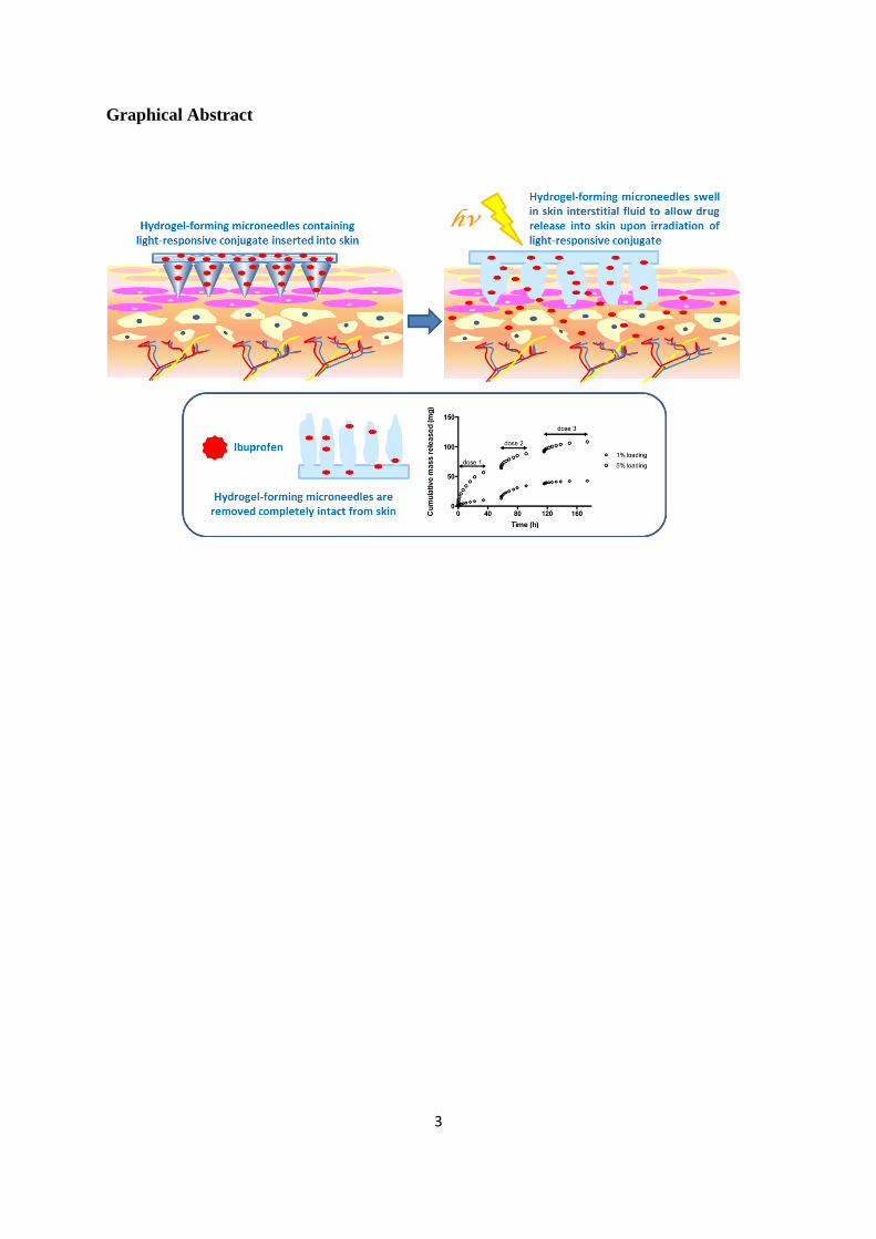
We describe, for the first time, stimuli-responsive hydrogel-forming microneedle (MN)
arrays that enable delivery of a clinically-relevant model drug (ibuprofen) upon application of
light. MN arrays where prepared using a polymer prepared from 2-hydroxyethyl methacrylate
(HEMA) and ethylene glycol dimethacrylate (EGDMA) by micromolding. The obtained MN
arrays showed good mechanical properties. The system was loaded with up to 5% (w/w)
ibuprofen included in a light-responsive 3,5-dimethoxybenzoin conjugate. Raman
spectroscopy confirmed the presence of the conjugate inside the polymeric MN matrix. In
vitro, this system was able to deliver up to three doses of 50 mg of ibuprofen upon
application of an optical trigger over a prolonged period of time (up to 160 hours). This
makes the system appealing as a controlled release system for prolonged periods of time. We
believe that this technology has potential for use in “on-demand” delivery of a wide range of
drugs in a variety of applications relevant to enhanced patient care.
Keywords: Microneedle, hydrogel, stimuli-responsive, light-triggered
Page 3
3
Graphical Abstract
Page 4
4
Introduction
Microneedle arrays (MN) are minimally-invasive devices that painlessly, and without
drawing blood, penetrate the skin’s stratum corneum barrier (1-8). MN have been extensively
investigated in recent years for enhanced transdermal and intradermal delivery of vaccines
and drug substances (1, 9-14). MN have generally been employed for delivery of bolus doses
in relatively short periods of times, in contrast to conventional transdermal patches, which are
used for sustained drug delivery, often over several days. Nevertheless we have recently
described hydrogel-forming MN that rapidly imbibe skin interstitial fluid upon insertion to
form discrete in situ hydrogel bulbs, which then control drug administration. In this case,
release from the swollen MN is determined by the crosslink density of the swollen hydrogel
network formed and delivery over several days is possible (15-20). In other work, MN have
been combined with iontophoresis (the application of a small electric current, typically ≤ 0.5
mA / cm2, to drive ionic and polar molecules across the skin) to facilitate enhanced rates of
drug delivery across skin pre-punctured with MN (21-25). Most studies involve pre-treating
skin with MN puncture and then application of an iontophoretic patch, which is not
particularly convenient. However, iontophoresis set-ups (electrodes, power source, controller)
are typically complex, expensive and quite bulky. Accordingly, very few iontophoretic
patches are on the market currently (13), with those that are being very much niche products.
As a result, it is difficult to imagine even more complex MN-iontophoresis combination
products being a priority for development by industry. If “on-demand” or pulsatile delivery
from a MN system is to be developed into a patient-friendly, low cost product, an external
stimulus for drug delivery from an otherwise benign delivery system would be very useful. In
order to address the lack of “on demand” transdermal delivery systems some research groups
are starting to develop MN-based stimulus responsive delivery systems (26-28).
Page 5
5
In combination with our previous works dealing developing hydrogel-forming MN arrays we
have previously developed materials impregnated with light-responsive drug conjugates (29,
30). Such conjugates are able to latently retain drug until light is applied, and the dose of drug
liberated can be precisely controlled by the total energy applied, in line with the kinetics of
photolysis of the conjugate. Therefore in this study, we investigated the combination of these
two technologies to develop hydrogel-based MN arrays containing a light-responsive drug
conjugate for light-triggered transdermal drug delivery.
Materials and Methods
Chemicals
Ibuprofen, dichloromethane, dimethylaminopyridine (DMAP), dicyclohexylcarboiimide
(DCC), acetone, hexane, 2-hydroxyethylmethacrylate, ethylene glycol dimethacrylate, benzyl
peroxide, phosphate buffer tablets, silica and silica plates were all obtained from Aldrich,
Gillingham, Dorset, UK. Dichloromethane (DCM), ethanol (EtOH), ethyl acetate (EtOAc),
hexane, acetonitrile and acetone were all obtained from BDH Laboratories, Poole, Dorset,
UK. All other chemicals used were of analytical reagent grade.
Synthesis and characterisation of light-responsive ibuprofen conjugates
2-(3,5-dimethoxyphenyl)-2-hydroxy-1-phenylethanone (3,5-dimethoxybenzoin, 1.00 g, 3.67
mmol) and ibuprofen (0.76 g, 3.67 mmol) were added to dried dichloromethane (15 ml), to
which 4-dimethylaminopyridine (DMAP) (0.03g, 0.2mmol) was added. The solution
temperature was maintained at 0 oC using an ice bath. N,N'-dicyclohexylcarbodiimide (DCC)
(0.76 g, 3.67 mmol) was added to the solution. The solution was then stirred for 24 hrs at 0
Page 6
6
oC, with a drying tube to prevent any water entering the solution. After 24 hrs the
dicyclohexylurea (DCU) by-product was removed by filtration and the volatiles removed
under vacuum. The product was purified by flash chromatography on a silica gel column
using ethyl acetate:hexane (3:2 v/v) as the eluent. The product, a light-responsive ibuprofen
conjugate was a viscous yellow oil (1.29 g, 73 %, 2.80 mmol). (CDCl3): 7.84-7.87 (Ar-1-2-
H9, s, dd, d, J = 6.25 Hz), 7.42 ( Ar-1-2 – H11, m ), 7.30-7.34 (Ar-1- H10, m ), 7.20-7.25 ( Ar-
3- H5, m), 7.14-7.19( Ar-3-, H3, dd, J = 5.0), 6.75 (H14, d, J = 1.9 Hz), 6.5-6.55 ( Ar- 1-2-
H8, dd, J = 1.0 Hz), 6.32-6.39 ( Ar-2- H13, m), 4.12-4.18 (H7 –CH, m), 3.71-3.79 (Ar-2-
H12 -OCH3 d, J = 1.0 Hz), 2.41-2.48 (H4 - CH2, m), 1.82-1.87 (H2 – CH, m), 1.48-1.59 (H6 -
CH3 , m ), 0.75-0.82 (H1 – CH3, m J = 5.0 Hz). IR max/cm-1
(KBr): 2945 methine group
(CH), 1670 ketone group (R-C=O-R), 1730 ester (R-C=O-OR). m/z(%): 461.2 (M+, 59),
255.3 (434), 920.8 (100), 919.3 (24), 942.9 (12), 413.4 (9), 212.3 (3), 227.3 (8), 256.3(7).
Elemental analysis: C29H32O5 requires: C 75.63%, H 7%, O 17.37%. Found: C 75.62%, H
7.01%, O 17.35%.
Preparation and physicochemical characterisation of drug loaded MN arrays
Laser-engineered silicone micromould templates were used in micromoulding of MN arrays
and were microfabricated using a previously-reported approach (31). The moulds were
composed of 9 (3x3) or 121 (11x11) needles perpendicular to the base, of conical shape and
600 μm in height, with base width of 300 μm and interspacing of 300 μm. 2-hydroxyethyl
methacrylate (HEMA, 98.6% w/w), ethylene glycol dimethacrylate (EGDMA, 1% w/w) and
BPO (0.4%, w/w) and the light-responsive ibuprofen conjugate (1 to 5% w/w) were mixed
with a magnetic stirrer until all of initiator was dissolved. MN arrays were prepared only
with the HEMA polymer to evaluate the ability of this type of polymer to form MN. For this
Page 7
7
purpose MN moulds were filled to the top with the HEMA, EGDMA and BPO solution (0.5
g), centrifuged at 3000 rpm, refilled to replace part of the evaporated solution and placed in
the oven at 90 oC for two hours.
Additionally swelling kinetics of hydrogels obtained by crosslinking HEMA (pHEMA) was
evaluated. Hydrogels squares (0.81 ± 0.05 g; 12.6 ± 0.4 x 16.6 ± 0.4 x 5.2 ± 0.2 mm) were
weighed as mo and then swollen in 50 mL of pH 7 phosphate buffer solution (PBS) for 24 h at
room temperature. At regular intervals, the films were removed, dried with filter paper to
eliminate excess surface water and weighed as mt (hydrogels). The percentage swelling, was
calculated, respectively, by using Equation 1.
% Swelling =100 x (mt – mo) / mo (1)
In order to prepare MN for mechanical testing and drug delivery, the polymer mixture was
poured into the laser drilled 3x3 or 11x11 silicon moulds at weight of 0.5 g per array, the
moulds were then centrifuged for 10 minutes to remove any air bubbles at 3000 rpm and then
placed in the oven at 90 oC for two hours, the MN moulds were removed from the oven,
allowed to cool and then the MNs were removed from the moulds. The “sidewalls” formed
by the moulding process were removed using a heated blade, as described previously (31). It
is important that these moulds remain covered to protect them from light. Uniformity with
regards to height and width of each batch of MN arrays was examined by a digital
microscope under the magnification 180x, using the ruler function of the microscope
software.
Page 8
8
Raman spectroscopy was carried out using on hydrated 1 cm2 squares of pHEMA and on
pHEMA incorporated with light responsive conjugate. Raman spectra and maps were
obtained using an Avalon Instruments Raman Station R3 coupled to an Olympus BX 50
microscope. Samples were placed on a microscope slide and Raman scattered light from a
785nm laser operating at 300mW focused on the surface of the sample was collected between
400-3200cm-1
at a resolution of 2 cm-1
, and with a total collection time of 120 s.
MN characterisation
Formed MN arrays were visualised using a Keyence VHX-700F Digital Microscope
equipped with a VH-Z20R lens (Keyence, Osaka, Japan), which allows automatic sizing of
multiple MN, facilitating quality control in terms of efficiency and reproducibility of MN
formation within and between individual arrays. Additionally needle dimensions where
evaluated using an Aigo Digital Viewer GE-5 (Aigo, Beijing, China). MN arrays were also
subjected to compression tests in order to ascertain their mechanical strength. Mechanical
properties were evaluated using a TA-XT2 Texture Analyser (Stable Microsystems,
Haslemere, UK) in compression mode, as described previously [16]. MN arrays were
visualised before and after application of the compression load using a desktop light
microscope (GXMGE-5 digital microscope, Laboratory Analysis Ltd., Devon, UK).
In vitro drug delivery studies
These experiments were performed with a system employing Franz-diffusion cells. pHEMA
MNs with 1% or 5% loading of the photoresponsive drug conjugate were manufactured as
detailed in MN preparation methodology. The MNs were then punctured through a synthetic
skin membrane. At this stage they were visualised under a microscope to check no damage
had occurred to the MNs and that they had all punctured through the synthetic membrane.
Page 9
9
The MN arrays were then irradiated with a 15W Hg discharge UV lamp source at 365 nm at a
fixed distance of 10 mm from the light source for 1 hr. At the 1 hr time point they were then
attached to the receptor compartment of the Franz-diffusion cells, and the receptor phase
volume was recorded. The surface area of the MN array exposed to the receptor compartment
was 2.96 cm2. The receptor medium was PBS. A measured volume of receptor solution (3
ml) was added to each receptor compartment with a magnetic stirring bead. The diffusion
cells were placed on a magnetic stirring plate in a water bath kept at 36 ºC, resulting in skin
temperature at the membrane surface of approximately 32 ºC during the experiment. Using a
needle, samples (3 ml) were collected from the receptor compartment at defined time
intervals and were immediately refilled by fresh receptor solution. These samples were then
assayed by UV spectroscopy at a wavelength of 365 nm. The experiment included 3
replicates of pHEMA arrays (3x3 and 11x11 MN arrays) loaded with the photoresponsive
drug conjugate and 3 replicates of pHEMA arrays without any photoresponsive drug
conjugate as the experimental control. Repeated sampling occurred over a 24 hr time frame,
with repeated 1hour irradiations.
Statistical analysis
Where appropriate, data was analyzed using a one-way ANOVA with post-hoc comparisons
performed using the Tukey-Kramer test. In all cases, p < 0.05 denoted significance. Statistical
Package for the Social Sciences, SPSS 18.0 version 2.0 (SPSS, Inc., Chicago, IL, USA), was
used for all analyses.
Results
Page 10
10
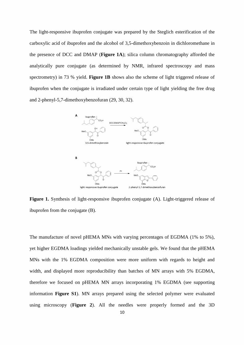
The light-responsive ibuprofen conjugate was prepared by the Steglich esterification of the
carboxylic acid of ibuprofen and the alcohol of 3,5-dimethoxybenzoin in dichloromethane in
the presence of DCC and DMAP (Figure 1A); silica column chromatography afforded the
analytically pure conjugate (as determined by NMR, infrared spectroscopy and mass
spectrometry) in 73 % yield. Figure 1B shows also the scheme of light triggered release of
ibuprofen when the conjugate is irradiated under certain type of light yielding the free drug
and 2-phenyl-5,7-dimethoxybenzofuran (29, 30, 32).
Figure 1. Synthesis of light-responsive ibuprofen conjugate (A). Light-triggered release of
ibuprofen from the conjugate (B).
The manufacture of novel pHEMA MNs with varying percentages of EGDMA (1% to 5%),
yet higher EGDMA loadings yielded mechanically unstable gels. We found that the pHEMA
MNs with the 1% EGDMA composition were more uniform with regards to height and
width, and displayed more reproducibility than batches of MN arrays with 5% EGDMA,
therefore we focused on pHEMA MN arrays incorporating 1% EGDMA (see supporting
information Figure S1). MN arrays prepared using the selected polymer were evaluated
using microscopy (Figure 2). All the needles were properly formed and the 3D
Page 11
11
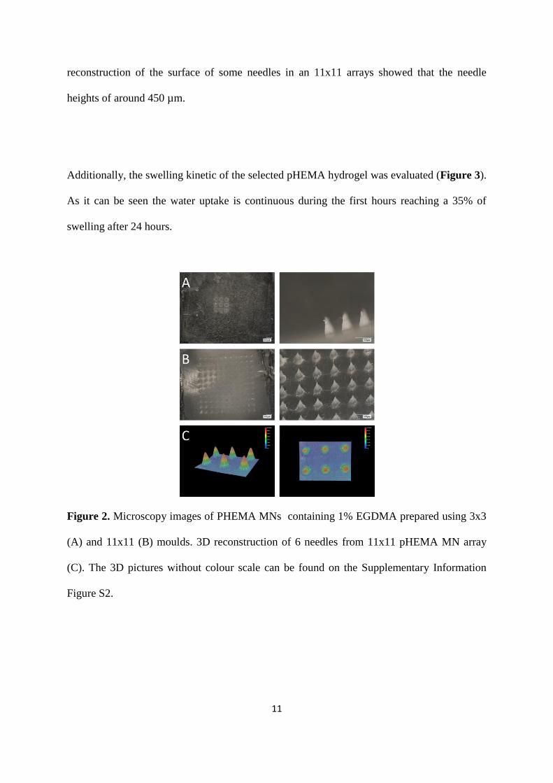
reconstruction of the surface of some needles in an 11x11 arrays showed that the needle
heights of around 450 µm.
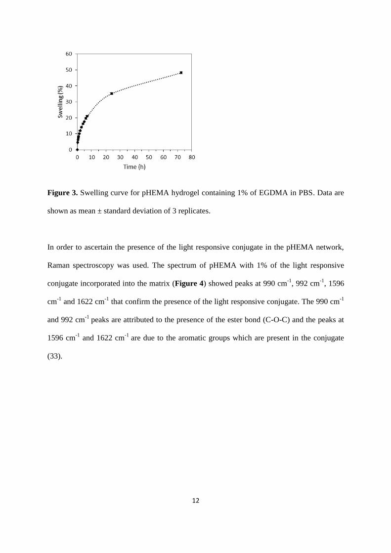
Additionally, the swelling kinetic of the selected pHEMA hydrogel was evaluated (Figure 3).
As it can be seen the water uptake is continuous during the first hours reaching a 35% of
swelling after 24 hours.
Figure 2. Microscopy images of PHEMA MNs containing 1% EGDMA prepared using 3x3
(A) and 11x11 (B) moulds. 3D reconstruction of 6 needles from 11x11 pHEMA MN array
(C). The 3D pictures without colour scale can be found on the Supplementary Information
Figure S2.
Page 12
12
Figure 3. Swelling curve for pHEMA hydrogel containing 1% of EGDMA in PBS. Data are
shown as mean ± standard deviation of 3 replicates.
In order to ascertain the presence of the light responsive conjugate in the pHEMA network,
Raman spectroscopy was used. The spectrum of pHEMA with 1% of the light responsive
conjugate incorporated into the matrix (Figure 4) showed peaks at 990 cm-1
, 992 cm-1
, 1596
cm-1
and 1622 cm-1
that confirm the presence of the light responsive conjugate. The 990 cm-1
and 992 cm-1
peaks are attributed to the presence of the ester bond (C-O-C) and the peaks at
1596 cm-1
and 1622 cm-1
are due to the aromatic groups which are present in the conjugate
(33).
Page 13
13
Figure 4. Raman spectra of pHEMA and pHEMA with 1% of the light responsive conjugate
incorporated into the matrix.
Mechanical properties of the materials containing the light responsible conjugate were
evaluated. We observed that the presence of the light responsive conjugate within the
hydrogels resulted in slight modifications to the bulk mechanical properties of the gels when
exposed to tensile stress (see supporting information Table S1), however ANOVA showed
these different results to be statistically insignificant. Furthermore, the heights and widths
distribution of the MNs seem to be independent of the photoresponsive conjugate
concentration (Table 1). Furthermore, it is noticeable that the obtained needles were shorter
than expected (ca. 425 µm instead of 600 µm). This behaviour has not been obtained in our
group before in the production of Gantrez®
hydrogel-forming MN arrays (16, 19).
Nevertheless the manufacturing process was totally different and in this case it can be due to
the different crosslinking process.
Page 14
14
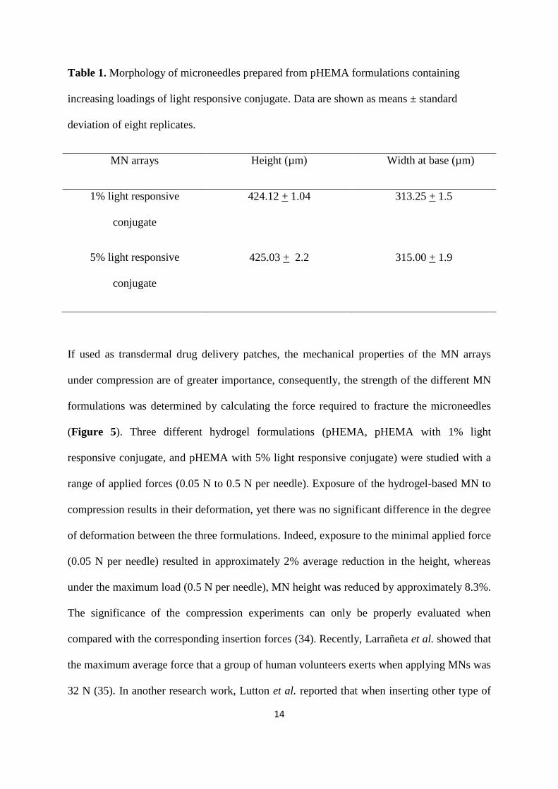
Table 1. Morphology of microneedles prepared from pHEMA formulations containing
increasing loadings of light responsive conjugate. Data are shown as means ± standard
deviation of eight replicates.
MN arrays Height (µm) Width at base (µm)
1% light responsive
conjugate
424.12 + 1.04 313.25 + 1.5
5% light responsive
conjugate
425.03 + 2.2 315.00 + 1.9
If used as transdermal drug delivery patches, the mechanical properties of the MN arrays
under compression are of greater importance, consequently, the strength of the different MN
formulations was determined by calculating the force required to fracture the microneedles
(Figure 5). Three different hydrogel formulations (pHEMA, pHEMA with 1% light
responsive conjugate, and pHEMA with 5% light responsive conjugate) were studied with a
range of applied forces (0.05 N to 0.5 N per needle). Exposure of the hydrogel-based MN to
compression results in their deformation, yet there was no significant difference in the degree
of deformation between the three formulations. Indeed, exposure to the minimal applied force
(0.05 N per needle) resulted in approximately 2% average reduction in the height, whereas
under the maximum load (0.5 N per needle), MN height was reduced by approximately 8.3%.
The significance of the compression experiments can only be properly evaluated when
compared with the corresponding insertion forces (34). Recently, Larrañeta et al. showed that
the maximum average force that a group of human volunteers exerts when applying MNs was
32 N (35). In another research work, Lutton et al. reported that when inserting other type of
Page 15
15
hydrogel-forming MN arrays (32x32 needles; 600 µm of needle length) in a skin simulant
using this force (0.16 N/needle) the needle height reduction was ca. 3% (36). These values
are consistent with those obtained in this work.
Figure 5. Images of different MN arrays after 0N, 0.05N, 0.1N, 0.3N and 0.5N have been
applied (force per needle). A graph depicting the average % reduction in height of three
different type of pHEMA microneedle array formulations (pHEMA, pHEMA loaded with 1%
conjugate 1 and pHEMA loaded with 5%) as different forces ranging from 0.05N to 0.5N are
applied to the microneedle arrays. Data are shown as mean ± standard deviation of five
replicates.
To demonstrate the ability of these hydrogel-based MNs to deliver a clinically relevant model
therapeutic agent (ibuprofen) in vitro, two different MN array designs (3x3 and 11x11
arrays), and two different loadings of the light responsive conjugate (1% and 5%) were
studied. The drug release profiles of a 3x3 MN arrays loaded with the light responsive
Page 16
16
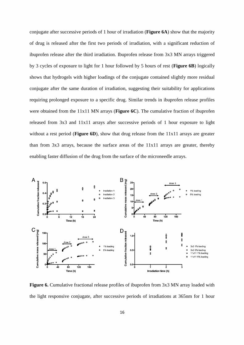
conjugate after successive periods of 1 hour of irradiation (Figure 6A) show that the majority
of drug is released after the first two periods of irradiation, with a significant reduction of
ibuprofen release after the third irradiation. Ibuprofen release from 3x3 MN arrays triggered
by 3 cycles of exposure to light for 1 hour followed by 5 hours of rest (Figure 6B) logically
shows that hydrogels with higher loadings of the conjugate contained slightly more residual
conjugate after the same duration of irradiation, suggesting their suitability for applications
requiring prolonged exposure to a specific drug. Similar trends in ibuprofen release profiles
were obtained from the 11x11 MN arrays (Figure 6C). The cumulative fraction of ibuprofen
released from 3x3 and 11x11 arrays after successive periods of 1 hour exposure to light
without a rest period (Figure 6D), show that drug release from the 11x11 arrays are greater
than from 3x3 arrays, because the surface areas of the 11x11 arrays are greater, thereby
enabling faster diffusion of the drug from the surface of the microneedle arrays.
Figure 6. Cumulative fractional release profiles of ibuprofen from 3x3 MN array loaded with
the light responsive conjugate, after successive periods of irradiations at 365nm for 1 hour
Page 17
17
(A). Data are shown as mean ± standard deviation of six replicates. Ibuprofen release from
3x3 MN array with release triggered by 3 cycles of exposure to light for 1 hour (B).
Ibuprofen release from 11x11 MN array with release triggered by 3 cycles of exposure to
light for 1 hour (C). Total cumulative fraction of ibuprofen released from 3x3 and 11x11
arrays after successive periods of exposure to light (D). Data are shown as mean ± standard
deviation of six replicates.
Discussion
The capability to control the quantity, location and time of drug dosing is one of the main
objectives in the design of drug delivery systems (37). For these purposes systems responsive
to different stimulus, such as temperature, pH or light among others have been prepared (38-
41). A light triggered MN drug delivery system have been designed and successfully tested in
vitro for the release of a clinical relevant model drug: ibuprofen. This systems combines the
advantages of MN transdermal delivery systems with a light-controlled drug liberation
reaction allowing a high level of control for the dose, the timing of the release process, and its
location.
Ibuprofen was selected as a model drug for this study. It was conjugated with 3,5-
dimethoxybenzoin to form a light responsible compound. This type of conjugate has been
previously synthetized and tested using different model molecules such as acetyl salicylic
acid, ibuprofen, and ketoprofen (29). When irradiated with light of specific wavelength the
conjugate is broken yielding the free drug and 2-phenyl-5,7-dimethoxybenzofuran (Figure
1B) (29, 30, 32). The ibuprofen conjugate was successfully loaded in pHEMA hydrogels
crosslinked using EGDMA. Nevertheless, prior to the incorporation of the conjugate inside
the hydrogel matrix the optimal composition of HEMA and EGDMA for MN preparation
Page 18
18
was selected. The results suggested that the use of a lower amount of EGDMA (1%) leads to
the formation of more homogenous distribution of needle length in the array (see supporting
information Figure S1). Therefore this composition was selected over the one containing 5%
of EGDMA. Additionally the swelling kinetics of this type of hydrogel was evaluated
(Figure 3). The swelling process of this type of material requires more than 24 hours to reach
maximum swelling. The swelling process for this type of material is slower in comparison to
other materials used for the preparation of hydrogel-forming MN arrays such as Gantrez®
AN-139 or Gantrez® S-97 that are able to reach maximum swelling degrees in less than 24
hours (20, 42). Moreover, the maximum amount of water that pHEMA hydrogels are able to
uptake is substantially smaller than the previously described materials. The pHEMA hydrogel
shows maximum swelling degrees of around 50% while for Gantrez® hydrogels the values
are higher than 1000% after 24h (20, 42). The lower swelling degree combined with the
slower swelling process makes this type of material more suitable for the production of
prolonged drug delivery systems. Besides, the crosslinking time required to prepare pHEMA
MN arrays is shorter than for Gantrez®
ones (2 h at 90°C vs. 24 h at 80°C). This process can
be accelerated by using alternative heating sources such as microwave radiation (42). The
optimization of the crosslinking time will be mandatory for the development of an industrial
manufacturing process.
The next step was the incorporation of the ibuprofen conjugate in the pHEMA hydrogel
matrices. The conjugate was incorporated in the solution containing HEMA monomer,
EGDMA and the initiator. After the polymerization reaction, the presence of the conjugate
was confirmed using Raman spectroscopy (Figure 4). The conjugate was attached to the
polymer matrix through non-covalent interactions and consequently immobilized until the
covalent ester that bound ibuprofen with the 3,5-dimethoxybenzoin was broken (43). The
Page 19
19
incorporation of the conjugate has no statistical significant effect on the mechanical
properties of the MN arrays (Figure 5).
Finally, MN arrays loaded with the light responsible conjugate were tested in vitro for
transdermal light-triggered delivery of ibuprofen. The experiment was carried out using Franz
Cells and Silescol® membrane as skin simulant. The reason for the use of the Silescol
membrane was that the experiment takes several days to be complete and the integrity of
excised skin during this time will be compromised. As can be seen in Figure 5, the
irradiation of the MN arrays with 365nm light triggers the release of ibuprofen. This process
can be repeated at least three times in a consecutive way. The amount of ibuprofen released
can be tailored by modifying the number of needles in the array, and the concentration of the
drug in the polymeric matrix. Additionally the system allows the release of ibuprofen during
prolonged periods of time (up to 160 h). Additionally it is important to note that despite of the
fact that the conjugate is not covalently bound to the polymer matrix there is no significant
release from pHEMA matrices of the other major product obtained after irradiation of the
conjugate (2-phenyl-5,7-dimethoxybenzofuran) (Figure 1B) due mainly to its hydrophobicity
(32). Nevertheless, in future studies this fact should be evaluated deeply in order to ascertain
the safety of the device.
It is important to notice that UV radiation is shown to be the major etiologic agent in the
development of skin cancers (44). Therefore this system cannot be used directly into patients
in the way it is described here. The work presented here is a probe of concept and a modified
version of the system should be used. Based on our previous research on hydrogel forming
MN arrays we propose a hydrogel-forming MN array system (16, 20) containing a backing
layer made with the material described in this work. Between the MN array and the backing
Page 20
20
layer a porous layer of a material that blocks UV radiation should be included. Therefore
radiation can be applied to the system without reaching the skin surface.
Triggerable systems such as the system presented here facilitate multiple drug dosages with a
single administration. This type of delivery system is of great interest especially for the
treatment of pain (45). A good example of this situation are cancer patients that often suffer
from various levels of pain as symptom of the cancer or as side effect of the cancer treatment.
To the date only a few authors have dealt with the combination of MN arrays and stimuli-
responsive systems (26-28). Huang et al. developed a biochip for real-time measurement of
glucose concentration and automatic insulin injection (28). This system was based on
electronic systems rather than in stimuli-responsible materials. Recently, Chen et al.
developed a MN system loaded with silica-coated lanthanum hexaboride nanoparticles that
are capable to release a model molecule (Rhodamine 6G) on demand when the array was
irradiated with near infrared radiation (26). The MN array was made of polycaprolactone.
When the system was irradiated with infrared radiation, the presence of light-sensitive
nanostructures caused a temperature increase of the array. This temperature increase leads to
microneedle melting and enabling drug release. After 132 minutes and multiple infrared
stimulated release cycles the system was able to release up to 80% of its cargo. The system
described in the present paper was able to release ibuprofen during longer periods of time
(Figure 6) making our system more appealing for long term drug administration.
Conclusion
A light-responsive MN drug delivery system has been designed and successfully tested in
vitro for the release of a model drug, ibuprofen. The coupling of the application of light to the
release of a predictable dose of drug has been demonstrated. The selected polymer for MN
Page 21
21
fabrication, pHEMA crosslinked with EGDMA, presented good mechanical properties and
could be used to form MN arrays successfully using a micromolding technique. This
technology has considerable potential for use in situations where “on-demand” drug delivery
is required, with patient- or physician-controlled analgesia an obvious example. Our next
steps will be in vivo studies aimed at ascertaining the efficacy of the system in a suitable
animal model. Due to the potentially harmful effect of UV radiation on the skin, the system
will be modified before its application in vivo, perhaps by including the drug and light-
sensitive conjugate in the baseplate and having the needles protrude through an opaque
barrier membrane into the skin. Moreover, as we take the technology forwards, we will also
consider optimization of the crosslinking time using microwave radiation and the minimising
the potential for release of any free 2-phenyl-5,7-dimethoxybenzofuran from the array into
skin following irradiation.
Acknowledgements
We acknowledge funding support from the Engineering and Physical Sciences Research
Council (EPSRC) grant number EP/H012249/1. The microneedles aspects of this study were
supported by BBSRC grant numbers BB/FOF/287 and BB/E020534/1.
Page 22
22
Supporting Information
Figure S1. Morphology of microneedles prepared from the different PHEMA formulations:
1% EGDMA, height 425.07 ± 2.82 μm and width 313.25 ± 1.23 μm (A); 5% EGDMA,
height 425.00 ± 12.34 μm and width 323.00 ± 14.53 μm (B). Data are shown as means ±
standard deviation of eight replicates.
Figure S2. 3D reconstruction of 6 needles from 11x11 pHEMA MN array
Page 24
24
Table S1. Influence of the incorporation of the light responsive conjugate on the bulk
mechanical properties of the hydrogels.
Sample UTS (MPa) Young’s Modulus
(GPa)
% Elongation at
break
pHEMA 0.40 + 0.09 0.72 + 0.06 154.0 + 16.8
pHEMA 1% light
responsive conjugate
0.37 + 0.13 1.13 + 0.21 155.5 + 8.9
pHEMA 5% light
responsive conjugate
0.32 + 0.10 1.42 + 0.08 162.5 + 7.3
References
1. Donnelly, R.F.; Singh, T.R.R.; Morrow, D.I.J.; Woolfson, A.D. Microneedle-mediated
Transdermal and Intradermal Drug Delivery. Wiley: 2012;
2. Singh, T.R.R.; Garland, M.J.; Cassidy, C.M.; Migalska, K.; Demir, Y.K.; Abdelghany, S.;
Ryan, E.; Woolfson, A.D.; Donnelly, R.F.; Singh, T.R.R. Microporation techniques for
enhanced delivery of therapeutic agents. Recent Pat. Drug. Deliv. Formul. 2010, 4, 1-17.
3. Prausnitz, M.R.; Mitragotri, S.; Langer, R. Current status and future potential of
transdermal drug delivery. Nat Rev Drug Discov 2004, 3, 115-124.
4. Kim, Y.C.; Park, J.H.; Prausnitz, M.R. Microneedles for drug and vaccine delivery. Adv.
Drug Deliv. Rev. 2012, 64, 1547-1568.
5. Prausnitz, M.R. Microneedles for transdermal drug delivery. Adv. Drug Deliv. Rev. 2004,
56, 581-587.
6. Prausnitz, M.R. and Langer, R. Transdermal drug delivery. Nat. Biotechnol. 2008, 26,
1261-1268.
7. Birchall, J.C. Microneedle array technology: the time is right but is the science ready?
Expert Rev. Med. Devices. 2006, 3, 1-4.
8. Coulman, S.; Allender, C.; Birchall, J. Microneedles and other physical methods for
overcoming the stratum corneum barrier for cutaneous gene therapy. Crit. Rev. Ther. Drug.
2006, 23, 205-258.
Page 25
25
9. Henry, S.; McAllister, D.V.; Allen, M.G.; Prausnitz, M.R. Microfabricated microneedles: a
novel approach to transdermal drug delivery. J. Pharm. Sci. 1998, 87, 922-925.
10. Pierre, M.B. and Rossetti, F.C. Microneedle-based drug delivery systems for transdermal
route. Curr. Drug Targets 2014, 15, 281-291.
11. Tuan-Mahmood, T.M.; McCrudden, M.T.; Torrisi, B.M.; McAlister, E.; Garland, M.J.;
Singh, T.R.; Donnelly, R.F. Microneedles for intradermal and transdermal drug delivery. Eur.
J. Pharm. Sci. 2013, 50, 623-637.
12. Liu, S.; Jin, M.N.; Quan, Y.S.; Kamiyama, F.; Katsumi, H.; Sakane, T.; Yamamoto, A.
The development and characteristics of novel microneedle arrays fabricated from hyaluronic
acid, and their application in the transdermal delivery of insulin. J. Control. Release 2012,
161, 933-941.
13. Matsuo, K.; Yokota, Y.; Zhai, Y.; Quan, Y.S.; Kamiyama, F.; Mukai, Y.; Okada, N.;
Nakagawa, S. A low-invasive and effective transcutaneous immunization system using a
novel dissolving microneedle array for soluble and particulate antigens. J. Control. Release
2012, 161, 10-17.
14. Naito, S.; Ito, Y.; Kiyohara, T.; Kataoka, M.; Ochiai, M.; Takada, K. Antigen-loaded
dissolving microneedle array as a novel tool for percutaneous vaccination. Vaccine 2012, 30,
1191-1197.
15. Hong, X.; Wu, Z.; Chen, L.; Wu, F.; Wei, L.; Yuan, W. Hydrogel Microneedle Arrays for
Transdermal Drug Delivery. Nano-Micro Lett. 2014, 6, 191-199.
16. Donnelly, R.F.; Singh, T.R.R.; Garland, M.J.; Migalska, K.; Majithiya, R.; McCrudden,
C.M.; Kole, P.L.; Mahmood, T.M.T.; McCarthy, H.O.; Woolfson, A.D. Hydrogel-Forming
Microneedle Arrays for Enhanced Transdermal Drug Delivery. Adv. Funct. Mater. 2012, 22,
4879-4890.
17. Kim, M.; Jung, B.; Park, J.H. Hydrogel swelling as a trigger to release biodegradable
polymer microneedles in skin. Biomaterials 2012, 33, 668-678.
18. Donnelly, R.F.; Singh, T.R.; Alkilani, A.Z.; McCrudden, M.T.; O'Neill, S.; O'Mahony,
C.; Armstrong, K.; McLoone, N.; Kole, P.; Woolfson, A.D. Hydrogel-forming microneedle
arrays exhibit antimicrobial properties: potential for enhanced patient safety. Int. J. Pharm.
2013, 451, 76-91.
19. Donnelly, R.F.; Morrow, D.I.; McCrudden, M.T.; Alkilani, A.Z.; Vicente-Pérez, E.M.;
O'Mahony, C.; González-Vázquez, P.; McCarron, P.A.; Woolfson, A.D. Hydrogel-forming
and dissolving microneedles for enhanced delivery of photosensitizers and precursors.
Photochem. Photobiol. 2014, 90, 641-647.
20. Donnelly, R.F.; McCrudden, M.T.C.; Zaid Alkilani, A.; Larrañeta, E.; McAlister, E.;
Courtenay, A.J.; Kearney, M.; Singh, T.R.R.; McCarthy, H.O.; Kett, V.L.; Caffarel-Salvador,
E.; Al-Zahrani, S.; Woolfson, A.D. Hydrogel-Forming Microneedles Prepared from "Super
Page 26
26
Swelling"• Polymers Combined with Lyophilised Wafers for Transdermal Drug Delivery.
PLoS ONE 2014, 9, e111547.
21. Garland, M.J.; Caffarel-Salvador, E.; Migalska, K.; Woolfson, A.D.; Donnelly, R.F.
Dissolving polymeric microneedle arrays for electrically assisted transdermal drug delivery.
J. Control. Release 2012, 159, 52-59.
22. Pawar, K.R.; Smith, F.; Kolli, C.S.; Babu, R.J. Effect of lipophilicity on microneedle-
mediated iontophoretic transdermal delivery across human skin in vitro. J. Pharm. Sci. 2013,
102, 3784-3791.
23. Wing, D.; Prausnitz, M.R.; Buono, M.J. Skin pretreatment with microneedles prior to
pilocarpine iontophoresis increases sweat production. Clin. Physiol. Funct. Imaging 2013, 33,
436-440.
24. Singh, N.D. and Banga, A.K. Controlled delivery of ropinirole hydrochloride through
skin using modulated iontophoresis and microneedles. J. Drug Target. 2013, 21, 354-366.
25. Vemulapalli, V.; Bai, Y.; Kalluri, H.; Herwadkar, A.; Kim, H.; Davis, S.P.; Friden, P.M.;
Banga, A.K. In vivo iontophoretic delivery of salmon calcitonin across microporated skin. J.
Pharm. Sci. 2012, 101, 2861-2869.
26. Chen, M.C.; Ling, M.H.; Wang, K.W.; Lin, Z.W.; Lai, B.H.; Chen, D.H. Near-infrared
light-responsive composite microneedles for on-demand transdermal drug delivery.
Biomacromolecules 2015, 16, 1598-1607.
27. Cahill, E.M. and O'Cearbhaill, E.D. Toward Biofunctional Microneedles for Stimulus
Responsive Drug Delivery. Bioconjug. Chem. 0, 0, nu.
28. Huang, C.J.; Chen, Y.H.; Wang, C.H.; Chou, T.C.; Lee, G.B. Integrated microfluidic
systems for automatic glucose sensing and insulin injection. Sensors Actuators B: Chem.
2007, 122, 461-468.
29. McCoy, C.P.; Rooney, C.; Edwards, C.R.; Jones, D.S.; Gorman, S.P. Light-triggered
molecule-scale drug dosing devices. J. Am. Chem. Soc. 2007, 129, 9572.
30. McCoy, C.P.; Rooney, C.; Jones, D.S.; Gorman, S.P.; Nieuwenhuyzen, M. Rational
design of a dual-mode optical and chemical prodrug. Pharm. Res. 2007, 24, 194-200.
31. Donnelly, R.F.; Majithiya, R.; Singh, T.R.; Morrow, D.I.; Garland, M.J.; Demir, Y.K.;
Migalska, K.; Ryan, E.; Gillen, D.; Scott, C.J.; Woolfson, A.D. Design, optimization and
characterisation of polymeric microneedle arrays prepared by a novel laser-based
micromoulding technique. Pharm. Res. 2011, 28, 41-57.
32. Rooney, C.; Hayes, G.; McCoy, C.P. Photolytic release of ibuprofen from a 3,5-
dimethoxybenzoin ester within a p(HEMA-co-MMA) polymer matrix. J. Pharm. Pharmacol.
2003, 55, S43.
Page 27
27
33. Ren, Y.; Matsushita, A.; Matsukawa, K.; Inoue, H.; Minami, Y.; Noda, I.; Ozaki, Y. Two-
dimensional Fourier-transform-Raman and near-infrared correlation spectroscopy studies of
poly(methyl methacrylate) blends. Vib. Spectrosc. 2000, 23, 207-218.
34. Lutton, R.E.M.; Moore, J.; Larrañeta, E.; Ligett, S.; Woolfson, A.D.; Donnelly, R.F.
Microneedle characterisation: the need for universal acceptance criteria and GMP
specifications when moving towards commercialisation. Drug Deliv. Transl. Res. 2015, 1-19.
35. Larrañeta, E.; Moore, J.; Vicente-Pérez, E.M.; González-Vázquez, P.; Lutton, R.;
Woolfson, A.D.; Donnelly, R.F. A proposed model membrane and test method for
microneedle insertion studies. Int. J. Pharm. 2014, 472, 65-73.
36. Lutton, R.E.M.; Larrañeta, E.; Kearney, M.C.; Boyd, P.; Woolfson, A.D.; Donnelly, R.F.
A novel scalable manufacturing process for the production of hydrogel-forming microneedle
arrays. Int. J. Pharm. 2015, 494, 417-429.
37. Langer, R. Drug delivery. Drugs on target. Science 2001, 293, 58-59.
38. Gillies, E.R. and Fréchet, J.M.J. Development of acid-sensitive copolymer micelles for
drug delivery. Pure Appl. Chem. 2004, 76, 1295-1307.
39. Moreno, E.; Schwartz, J.; Larrañeta, E.; Nguewa, P.A.; Sanmartín, C.; Agüeros, M.;
Irache, J.M.; Espuelas, S. Thermosensitive hydrogels of poly(methyl vinyl ether-co-maleic
anhydride) - Pluronic(®) F127 copolymers for controlled protein release. Int. J. Pharm. 2014,
459, 1-9.
40. Sershen, S.R.; Mensing, G.A.; Ng, M.; Halas, N.J.; Beebe, D.J.; West, J.L. Independent
Optical Control of Microfluidic Valves Formed from Optomechanically Responsive
Nanocomposite Hydrogels. Adv. Mater. 2005, 17, 1366.
41. Li, S.K. and D'Emanuele, A. On-off transport through a thermoresponsive hydrogel
composite membrane. J. Control. Release 2001, 75, 55-67.
42. Larrañeta, E.; Lutton, R.E.M.; Brady, A.J.; Vicente-Pérez, E.M.; Woolfson, A.D.;
Thakur, R.R.S.; Donnelly, R.F. Microwave-Assisted Preparation of Hydrogel-Forming
Microneedle Arrays for Transdermal Drug Delivery Applications. Macromol. Mater. Eng.
2015, 300, 586-595.
43. McCoy, C.P.; Gorman, S.P.; Jones, D.S. Drug delivery composition. 2011,
WO2009147372.
44. Narayanan, D.L.; Saladi, R.N.; Fox, J.L. Review: Ultraviolet radiation and skin cancer.
Int. J. Dermatol. 2010, 49, 978-986.
45. Timko, B.P.; Arruebo, M.; Shankarappa, S.A.; McAlvin, J.B.; Okonkwo, O.S.; Mizrahi,
B.; Stefanescu, C.F.; Gomez, L.; Zhu, J.; Zhu, A.; Santamaria, J.; Langer, R.; Kohane, D.S.
Near-infrared-actuated devices for remotely controlled drug delivery. Proc. Natl. Acad. Sci.
U. S. A. 2014, 111, 1349-1354.


![NATURE-INSPIRED STIMULI-RESPONSIVE SELF-FOLDING … · materials. The example of the first case is the bending of polyelectrolyte hydrogel during electrolysis [8]. The examples of](https://static.documents.pub/doc/80x56/5f0b512a7e708231d42feb18/nature-inspired-stimuli-responsive-self-folding-materials-the-example-of-the-irst.jpg)

![Hydrogel-forming microneedle arrays: Potential for use in ... · agents, lithium is still considered the ‘gold standard’ treatment for bipolar (BP) disorder [1,2]. As a pharmacological](https://static.documents.pub/doc/80x56/5fb0a003d903040a937179a3/hydrogel-forming-microneedle-arrays-potential-for-use-in-agents-lithium-is.jpg)