50
2012.12.08
| Date post: | 15-Jul-2015 |
| Category: |
Health & Medicine |
| Upload: | wasula-rathnaweera |
| View: | 34 times |
| Download: | 0 times |

2012.12.08
84–amino acid polypeptide hormone
responsible for maintaining ECF [Ca2+]
secretion is gulated directly by the ECF
[Ca2+]
stimuli
Decreased serum [Ca2+]
Mild decreases in serum [Mg2+]
An increase in serum phosphate
only hormone which is up regulated when the
stimulus is low

increase ECF [Ca2+] by
increasing the release of calcium and phosphate
from bone matrix
increasing calcium reabsorption by the kidney
increasing renal production of 1,25-
dihydroxyvitamin D-3 (calcitriol), which increases
intestinal absorption of calcium
causes phosphaturia, decreasing serum
phosphate levels


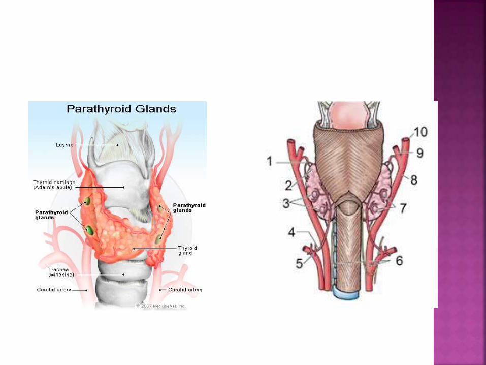
4 glands - posterior to the thyroid gland Superior 2, inferior 2
some times 3, 5, or, occasionally, more glands
inferior glands - derived from the third pharyngeal pouch with the thymus migrate along with the thymus
become situated more inferiorly than the superior glands
usually located near the inferior pole of the thyroid
Can go in to superior mediastinum

superior glands - more consistent in location
just superior to the intersection of the inferior
thyroid artery and the recurrent laryngeal nerve
derived from the fourth pharyngeal pouch
occasionally found within the substance of the
thyroid gland.


Primary
Secondary
Tertiary
unregulated overproduction of parathyroid
hormone (PTH) resulting in abnormal calcium
homeostasis
21 cases per 100,000 person-years.
The mean age at diagnosis - between 52 and
56 years
female-to-male ratio of 3:1

85% of cases, primary hyperparathyroidism is
caused by
single adenoma
hyperplasia
15% of cases, multiple glands are involved
Rarely, primary hyperparathyroidism is
caused by parathyroid carcinoma.
aetiology of adenomas or hyperplasia is
unknown in most cases

Familial
multiple endocrine neoplasia syndromes (MEN 1
or MEN 2a)
hyperparathyroid-jaw tumor (HPT-JT) syndrome
familial isolated hyperparathyroidism (FIHPT)
familial hypocalciuric hypercalcemia
neonatal severe hyperparathyroidism

normal feedback on parathyroid hormone
production by extracellular calcium is lost
increase in the cell numbers is probably the
cause.
chronic excessive resorption of calcium from
bone result in osteopenia
may result in osteitis fibrosa cystica
subperiosteal resorption of the distal phalanges
tapering of the distal clavicles
salt-and-pepper appearance of the skull
brown tumors of the long bones
chronically hypercalciuria predisposes to the
formation of renal stones.
symptoms of hyperparathyroidism are due to the hypercalcemia
muscle weakness
fatigue
volume depletion
nausea and vomiting
and in severe cases, coma and death
neuropsychiatric manifestations depression
confusion
increase gastric acid secretion peptic ulcer disease
rare cases of pancreatitis

bones, stones, abdominal groans, and psychic
moans
severe bone disease, kidney stones to
asymptomatic hypercalcemia
Skeletal manifestations
selective cortical bone loss
bone and joint pain
pseudogout
chondrocalcinosis
osteitis fibrosa cystica

Renal manifestations
polyuria
kidney stones
hypercalciuria
nephrocalcinosis.
Gastrointestinal manifestations
anorexia
nausea
vomiting
abdominal pain
constipation
peptic ulcer disease
acute pancreatitis.
Cardiovascular manifestations
hypertension
bradycardia
shortened QT interval
left ventricular hypertrophy
Physical examination findings
usually noncontributory

causes of hypercalcemia + elevated
parathyroid hormone level are few
familial benign (hypocalciuric) hypercalcemia
(FHH) (see Related disorders)
lithium-induced hypercalcemia
tertiary hyperparathyroidism.

all potential causes of secondary hyperparathyroidism should be excluded low calcium intake
gastrointestinal disorders
renal insufficiency
vitamin D deficiency
hypercalciuria of renal origin
secondary and tertiary hyperparathyroidism are typically diagnosed based on their clinical context
cancer-induced hypercalcemia low parathyroid hormone level
possibly a high parathyroid hormone-related peptide level

Laboratory studies
total serum calcium and albumin levels or ionized calcium levels
hypercalcemia should be documented on more than one occasion
intact parathyroid hormone level is the core of the diagnosis
elevated intact parathyroid hormone level with an elevated ionized serum calcium level is diagnostic of primary hyperparathyroidism
24-hour urine calcium measurement is necessary to rule out FHH.
other biochemical abnormalities
mild hyperchloremic acidosis
hypophosphatemia
mild-to-moderate increase in urinary calcium
excretion rate.
Imaging studies
make a decision about whether to pursue
surgical therapy
If a limited parathyroid exploration is to be
attempted, a localizing study is necessary

USS of the neck
capable of a high degree of accuracy
operator dependent
not been reliable in detecting multigland
disease.
Nuclear medicine scanning with radiolabeled
sestamibi


CT scanning and MRI
locate abnormal parathyroid glands
Standard CT scanning has inadequate sensitivity.
Newer techniques of CT scanning with dynamic
contrast images (4D-CT) accuracy 88%.
MRI - particularly in cases of
recurrent
persistent disease
ectopic locations such as the mediastinum.

dual-energy radiographic absorptiometry
demonstrate the skeletal involvement in primary
hyperparathyroidism
Hyperparathyroidism affects the cortical bone at
the radius (distal third)
skeletal radiographs
salt-and-pepper degranulation in the skull
subperiosteal bone resorption in the phalanges.
Procedures
Bilateral internal jugular vein sampling
localize ectopic parathyroid adenomas

surgical excision of the abnormal parathyroid
glands
the only permanent, curative treatment for
primary hyperparathyroidism.
surgical treatment should be offered to all
patients with symptomatic disease.

The indications for surgery
1 mg/dL above the upper limit of the reference
range for serum calcium
24 hour urinary calcium excretion greater than
400 mg
30% reduction in creatinine clearance
bone mineral density T-score below -2.5 at any
site
age younger than 50 years

monitoring of patients with asymptomatic
hyperparathyroidism
serum calcium and creatinine levels every 6
months
annual bone mineral density

Management of severe hypercalcemia in the
acute setting
IV volume expansion
sodium chloride and loop diuretics, once the
intravascular volume is restore
Drugs (temporary measure prior to surgical
treatment )
calcitonin
IV bisphosphonate

Nonsurgical care
should be carefully monitored
maintain a moderate daily elemental calcium
intake of 800-1000 mg
vitamin D intake appropriate for their age
and sex.
participation in regular exercise activity
avoid immobilization
avoid thiazides, diuretics, and lithium

Pharmacotherapy
Estrogen therapy in postmenopausal women
Selective estrogen receptor modulators
raloxifene
Bisphosphonates
Calcimimetic drugs
activate the calcium-sensing receptor and inhibit
parathyroid cell function - cinacalcet

Other treatments
Percutaneous alcohol injection to parathyroids
ablation with ultrasound energy
surgical care
should be offered to most patients
standard operative approach is complete neck
exploration,identification of all parathyroid
glands and removal of all abnormal glands.
85% of cases caused by a single adenoma
full neck exploration might be an unnecessary
dissection
directed parathyroidectomy
preoperative imaging studies to localize the abnormal
gland
removal only that gland

Localisation
sestamibi scanning or ultrasonography.
intraoperative parathyroid hormone assay
radio-guided parathyroidectomy
detecting the labeled sestamibi in the abnormal
gland using a handheld probe
for familial disease
total parathyroidectomy with
autotransplantation to the forearm and
cryopreservation of some parathyroid tissue

in 4-gland hyperplasia
3.5-gland (subtotal) parathyroidectomy
50-70 mg of the most normal-appearing tissue is
left

Complications and postoperative care
calcium levels must be monitored
every 12 hours until stabilization
many become hypocalcemic
few become symptomatic
treatment for hypocalcemia
severe
Symptomatic
hypocalcemia after parathyroid surgery may
be due to hungry bone syndrome
calcium and phosphorus are rapidly deposited in
the bone
If hypoparathyroidism persists
oral supplementation
calcium
vitamin

overproduction of parathyroid hormone secondary to a chronic abnormal stimulus for its production
Typically
chronic renal failure
vitamin D deficiency
Secondary hyperparathyroidism (SHPT) develops early in CKD before dialysis is required
In chronic kidney disease, overproduction of parathyroid hormone occurs in response
Hyperphosphatemia
Hypocalcemia
impaired 1,25-dihydroxyvitamin D production

Medical management is the mainstay
Correcting vitamin D deficiency
Dietary phosphate restriction
Phosphate binders calcium-based phosphate binders
calcium carbonate
calcium acetate
non-calcium-based phosphate binders sevelamer hydrochloride
lanthanum carbonate
Calcium supplementation should be limited to less than 2 g/d

Indications for surgery
bone pain or fracture
Pruritus
Calciphylaxis
Extraskeletal nonvascular calcifications
elevated parathyroid hormone levels despite
appropriate medical therapy
severe hyperparathyroidism
persistent serum levels of intact parathyroid
hormone greater than 800 pg/mL
Medical treatment is successful in most
patients
Patients who require parathyroidectomy have
a 10% risk of recurrent or persistent disease
development of autonomous hypersecretion
of parathyroid hormone causing
hypercalcemia
aetiology is unknown
may be due to monoclonal expansion of
parathyroid cells
four-gland involvement occurs in most patients.
Pathophysiology
observed in patients with chronic secondary
hyperparathyroidism and often after renal
transplantation.
hypertrophied parathyroid glands fail to
return to normal
continue to oversecrete despite serum
calcium levels normal or elevated
dngerous - phosphate level is often elevated.
diffuse calcinosis may occur.

Treatment
Total parathyroidectomy with
autotransplantation
subtotal parathyroidectomy

Familial benign (hypocalciuric)
hypercalcemia
loss-of-function mutation of one allele of the
gene for the calcium-sensing receptor
hypercalcemia, hypophosphatemia, and
hypermagnesemia
can be distinguished from primary
hyperparathyroidism by low 24-hour urinary
calcium excretion
Persons with FHH are asymptomatic.
parathyroidectomy is not indicated


Hypercalcemia of malignancy
caused by
tumor release of parathyroid hormone -related
peptide
over production of 1,25-dihydroxyvitamin D
local osteolytic lesions
low or undetectable intact parathyroid
hormone level

Calciphylaxis
= uremic gangrene syndrome
observed in patients with renal failure and
secondary or tertiary hyperparathyroidism.
characterized by ischemic necrosis of the
skin due to calcium phosphate crystal
deposition and subsequent inflammation in
small-to-medium–sized vessels