37
HYPERBARIC OXYGEN THERAPY: Nursing Considerations Julio R. Garcia CHT, ACHRN Program Director Springhill Medical Center

HYPERBARIC OXYGEN THERAPY:Nursing Considerations
Julio R. Garcia CHT, ACHRNProgram DirectorSpringhill Medical Center

Disclosures
Speaker’s Bureau:
• none
Honorarium:
• none
Consultant:
• Wound Care Education Partners
Stockholder:
• none
Grant/Research Support:
• none
Medical/Scientific Boards:
• Editorial Board: Wound Care and Hyperbaric Medicine Journal

HYPERBARIC OXYGEN THERAPY
• Clinical definition as defined by the UHMS :• An intervention in which an individual breathes near 100%
oxygen intermittently while inside a hyperbaric chamber pressurized to greater 1.4 ATA.
• The National Fire Protection Agency (NFPA) classifies them into three categories:
• Class A – Human; multiple occupancy
• Class B – Human; single occupancy
• Class C – Animal; no human occupancy
HYPERBARIC OXYGEN THERAPY
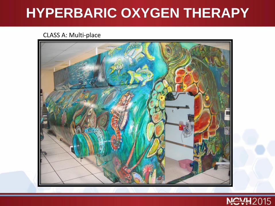
CLASS A: Multi-place

HYPERBARIC OXYGEN THERAPY
Class B: Monoplace
HYPERBARIC OXYGEN THERAPY
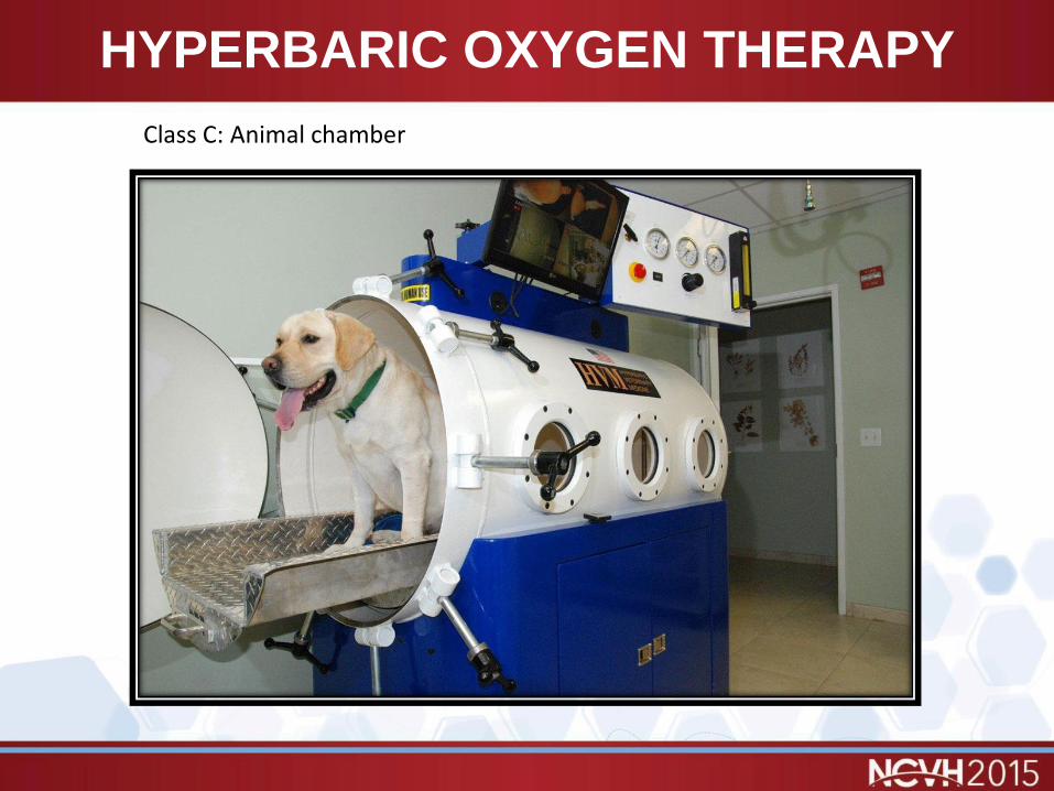
Class C: Animal chamber

Physiologic Effects
There are only two basic primary effects of hyperbaric oxygen on the human body.
◦ The mechanical effect in reduction of bubble size.
A gas bubble compressed to 6ATA is 16% its volume from surface
a spherical bubble decreases its diameter by only 1/2 at 6ATA
◦ The increasing of partial pressure of oxygen in all the tissues of the body. (Henry’s Law of Gas)
Placing a patient in a chamber can raise the oxygen tensions 10 to 13 times above normal levels. At 2.8 ATA (60ft sw), six volumes percent of oxygen is dissolved in the plasma meeting the the needs of the body’s tissue. The overall effect is that the hemoglobin is fully saturated on the venous side.
(Kindwall, Hyperbaric Medicine Practice, Page 18-19)

Increased Oxygen Partial Pressures

Mechanical Effects
• Effects on bubble size• Boyle’s Law of gas states that volume is inversely
proportional to the absolute pressure.
• Two types of intravascular bubbles• Spherical- no damage
• Cylindrical- block blood vessels, activate platelets impinging on vessel walls
• Charles Law
• Gay Lussac’s Law
• Pascal’s Principle(Kindwall, Hyperbaric Medicine Practice, Page 18-19)

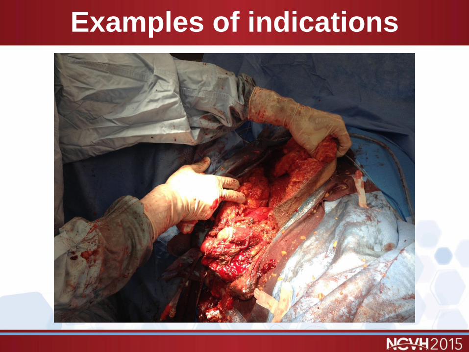
Indications for HBOT• Arterial Gas
Embolism (AGE)
• Carbon Monoxide Poisoning
• ClostridialMyonecrosis (Gas Gangrene)
• Crush Injuries and other acute traumatic peripheral ischemia
• Refractory Osteomyelitis
• Exceptional Blood Loss Anemia
• Decompression Illness (Bends)
• Enhancement in Healing in Selected problem wounds
• Necrotizing Soft Tissue Infections
• Radiation Tissue Injuries
• Skin Grafts and Flaps (compromised)
• Thermal Burns
• Intracranial Abscess
• CRAO

Nursing Considerations
• Absolute Contraindication
• Untreated Pneumothorax
• Contraindications• Doxorubicin
(Adriamycin)• Disulfiram
(Antabuse)• Cis-Platinum• Mafenide Acetate
(Sulfamylon)• Amiodorone
• Relative Contraindications
• Known Malignancies• Pregnancy• Upper Respiratory
Infections• Seizure Disorders• Emphysema with
CO2 Retention• High Fevers• Hx of spontaneous
pneumothorax• History of CHF

Nursing Considerations
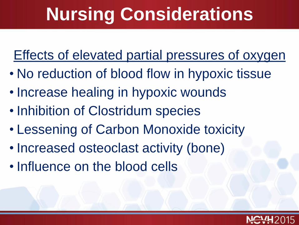
Effects of elevated partial pressures of oxygen
• Reduction in blood flow in hyperoxic tissues
• Hemodynamic changes include:
• decreases in cardiac output by 24-35%
• increases in afterload by 30-60%
• decreases in left ventricular index by 11-30%
Nursing Considerations
Effects of elevated partial pressures of oxygen
• No reduction of blood flow in hypoxic tissue
• Increase healing in hypoxic wounds
• Inhibition of Clostridum species
• Lessening of Carbon Monoxide toxicity
• Increased osteoclast activity (bone)
• Influence on the blood cells

Nursing Considerations
Effects of elevated partial pressures of oxygen
• Increases neovascularization formation
• Reduces edema
• Enhanced fibroblast proliferation
• Enhanced collagen formation
• Enhanced Polymorphonuclear cell function in the destruction of invading bacteria
• Mitigation of reperfusion injury by prevention of leukocyte adherence to endothelium
Nursing Considerations
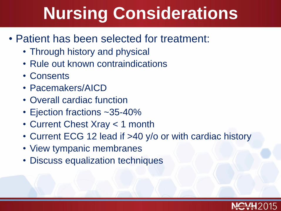
• Patient has been selected for treatment:• Through history and physical
• Rule out known contraindications
• Consents
• Pacemakers/AICD
• Overall cardiac function
• Ejection fractions ~35-40%
• Current Chest Xray < 1 month
• Current ECG 12 lead if >40 y/o or with cardiac history
• View tympanic membranes
• Discuss equalization techniques
Nursing Considerations
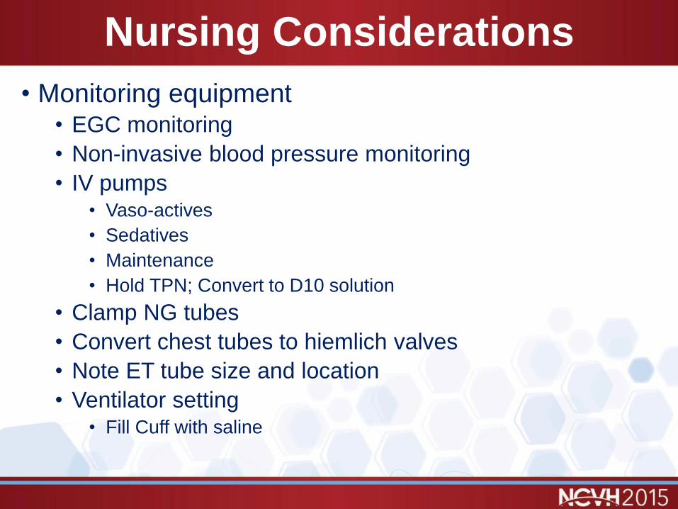
• Monitoring equipment• EGC monitoring
• Non-invasive blood pressure monitoring
• IV pumps• Vaso-actives
• Sedatives
• Maintenance
• Hold TPN; Convert to D10 solution
• Clamp NG tubes
• Convert chest tubes to hiemlich valves
• Note ET tube size and location
• Ventilator setting• Fill Cuff with saline

Nursing Considerations
Nursing Considerations

Nursing Considerations
Nursing Considerations

Examples of indications
Examples of indications
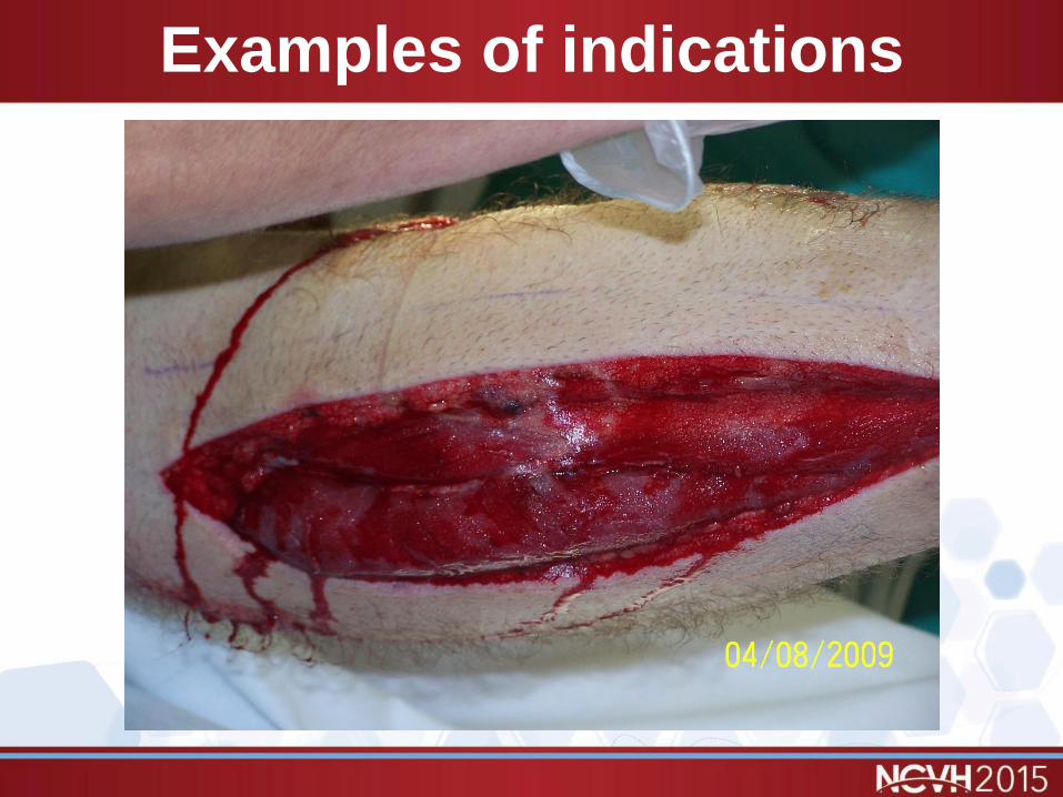
Examples of indications

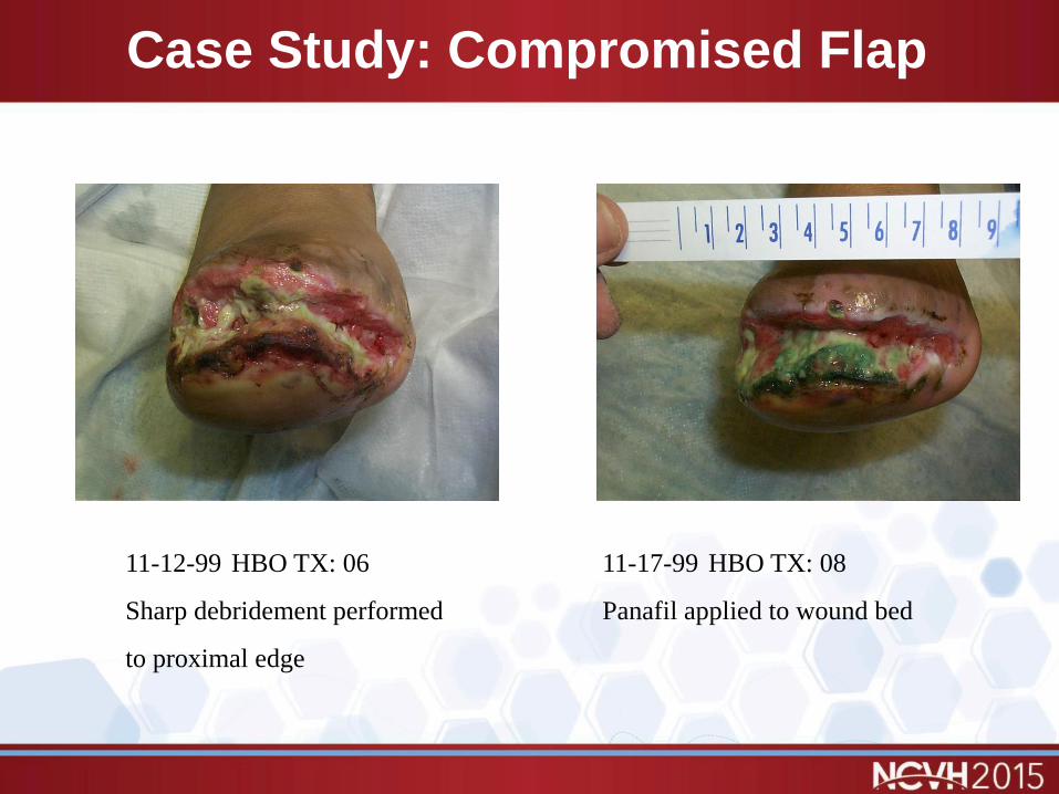
Case Study: Compromised Flap
11-12-99 HBO TX: 06
Sharp debridement performed
to proximal edge
11-17-99 HBO TX: 08
Panafil applied to wound bed

Case Study: Compromised Flap
11-29-99 HBO TX: 15
Aggressive sharp debridement
performed. Continued with Panafil
for wound care dressing
12-22-99 HBO TX: 30
HBO held at this time.
Continued with Panafil.

Case Study: Compromised Flap
12-29-99 HBO: Hold Rich
Granulation Bed Growth factor
therapy initiated (Regranex)
01-04-00 HBO: Hold
Continued with Regranex therapy

Case Study:Compromised Flap
01-24-00 HBO: Hold
Granulation bed is becoming dusky
progression of wound slowing
significantly. Initiated HBO TX's
again.
02-08-00 HBO:09
Progression of wound healing
began again. Initiated hydrogel
therapy
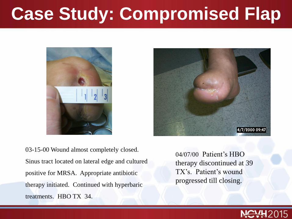
Case Study: Compromised Flap
03-15-00 Wound almost completely closed.
Sinus tract located on lateral edge and cultured
positive for MRSA. Appropriate antibiotic
therapy initiated. Continued with hyperbaric
treatments. HBO TX 34.
04/07/00 Patient’s HBO
therapy discontinued at 39
TX’s. Patient’s wound
progressed till closing.
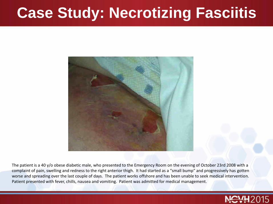
Case Study: Necrotizing Fasciitis
The patient is a 40 y/o obese diabetic male, who presented to the Emergency Room on the evening of October 23rd 2008 with a complaint of pain, swelling and redness to the right anterior thigh. It had started as a “small bump” and progressively has gotten worse and spreading over the last couple of days. The patient works offshore and has been unable to seek medical intervention. Patient presented with fever, chills, nausea and vomiting. Patient was admitted for medical management.

Case Study: Necrotizing Fasciitis
Initial surgical debridement was performed on 10.24.08. Post operative diagnosis synergistic cellulitis with necrotizing fasciitis. Aggressive debridement of the necrosis resulted in a subsequent wound that measured ~46 x 21 x 6cm to the depth of the fascia plane. Emergent hyperbaric oxygen therapy was initiated, along IV antibiotics and negative pressure therapy utilizing the KCI Wound VAC ®. A total of 5 surgical debridements were performed from 10.24.08 – 10.31.08.

Case Study: Necrotizing Fasciitis
Hyperbaric Oxygen Therapy was continued along with IV antibiotics and medical management as an inpatient. Wound VAC dressings changes were performed in the OR due to the extent of the wound and pain management. On 11.14.2008, the wound had progressed to the following presentation.

Case Study: Necrotizing Fasciitis
On 11.25.08, placement of fetal bovine collagen, 2 pieces hydrated and meshed, were placed on the wound bed. The pieces utilized were 10 x 25 cm and 20 x 25. A Wound VAC ® dressing was applied post-application.

Case Study: Necrotizing Fasciitis
On the 30th of December, the patient was admitted for closure utilizing a split-thickness skin graft. The defect was significantly reduced at the time. The following two pictures are post split-thickness skin graft and the final closure.
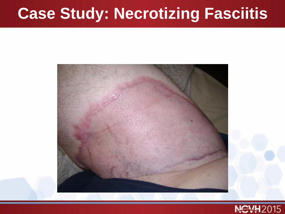
Case Study: Necrotizing Fasciitis

Closing Remarks / Thank You

HYPERBARIC OXYGEN THERAPY:Nursing Considerations
Julio R. Garcia CHT, ACHRNProgram DirectorSpringhill Medical Center


![90-0076F T.Ox Nursing Inservice.ppt [Read-Only] - …€¢ T.Ox Measures Tissue Oxygen saturation ... console by lining up the green arrows. ... 90-0076F T.Ox Nursing Inservice.ppt](https://static.documents.pub/doc/80x56/5b0014ff7f8b9a84338c1a54/90-0076f-tox-nursing-read-only-tox-measures-tissue-oxygen-saturation.jpg)


