120
Hypertensive Disorders in Pregnancy Dr. Jograjiya G.R. Post Graduate Student ESI-PGIMSR, Basaidarapur, New Delhi.
| Date post: | 14-Jul-2015 |
| Category: |
Healthcare |
| Upload: | jograjiya-gelabhai-raghubhai |
| View: | 910 times |
| Download: | 3 times |

Hypertensive Disorders in Pregnancy
Dr. Jograjiya G.R.
Post Graduate Student
ESI-PGIMSR, Basaidarapur, New Delhi.

INTRODUCTION
It is associated with severe maternal obstetric
complications.
Incidence is 5-10%.
The most frequent cause of iatrogenic
prematurity.
Preterm delivery
Intrauterine growth restriction (IUGR)
Perinatal death
Maternal cerebrovascular accidents
Placental abruption

Hypertension in Pregnancy
Systolic B.P. ≥ 140 mmHg
and/or
Diastolic B.P. ≥ 90 mmHg
Documented on two occasions
At least 6 hours apart
Not more than 7 days apart
Readings should be confirmed using appropriate
measurement technique, and should be remeasured after
10-15 minutes of rest.
Other Criteria (Not part of definition currently)
SBP increased by 30mmHg
DBP increased by 15mmHg
Mean Arterial Pressure increased by 20mmHg

SEVERITY HYPERTENSION
Non-severe hypertension: SBP
140-159 mmHg or DBP 90-109
mmHg.
Mild: SBP 140-149 mmHg or
DBP 90-99mmHg.
Moderate: SBP 150-160 mmHg
or DBP 100-110 mmHg.
Severe hypertension: SBP >
160 mm Hg or DBP > 110 mmHg
or both.

How to Measure Blood
Pressure
Sitting Position
Patient Relaxed
Arm well supported
Measured in right arm
Cuff at heart level
Proper cuff size (80% of arm
circumference)
Slow deflation of bladder
(2mmHg/s)
From start of Korotkoff I to end
of Korotkoff V

Normal Blood Pressure
changes in Pregnancy
• Decreases during the first
trimester,
• Reaching its lowest point at 20
weeks,
• Returns to pre-pregnancy
levels during the third
trimester.

What is Significant
Proteinuria in Pregnancy
Total protein in 24 hours urine
> 300mg
Protein : Creatinine ratio in
random sample > 0.1

Calcification and
Definitions

Classification
2. Pre-eclampsia
4. Eclampsia
3. Preeclampsia superimposed
on chronic hypertension
5. Chronic hypertension
with pregnancy
1. Gestational hypertension

GESTATIONAL HYPERTENSION
New onset of hypertension after
20 weeks of gestation without
proteinuria or other features of
preeclampsia, followed by return
of B.P. to normal within 12
weeks post-partum.
This terminology replaces the term
“Pregnancy Induced Hypertension.”

Gestational HTN:
DIAGNOSIS
Determine the severity of
hypertension
Measure protein excretion
24-hour urine collection
Evaluate for signs/symptoms of
severe preeclampsia
Perform laboratory evaluation
+/- end - organ
involvement

Gestational HTN: DIAGNOSIS
CRITERIA FOR MILD GESTATIONAL HYPERTENSION
Blood Pressure > 140 to < 160 mm Hg, systolic
> 90 to < 110 mm Hg, diastolic
Proteinuria < 300 mg per 24-hr collection
Platelet count > 100,000/mm3
Liver enzymes Normal
Maternal symptoms Absent
IUGR / Oligohydramnios Absent

Gestational HTN: MANAGEMENT
Mild Gestational HTN
Managed as outpatients (weekly antepartum
visits)
Daily fetal movement/kick counting
NST + AFI OR BPS
Fetal growth monitoring every 3-4 weeks
No antihypertensive therapy
No antenatal corticosteroids
Deliver patients no later than their EDD

Gestational HTN: MANAGEMENT
Severe Gestational HTN
SBP ≥160 mmHg or DBP
≥110 mmHg is treated with
antihypertensive agents
> 34 wks AOG DELIVER!
< 34 wks AOG give
steroids


Gestational HTN:
Risk of Progression to Preeclampsia
15-25% risk
Women with early onset of
gestational hypertension
are more likely to progress
to preeclampsia than
women with late onset

Gestational HTN:
RECURRENCE
Prevalence: 22 - 47 % (2nd
pregnancy)
tends to recur with
subsequent pregnancies

Gestational HTN:
LONG-TERM PROGNOSIS
associated with
development of HTN later
in life
associated with
development of diseases
related to hypertension
(CVD, CKD,DM)

PREECLAMSIA
New onset of hypertension after 20
weeks of gestation along with properly
documented proteinuria or end-organ
dysfunction symptoms, followed by
return of B.P. to normal within 12
weeks post-partum.
Preeclamsia Gestational Hypertension Proteinuria

Note it……………………..
Preeclampsia can also occur
without proteinuria, with end-
organ dysfunction manifestations.
Edema is no longer considered a
specific diagnostic criterion for
preeclampsia.


Risk Factors
Genetic
Age & parity
Partner factors
Pregnancy Factors
Underlying Medical Conditions
Others
Risk Factors

Risk Factors: Cont.
Genetic
Genetic Predisposition
Family History
Race & Ethnicity
More Common in black & Asians
Pregnancy by ovum donation
Age &Parity
Teenage pregnancy <18 yrs
Age>35 yrs
Long interval between
pregnancy >10 years
Nulliparity
Partner Factors
Change of partner
Limited sperm exposure
Pregnancy by donor
insemination
Partner fathered an eclamptic pregnancy

Risk Factors: Cont.
Pregnancy Factors
Multiple pregnancy
Hydatiform mole
Hydrops fetalis
Fetal chromosomal anomaly
(trisomy 13)
Underlying Medical Diseae
Chronic hypertension
Diabetes mellitus
Renal Disease
Cardiovascular disease
Hyperthyroidism
Metabolic Syndrome
Others
Obesity BMI> 35 kg/m2
Psychological stress & strain
Smoking
Previous history of preeclamsia
• Hyperhomocysteinemia ,
• Autoimmune disease
• Antiphospholipid antibodies,
• Thrombophilia

PATHOPHYSIOLOGY
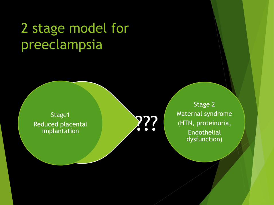
2 stage model for
preeclampsia
Stage 2
Maternal syndrome
(HTN, proteinuria,
Endothelial dysfunction)
Stage1
Reduced placental implantation ???

Reduced placental
implantation –Stage-1
PREDISPOSING FACTORS:
Abnormal implantation
Association with microvascular
diseases (diabetes,
hypertension etc.)
Association with large
placentas (hydrops, multiple
gestation, hydatidiform mole)


Net effect
Replacement of endothelial lining & muscular arterial wall by trophoblast cells
Distended tortuous spiral arteries
Low resistence, low pressure, high flow system

ETIOLOGICAL FACTORS
Placental hypoxia
Immunological factors
Placental enzymes
Genetic factors (MTHFR, F5,)
Oxidative stress

What causes maternal
syndrome
Stage 2
Maternal syndrome
(HTN, proteinuria,
Endothelial dysfunction)
Stage1
Reduced placental implantation
What gets into maternal circulation

Maternal Syndrome
stage-II
Not just hypertension and
proteinuria
But also involves different end
organs

Physiology of maintain
uteroplacental flow in
Normal pregnancy Placenta releases angiotensinase
destruction of angiotensin-II(a
potent vasoconstrictor) BP
stabilized
Vascular synthesis of PGI-2 and
NO in excess vasodilation BP
stabilized & uteroplacental flow
maintains
Release of VEGF restores
uteroplacental flow

Normal balance of agonist &
anta-gonistic factors:
1.vasodialator &
vasoconstrictor
2. angiogenic and
antiangiogenic factors
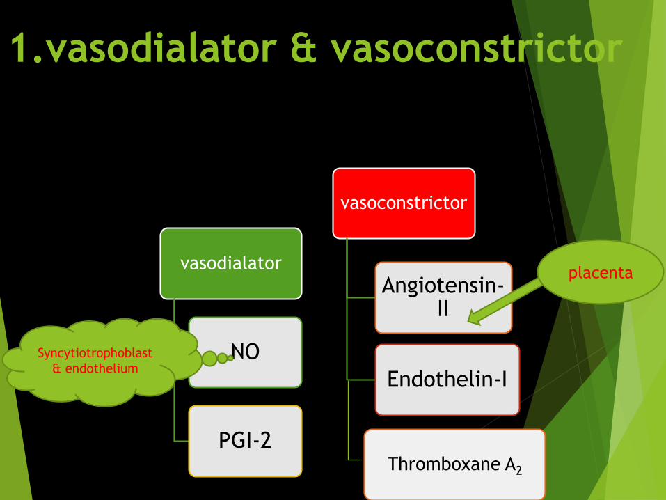
1.vasodialator & vasoconstrictor
vasodialator
NO
PGI-2
vasoconstrictor
Angiotensin-II
Endothelin-I
Thromboxane A2
placenta
Syncytiotrophoblast
& endothelium

2. angiogenic and
antiangiogenic factors
Angiogenicfactor
• VEGF
• TFG-beta• PlGF
Antiangiogenicfactor
• sFlt-1
• sEng


Gestational Hypertension
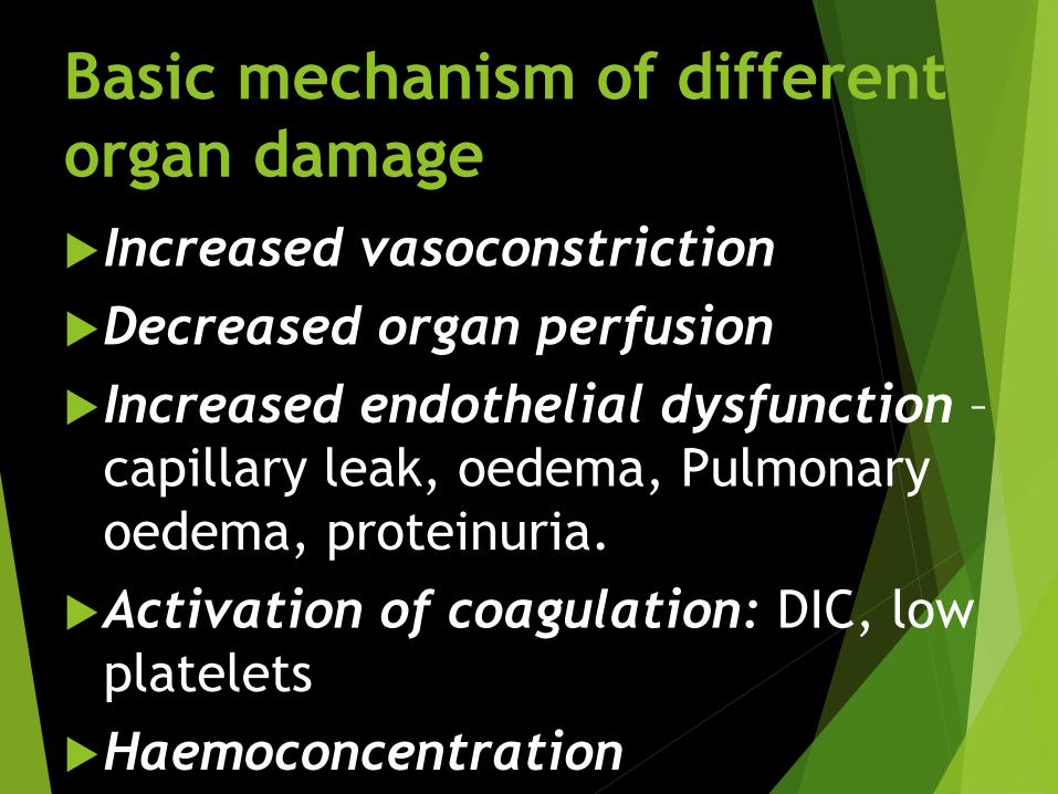
Basic mechanism of different
organ damage
Increased vasoconstriction
Decreased organ perfusion
Increased endothelial dysfunction –
capillary leak, oedema, Pulmonary
oedema, proteinuria.
Activation of coagulation: DIC, low
platelets
Haemoconcentration

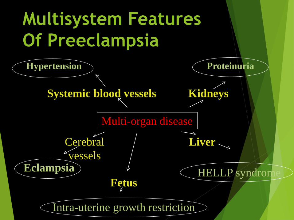
Multisystem Features
Of Preeclampsia
Hypertension Proteinuria
Eclampsia HELLP syndrome
Intra-uterine growth restriction
Multi-organ disease
Cerebral
vessels
Fetus
Liver
Systemic blood vessels Kidneys

Organ Damage
utero-placenta IUGR
Hematological Epistaxis, DIC like features,
hemoconcentration
CNS Cerebral edema, cerebral hge seizures
Heart Subendothelial hge , focal necrosis & hge,
cardiomyopathy, heart failure
Lungs Pulmonary edema, hemorrhagic
brochopneumonia
Kidneys glomerular endotheliosis, oliguria
liver Subcapsular hge, ischaemiaperiportal
necrosis, HELLP

CVS involvement:
• ↑afterload↑ed peripheral resistance
•↓preload ↓ed pregnancy induced
hypervolumia
•Pulmonary leak edemaalveolar endothelial damage & ↓ed plasma
oncotic pr
•hemoconcentration & ↑edhematocrit
↓ed blood volume than normal pregnancy(16%
vs 50%):
Heart failure
↓cardiac output

Hematological system
Thrombocytopenia & other PL
abnormality:
• ↑ed PL activation & degranulation,
• ↓ed life span.
• Corelates well wthdisease severity.
Intravascular hemolysis
• endothelial damage & altered fluidity of erythrocyte membrane d/t change in serum lipid content →↑ed LDH, spherocytosis, reticulocytosis
• microangiopathichemolysis
↑ed coagulation & fibrinolysis
• Feature like DIC
• Release of thromboplastin
• ↓fibrinogen
• AT-III
• plasminogen

Renal system involvement:
↓ed renal perfusion :(d/t ↓ed blood volume & ↑ed
afferent arteriolar pr.)
↓ed GFR : d/t
glomerular capillary endotheliosis
Endothelial dysfunction + mesangial swelling + BM
disruption
(but podocyte disruption minimal)
Oliguria
↑ed creatinine level
↑ed uric acid

Hepatic involvement:
Periportalhemorrhagic
necrosis
hematoma formation
Stretch/Rupture
epigastric pain

Brain involvement:
Acute severe HTN
cerebrovascular overregulation
Vasospasm
Parenchymal ischemia
Cytotoxic edema
sudden ↑↑SBP
exceeds normal range of cerebrovascular autoregulation
Forced vasodilation + hyperperfusion
Vasogenic edema

Lungs involvement:
High SBP
↑ed arteriolar pr
↑ed extravasation of blood into alveoli + rupture of arteriole
Pulmonary edema, hemorrhagic brochopneumonia

PREECLAMPSIA PREDICTION
There are many test during early pregnancy—
or across pregnancy—of various biological,
biochemical, and biophysical markers
implicated for preeclampsia prediction.
The most predictive investigative procedures
are cumbersome, time-consuming and with
poor sensitivity and with poor positive
predictive value for preeclampsia.
The efficacy of the preventive methods is
questionable too
Currently, no screening tests are predictably
reliable, valid, and economical
(Kleinrouweler, 2012).

Endothelial Dysfunction/Oxidant Stress
Feto-Placental unit Endocrine Dysfunction
Renal Dysfuntion Misc
Placental Perfusion/ Vascular Resistance related Tests
Uterine Artery Doppler Velocimetry
AT- III
ANPFree fetal DNA
Adapted from Conde-Agudelo and associates (2009)
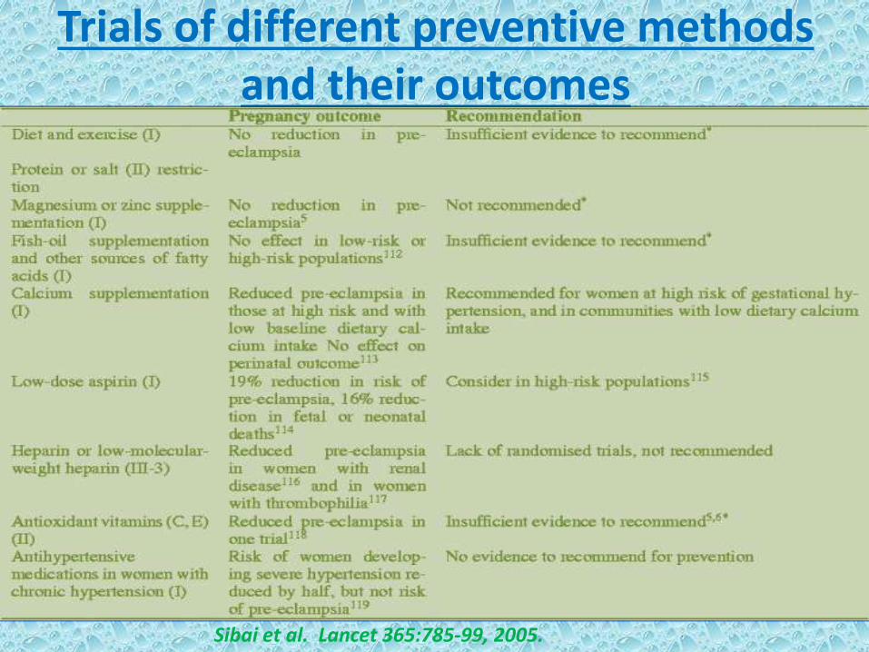
Trials of different preventive methods and their outcomes
Sibai et al. Lancet 365:785-99, 2005.

Provocative
Pressure Tests
Three tests have been extensively
evaluated to assess the blood pressure
rise in response to a stimulus.

1.Roll-over test
After resting in the left lateral
position turning to a supine
position induces a rise in
diastolic pressure of 20 mmHg
or more is a positive test
indicative of tendency to
develop pre-eclampsia.
Perform at 28 to 32 weeks
pregnancy.
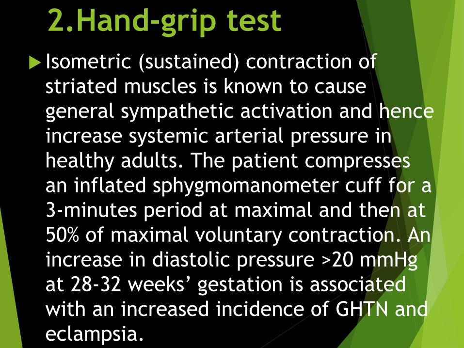
2.Hand-grip test
Isometric (sustained) contraction of
striated muscles is known to cause
general sympathetic activation and hence
increase systemic arterial pressure in
healthy adults. The patient compresses
an inflated sphygmomanometer cuff for a
3-minutes period at maximal and then at
50% of maximal voluntary contraction. An
increase in diastolic pressure >20 mmHg
at 28-32 weeks’ gestation is associated
with an increased incidence of GHTN and
eclampsia.

3. Angiotensin II sensitivity
Sensitivity to infused angiotensin II: is
increased may be due to alteration in
vascular smooth muscle A II receptors.
Sensitivities of all above three tests to
range from 55 to 70 percent, and
specificities approximated 85 percent.
(metaanalysis, Conde-Agudelo and
associates 2014)

• is most promising, but currently, none of them is completely suitable for clinical use. (Conde-Agudelo, 2014; Kleinrouweler, 2012; Myatt, 2012a).
• These have value for fetal-growth restriction but not preeclampsia (ACOG, 2013a).
• As a result of these trials, some methods to prevent Preeclampsia have been theorized…
Uterine Artery Doppler Velocimetry (abnormal flow resistance/ diastolic notch in
2nd/ 3rd trimester)

uterine artery DOPPLER
In preeclamptic mother:
Showing early diastolic NOTCH
Decreased EDF
(due to high resistance)
In normal mother
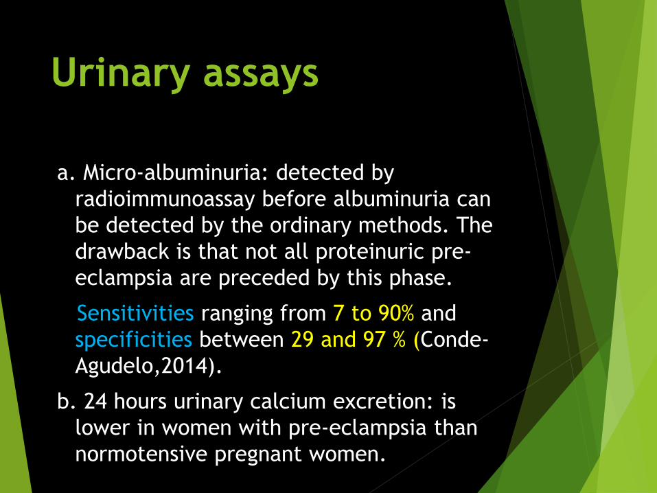
Urinary assays
a. Micro-albuminuria: detected by
radioimmunoassay before albuminuria can
be detected by the ordinary methods. The
drawback is that not all proteinuric pre-
eclampsia are preceded by this phase.
Sensitivities ranging from 7 to 90% and
specificities between 29 and 97 % (Conde-
Agudelo,2014).
b. 24 hours urinary calcium excretion: is
lower in women with pre-eclampsia than
normotensive pregnant women.
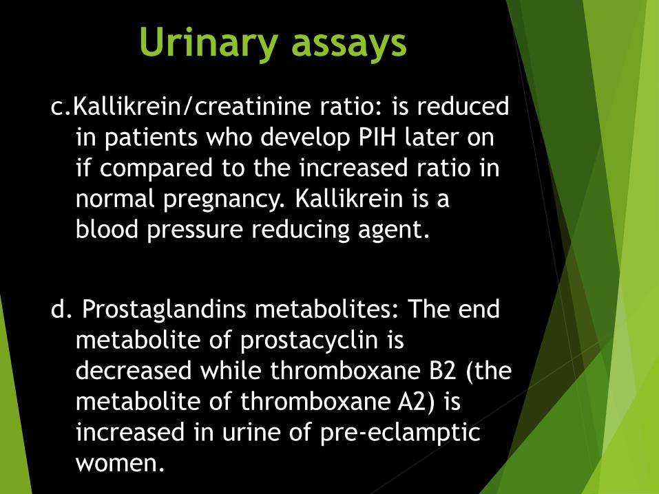
Urinary assays
c.Kallikrein/creatinine ratio: is reduced
in patients who develop PIH later on
if compared to the increased ratio in
normal pregnancy. Kallikrein is a
blood pressure reducing agent.
d. Prostaglandins metabolites: The end
metabolite of prostacyclin is
decreased while thromboxane B2 (the
metabolite of thromboxane A2) is
increased in urine of pre-eclamptic
women.

Blood tests
a. Plasma urate: serial increase is a
warning of PIH before appearance
of other clinical features.
b.Platelet count: a reduction
occurs early in pre-eclampsia.
c. Anti-thrombin - III activity: begin
to decline as much as 13 weeks
prior to the development of clinical
manifestations of pre-eclampsia.
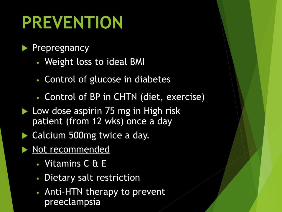
PREVENTION
Prepregnancy
• Weight loss to ideal BMI
• Control of glucose in diabetes
• Control of BP in CHTN (diet, exercise)
Low dose aspirin 75 mg in High risk patient (from 12 wks) once a day
Calcium 500mg twice a day.
Not recommended
• Vitamins C & E
• Dietary salt restriction
• Anti-HTN therapy to prevent preeclampsia
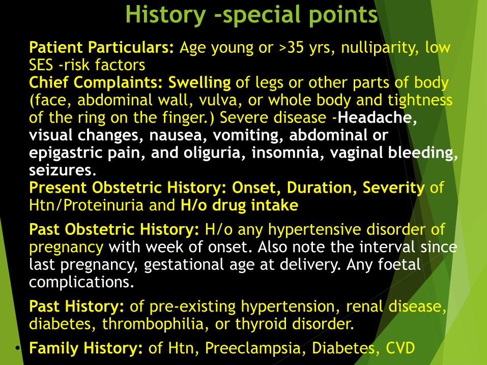
History -special points
• Patient Particulars: Age young or >35 yrs, nulliparity, low SES -risk factors
• Chief Complaints: Swelling of legs or other parts of body (face, abdominal wall, vulva, or whole body and tightness of the ring on the finger.) Severe disease -Headache, visual changes, nausea, vomiting, abdominal or epigastric pain, and oliguria, insomnia, vaginal bleeding, seizures.
• Present Obstetric History: Onset, Duration, Severity of Htn/Proteinuria and H/o drug intake
• Past Obstetric History: H/o any hypertensive disorder of pregnancy with week of onset. Also note the interval since last pregnancy, gestational age at delivery. Any foetal complications.
• Past History: of pre-existing hypertension, renal disease, diabetes, thrombophilia, or thyroid disorder.
• Family History: of Htn, Preeclampsia, Diabetes, CVD

Physical Examination:● Obesity/BMI >35 kg/m2
● Weight (serial measurements): Gain in wt at the rate of >500gs a week or
2.5kgs a month in the later months of pregnancy may be the earliest sign
of preeclampsia.
● Oedema (all sites): has to be pathological, meaning visible pitting edema
demonstratable over the ankles after 12 hrs bed rest.
● Pulse
● B.P.:
○ right arm, sitting/supine, arm at level of heart, cuff length=1.5
times of arm circumference, diastolic BP is the disappearance of
Korotkoff sounds (phase V)
○ taken on 2 occasions at least 6 hrs apart for confirmation of
diagnosis.
● CVS examination: auscultation for heart rate, rhythm, splitting of S2,
murmurs.
● Ophthalmic examination: retinal haemorrage, nicking of veins,
arteriole/vein ratio 3:1 from 3:2, papilloedema
● Deep tendon reflexes: hyperreflexia/presence of clonus

Obstetric Examination:
Nothing special is found except features of IUGR, oligohydramnios in some cases.

Maternal Investigations:
Tests may be abnormal even when BP elevation is minimal.
• Urine dipstick testing for proteinuria
o Quantitation by laboratory methods if ≥1+ on dipstick testing
o Urinary ACR(albumin-creatinine ratio) to detect significant
proteinuria (≥30mg/mmol)
o 24 hour urine collection is not necessary in routine clinical
management
• Routine Blood Examination: TLC, DLC, Peripheral Smear, BT, CT,
Hb%
• Serum Urea, creatinine, electrolytes including lactate
dehydrogenase (LDH) and uric acid.
• Liver function tests (LFT) -AST, ALT >70 IU/l
• Skiagram of chest –PA view, Pulmonary Capillary Wedge
Pressure (PCWP), Brain Natriuretic Peptide (BNP) for
detection of pulmpnary oedema

Diagnosing Preeclampsia-Eclampsia:• Blood pressure ≥ 140/90 mm of Hg (at
or after 20 weeks of gestation) on 2
occasions at least 6 hours apart during
bed rest. (160/90 mm of Hg is severe
disease)
• accompanied by one or more of:
o significant proteinuria
-urinary dipstick 1+
-random urinary
protein/creatinine
ratio ≥ 30 mg/mmol
-24 hour urine excretion ≥300
mg/24 hrs
o renal involvement
-serum creatinine ≥ 90 mmol/L
or
-oliguria (<400 ml in 24 hrs)
o haematological involvement
-platelet count <1 lakh
o liver involvement
-raised AST, ALT (>70 IU/l)
-severe upper abdominal pain
o neurological involvement
-severe headache
-persistent visual disturbances
-hyperreflexia with sustained
clonus
-convulsions (eclampsia)
-stroke
o pulmonary oedema
o fetal growth restriction
o placental abruption
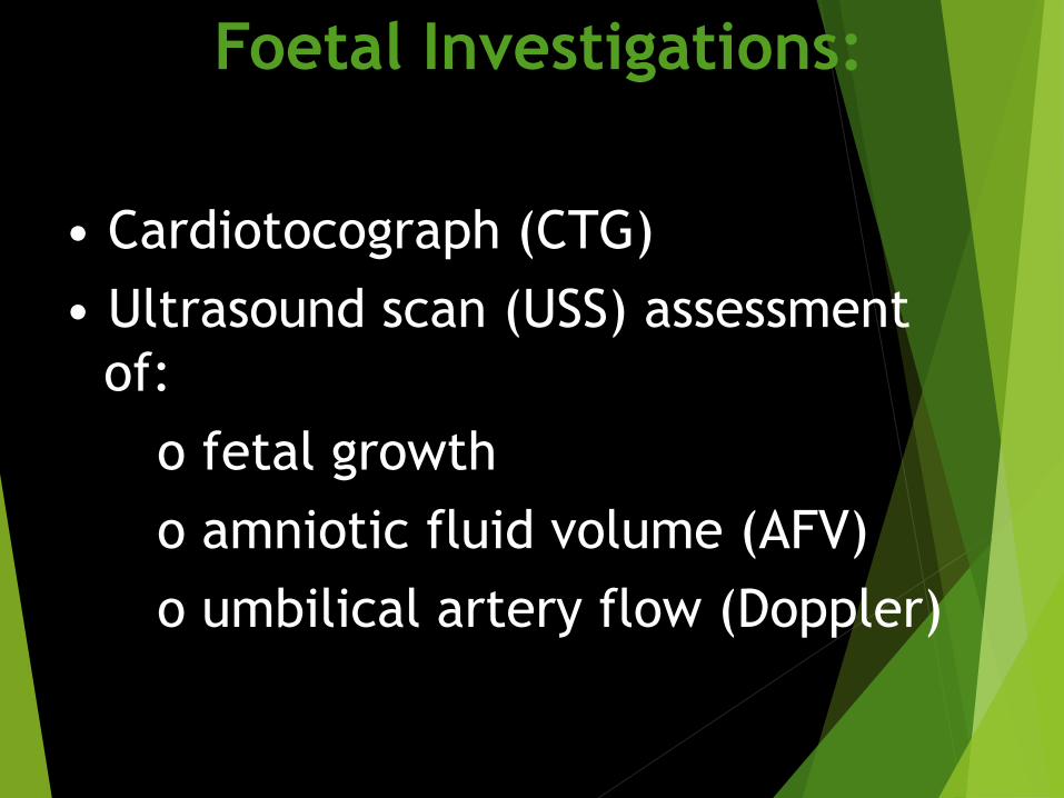
Foetal Investigations:
• Cardiotocograph (CTG)
• Ultrasound scan (USS) assessment
of:
o fetal growth
o amniotic fluid volume (AFV)
o umbilical artery flow (Doppler)

Differential Diagnosis
Pre-existing hypertension
New/gestational hypertension
Pre-eclampsia
Eclampsia
Exacerbation of underlying renal disease/Superimposed pre-eclampsia-eclampsia
SLE

N.B: Grades of proteinuria (in g/L): Trace=0.1, 1+=0.3, 2+=1, 3+=3,
4+=10
Hypertensive Disorders in Pregnancy
Gestational HTN
● BP ≥ 140/90mmHg
●No evidence of underlying cause of HTN
●No associated symptoms
●Comes to normal within 12 wks of delivery
Pre-eclampsia
Non Severe Severe
Eclampsia
PreEclamsia
+
Convulsion
±
Coma
N.B: Pre-eclampsia is
principally a syndrome of
signs and when symptoms
appear it is usually late.

Indicators of severity of Pre-eclampsiaABNORMALITIES NONSEVERE SEVERE
Blood pressure ≥140/90mmHg but
<160/110mmHg
≥160/110mmHg
Proteinuria ≤2+ ≥3+
Oliguria Absent <400ml/day
Headache Absent Present
Visual disturbances Absent Present
Platelet count Normal Thrombocytopenia
(<100,000/mm3)
HELLP syndrome Absent May be present
ALT,AST >70 IU/L
LDH>600 IU/L
Bilirubin >1.2g/L
Serum transaminases(AST,ALT) Normal (<40 IU/L) Elevated
Serum Creatinine Normal Elevated
Epigastric pain Absent Present
Fetal growth restriction Absent Obvious
Pulmonary oedema Absent present

MANAGEMENT
Definitive treatment: DELIVERY!
Based on:
AOG
Severity of PE
Maternal / Fetal condition

NONSEVERE PE: MANAGEMENT
Deliver at ≥37 weeks of
gestation
Labor induction encouraged

For Nonsevere - controlled disease :
There after induction may be done at term depending on cervical condition
Can be managed expectantly till term at home/hospital and continued till term.
74

What is EXPECTANT MANAGEMENT?
NO
YES
Neither forced nor restricted

EXPECTANT ANTEPARTUM MANAGEMENT OF
NONSEVERE PREECLAMPSIA
Inpatient vs outpatient care
Close maternal monitoring upon diagnosis
of preeclampsia is important to establish
disease severity and the rate of
progression
Hospitalization is useful for making these
assessments and facilitates rapid
intervention in the event of rapid
progression
Outpatient care is a cost-effective option
for women with stable mild preeclampsia
after initial dx evaluation

EXPECTANT ANTEPARTUM MANAGEMENT
OF NONSEVERE PREECLAMPSIA
Laboratory follow – up
platelet count, serum creatinine,
serum AST
1-2x/wk, assess disease
progression
Assessment of fetal well-being
daily fetal movement count
twice weekly fetal NST with AFI
or
twice weekly BPS
UMA Doppler indices evaluation

EXPECTANT ANTEPARTUM MANAGEMENT
OF NONSEVERE PREECLAMPSIA
Assessment of fetal growth
Sonographic estimation of fetal weight
done to look for growth restriction and
oligohydramnios at the time of diagnosis
of PE , repeated every 3 weeks if the
initial examination is normal
Antenatal corticosteroids
< 34 weeks AOG

But wait…can antihypertensives be used in expectant management???
• In non-severe Pregnancy hypertension – No clear
Evidence of benefit other than to reduce
The Frequency of Episodes of Severe
hypertension
• May Adversely Effect Fetal Growth velocity

Fetal considerations
Prematurity
Stillbirth
Newborn asphyxia
Maternal considerations
Worsening of disease
Complications
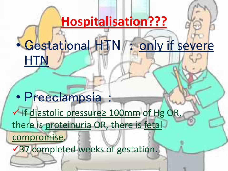
Hospitalisation???
• Gestational HTN : only if severe HTN
• Preeclampsia : If diastolic pressure≥ 100mm of Hg OR, there is proteinuria OR, there is fetal compromise.37 completed weeks of gestation.

INTRAPARTUM MANAGEMENT
Intrapartum monitoring
Fluids
monitored closely to avoid
excessive administration,
since women with severe
disease are at risk of
pulmonary edema and
significant third-spacing

DELIVERY CARE
• For any HDP, vaginal delivery should be considered unless a CS is required for the usual obstetric indications.
• Antihypertensives : continued throughout labour to maintain BP < 160/110 mmHg .
• 3rd Stage : actively managed with 10 units IM, particularly in the presence of thrombocytopenia or coagulopathy. (I-A)
• Ergometrine should NOT be given

PES: MANAGEMENTDeliver regardless of gestational age
if proteinuria ( ≥5 grams) is the
only criteria for severe disease
managed as nonsevere PE
mild fetal growth restriction with
reassuring Doppler velocimetry
treat conservatively *
severe hypertension treat
conservatively *
NOTE: * remote from term

For early onset severe preeclampsia:
• Controversy regarding termination in early onset disease
• But there is no beneficial role for mother, as well as perinatal mortality is also high instead of conservative management
• So…
85
termination is seriously considered

suspected
severe
preeclampsia
at < 34 weeks


Acute Management of PESSet 1: Labetalol first protocol
•Notify OB provider when patient presents with severe HTN (systolic ≥ 160 mmHg or diastolic > 110 mmHg)
• Initiate appropriate fetal surveillance
• Labetalol 20 mg IV over 2 min; recheck BP in 10 min; if still above either threshold then:
• Labetalol 40 mg IV over 2 min; recheck BP in 10 min; if still above either threshold then:
• Labetalol 80 mg IV over 2 min; recheck BP in 10 min; if still above either threshold then:
• Hydralazine 10 mg IV over 2 min; recheck BP in 20 min; if still above either threshold then:
• Emergency consultation with maternal fetal medicine (MFM), anesthesia, internal medicine, critical care specialist.
• Give additional antihypertensive medication per specific order.
• Once the aforementioned BP thresholds are achieved, repeat BP measurement every 10 minutes for 1 hour, then every15 minutes for 1 hour, then every 30 minutes for 1 hour, and then every hour for 4 hours.
• Institute additional BP timing per specific order.

Acute Management of PES
Set 2: Hydralazine first protocol
• Notify OB provider when patient presents with severe HTN (systolic ≥ 160 mmHg or diastolic ≥ 110 mmHg)
• Initiate appropriate fetal surveillance
• Hydralazine 5 or 10 mg IV over 2 min; recheck BP in 20 min; if still above either threshold then:
• Hydralazine 10 mg IV over 2 min; recheck BP in 20 min; if still above either threshold then:
• Labetalol 20 mg IV over 2 min; recheck BP in 10 min; if still above either threshold then:
• Labetalol 40 mg IV over 2 min and;
• Obtain emergency consultation with MFM, anesthesia, internal medicine, critical care specialist.
• Give additional antihypertensive medication per specific order.
• Once the aforementioned BP thresholds are achieved, repeat BP measurement every 10 minutes for 1 hour, then every 15 minutes for 1 hour, then every 30 minutes for 1 hour, and then every hour for 4 hours
• Institute additional BP timing per specific order.
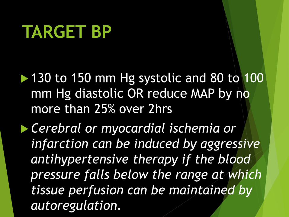
TARGET BP
130 to 150 mm Hg systolic and 80 to 100
mm Hg diastolic OR reduce MAP by no
more than 25% over 2hrs
Cerebral or myocardial ischemia or
infarction can be induced by aggressive
antihypertensive therapy if the blood
pressure falls below the range at which
tissue perfusion can be maintained by
autoregulation.
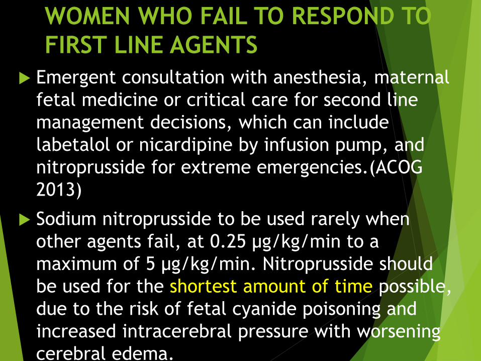
WOMEN WHO FAIL TO RESPOND TO
FIRST LINE AGENTS
Emergent consultation with anesthesia, maternal
fetal medicine or critical care for second line
management decisions, which can include
labetalol or nicardipine by infusion pump, and
nitroprusside for extreme emergencies.(ACOG
2013)
Sodium nitroprusside to be used rarely when
other agents fail, at 0.25 μg/kg/min to a
maximum of 5 μg/kg/min. Nitroprusside should
be used for the shortest amount of time possible,
due to the risk of fetal cyanide poisoning and
increased intracerebral pressure with worsening
cerebral edema.

Seizure Prophylaxis
MgS04 given to mild / severe PE
Loading dose: 4-6 g, slow IV
push, over 15-20 mins
Continuous infusion: 1-2 g/hr
OR 5g IM into each buttock
(total 10 g) followed by 5 g IM,
alternate buttocks ever 4h
continued for 24 hours after last
convulsion or delivery.

MgS04 Toxicity
1. Impaired breathing(@8-10meq/L)
2. Arrythmia and Asystole ( @10-13 mEq/L)
3. Decreased/absent deep tendon reflex
(Hyporeflexia at 4 mEq/L, loss of patellar reflex at 7-10 mEq/L)
4. Shock (>13 mEq/L)
• For a maintenance dose following must be present -
Serum Mg level 4-7meq/l(twice daily)
Having Patellar reflex
Urine output >30ml/hr
RR>12/min

WHAT If magnesium toxicity is suspected???
Administration of 10mL of 10% calcium gluconate (1 g in total) as a slow intravenous push.
Serum magnesium level obtained.
Magnesium infusion should be discontinued, supplemental oxygen administered,

For severe-uncontrolled disease:
LUCS OR In case of very severe uncontrolled disease elective LUCS may be done without induction
Preinduction
Cervical ripening with prostaglandin followed by induction
Termination is considered
95
If failed

Postpartum Management
NSAIDs
for pain control should be avoided in
women with poorly controlled
hypertension, oliguria, renal
insufficiency, or thrombocytopenia
patient controlled fentanyl or
remifentanil analgesia or epidural.
Monitor VS q 2h while on MgS04
Treat PES
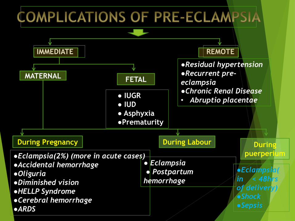
IMMEDIATE REMOTE
MATERNALFETAL
● IUGR
● IUD
● Asphyxia
●Prematurity
During Pregnancy During Labour During
puerperium●Eclampsia(2%) (more in acute cases)
●Accidental hemorrhage
●Oliguria
●Diminished vision
●HELLP Syndrome
●Cerebral hemorrhage
●ARDS
● Eclampsia
● Postpartum
hemorrhage
●Eclampsia(
in < 48hrs
of delivery)●Shock
●Sepsis
●Residual hypertension
●Recurrent pre-
eclampsia●Chronic Renal Disease
• Abruptio placentae

HELLP SyndromeThis is an acronym for Hemolysis, Elevated Liver
enzymes, and Low Platelet count.
It is a rare multisystem disorder that complicates
pregnancy with lab evidences of micro-
angiopathic hemolysis, hepatic dysfunctioning &
thrombocytopenia.
It is a complication mostly associated with Pre-
eclampsia but can also be diagnosed (rarely
though) in the absence of these disorders.
Incidence 20% of women with severe
preeclampsia.
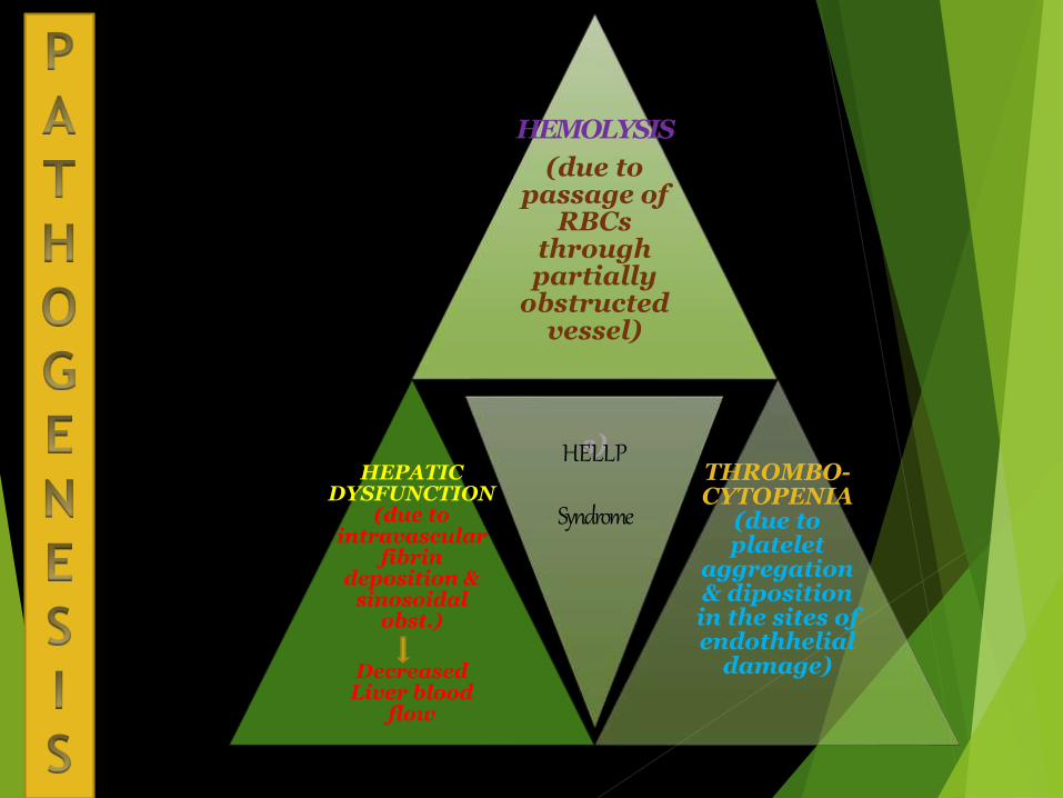
HEMOLYSIS
(due to passage of
RBCs through partially
obstructed vessel)
s)HEPATIC
DYSFUNCTION(due to
intravascular fibrin
deposition & sinosoidal
obst.)
Decreased Liver blood
flow
HELLP
Syndrome
THROMBO-CYTOPENIA
(due to platelet
aggregation & dipositionin the sites of endothhelial
damage)

Diagnosis
Hemolysis (Hallmark of
the triad)
Elevated Liver Enzymes Low Platelet Count
LDH>600IU/L Liver Enzymes (<100,000/cu.mm)
Low serum haptoglobin
High serum bilirubin
(>1.2 mg/dl)
High ALT & AST
(>70 IU/L)
Abnormal PBS
(Schistocytes {helmet
cells} , burr cells
{Echinocytes})
Later-low Hb%
• ●Epigastric /Right Upper Quadrant pain
• ●Nausea, Vomiting1. Clinical Features:
2. Lab Investigation:

HELLP Syndrome
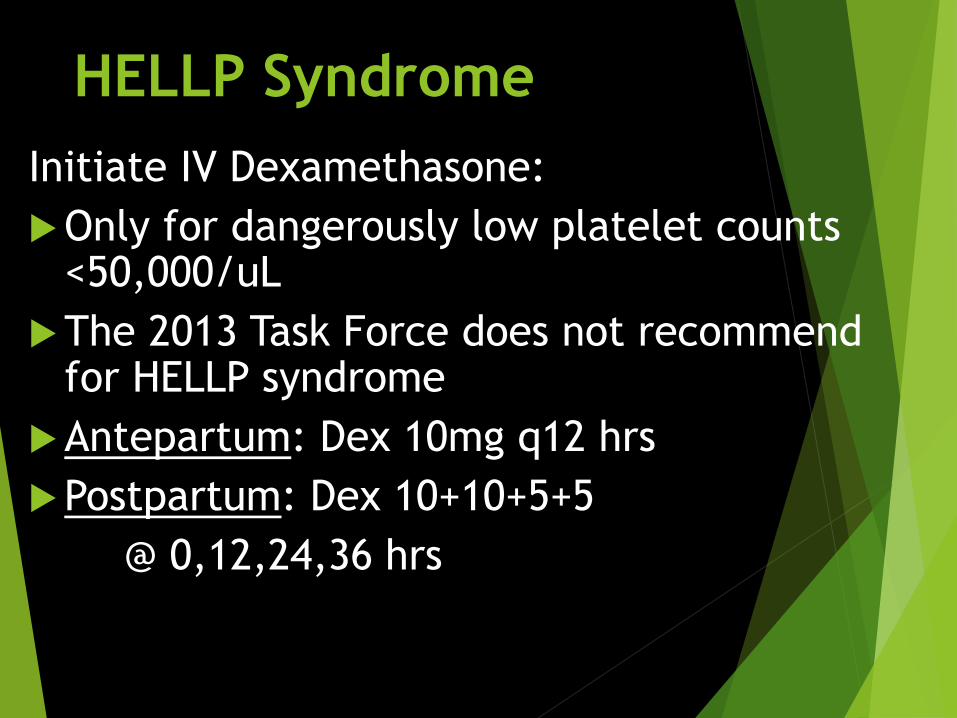
Initiate IV Dexamethasone:
Only for dangerously low platelet counts <50,000/uL
The 2013 Task Force does not recommend for HELLP syndrome
Antepartum: Dex 10mg q12 hrs
Postpartum: Dex 10+10+5+5
@ 0,12,24,36 hrs

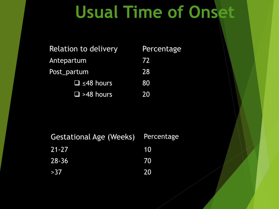
Usual Time of Onset
Relation to delivery Percentage
Antepartum 72
Post_partum 28
≤48 hours 80
>48 hours 20
Gestational Age (Weeks) Percentage
21-27 10
28-36 70
>37 20
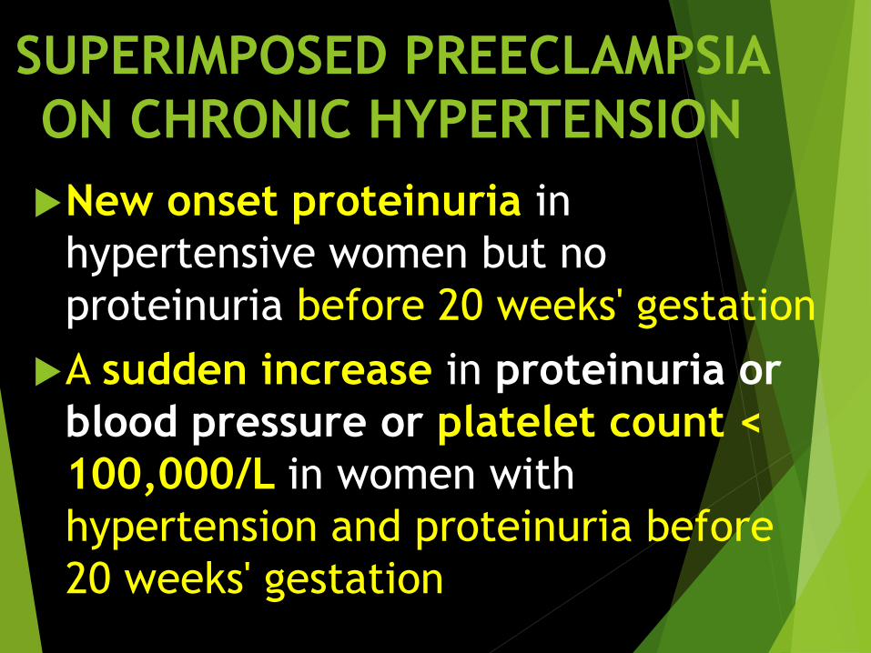
SUPERIMPOSED PREECLAMPSIA
ON CHRONIC HYPERTENSION
New onset proteinuria in
hypertensive women but no
proteinuria before 20 weeks' gestation
A sudden increase in proteinuria or
blood pressure or platelet count <
100,000/L in women with
hypertension and proteinuria before
20 weeks' gestation

ECLAMPSIA
Generalized tonic-clonic seizure in a
patient with Preeclampsia not attributed
to any other cause.
If seizures occur beyond 48-72 hours postpartum
causes may be more likely other than eclampsia.
occurs in 2 to 3 percent of severely preeclamptic
women not receiving anti-seizure prophylaxis
Eclampsia Preeclampsia
Seizure/
Convulsion/
Coma

D/D ECLAMPSIA
Epilepsy,
Intracranial
haemorrhage/thrombosis,
Meningitis,
Cerebral malaria,
Amniotic fluid embolism
can mimic eclampsia.

PATHOGENESIS OF SEIZURES
1. Cerebral overregulation in
response to high systemic blood
pressure
vasospasm of cerebral arteries
underperfusion of the brain
localizedischemia/infarction
cytotoxic (intracellular) edema
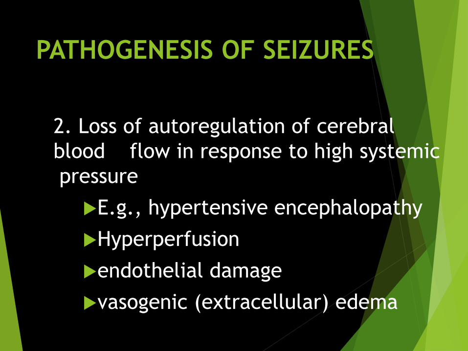
PATHOGENESIS OF SEIZURES
2. Loss of autoregulation of cerebral
blood flow in response to high systemic
pressure
E.g., hypertensive encephalopathy
Hyperperfusion
endothelial damage
vasogenic (extracellular) edema

MANAGEMENT
Iinitial Mx: Maintenance of airway
patency and prevention of
aspiration
Gravida rolled onto her left side
Protect from trauma
Supplemental O2 (8-10L/min via
face mask)

Management of severe hypertension,
if present
Prevention of recurrent seizures
Evaluation for prompt delivery
definitive treatment of eclampsia
is delivery, irrespective of
gestational age
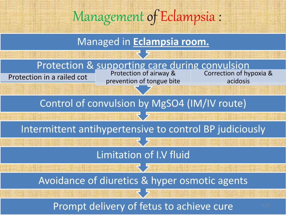
Management of Eclampsia :
Prompt delivery of fetus to achieve cure
Avoidance of diuretics & hyper osmotic agents
Limitation of I.V fluid
Intermittent antihypertensive to control BP judiciously
Control of convulsion by MgSO4 (IM/IV route)
Protection & supporting care during convulsionProtection in a railed cot
Protection of airway & prevention of tongue bite
Correction of hypoxia & acidosis
Managed in Eclampsia room.
113

to control convulsion
“It is the most effective drug to control even recurrent seizures without any central nervous system depression to mother & fetus”
114
Magnesium sulphate

Dosages
→Paralysing agent & Intubation
→Amobarbital 250mg I.V over 3 min
In case of uncontrolled recurrent seizure (10-15%) : →additional 2-4g of 20% solution IV @ <1g/min
→4gm of 20% solution IV slowly(@ <1g/min) + 10g of 50% solution deep IM in upper & outer quadrant of buttock by a wide bore needle then 5g
of 50% solution IM 4hrly similarly
IM regime (Pritchard protocol):1955
→4 gm loading in 100ml of IVF over 15-20 min followed by 2-3g/hr in 100 ml IVF as maintenance
I.V regime (Sibai protocol):1990
IM doses are as active as IV doses in controlling seizures
115

• Duration : 24 hrs from last convulsion or from delivery which one is longer.
116

MATERNAL FETAL
●Asphyxia
●Prematurity
●Hypoxia & IUD
Injuries Systemic
●Tongue bite
●Injuries due
to fall
●Bed sore
●PULMONARY: edema,
pneumonia, ARDS,
embolism
●CARDIAC: acute left
ventricular failure
●RENAL: renal failure
●HEPATIC: necrosis,
subcapsular hematoma
●CNS: cerebral
hemorrhage,
edema(vasogenic)
Vision
●Diminished
vision due to
retinal
detachment or
occipital lobe
ischemia
Hematology
●Low platelet
count
●Disseminated
Intravascular
Coagulation
Postpartum
●Shock
●Sepsis
●Psychosis

CHRONIC HYPERTENSION IN
PREGNANCY
Hypertension before pregnancy /
Diagnosed before 20 weeks of
pregnancy not due to gestational
trophoblastic disease.
Hypertension diagnosed after 20
weeks but persistent after 12
weeks postpartum

Chronic HTN & Pregnancy
Etiology :
1. Essential HTN (Most Common)
2. Secondary HTN :
1. Genetic: Glucocorticoid remediable aldosteronism,
Liddle Syndrome
2. Renal : Parenchymal, Renovascular
3. Endocrine : Primary hyperaldosteronism, cushing
syndrome, Pheochromocytoma
4. Vascular : Aortic coarctation, Estrogen use
5. Others
Thank you…


