IS FACE RECOGNITION NOT SO UNIQUE AFTER ALL? Isabel Gauthier Department of Diagnostic Radiology, Yale School of Medicine, New Haven, CT, USA Nikos K. Logothetis Max Planck Institute for Biological Cybernetics, Tuebingen, Germany In monkeys, a number of different neocortical as well as limbic structures have cell populations that re- spond preferentially to face stimuli. Face selectivity is also differentiated within itself: Cells in the infe- rior temporal and prefrontal cortex tend to respond to facial identity, others in the upper bank of the superior temporal sulcus to gaze directions, and yet another population in the amygdala to facial expres- sion. The great majority of these cells are sensitive to the entire configuration of a face. Changing the spatial arrangement of the facial features greatly diminishes the neurons’ response. It would appear, then, that an entire neural network for faces exists which contains units highly selective to complex con- figurations and that respond to different aspects of the object “face.” Given the vital importance of face recognition in primates, this may not come as a surprise. But are faces the only objects represented in this way? Behavioural work in humans suggests that nonface objects may be processed like faces if sub- jects are required to discriminate between visually similar exemplars and acquire sufficient expertise in doing so. Recent neuroimaging studies in humans indicate that level of categorisation and expertise interact to produce the specialisation for faces in the middle fusiform gyrus. Here we discuss some new evidence in the monkey suggesting that any arbitrary homogeneous class of artificial objects—which the animal has to individually learn, remember, and recognise again and again from among a large number of distractors sharing a number of common features with the target—can induce configurational selec- tivity in the response of neurons in the visual system. For all of the animals tested, the neurons from which we recorded were located in the anterior inferotemporal cortex. However, as we have only re- corded from the posterior and anterior ventrolateral temporal lobe, other cells with a similar selectivity for the same objects may also exist in areas of the medial temporal lobe or in the limbic structures of the same “expert” monkeys. It seems that the encoding scheme used for faces may also be employed for other classes with similar properties. Thus, regarding their neural encoding, faces are not “special” but rather the “default special” class in the primate recognition system. INTRODUCTION The current debate on whetherfaces are “special” or not (Farah, 1996; Tovée, 1998) is firmly rooted in research on humans. The evidence that face recog- nition in humans may be qualitatively different from the recognition of other objects comes from brain lesion studies (e.g. Farah, Levinson, & Klein, 1995a; Moscovitch, Winocur, & Behrmann, 1997; Yin, 1969), behavioural studies (e.g. Farah, Wil- son, Drain, & Tanaka, 1998; Young, Hellawell, & Hay, 1987) and neuroimaging studies (Clark et al., COGNITIVE NEUROPSYCHOLOGY, 2000, 17 (1/2/3), 125–142 Ó 2000 Psychology Press Ltd http://www.tandf.co.uk/journals/pp/02643294.html 125 Requests for reprints should be addressed to Nikos K. Logothetis, Max Planck Institute for Biological Cybernetics, Spemannstr. 38, 72076 Tuebingen, Germany (Tel: +49 7071 601-650; Fax: +49 7071 601-660; Email: [email protected]).
Transcript
IS FACE RECOGNITION NOT SO UNIQUEAFTER ALL?
Isabel GauthierDepartment of Diagnostic Radiology, Yale School of Medicine, New Haven, CT, USA
Nikos K. LogothetisMax Planck Institute for Biological Cybernetics, Tuebingen, Germany
In monkeys, a number of different neocortical as well as limbic structures have cell populations that re-spond preferentially to face stimuli. Face selectivity is also differentiated within itself: Cells in the infe-rior temporal and prefrontal cortex tend to respond to facial identity, others in the upper bank of thesuperior temporal sulcus to gaze directions, and yet another population in the amygdala to facial expres-sion. The great majority of these cells are sensitive to the entire configuration of a face. Changing thespatial arrangement of the facial features greatly diminishes the neurons’ response. It would appear,then, that an entire neural network for faces exists which contains units highly selective to complex con-figurations and that respond to different aspects of the object “face.” Given the vital importance of facerecognition in primates, this may not come as a surprise. But are faces the only objects represented inthis way? Behavioural work in humans suggests that nonface objects may be processed like faces if sub-jects are required to discriminate between visually similar exemplars and acquire sufficient expertise indoing so. Recent neuroimaging studies in humans indicate that level of categorisation and expertiseinteract to produce the specialisation for faces in the middle fusiform gyrus. Here we discuss some newevidence in the monkey suggesting that any arbitrary homogeneous class of artificial objects—which theanimal has to individually learn, remember, and recognise again and again from among a large numberof distractors sharing a number of common features with the target—can induce configurational selec-tivity in the response of neurons in the visual system. For all of the animals tested, the neurons fromwhich we recorded were located in the anterior inferotemporal cortex. However, as we have only re-corded from the posterior and anterior ventrolateral temporal lobe, other cells with a similar selectivityfor the same objects may also exist in areas of the medial temporal lobe or in the limbic structures of thesame “expert” monkeys. It seems that the encoding scheme used for faces may also be employed forother classes with similar properties. Thus, regarding their neural encoding, faces are not “special” butrather the “default special” class in the primate recognition system.
INTRODUCTION
The current debate on whetherfaces are “special” ornot (Farah, 1996; Tovée, 1998) is firmly rooted inresearch on humans. The evidence that face recog-nition in humans may be qualitatively different
from the recognition of other objects comes frombrain lesion studies (e.g. Farah, Levinson, & Klein,1995a; Moscovitch, Winocur, & Behrmann, 1997;Yin, 1969), behavioural studies (e.g. Farah, Wil-son, Drain, & Tanaka, 1998; Young, Hellawell, &Hay, 1987) and neuroimaging studies (Clark et al.,
Ó 2000 Psychology Press Ltdhttp://www.tandf.co.uk/journals/pp/02643294.html
125
Requests for reprints should be addressed to Nikos K. Logothetis, Max Planck Institute for Biological Cybernetics, Spemannstr.38, 72076 Tuebingen, Germany (Tel: +49 7071 601-650; Fax: +49 7071 601-660; Email: [email protected]).
1996; Kanwisher, McDermott, & Chun, 1997;McCarthy, Puce, Gore, & Allison, 1997; Puce,Allison, Gore, & McCarthy, 1995; Sergent, Ohta,& MacDonald, 1992; Sergent & Signoret, 1992).In parallel, we have known of the existence of“face cells” in the monkey brain for many years(Gross, Bender, & Rocha-Miranda 1969). Mon-keys’ face-recognition performance is remarkablysimilar to that of humans (Bruce, 1982; Hamilton& Vermeire, 1983; Lutz, Lockard, Gunderson, &Grant, 1998; Mendelson, Haith, & Goldman-Rakic, 1982; Nahm, Perret, Amaral, & Albright1997; Rosenfield & Van Hoesen, 1979; Wright &Roberts, 1996). It is not surprising, therefore, that agreat deal of neural tissue is devoted to the process-ing of facial information in this species, too. How-ever, perhaps because the techniques are sodifferent, evidence from the animal and human lit-eratures is not fully integrated. The physiologicalevidence from animal research may considerablyenrich the debate and offer information that is lack-ing in humans because of technical and ethical con-straints. On the other hand, the monkey andhuman work may be difficult to compare because oflarge methodological differences. Here we brieflyreview the issues that are most debated regardingthe possibility of face-specific mechanisms inhumans and we consider relevant evidence fromsome recent neurophysiological work in themonkey.
During the last 15 years, the interpretation ofvirtually every piece of evidence for a face-specificsystem in humans has been contested. Newbornsshow a preference for facelike patterns (Johnson &Morton, 1991; Valenza, Simion, Macchi Cassia, &Umilta, 1996): However, this preference appears todepend on a crude subcortical mechanism termedCONSPEC, whereas cortical circuits specialisedfor identifying faces (CONLERN) and responsiblefor adult-like face recognition are thought to ariseat around 2 months of age, presumably throughrepeated exposure to faces (Morton & Johnson,1991; Simion, Valenza, Umilta, & Dalla Barba,1998). A stronger inversion effect was found forfaces (i.e., face recognition is more dramaticallyimpaired by inversion than the recognition of otherobjects, Yin, 1969) but this effect was replicated
with dog experts (Diamond & Carey, 1986) andlater on with handwriting experts (Bruyer &Crispeels, 1992). Faces seemed to be processed in amore configural (or “holistic”) manner than otherobjects (Farah, 1996; Farah et al., 1995b; Young etal., 1987) but these configural effects have nowbeen replicated with subjects trained to expertisewith novel objects (Gauthier & Tarr, 1997;Gauthier, Williams, Tarr, & Tanaka, 1998).Patients with a selective deficit for faces(prosopagnosia; Bodamer, 1947) have beenreported (De Renzi, 1986; Farah et al., 1995a), butrecent evidence suggests that past studies havefailed to control adequately for the dramaticimpairment shown by such patients in the discrimi-nation of visually similar nonface objects (Gauthier,Behrmann, & Tarr, 1999b; see also Damasio,Damasio, & Van Hoesen, 1982). A prosopagnosicpatient was found to be significantly better withinverted faces than upright faces, contrary to theinversion effect obtained with normal control sub-jects (Farah et al., 1995a). This was interpreted asevidence for a face-specific recognition moduleuntil another prosopagnosic patient (de Gelder,Bachoud-Levi, & Degos, 1998) showed the same“reversed” inversion effect for…shoes! In neuro-imaging, the existence of a cortical area thatresponds preferentially to faces in the right fusiformgyrus has been well established (Kanwisher et al.,1997; McCarthy et al., 1997; Sergent & Signoret,1992). Recent studies (Gauthier & Tarr, 1997;Gauthier et al., this issue) indicate that the samearea can be activated for nonface objects when theyare processed at a specific (or subordinate) level(e.g. Honda rather than car) and that relativelyshort-term expertise with novel objects can alsorecruit the “face area” (Gauthier, Tarr, Anderson,Skudlarski, & Gore, 1999a).
The question of a special status for faces is com-plicated by the fact that “special” does not mean thesame thing for everybody. Hay and Young (1982)dissociated two different aspects of this question:first, the possibility of a specific part of the brainprocessing faces (specificity), and second, the issueof whether or not faces are recognised in a qualita-tively different way (uniqueness). We will considerhow neurophysiological evidence in monkeys may
GAUTHIER AND LOGOTHETIS
126 COGNITIVE NEUROPSY CHOLOGY, 2000, 17 (1/2/3)
inform the debate on each of these issues. First,however, we offer a summary of the anatomy of facerecognition in the monkey and discuss the responseproperties of face cells in different cortical areas (formore details, see Logothetis & Sheinberg, 1996; orLogothetis, 1998).
THE ANATOMY OF THE FACERECOGNITION SYSTEM IN THEMONKEY
The cortical pathway that originates in the primaryvisual cortex and stretches through the extrastriateareas V2 and V4 to the temporal cortices is knownto be involved in pattern perception and recogni-tion. In this pathway, the hierarchically highestassociation area that is exclusively visual is the infe-rior temporal cortex (IT).
Based on cytoarchitectonic criteria (Von Bonin& Bailey, 1947) and later also on the deficits thatfollow focal lesions (Iwai & Mishkin, 1969), ITwas initially subdivided into a posterior (TEO)and anterior (TE) part. On the basis of both cyto-architectonic and myeloarchitectonic criteria andof afferent cortical connections, the area TE waslater subdivided further into five more or less paral-lel, rostrocaudally oriented cortical sectors termedareas TE1, TE2, TE3, TEm, and TEa (Seltzer &Pandya, 1978). Input to the area TE comes pri-marily from the area TEO (Desimone, Fleming, &Gross, 1980; Distler, Boussaoud, Desimone, &Ungerleider, 1993; Shiwa, 1987; Webster,Ungerleider, & Bachevalier, 1991), but alsodirectly from V4 (Shiwa, 1987). Areas TE andTEO possess many other sparser inputs, send feed-back projections to other visual areas and medialtemporal lobe structures, and project to areas inprefrontal cortex, the limbic system, and to a largenumber of subcortical structures (see Logothetis,1998).
Not surprisingly, many of the TE and TEO sub-divisions contain cells that have different physio-logical properties. The area TEO has a coarsevisuotopic organisation. Its receptive fields arelarger than those of the neurons in area V4
(Boussaoud, Desimone, & Ungerleider, 1991).The cells here respond to moderately complex pat-terns (K. Tanaka, 1996). The areas TEa, TEm, andTE1-3 are primarily visual and can be activated bystationary stimuli of various complexity (Baylis,Rolls, & Leonard, 1987). Areas in the ante-rior-dorsal part of STS show sensitivity to motion,whereas cells in the areas TPO, PGa, and IPa aremultimodal.
Face cells were discovered by Charles Gross atthe beginning of the 1970s (Gross et al., 1969;Gross, Roche-Miranda, & Bender, 1972). In theirseminal studies the authors reported a few cells thatresponded best to complex shapes, such as hands,trees, and human and monkey faces, providing thefirst evidence for a neurophysiological correlate forKonorski’s gnostic (Konorski, 1967). A large num-ber of investigations confirmed and extended theseinitial findings. Face neurons have been foundmainly in the inferotemporal areas TEa and TEm(lower bank of the STS—within an area also calledIT) as well as in areas TPO1 and TPO2 (upperbank of the STS—also called superior temporalsensory area or STP) (Baylis et al., 1987; Desimone,Albright, Gross, & Bruce, 1984). Face cells tend tocluster in small patches of 0.5 to 2.5mm across.Face selective cells were also found outside of theSTS in the amygdala (Rolls, 1992), the ventralstriatum, which receives a projection from theamygdala (Williams, Rolls, Leonard, & Stern1993), and the inferior convexity of the prefrontalcortex (Wilson, Ó Scalaidhe, & Goldman-Rakic,1993; Ó Scalaidhe, Wilson, & Goldman-Rakic,1997).
RELATION TO THE ANATOMY OFFACE RECOGNITION IN MAN
The presence of face cells in several parts of themonkey brain may appear inconsistent with thepredominant story in the human of a single “facearea” in the right fusiform gyrus (Kanwisher et al.,1997; McCarthy et al., 1997). However, corticalresponses to faces in humans are not limited to theright fusiform gyrus. In PET studies, several
COGNITIVE NEUROPSY CHOLOGY, 2000, 17 (1/2/3) 127
IS FACE RECOGNITION NOT SO UNIQUE?
regions have been implicated in face processing, inareas of the occipital, temporal, and frontal lobes,although the control conditions in many of thesestudies make it difficult to know whether theresponses are highly selective to faces (seeUngerleider, 1995, for a review). In fMRI studiesof face recognition, the fusiform “face area” isoften identified using a functional definition(Gauthier et al., this issue; Kanwisher et al., 1997;McCarthy et al., 1997). In such a design, a com-parison of passive viewing for faces vs. nonfaceobjects is used by experimenters to define in eachsubject the part of the fusiform gyrus that is highlyselective for faces. The strongest activation in thiscase is typically an area within the right fusiformgyrus. However, several other areas are routinelyfound to be more activated for faces than objects,including areas within the left fusiform gyrus,bilateraly in the anterior fusiform gyrus (Gauthieret al., 1999a; Sergent & Signoret, 1992), the leftposterior inferior temporal gyrus (Gauthier et al.,this issue), and in the medial occipital lobe(Gauthier, personal observation). Recently, Puce,Allison, Bentin, Gore, and McCarthy (1998) haveidentified an area of the human superior temporalsulcus (STS) that responds to gaze direction andmouth movements.
The multiplicity of areas that show somedegree of selectivity for faces in both the humanand monkey makes the task of finding homologueregions particularly difficult. (This is not just aproblem limited to high-level visual areas—seeKaas, 1995.) Because of the unavailability ofcytoarchitectonic and connectivity data inhumans, the evidence is mostly restricted to thefunctional properties of different areas. Given thislimited information, we will consider two possiblehomologies between the human and monkey faceprocessing systems. The first is a region in theSTS of both humans and monkeys, which appearsto be important for the processing of eye gaze andother facial expressions. The second is an area ofthe fusiform gyrus in humans and its putativehomologue in areas TEa and TEm, which may beimportant for the identification of individualfaces.
FACE CELLS IN THE UPPER BANKOF STS
In general, cells that respond to facial expressionsand gaze direction are mostly located in the upperbank and fundus of the STS (Hasselmo et al., 1989;Perrett, Hietanen, Oram, & Benson, 1992; Perrettet al., 1991). Most of these face neurons were foundto be 2 to 10 times more sensitive to faces than tosimple geometrical stimuli or three-dimensionalobjects (Perrett, Oram, Hietanen, & Benson 1994;Perrett, Rolls, & Caan, 1979, 1982). They showconsiderable translation and position invariance,but their response is affected when a three-dimensional head is rotated around the vertical axis(they are somewhat insensitive to rotations in thepicture plane). A detailedanalysis by Perrett and hiscolleagues (Perrett et al., 1985, 1994) revealed atotal of five types of cells in STS, each maximallyresponsive to one view of the head. The five types ofcells were separately tuned for full face, profile, backof the head, head up, and head down. In addition,two subtypes have been discovered that respondonly to left profile or only to right profile, suggest-ing that these cells are involved in visual analysisrather than representing specific behavioural oremotional responses. The viewpoint selectivity ofthese neurons is preserved independently of verylarge changes in lighting. For instance, a cell mayrespond more to a front view than a profile viewregardless of whether the faces are illuminated froma front, top, bottom, or side light source (Hietanen,Perrett, Oram, Benson, & Dittrich, 1992).Masking out or presenting parts of the face in isola-tion revealed that different cells respond to differ-ent features or subsets of features. For most cells inthe upper bank of the STS, different faces fail toelicit differentiated activity of the cells, suggestingthat this cell population was encoding the object“face” rather than specifying the presence of partic-ular faces. However, a small proportion (10%) ofthe view-selective face cells in this area appear toshow some sensitivity to differences between indi-vidual faces (Hietanen et al., 1992).
Lesion experiments in monkeys (Heywood &Cowey, 1992) first revealed that removal of the cor-
GAUTHIER AND LOGOTHETIS
128 COGNITIVE NEUROPSY CHOLOGY, 2000, 17 (1/2/3)
tex in the banks and floorof the rostral STS of mon-keys results in deficits in the perception of gazedirections and the facial expression, but not in faceidentification. A later study (Eacott, Heywood,Gross, & Cowey, 1993) found that similar lesionscan result in a marked impairment in learning novelvisual discriminations (rather than for performingpreoperatively learned discriminations as in the1992 study), but this deficit was not selective forface or eye gaze discriminations.
Perrett and colleagues (1992) have suggestedthat STS face cells may signal “social attention,” orthe direction of another individual’s attention,information clearly crucial in the social interactionsof primates. A possible human homologue for thispopulation of face cells has recently been describedby Puce et al. (1998). These authors found that anarea in the human STS (posterior portion of thestraight segment of the STS) is involved in the per-ception of gaze direction and mouth movements,but not the perception of comparable nonfacialmotion. Puce et al. also note that a number ofneuroimaging studies have reported activation inadjacent areas for the perception of different typesof biological motion (e.g. lip-reading or bodymovements).
FACE CELLS IN THE LOWER BANKOF STS
In general, face-selective neurons responsive to theidentity of faces are found in a region straddling thelower lip of the STS, in areas TEa/m (Hasselmo,Rolls, & Baylis 1989; Young & Yamane, 1992).These face cells generalise over retinal position butare sensitive to orientation and size to a largerextent than cells in the upper bank of the STS. Theyshow the same type of orientation tuning as Elabo-rate cells (K. Tanaka, Saito, Fukada, & Moriya,1991), which respond to moderately complex fea-tures such as a vertically striped triangle. To theextent that Elaborate cells may be thought of asshape primitives appropriate to represent nonfaceobjects, the face cells interspersed among them may
be thought of as features appropriate to the repre-sentation of different faces.
Hasselmo et al. (1989) studied face cells with aset of nine stimuli consisting of three differentmonkeys each displaying three different expres-sions. Neurons were found to respond to eitherdimension independently of the other. Interest-ingly, cells responding to expressions clustered inthe STS whereas cells responding to identity clus-tered in area TE. Cells in area TEm showed effectsof both dimensions. A quantitative study using cor-relation analysis between the quantified facial fea-tures and the neurons’ responses showed thatanterior IT face neurons can detect combinations ofthe distances between facial parts such as eyes,mouth, eyebrows, and hair (Young & Yamane,1992). These cells show a remarkable redundancyof coding characteristics, as becomes evident fromthe fact that two dimensions were already found tobe enough to explain most of the variance in a popu-lation of studied neurons. For example, all thewidth measurements, such as the width of the eyesor the mouth, the interocular distance, etc., covarywith the general width of the face. Moreover, theneurons responsive to faces exhibited gradedresponses with respect to the face stimuli, with eachcell appearing to participate in the representation ofmany different faces (Young & Yamane, 1992). Incomparison, a population of face neurons in theupper bank of STS also exhibited a graded repre-sentation of the face stimuli but this populationseemed to encode familiarity with the faces (andpossibly some other social properties of the stimuli,such as dominance) rather than their physical char-acteristics. Face-selective neurons are remarkablysensitive to changes in facial configuration, andtheir response diminishes significantly if facial fea-tures are reduced or their spatial relationship ischanged. Faces are not the only objects that elicitselective responses in this area. For instance, somecells in inferotemporal cortex also respond to thesight of the entire human body or of body parts(Wachsmuth, Oram, & Perrett, 1994). About 90%of these neurons responded to the human body withresponses being selective for certain views, whereasthe rest responded equally well to any view of the
COGNITIVE NEUROPSY CHOLOGY, 2000, 17 (1/2/3) 129
IS FACE RECOGNITION NOT SO UNIQUE?
stimulus. An intriguing finding, which may leadone to question the simplistic view of “social” facecells in the upper bank of the STS and identity facecells in the lower bank of the STS, is that of cells inarea TEa that seem to code for actions. These cellswere selectively activated for different instances ofcertain actions of the hand (e.g. only for manipu-late, pick, or tear), and for many of the cells, theresponses were independent of the object actedupon (Perrett et al., 1989).
In summary, face cells respond to faces signifi-cantly more than to any other visual stimulus (theyrespond at least twice as much, and often more, tofaces compared to the best nonface stimuli).Although they show considerable position andtranslation invariance, they also exhibit selectivityfor rotations in depth or in the picture plane.Most importantly, they appear to encode holisticinformation, as the entire configuration of a face isoften critical for the neuron to discharge actionpotentials. At this point, population of face cellsin TEa/m (lower bank of STS) represents themost likely homologue of the human fusiform facearea, since these cell populations are thought toprovide distributed representations about faceidentity (Rolls & Tovée, 1995; Young & Yamane,1992).
METHODOLOGICAL ISSUES
A few technical aspects of single-cell recording maybe worth pointing out to some readers who may pri-marily be familiar with brain imaging techniques inhumans. A limitation of single-cell recording is thatresearchers are limited to recording from only asmall part of the brain at any one moment (in con-trast to brain imaging techniques with poorer spa-tial resolution but a much larger field of view). Inaddition, there is no way to record systematicallyfrom a large and representative sample of neuronsof a given brain area: One is more or less dropping amicrophone slowly into a pool of firing cells until asingle voice can be heard and isolated as an individ-ual cell. Then, an experiment can begin in whichthe response of the cell is examined under a variety
of conditions (for instance, its response to variousvisual stimuli). The experiment with this particularcell can proceed until the cell is lost (usually becauseof cell injury), in which case the experimenter canstart looking for another “subject.” These technicalaspects are important because they limit some of theinterpretation of the findings obtained by sin-gle-cell recording. That is, to characterise theresponse of a brain area that would be very homoge-neous and would contain cells with identical prop-erties, interrogating just a small number of themwould be sufficient. Unfortunately, most brainareas are not homogeneous: In particular, theorganisation of IT has been shown to be stronglymodular. For instance, the preferred stimuli of dif-ferent cells within a small cortical column of cellstend to be similar and there is a wide range of opti-cal stimuli for different cortical columns in the samearea. Even in areas TEa and TEm, only about 20%of the cells respond to faces. This makes it difficultto record from a large number of face cells. Giventhat faces are only one of the several categories thatan animal may encounter, 20% is a very large repre-sentation and this could be due to the particularimportance of faces to primates. The approachtaken in the experiments described later on is toprovide monkeys with extensive training at dis-criminating members of a particular object cate-gory. As the category gains importance for themonkey and as an animal becomes capable of veryfine discriminations, this may lead to a more impor-tant representation of this category in IT.
Another methodological constraint is that themeasured selectivity of any cell depends directly onthe set of stimuli that it is confronted with. It is pos-sible faces are over-represented in the sets of stimuliused in many experiments. As an example, Mikami,Nakamura, and Kubota (1994) report having used411 photographs of human faces, 308 photographsof monkey faces, and 35 nonface objects as stimuli.They found that 45% of stimulus-selective neurons(responding to less than 20% of the stimuli tested)responded to human faces, 29% to a monkey face,7% to food, 9% to a nonfood object, and 10% tosimple geometric shapes. It is difficult to knowwhat to make of these numbers given the biasedrepresentation of faces in the stimulus set.
GAUTHIER AND LOGOTHETIS
130 COGNITIVE NEUROPSY CHOLOGY, 2000, 17 (1/2/3)
NEURONS SELECTIVE FORCOMPLEX VIEWS OTHER THANFACES
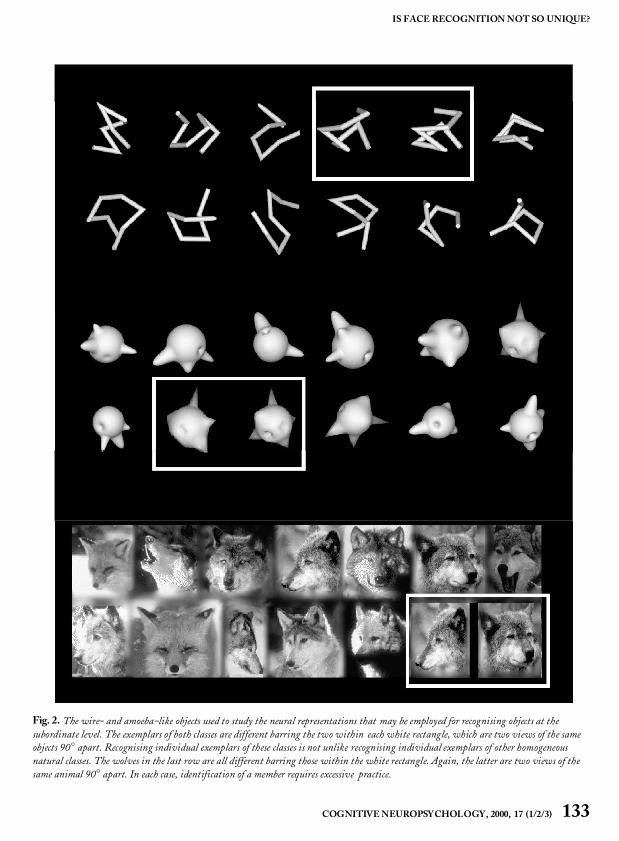
Face cells may be greatly represented within ITbecause faces are one of the few categories of visu-ally similar objects that a monkey needs to discrimi-nate. Consistent with this idea, more face cells inlab-reared monkeys are found to respond to humanfaces than monkey faces and cells often show betterresponses to familiar than unfamiliar humans(Mikami et al., 1994). This anecdotal evidence sug-gests that experience in discriminating visually sim-ilar objects of a novel category could lead to moreneurons being devoted to this category. Logothetisand Pauls (1995) and Logothetis, Pauls, andPoggio (1995) addressed this question by generat-ing expert monkeys on two different object classes.They used the same wire-like and spheroidalobjects (Fig. 1) that had been studied previously inhuman psychophysical experiments (Buelthoff &Edelman, 1992; Edelman & Buelthoff, 1992).
The animals were trained to recognise novelobjects presented from one view and were thentested for their ability to generalise recognition toviews generated by rotating the objects mathemati-cally around arbitrary axes. More specifically, suc-cessful fixation of a central light spot was followedby the learning phase, during which the monkeyswere allowed to inspect an object, the target, from agiven viewpoint arbitrarily called the zero view ofthe target. The learning phase was followed by ashort fixation period, after which the testing phasestarted. Each testing phase consisted of up to 10trials. The beginning of a trial was indicated by alow-pitched tone, immediately followedby the pre-sentation of the test stimulus, a shaded, static viewof either the target or a distractor. Target views weregenerated by rotating the object around one of fouraxes: the vertical, the horizontal, the right oblique,or the left oblique. Distractors were other objectsfrom the same or a different class. Two levers wereattached to the front panel of the monkey chair, andreinforcement was contingent upon pressing theright lever each time the target was presented.Pressing the left lever was required upon presenta-tion of a distractor.
After the monkeys mastered the task, they weretested for generalising recognition with a variety ofobjects, including pictures of real objects (e.g. cars,airplanes, fruits), and wire-like and spheroidobjects. In contrast to real objects, the recognitionof the novel objects was strictly view-dependent.The monkey could correctly identify the views ofthe target around the trained view, whereas its per-formance dropped to chance levels for disparitieslarger than approximately 40° of rotation in depth.For many wire-like objects the animal’s recognitionwas found to exceed criterion performance forviews that resembled “mirror-symmetrical,” two-dimensional images of each other, due to accidentallack of self-occlusion. Initially, the animal’s gener-alisation of recognition was also view-dependentfor rotations in the picture plane. However, in thelatter case recognition performance improved, andin a few sessions it became rotation-invariant.
Recording from the anterior inferotemporalcortex (mostly in the upper bank of the anteriormedial temporal sulcus) during this recognitiontask revealed a number of cells that were highlyselective to familiar views of these recently learnedobjects (Logothetis & Pauls, 1995; Logothetis etal., 1995). These cells exhibit a selectivity forobjects and viewpoints that is similar to that foundin face cells. The response of many object-selectiveneurons was invariant for translations within thefoveal region (centre 5°) and large changes in size(often by a factor of four in a linear dimension).
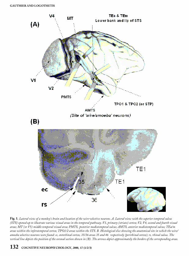
To determine the features driving the neuralresponses, Jon Pauls developeda method in our lab-oratory of eliminating, scrambling, or occluding thedisplayed wire segments (Pauls, 1997). By system-atically reducing the complexity of the stimuluswith this technique, Pauls found that some cellswere actually selective to a simple feature such as anangle, rather than to the entire wire configuration.In sharp contrast to such cells, however, otherwire-selective neurons exhibited extreme sensitivityto alterations of the stimulus configuration. Inother words, reductionof the stimulus was impossi-ble without significantly reducing the unit’sresponse. Almost all view-selective neurons wererecorded around the anterior mediotemporal sulcus(Fig. 3).
COGNITIVE NEUROPSY CHOLOGY, 2000, 17 (1/2/3) 131
IS FACE RECOGNITION NOT SO UNIQUE?
GAUTHIER AND LOGOTHETIS
132 COGNITIVE NEUROPSY CHOLOGY, 2000, 17 (1/2/3)
Fig. 1. Lateral view of a monkey’s brain and location of the wire-selective neurons. A. Lateral view with the superior temporal sulcus(STS) opened up to illustrate various visual areas in the temporal pathway. V1, primary (striate) cortex; V2, V4, second and fourth visualareas; MT (or V5) middle temporal visual area; PMTS, posterior mediotemporal sulcus; AMTS, anterior mediotemporal sulcus; TEa/mareas within the inferotemporal cortex; TPO1/2 areas within the STS. B. Histological slice showing the anatomical site in which the wire/amoeba selective neurons were found: ec, entorhinal cortex, 35/36 areas 35 and 46 respectively (perirhinal cortex); rs, rhinal sulcus. Thevertical line depicts the position of the coronal section shown in (B). The arrows depict approximately the borders of the corresponding areas.
COGNITIVE NEUROPSY CHOLOGY, 2000, 17 (1/2/3) 133
IS FACE RECOGNITION NOT SO UNIQUE?
Fig. 2. The wire- and amoeba-like objects used to study the neural representations that may be employed for recognising objects at thesubordinate level. The exemplars of both classes are different barring the two within each white rectangle, which are two views of the sameobjects 90° apart. Recognising individual exemplars of these classes is not unlike recognising individual exemplars of other homogeneousnatural classes. The wolves in the last row are all different barring those within the white rectangle. Again, the latter are two views of thesame animal 90° apart. In each case, identification of a member requires excessive practice.
GAUTHIER AND LOGOTHETIS
134 COGNITIVE NEUROPSY CHOLOGY, 2000, 17 (1/2/3)
Fig. 3. Responses of single units in the inferior temporal cortex of the monkey. The upper row shows responses to wire-like objects and themiddle row to amoeba-like objects. The neuron responds best to a recently learned object -view and its response diminishes as the object isrotated in depth. For objects that the monkey could recognise from all vantage points more than one unit was found that responded todifferent views of the same object. Systematic decomposition of the wire objects showed that while some neurons could also be activated byparts of the object (e.g. an angle), others required the entire configuration, strongly diminishing their response even when only a singlewire-segment was removed (Pauls, 1997). The bottom row shows responses of a face-selective neuron recorded in the upper bank of the STS.“Wire” and “amoeba” cells display view tuning similar to that of the face cells.
IS FACE PROCESSING UNIQUE?
The finding of “expert” cells in monkeys trained todiscriminate among amoebas and wires suggestthat face recognition may find its homologue in thebrain under the right circumstances. In Hay andYoung’s (1982) framework, one way in which facesmay be special is that they could be represented in adifferent manner to nonface objects. In humans,evidence for unique face processing comes from anumber of behavioural effects that are obtainedwith faces but not with nonface control stimuli suchas houses and even inverted faces. Most of thesebehavioural effects measure some aspect of what iscalled holistic or configural processing. Simplystated, face recognition is often found to be moresensitive than nonface recognition to the disruptionof the configuration of features: for instance, mov-ing the eyes slightly apart or inverting the entireface so that relations such as “top of ” or “right of”are changed (for reviews, see Farah, 1996; J.W.Tanaka & Gauthier, 1997). Evidence against faceprocessing being unique comes from experimentswhere the same configural effects are obtained withnonface objects when subjects are experts withthese categories (Diamond & Carey, 1986;Gauthier & Tarr, 1997; J.W. Tanaka & Gauthier,1997). This suggests that configural sensitivity isnot restricted to faces and that it is the particularexperience with an object category, rather than itssuperficial properties, which determines the pro-cessing of its exemplars. Here, we consider whetherIT cells may be thought to represent faces in a dif-ferent way to other objects.
Face Cells Show a High Degree ofSelectivity to the Face Category
Face cells in anterior IT are sensitive to configura-tion of features (Young & Yamane, 1992) and maybe mediating the configural sensitivity that is a hall-mark of upright face recognition. In a paper dis-cussing face specificity in humans, Farah et al.(1998) cite the existence of face cells as convergingevidence for faces being represented in a differentfashion, because “the selectivity and strength ofsuch responses [to nonface objects] are weaker
[than to faces]”. In a recent review article, Tovée(1998) notes that face cells are resistant to a stimu-lus simplification protocol (K. Tanaka, 1997)whereas the selectivity of most other IT cells can bereduced to rather simple stimuli. Tovée argues that“The ‘specialness’ of the face processing system willrest upon the determination of whether the faceprocessing cells in IT have no functional equivalentcounterparts for object processing, either in IT orelsewhere.”
The single-cell recording experiments describedin this paper may provide some evidence fornonface object cells that are the functional equiva-lent of face cells. A remarkable similarity existsbetween the properties of the face cells and those ofthe wire- or amoeba-selective neurons recordedfrom expert monkeys (Logothetis & Pauls, 1995;Logothetis et al., 1995). The latter type of neuronsshow selectivity to complex configurations thatcannot be reduced without diminishing the cells’response to specific views and to views that appearto be mirror symmetrical. They also exhibit posi-tion and scale invariance, and are clustered in a spe-cific brain location. This evidence is consistent withthe possibility that the responses of IT cells are builtfrom experience and adapted to the interactions ofan animal with objects. In most cases, animals needto recognise most objects at a categorical level (e.g.cage, ball, tree) and faces at the exemplar level.However, if animals need to treat other objects likefaces and discriminate visually similar exemplars, anumber of cells within IT may begin to representthe features that are best suited to this task.
Face Cells Represent Face Identity in aSparse Fashion
Several authors (Rolls & Tovée, 1995; Young &Yamane, 1992) have suggested that IT face cellsmay be representing face identity using sparse cod-ing. On a continuum from “grandmother” repre-sentations (where a single cell represents a singleobject) to highly distributed processing (in which avery large number of cells contribute to the repre-sentation, each one carrying an infinitely smallamount of useful information), sparse coding con-stitutes a case where the firing of each neuron
COGNITIVE NEUROPSY CHOLOGY, 2000, 17 (1/2/3) 135
IS FACE RECOGNITION NOT SO UNIQUE?
strongly biases the probability of a response to anobject. Face cell populations are thought to usesparse rather than distributed coding because eachface cell at least carries a lot of information at thelevel of the stimulus class, responding more to anyface than to nonface stimuli. Within the class offaces, however, the cells respond to many of thefaces in a more distributed fashion. This type ofrepresentationhas been suggested to be ideal for thediscrimination of faces (Rolls & Tovée, 1995).Note that such conclusions are based on what iscalled information theoretic analyses, in whichface-selective cells are first selected and later shownto provide more information about faces than aboutnonface stimuli. A comparable analysis for nonfaceobjects would first require the selection of a popula-tion of cells that respond best to a certain class ofnonface objects than to other stimuli. As discussedpreviously, this may be impractical for nonface cat-egories of no particular relevance to an animal butmay be feasible after an animal has been trained todiscriminate among visually similar objects.
Some authors emphasise the similaritiesbetween face cells and other IT cells selective forelaborate features. For instance, Perrett and Oram(1993) note that in the anterior temporal cortex,both face cells and Elaborate cells do not generaliseacross orientation and size (whereas face cells inSTP do). In both cases a rotation of 90° in the pic-ture plane reduces the response by more than 50%.However, other authors have contrasted the appar-ent sparse coding for faces to the more distributedcoding by which nonface objects appear to be repre-sented. K. Tanaka (1997) has suggested thatnonface objects are represented by distributedcoding over a large number of IT columns, eachcontaining cells selective for moderately complexfeatures. In this framework, each shape primitivecarries very little information about the identity ofthe object and the representation of nonface objectsmay be argued to be qualitatively different fromthat of faces, in that it would be considerably moredistributed.
Recently, however, Kobatake, Wang, andTanaka (1998) have trained monkeys to recognise28 moderately complex stimuli (mostly combina-tions of 2 simple geometric shapes, these stimuli
were less homogeneous than wires or amoebas) andfound a greater proportion of cells responsive to thetrained stimuli in trained than untrained monkeys.Furthermore, many of these cells responded tomultiple members of the training stimuli, notunlike face cells. The discriminations learned by themonkeys may be supported by sparse representa-tions and the number of cells that respond to a cer-tain object may be partly determined by an animal’sexperience with this category (see also Booth &Rolls, 1998). However, experience with a visuallyhomogeneous class of objects (e.g. the wires andameobas) may be necessary to build up a populationof cells that will generalise to novel exemplars of thecategory. When humans are trained with severalobjects of an homogeneouscategory, their expertisegeneralises to novel exemplars (for instance,configural sensitivity is found for untrainedobjects—Gauthier & Tarr, 1997). Given the simi-larity of behavioural performance in object recogni-tion tasks between man and monkey (Logothetis &Pauls, 1995), we can hypothesise that expertise inmonkeys would also generalise to novel exemplarsof a trained class. However, such generalisationcould be expected in monkeys trained with ameobasand wires, but not necessarily for animals trainedwith less homogeneous stimulus sets.
IS FACE PROCESSING SPECIFIC?
Even if we found that faces and objects are repre-sented by common mechanisms in IT, faces couldstill be special in that they could be processed in adistinct and separate neural system. It may be thatspecificity (Hay & Young, 1982) in the location ofcells for any object category is not a sufficient crite-rion to designate this category “special” (Tovée,1998), presumably because specificity would not beunique to a single category (i.e. if face cells are sepa-rated from wire cells, then wire cells are also sepa-rated from face cells). However, regardless of thedebate on faces, to consider the spatial organisationof object-selective cells is essential to the under-standing of the temporal cortex organisation.
The area where wire and amoeba cells werefound, the AMTS, is anterior to area TE and more
GAUTHIER AND LOGOTHETIS
136 COGNITIVE NEUROPSY CHOLOGY, 2000, 17 (1/2/3)
ventral than areas where face cells are typicallyfound in other studies. What this means is some-what difficult to interpret, given the methodologi-cal constraints of single-cell recording. As in anysingle-cell study where there is no prior knowledgeof precisely where selective responses are expected,Logothetis and colleagues (Logothetis & Pauls,1995; Logothetis et al., 1995) recorded systemati-cally from posterior to anterior areas of the tempo-ral lobe, moving to a new area after a week or soof fruitless explorations. Once a first wire- oramoeba-selective cell was found in AMTS, theresearchers kept on recording in this area withoutgoing back to more posterior regions. In addition,the AMTS was not systematically tested with facesin this experiment. In other words, the current evi-dence suggests that populations of expert objectcells are found in a different area than populationsof face cells with comparable properties, but thisevidence is not as strong as it would be if it camefrom a neuroimaging experiment in which all areasof the visual system had been equally sampled at alltime-points.
Evidence that face processing may be segregatedfrom object processing in the human brain mainlycomes from two different sources. The first is evi-dence from patients with selective deficits in faceprocessing (De Renzi, 1986; Farah et al., 1995a).The selectivity of face agnosia is controversial, asmany prosopagnosic patients also report difficultieswith other visually similar categories (Bornstein,Sroka, & Munitz, 1969; Damasio et al., 1982;Shuttleworth, Syring, & Norman, 1982). Even inthe case of patients who believe that their deficitapplies only to faces, recent work has revealed amore general impairment for subtle, subordinate-level discriminations (Gauthier et al., 1999b).A second source of evidence comes from neuro-imaging studies in which activation in the middlefusiform gyrus is found when subjects are viewingfaces as opposed to nonface objects (Kanwisher etal., 1997; McCarthy et al., 1997; Sergent &Signoret, 1992). To address this evidence andinspired by the fact that prosopagnosic patientsoften have difficulties discriminating objects withinthe same category, Gauthier et al. (1998) comparedbrain activation when normal subjects verified the
subordinate identity of a picture (e.g. pelican) vs. thebasic level (e.g. bird). They found activation in ven-tral temporal areas described as face-sensitive inprior studies. In this issue, a new study (Gauthier etal., this issue) verified that subordinate-level pro-cessing of nonface objects activates the small areathat can be defined as face-specific in each subject.Thus, the presentation of faces is not necessary toengage what is often called the “face area.” Thisregion can be differentially engaged when the samenonface object is recognised at the subordinate vs.the basic level. However, faces appear to activateonly a portion of ventral cortex dedicated to subor-dinate-level processing. These studies, which sug-gest that subordinate level processing accounts forsome of the activation in the face area, are not neces-sarily incompatible with other work suggesting thatnot all of the activation in the face area can beaccounted for by subordinate-level classification(Kanwisher et al., 1997). What may be happeningis that the former studies focus on the fact that thereis difference between basic level and subordinatelevel recognition of nonface objects in the face area,whereas the latter studies account for a differentpart of the data, pointing out that there is still moreevidence for subordinate-level recognition of facesthan subordinate-level recognition of nonfaceobjects. A recent fMRI study (Gauthier et al.,1999a) has revealed that expertise with subordi-nate-level discrimination of novel objects (similartraining experience as the monkeys in Logothetis &Pauls, 1995; Logothetis et al., 1995) leads toincreased activation localised in the “face area.”This suggests that the interaction of two factors,level of categorisation and expertise, may interact toproduce the specialisation for faces found in themiddle fusiform face area. In the next section, weconsider how what we know of the monkey visualsystem can help resolve the role of these two factors.
Level of Categorisation and Expertise
Given the importance of level of categorisationdemonstrated in behavioural (J.W. Tanaka & Tay-lor, 1993) and fMRI studies in humans (Gauthieret al., 1998, this issue), one may ask whether thereis any evidence that this factor is important in
COGNITIVE NEUROPSY CHOLOGY, 2000, 17 (1/2/3) 137
IS FACE RECOGNITION NOT SO UNIQUE?
determining the responses of IT cells. Unfortu-nately, no single-cell recording study has comparedthe responses of cells to the same stimuli when ani-mals are requested to recognise it at different levelsof abstraction. However, Logothetis and Pauls(1995) have trained monkeys to recognise objectseither at the basic level (among distractors differinglargely in shape, such as a wire vs. an amoeba) or atthe subordinate level (for instance, discriminatingbetween two wires). They found that the animals’behavioural performance was viewpoint-dependent in the case of subordinate-level judge-ments and viewpoint-independent in the case ofbasic-level judgements. This suggests that level ofcategorisation may at least have a similar impor-tance for monkey and human visual recognition.
Two recent studies provided monkeys withexperience with certain objects and later found cellsto be responsive to many of these trained objects(Booth & Rolls, 1998; Kobatake et al., 1998).However, these studies differ in an important wayfrom the wire-frame and amoeba study byLogothetis and colleagues: The different objectsdid not belong to what would be considered thesame “basic-level” category (Rosch, Mervis, Gray,Johnson, & Boyes-Braem, 1976). This is becausethey do not share common parts and could be dis-criminated by the presence of a single feature (e.g.the way that the presence of eyes is diagnostic todetect a face) or simple relationships between parts(e.g. as for the presence of a nose underneath twoeyes). In comparison, objects from homogeneouscategories share common parts as well as thefirst-order configuration of these parts (Diamond& Carey, 1986; Rhodes & McLean, 1990). Theycan onlybe distinguished using subtle differences inthe shape of their parts or subtle differences in theconfiguration of their parts (e.g. distances betweendifferent face features). It is expertise discriminat-ing between objects of such homogeneous catego-ries that is thought to mediate behaviouralconfigural effects and the increased recruitment ofthe fusiform face area (Gauthier & Tarr, 1997;Gauthier et al., 1999a). Again, there is yet no directcomparison using physiological measurements ofthe difference between basic and subordinate levelprocessing of objects, but the expertise of monkeys
discriminating between wires and amoebas may bemost relevant to the debate on face recognition inhumans.
In humans, recent fMRI results suggest thatexpertise with novel objects (Greebles) can recruitthe middle fusiform face area (Gauthier et al.,1999a). However, at least one area, in the lateraloccipital gyrus, showed a strong expertise effect,with more activation for Greeble experts than nov-ices, and even more for Greebles than for faces.This lateral occipital gyrus area did not behave likethe fusiform face area in all conditions: In particu-lar, this region responded more to inverted than toupright faces, whereas the face area respondedmoreto upright than to inverted faces. Thus, there maybe a complex system of areas within the temporallobe that is modified by experience with objects.This is consistent with the existence of face cells inmany areas of both the human and the monkeybrain. Similarly, AMTS may not be the only area ofexpert monkeys where wire and amoeba cells canbe found. At this point, it is likely that furtheradvances in comparing the man and monkey sys-tems will require the addition of novel techniquessuch as functional MRI in monkeys (Logothetis,Guggenberger, Peled, & Pauls, 1999) to thosealready available in both species.
CONCLUSIONS
Both humans and monkeys are extremely good atrecognising faces, a fact that is hardly surprising inview of the vital importance that face recognitionhas for the primate. An important neural systemexists in both species for the processing of facialinformation. In the human behavioural literature,starting with Diamond and Carey’s (1986) land-mark study of dog expertise, a consensus has grownthat nonface categories of objects can be processedin the same way as faces given similar task con-straints and subject expertise. However, in humanneuropsychological and neuroimaging studies,there is still an ongoing debate regarding the possi-bility that faces may be special.
Interestingly, the single-cell recording literaturealso converges to suggest that faces are not repre-
GAUTHIER AND LOGOTHETIS
138 COGNITIVE NEUROPSY CHOLOGY, 2000, 17 (1/2/3)
sented by IT cells in a unique fashion. Severalauthors, including C.G. Gross (1992), the pioneerin the domain of face cells, have suggested that facecells may appear more specialised than other ITcells only because face recognition happens to be anextremely demanding subordinate recognitiontask, and for nonhuman primates it may be the onlyidentification task performed in life. Clearly, suchan hypothesis leads to the prediction that a similarspecialisation may also arise when the identificationof members of other classes becomes the criticaltask at hand. This was tested in recent single-cellrecordingexperiments.A remarkable similarity wasfound between the properties of the face cells andthose of the wire- or amoeba-selective neuronsrecorded from expert monkeys (Logothetis &Pauls, 1995; Logothetis et al., 1995). The lattertype of neurons show selectivity to complex config-urations that cannot be reduced without diminish-ing the cells’ response to specific views and to viewsthat appear to be mirror-symmetrical. They exhibitposition and scale invariance, and are clustered in aspecific brain location. Since recordings have onlybeen made in the inferotemporal cortex and mostlyin AMTS, it is not currently known whether selec-tivity to these objects might not also be found inother brain structures.
Such results are consistent with behavioural andfMRI studies in humans showing that novel objectsare processed in a more configural manner withexpertise and can increasingly recruit parts of theventral temporal lobe. However, whereas fMRIresults in humans suggests that the very same areasare recruited for faces and nonface objects, sin-gle-cell studies in monkeys point to specialisationof different areas. These techniques are very differ-ent and it is important to note that fMRI could pro-vide more convincing evidence than single-cell datafor a dissociation between the location of face andobject expert processing. On the other hand, thebetter spatial resolution of single-cell recordingcould provide stronger support for an association inlocation (e.g. if the very same cells were found tomediate expert representations of different catego-ries). Paradoxically, the current data in fMRI sug-gests an association whereas single cell recordingsuggests a dissociation, albeit only in the location of
face and wire/amoeba cells within the anterior tem-poral lobe. Therefore, for both sources of evidencethe interpretation should be cautious. In any case,faces are not unique with regard to the type of neu-ral activity that can be recorded in a monkey’s brainwhen the animal is coping with other classes ofobjects in the same manner with which it deals withfaces.
REFERENCES
Baylis, G.C., Rolls, E.T., & Leonard C.M. (1987).Functional subdivisions of the temporal lobe neocor-tex. Journal of Neuroscience, 7, 330–342.
Bodamer, J. (1947). Die Prosopagnosie. Die Agnosiedes Physiognomieerkennes. Arch Psychiatr Nervenkr,179, 6–54.
Booth, M.C.A., & Rolls, E.T. (1998). View-invariantrepresentations of familiar objects by neurons in theinferior temporal visual cortex. Cerebral Cortex, 8,510–523.
Bornstein, B., Sroka, H., & Munitz, H. (1969).Prosopagnosia with animal face agnosia Cortex, 5,164–171.
Boussaoud, D., Desimone, R., & Ungerleider, L.G.(1991). Visual topography of area TEO in themacaque. Journal of Comparative Neurology, 306,554–575.
Bruce, C.J. (1982). Face recognition by monkeys:Absence of an inversion effect. Neuropsychologia, 20,515–521.
Bruyer, R., & Crispeels, G. (1992). Expertise in personrecognition. Bulletin of the Psychonomic Society, 30,501–504.
Buelthoff, H.H., & Edelman, S. (1992). Psycho-physical support for a two-dimensional view inter-polation theory of object recognition. Proceedings ofthe National Academy of Sciences USA, 89, 60–64.
Clark, V.P., Keil, K., Maisog, J.M., Courtney, S.M.,Ungerleider, L.G., & Haxby, J.V. (1996). Func-tional magnetic resonance imaging of human visualcortex during face matching: A comparisonwith posi-tron emission tomography. Neuroimage, 4, 1–15.
de Gelder, B., Bachoud-Lévi, A.C., & Degos, J.D.(1998). Inversion superiority in visual agnosia may becommon to a variety of orientation-polarised objectsbesides faces. Vision Research, 38, 2855–2861.
COGNITIVE NEUROPSY CHOLOGY, 2000, 17 (1/2/3) 139
IS FACE RECOGNITION NOT SO UNIQUE?
De Renzi, E. (1986). Slowly progressive visual agnosiaor apraxia without dementia. Cortex, 22, 171–180.
Desimone, R., Albright, T.D., Gross, C.G., & Bruce,C.J. (1984). Stimulus-selective properties of infe-rior temporal neurons in the macaque. Journal ofNeurosciences, 4, 2051–2062.
Desimone, R., Fleming, J.F.R., & Gross, C.G.(1980). Prestriate afferents to inferior temporal cor-tex: An HRP study. Brain Research, 184, 41–55.
Diamond, R., & Carey, S. (1986). Why faces are andare not special: An effect of expertise, Journal ofExperimental Psychology: General, 115, 107–117.
Distler, C., Boussaoud, D., Desimone, R., & Unger-leider, L.G. (1993). Cortical connections of inferiortemporal area TEO in macaque monkeys. Journal ofComparative Neurology, 334, 125–150.
Eacott, M.J., Heywood, C.A., Gross, C.G., & Cowey,A. (1993). Visual discrimination impairments fol-lowing lesions of the superior temporal sulcus are notspecific for facial stimuli. Neuropsychologia, 31,609–619.
Edelman, S., & Buelthoff, H.H. (1992). Orientationdependence in the recognition of familiar and novelviews of 3D objects. Vision Research, 32, 2385–2400.
Farah, M.J. (1996). Is face recognition ‘special’? Evi-dence from neuropsychology. Behavioural BrainResearch, 76, 181–189.
Farah, M.J., McMullen, P.A., & Meyer, M.M. (1991).Can recognition of living things be selectivelyimpaired? Neuropsychologia, 29, 185–193.
Farah, M.J., Wilson, K.D., Drain, H.M., & Tanaka,J.W. (1995b). The inverted face inversion effect inprosopagnosia: Evidence for mandatory, face-specificperceptual mechanisms. Vision Research, 35,2089–2093.
Farah, M.J., Wilson, K.D., Drain, M., & Tanaka, J.N.(1998). What is “special” about face perception? Psy-chological Review, 105, 482–498.
Gaffan, D., & Heywood,C.A. (1993). A spurious cate-gory-specific visual agnosia for living things in humanand nonhuman primates. Journal of Cognitive Neuro-science, 5, 118–128.
Gauthier, I., Behrmann, M., & Tarr, M.J. (1999). Canface recognition really be dissociated from object rec-ognition? Journal of Cognitive Neuroscience, 11,349–370.
Gauthier, I., & Tarr, M.J. (1997). Becoming a“Greeble’’ expert: Exploring the face recognitionmechanism. Vision Research, 37, 1673–1682.
Gauthier, I., Tarr, M.J.,Anderson, A.W.,Skudlarski, P.,& Gore, J.C. (1999a). Activation of the middlefusiform “face area” increases with experience in rec-ognizing novel objects. Nature Neuroscience, 2,568–573.
Gauthier, I., Williams, P., Tarr, M.J., & Tanaka,J.W. (1998). Training “Greeble’’ experts: A frame-work for studying expert obect recognition processes.Vision Research, 38, 2401–2428.
Gross, C. (1992). Representation of visual stimuli ininferior temporal cortex. Philosophical Transactions ofthe Royal Society of London, B, 335, 3–10.
Gross, C.G., Bender, D.B., & Rocha-Miranda, C.E.(1969). Visual receptive fields of neurons ininferotemporal cortex of the monkey. Science, 166,1303–1306.
Gross, C.G., Roche-Miranda, C.E., & Bender, D.B.(1972). Visual properties of neurons in infero-temporal cortex of the macaque. Journal of Neuro-physiology, 35, 96–111.
Hamilton, C.R., & Vermeire, B.A. (1983). Discrimi-nation of monkey faces by split-brain monkeys.Behavioural Brain Research, 9, 263–275.
Hasselmo, M.E., Rolls, E.T., & Baylis, G.C.(1989). The role of expression and identity in theface-selective responses of neurons in the temporalvisual cortex of the monkey. Behavioural BrainResearch, 32, 203–218.
Hay, D.C., & Young, A.W. (1982). The human face.In A.W. Ellis (Ed.), Normality and pathology in cogni-tive function. London: Academic Press.
Heywood, C.A., & Cowey, A. (1992). The role of the“face-cell” area in the discrimination and recognitionof faces by monkeys. Philosophical Transactions of theRoyal Society of London–Series B: Biological Sciences,335, 31–37.
Hietanen, J.K.,Perrett, D.I., Oram, M.W.,Benson, P.J.,& Dittrich, W.H. (1992). The effects of lightingconditions on the responses of cells selective for faceviews in the macaque temporal cortex. ExperimentalBrain Research, 89, 157–171.
Iwai, E., & Mishkin, M. (1969). Further evidence onthe locus of the visual area in the temporal lobe of themonkey. Experimental Neurology, 25, 585–594.
Johnson, M.H., & Morton, J. (1991). Biology and cog-nitive development: The case of face recognition. Oxford:Basil Blackwell.
GAUTHIER AND LOGOTHETIS
140 COGNITIVE NEUROPSY CHOLOGY, 2000, 17 (1/2/3)
Kaas, J.H. (1995). Progress and puzzles. Current Biol-ogy, 5, 1126–1128.
Kanwisher, N., McDermott, J., & Chun, M.M.(1997). The fusiform face area: A module in humanextrastriate cortex specialised for face perception.Journal of Neuroscience, 17, 4302–4311.
Kobatake, E., Wang, G., & Tanaka, K. (1998). Effectsof shape-discrimination training on the selectivity ofinferotemporal cells in adult monkeys. Journal ofNeurophysiology, 80, 324–330.
Konorski, J. (1967). Integrative activity of the brain.An interdisciplinary approach. Chicago: University ofChicago Press.
Logothetis, N.K. (1998). Object vision and visualawareness. Current Opinion in Neurobiology, 8,536–544.
Logothetis, N.K., Guggenberger, H., Peled, S., & Pauls,J. (1999). Functional imaging of the monkey brain.Nature Neuroscience, 2, 555–562.
Logothetis, N.K., & Pauls, J. (1995). Psychophysicaland physiological evidence for viewer-centered objectrepresentations in the primate. Cerebral Cortex, 5,270–288.
Logothetis, N.K.,Pauls, J., & Poggio,T. (1995). Shaperepresentation in the inferior temporal cortex of mon-keys. Current Biology, 5, 552–563.
Logothetis, N.K., & Sheinberg, D. (1996). Visualobject recognition, Annual Review of Neuroscience, 19,577–621.
Lutz, C.K., Lockard, J.S., Gunderson, V.M., & Grant,K.S. (1998). Infant monkeys’ visual responses todrawings of normal and distorted faces. AmericanJournal of Primatology, 44, 169–174.
McCarthy, G., Puce, A., Gore, J.C., & Allison, T.(1997). Face-specific processing in the fusiformgyrus, Journal of Cognitive Neuroscience, 9, 605–610.
Mendelson, M.J., Haith, M.M., & Goldman-Rakic,P.S. (1982). Face scanning and responsiveness tosocial cues in infant rhesus monkeys. DevelopmentalPsychology, 18, 222–228.
Mikami, A., Nakamura, K., & Kubota, K. (1994).Neuronal responses to photographs in the superiortemporal sulcus of the rhesus monkey. BehaviouralBrain Research, 60, 1–13.
Morton, J., & Johnson, M.H. (1991). CONSPEC andCONLERN:A two-process theory of infant face rec-ognition. Psychological Review, 98, 164–181.
Moscovitch, M., Winocur, G., & Behrmann, M.(1997). What is special in face recognition? Nineteenexperiments on a person with visual object agnosia
and dyslexia but normal face recognition. Journal ofCognitive Neuroscience, 9, 555–604.
Nahm, F.D., Perret, A., Amaral, D.G., & Albright,T.D. (1997). How do monkeys look at faces. Journalof Cognitive Neuroscience, 9, 611–623.
Ó Scalaidhe, S.P., Wilson, F.A.W., & Goldman-Rakic,P.S. (1997). Areal segregation of face-processingneurons in prefrontal cortex. Science, 278, 1135–1138.
Pauls, J. (1997). The representation of 3-dimensionalobjects in the primate visual system. Unpublished dis-sertation number 1–162. Baylor College of Medicine.
Perrett, D.I., Harries, M.H., Bevan, R., Thomas, S.,Benson, P.J., Mistlin, A.J., Chitty, J.K., Hietanen,J.K., & Ortega, J.E. (1989). Frameworks of analysisfor the neural representation of animate object andactions. Journal of Experimental Biology, 146, 87–113.
Perrett, D.I., Hietanen, J.K., Oram, M.W., & Benson,P.J. (1992). Organisation and functions of cellsresponsive to faces in the temporal cortex. Philosophi-cal Transactions of the Royal Society of London B: Biolog-ical Science, 335, 23–30.
Perrett, D.I., Oram, M.W., Harries, M.H., Bevan, R.,Hietanen, J.K., Benson, P.J., & Thomas, S. (1991).Viewer-centred and object-centred coding of heads inthe macaque temporal cortex. Experimental BrainResearch, 86, 159–173.
Perrett, D.I., Oram, M.W., Hietanen, J.K., & Benson,P.J. (1994). Issues of representation in object vision.In M.J. Farah, G. Ratcliff (Eds.), The neuro-psychology of high-level vision: Collected tutorialassays, (pp 33–62). Hillsdale NJ: Lawrence ErlbaumAssociates Inc.
Perrett, D.I., Rolls, E.T., & Caan, W. (1979). Tempo-ral lobe cells of the monkey with visual responsesselective for faces. Neuroscience Lettr Suppl, S3, S358.
Perrett, D.I., Rolls, E.T., & Caan, W. (1982). Visualneurones responsive to faces in the monkey temporalcortex. Experimental Brain Research, 47, 329–342.
Perrett, D.I., Smith, P.A., Potter, D.D., Mistlin, A.J.,Head, A.S., Milner, A.D., & Jeeves, M.A. (1985).Visual cells in the temporal cortex sensitive to faceview and gaze direction. Proceedings of the Royal Societyof London–Series B: Biological Sciences, 223, 293–317.
Puce, A., Allison, T., Bentin, S., Gore, J.C., & McCar-thy, G. (1998). Temporal cortex activation inhumans viewing eye and mouth movements. Journalof Neuroscience, 18, 2188–2199.
Puce, A., Allison, T., Gore, J.C., & McCarthy, G.(1995). Face-sensitive regions in human extrastriate
COGNITIVE NEUROPSY CHOLOGY, 2000, 17 (1/2/3) 141
IS FACE RECOGNITION NOT SO UNIQUE?
cortex studied by functional MRI. Journal ofNeurophysiology, 74, 1192–1199.
Rhodes, G., & McLean, I.G. (1990). Distinctivenessand expertise effects with homogeneous stimuli:Towards a model of configural coding. Perception, 19,773–794.
Rolls, E.T. (1992). Neurophysiology and functions ofthe primate amygdala. In J.P. Aggleton (Ed.),Theamygdala (pp 143–165). New York: Wiley-Liss.
Rolls, E.T., & Tovée, M.J. (1995). Sparseness of theneuronal representation of stimuli in the primatetemporal cortex. Journal of Neurophysiology, 73,713–726.
Rosch, E., Mervis, C.B., Gray, W.D., Johnson,D.M., &Boyes-Braem, P. (1976). Basic objects in naturalcategories. Cognitive Psychology, 8, 382–439.
Rosenfield, S.A., & Van Hoesen, G.W. (1979). Facerecognition in the rhesus monkey. Neuropsychologia,17, 503–509.
Seltzer, J.B., & Pandya, D.N. (1978). Afferent corticalconnections and architectonics of the superior tempo-ral sulcus and surrounding cortex in the rhesusmonkey. Brain Research, 149, 1–24.
Sergent, J., Ohta, S., & MacDonald, B. (1992). Func-tional neuroanatomy of face and object processing. Apositron emission tomography study. Brain, 115,15–36.
Sergent, J., & Signoret, J.L. (1992). Functional andanatomical decomposition of face processing: Evi-dence from prosopagnosia and PET study of normalsubjects. Philosophical Transactions of the Royal Societyof London B, 335, 55–62.
Shiwa, T. (1987). Corticocortical projections to themonkey temporal lobe with particular reference to thevisual processing pathways. Arch Ital Biol, 125,139–154.
Shuttleworth, E.C., Syring, V., & Norman, A. (1982).Further observations on the nature of prosopagnosia.Brain and Cognition, 1, 307–322.
Simion, F., Valenza, E., Umilta, C., & Dalla Barba, B.(1998). Preferential orienting to faces in newborns:A temporal-nasal asymmetry. JEP: HPP, 24,1399–1405.
Tanaka, J.W., & Gauthier, I. (1997). Expertise inobject and face recognition. In R.L. Goldstone, P.G.Schyns, & D.L. Medin (Eds.), Psychology of learningand motivation. San Diego, CA: Academic Press.
Tanaka, J.W., & Taylor, M. (1991). Object categoriesand expertise: Is the basic level in the eye of thebeholder? Cognitive Psychology, 23, 457–482.
Tanaka, K. (1996). Inferotemporal cortex and objectvision. Annual Review of Neuroscience, 19, 109–139.
Tanaka, K. (1997). Mechanisms of visual object recog-nition: Monkey and human studies. Current Opinionin Neurobiology, 7, 523–529.
Tanaka, K., Saito, H., Fukada, Y., & Moriya, M.(1991). Coding visual images of objects in theinferotemporal cortex of the macaque monkey. Jour-nal of Neurophysiology, 66, 170–189.
Toveé, M.J. (1998). Is face processing special? Neuron,21, 1239–1242.
Ungerleider, L.G. (1995). Functional brain imagingstudies of cortical mechanisms for memory. Science,270, 769–775.
Valenza, E., Simion, F., Macchi Cassia, V., & Umilta,C. (1996). Face preference at birth. JEP:HPP, 22,892–903.
Von Bonin, G., & Bailey, P. (1947). The neocortex ofmacaca mulatta. Urbana, IL: University of IllinoisPress.
Wachsmuth, E., Oram, M.W., & Perrett, D.I. (1994).Recognition of objects and their component parts:Responses of single units in the temporal cortex of themacaque. Cerebral Cortex, 4, 509–522.
Webster, M.J., Ungerleider, L.G., & Bachevalier, J.(1991). Connections of inferior temporal areas TEand TEO with medial temporal-lobe structures ininfant and adult monkeys. Journal of Neuroscience, 11,1095–1116.
Williams, G.V., Rolls, E.T., Leonard, C.M., & Stern,C. (1993). Neuronal responses in the ventralstriatum of the behaving macaque. Behavioural BrainResearch, 55, 243–252.
Wilson, F.A.W., Ó Scalaidhe, S.P., & Goldman-Rakic,P.S. (1993). Dissociation of object and spatial pro-cessing domains in primate prefrontal cortex. Science,260, 1955–1958.
Wright, A.A., & Roberts, W.A. (1996). Monkey andhuman face perception: Inversion effects for humanfaces but not for monkey faces or scenes. Journal ofCognitive Neuroscience, 8, 278–290.
Yin, R.K. (1969). Looking at upside-down faces, JEP,81, 141–145.
Young, A.W., Hellawell, D., & Hay, D. (1987).Configural information in face perception. Perception,10, 747–759.
Young, M.P., & Yamane, S. (1992). Sparse populationcoding of faces in the inferotemporal cortex. Science,256, 1327–1331.