I. Plasma Membrane Structure Plasma membrane – Boundary that separates living cells from their nonliving surroundings. - Apprx. 8 nm thick - Composed chiefly of lipids and proteins - Surrounds the cell and controls chemical traffic in/out of cell - Is semi-permeable Enables cells to maintain internal environment different from external environment
Transcript
I. Plasma Membrane Structure
Plasma membrane
– Boundary that separates living cells
from their nonliving surroundings.
- Apprx. 8 nm thick
- Composed chiefly of lipids and proteins
- Surrounds the cell and controls chemical traffic in/out of cell
- Is semi-permeable
Enables cells to maintain internal environment different from external environment
Phospholipid bilayer
-Composed of 2 layers of phospholipids
-Heads are hydrophillic
-Tails are hydrophobic
Membrane Structure (Fluid Mosaic Model)
• Membrane proteins embedded in phospholipid bilayer
• Give membrane ‘fluidity’ similar to salad oil
• Phospholipids & proteins can drift laterally (2 um / sec)
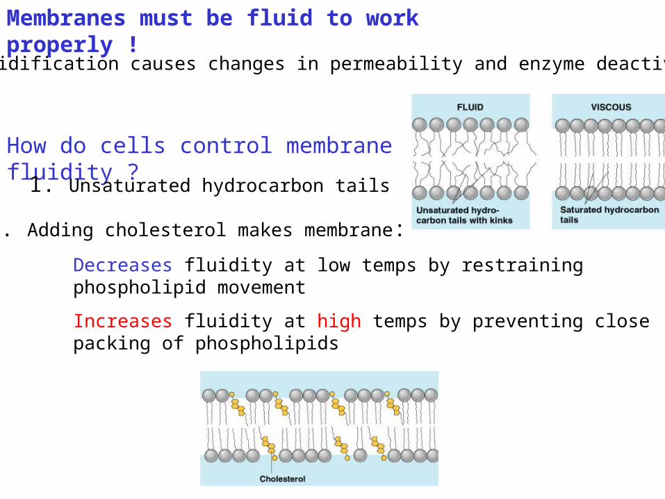
- solidification causes changes in permeability and enzyme deactivation
Membranes must be fluid to work properly !
How do cells control membrane fluidity ?
1. Unsaturated hydrocarbon tails
Decreases fluidity at low temps by restraining phospholipid movement
Increases fluidity at high temps by preventing close packing of phospholipids
2. Adding cholesterol makes membrane:
• Increase the percentage of cholesterol in phospholipids
• Prevents membrane from solidifying in cold weather
winter wheat
So, how do the plant overcome the winter?
Proteins in Plasma Membrane
- Mosaic of proteins ‘bobbing’ in a fluid lipid bilayer
- Proteins determine a membrane’s specific function:
Two types
1. Integral proteins (‘transmembrane’, or embedded)
2. Peripheral proteins (bound to surface of membrane)
Transport – protein provides channel across membrane for particular solutes
Enzymatic activity – proteins may be enzymes that catalyze steps in metabolic pathway
Signal transduction – protein is a receptor for chemical messenger (hormone). Conformational change in protein relays message to inside of cell
Intercellular joining – membrane proteins of adjacent cells join together for strength (epithelium)
Cell-cell recognition – glycoproteins act as I.D. tags that are recognized by other cells (e.g. RBCs)
Some Functions of Membrane Proteins
Regulating Traffic Across Membranes
II. Passive Transport: Diffusion and Facilitated diffusion
Diffusion : net movement
of a substance down
a concentration gradient.
• Solutes diffuse from high to low concentration.
• Continues until a dynamic equilibrium is reached.
• No requirement for energy expense (passive)
• Examples:
Uptake of O2 by cell performing respiration
Elimination of CO2 from cell
Diffusion of solutes across a membrane
Each dye diffuses down its own concentration gradient.
Facilitated diffusion
a) Channel protein : aquaporins, ion channels
b) Carrier protein
• Passive transport• Transport proteins speed the movement of molecules
across the plasma membrane.• Channel protein and Carrier protein required
Osmosis• Diffusion (passive transport) of
water across a selectively permeable membrane
• Direction of water movement is determined by the difference in total solute concentration, regardless of type or diversity of solutes.
• Water moves always from high concentration solution to low concentration solution.
Water balance of living cells
• Tonicity : the ability of a solution to cause a cell to gain or lose water Isotonic: no net movement of water across the membrane (normal). Hypertonic : the cell loses water to its environment (crenation). Hypotonic : the cell gains water from its environment (lysis).
QuestionsAn artificial cell consisting of an aqueous solution enclosed in a selectively permeable membrane has just been immersed in a beaker containing a different solution. The membrane is permeable to water and to the simple sugars glucose and fructose but completely impermeable to sucrose.
1. Glucose?2. Fructose?3. Hypotonic/
Hypertonic?4. Water?
Active Transport• Requires the cell to expend energy: ATP
• Transport proteins pump molecules across a membrane against their concentration gradient.
• “Uphill” transport
• Maintain steep ionic gradients across the cell membrane (Na+ , K+ , Ca++ , Mg++ , Cl-)
Na+
Na+
Na+
Na+
Na+Na+
Na+
Na+
Na+
Na+
inside outside
Na+
An Example of Active Transport: The Sodium-Potassium Pump
Passive and Active Transport
More examples of active transport
• Exocytosis– Removing large particles out of the cell with a vesicle
• Involves two steps:– Transcription – Translation
Genetic Information• Uses 2 main forms of genetic information:
– DNA Deoxyribonucleic Acid• Double stranded• Sugar: Deoxyribose• Stays in the nucleus• Bases: A T G C
– RNA Ribonucleic Acid• Single stranded• Sugar: Ribose• Can leave the nucleus• Bases: A U G C
Transcription
• DNA unwinds
• One strand of the double helix is used as a template
• Nucleotides line up along the DNA and form a copy, called mRNA
• Once completed, DNA winds back up and mRNA leaves
• mRNA must be spliced before it leaves the nucleus ( immature RNA)– Enzymes remove noncoding areas called
introns, and coding regions called exons are spliced back together
– The result is a shorter, coding strand of mRNA– Every 3 bases on mRNA is a codon
Codons
• Codes for amino acids
• 64 codons can code for 20 different amino acids
Translation
• mRNA binds to a ribosome• tRNA binds to ribosome along the codon and
reads which amino acid it codes for• tRNA finds the specific amino acids • For every codon, the tRNA brings the amino acids• Amino acids link together forming a proteins• Peptide bonds link each amino acid together.


![Plasma Membrane [7.2] Goals: Understand the concept of homeostasis in relation to the plasma membrane Demonstrate and understand how the plasma membrane.](https://static.documents.pub/doc/80x56/5697c01d1a28abf838cd0a9a/plasma-membrane-72-goals-understand-the-concept-of-homeostasis-in-relation.jpg)


