Matthieu Arlat,1,2 and Emmanuelle Lauber1*Laboratoire des Interactions Plantes Micro-organismes (LIPM), UMR CNRS-INRA 2594/441, F-31326 Castanet-Tolosan, France,1
and Universite de Toulouse, UPS, 118 Route de Narbonne, F-31062 Toulouse, France2
Received 29 October 2009/Accepted 4 January 2010
Xanthomonas campestris pv. campestris, the causal agent of black rot disease of brassicas, is known for itsability to catabolize a wide range of plant compounds. This ability is correlated with the presence of specificcarbohydrate utilization loci containing TonB-dependent transporters (CUT loci) devoted to scavenging spe-cific carbohydrates. In this study, we demonstrate that there is an X. campestris pv. campestris CUT systeminvolved in the import and catabolism of N-acetylglucosamine (GlcNAc). Expression of genes belonging to thisGlcNAc CUT system is under the control of GlcNAc via the LacI family NagR and GntR family NagQregulators. Analysis of the NagR and NagQ regulons confirmed that GlcNAc utilization involves NagA andNagB-II enzymes responsible for the conversion of GlcNAc-6-phosphate to fructose-6-phosphate. Mutants withmutations in the corresponding genes are sensitive to GlcNAc, as previously reported for Escherichia coli. ThisGlcNAc sensitivity and analysis of the NagQ and NagR regulons were used to dissect the X. campestris pv.campestris GlcNAc utilization pathway. This analysis revealed specific features, including the fact that uptakeof GlcNAc through the inner membrane occurs via a major facilitator superfamily transporter and the fact thatthis amino sugar is phosphorylated by two proteins belonging to the glucokinase family, NagK-IIA andNagK-IIB. However, NagK-IIA seems to play a more important role in GlcNAc utilization than NagK-IIBunder our experimental conditions. The X. campestris pv. campestris GlcNAc NagR regulon includes four genesencoding TonB-dependent active transporters (TBDTs). However, the results of transport experiments suggestthat GlcNAc passively diffuses through the bacterial envelope, an observation that calls into question whetherGlcNAc is a natural substrate for these TBDTs and consequently is the source of GlcNAc for this nonchiti-nolytic plant-associated bacterium.
Xanthomonas campestris pv. campestris, the causal agent ofblack rot disease of brassicas, produces extracellular plant cellwall-degrading enzymes which contribute to its pathogenicityby facilitating its spread through plant tissues and give thebacterium access to a ready source of nutrients via the carbo-hydrate utilization loci containing TonB-dependent transport-ers (CUT loci) (7, 16, 35). The CUT loci are characterized bythe presence of genes encoding regulators, degradative en-zymes, inner membrane transporters, and outer membraneTonB-dependent transporters (TBDTs), which have beenidentified as active carbohydrate transporters (7, 33, 44). How-ever, recently, an example of passive diffusion through a TBDTin Caulobacter crescentus was described (17). X. campestris pv.campestris has 72 TBDTs and belongs to a class of bacteria inwhich TBDTs are overrepresented (7). Our previous studysuggested that there are several CUT loci or systems in thisbacterium (7).
N-Acetylglucosamine (GlcNAc) is an amino sugar that isused for the synthesis of cell surface structures in bacteria and
plays an important role in supplying carbon and energy byentering the glycolytic pathway after it is converted into fruc-tose-6-phosphate (fructose-6P) (1, 9). In a recent comparativestudy of bacterial GlcNAc utilization pathways and regulatorynetworks, Yang and coworkers identified conserved and distinctfeatures of the GlcNAc utilization pathway in proteobacteria(48). The expression of X. campestris pv. campestris GlcNAc-specific genes was proposed to be controlled by NagR and NagQregulators belonging to the LacI and GntR families, respec-tively. In X. campestris pv. campestris strain ATCC 33913, onepredicted binding motif specific for NagQ (designated theNagQ box) consists of two imperfect repeats of the TGGTATTsequence separated by 4 bp and is located upstream of thenagQ gene (XCC3414) (Fig. 1A) (48). This gene is part of thenag cluster and is followed by genes encoding the major facil-itator superfamily (MFS) inner membrane transporter NagP(XCC3413), the regulator NagR (XCC3412), the GlcN-6Pdeaminase NagB-II (XCC3411), and the GlcNAc-6P deacety-lase NagA (XCC3410) (Fig. 1A). NagR boxes contain the pal-indromic sequence AATGACARCGYTGTCATT (bold typeindicates less highly conserved nucleotides) and are upstreamof genes encoding two glucokinase-like NagK-II proteins(XCC2886 [nagK-IIA] and XCC2943 [nagK-IIB]), as well as 5genes encoding TBDTs (XCC0531, XCC2887, XCC3045,XCC3408, and XCC2944 located downstream of XCC2943)(Fig. 1A). All of the X. campestris pv. campestris genes located
downstream of NagR or NagQ boxes were proposed to belongto a GlcNAc utilization pathway involved in uptake of GlcNActhrough the bacterial envelope and subsequent phosphory-lation, deacetylation, and deamination, which finally leads tothe common metabolic intermediate fructose-6-phosphate(Fig. 1B) (48). It was recently demonstrated that in C. cres-centus the TBDT CC0446 gene, which is clustered with othernag genes, is responsible for the uptake of GlcNAc (17). Thepresence of TBDTs in the GlcNAc regulon, which has beenobserved in Alteromonadales and Xanthomonadales (48),suggests that genes belonging to the GlcNAc utilizationpathway define a new CUT system.
Here we describe characterization of the X. campestris pv.campestris GlcNAc utilization pathway and regulatory net-work, which involves at least the repressors NagR and NagQ.TBDTs are associated with this pathway, confirming the pres-ence of a GlcNAc CUT system in X. campestris pv. campestris.In this bacterium, GlcNAc entry and catabolism imply thatnovel families containing a GlcNAc inner membrane trans-porter and GlcNAc kinases are involved.
MATERIALS AND METHODS
Bacterial strains, plasmids, and growth conditions. The X. campestris pv.campestris strains and plasmids used in this study are listed in Table 1. X.campestris pv. campestris cells were grown at 30°C in MOKA (7) or KADO (4)rich medium or in minimal medium (MME) (3). Sodium-free minimal mediumcontained 10.5 g/liter K2HPO4 and 4.5 g/liter KH2PO4. Escherichia coli cells weregrown on Luria-Bertani medium at 37°C. For solid media, agar was added at afinal concentration of 1.5% (wt/vol).
Antibiotics were used at the following concentrations: for X. campestris pv.campestris, 50 �g/ml rifampin, 50 �g/ml kanamycin, and 5 �g/ml tetracycline; forE. coli, 50 �g/ml ampicillin, 50 �g/ml kanamycin, and 10 �g/ml tetracycline.
Mutagenesis of X. campestris pv. campestris. X. campestris pv. campestrisinsertion mutants were constructed using the suicide plasmid pVO155 (34) with
a 300- to 500-bp PCR amplicon internal to each open reading frame (ORF)(Table 1). Deletion mutants were constructed by using the cre-lox system adaptedby Angot et al. (2) from the system of Marx and colleagues (30) or by using thesacB system (43). Deleted regions are indicated in Table 1. Oligonucleotideprimers used for PCR amplification will be provided upon request.
Plasmids were introduced into E. coli by electroporation and into Xanthomo-nas strains by triparental conjugation, as described by Turner et al. (45).
Plasmid constructs. DNA manipulations were performed as described previ-ously (42). For complementation studies, PCR amplicons (oligonucleotide prim-ers used for PCR amplification will be provided upon request) were cloned intopCZ917, a derivative of pFAJ1700 (15) containing a 2,094-bp fragment ofpSC150 (13) with the lacI gene, Ptac promoter, and T7 terminator.
Expression studies. Bacterial cultures grown in the appropriate medium wereharvested after 6 h of incubation for �-glucuronidase assays (25).
The methods used for quantitative reverse transcription-PCR (qRT-PCR)experiments were adapted from the methods of Blanvillain et al. (7). A 2-�gsample of RNA was treated with RNase-free DNase I (Amersham) for 30 min at37°C. After DNase inactivation (10 min at 75°C), RNAs were reverse transcribedwith Superscript II (Invitrogen) using random hexamers (Biolabs) for 10 min atroom temperature and then for 1 h at 42°C. Oligonucleotide primers used forquantitative PCR amplification will be provided upon request. 16S rRNA wasused as a control for real-time PCR (7, 32).
Growth curves. Growth curves were generated using a FLUOStar Omegaapparatus (BMG Labtech, Offenburg, Germany) with four replicates. Growthwas measured using 96-well flat-bottom microtiter plates with 200-�l prepara-tions inoculated at an optical density at 600 nm (OD600) of 0.1 from 4 indepen-dent washed overnight precultures. The microplates were shaken continuously at700 rpm using the double-orbital-shaking mode.
[14C]GlcNAc transport experiments. Transport experiments with radiolabeledGlcNAc (specific activity, 2.04 GBq/mmol; PerkinElmer) were performed aspreviously described (7). For competition experiments, unlabeled sugars wereadded to [14C]GlcNAc at final concentrations of 50 and 500 �M, and cells wereincubated for 1 h before collection. The initial concentration-dependent GlcNActransport was determined using the rapid dilution method as previously de-scribed (7, 33).
GlcNAc phosphorylation assays. GlcNAc kinase activity assays were per-formed using an enzyme-linked assay based on the NAD�/NADH ratio (19).Fifty milliliters of an overnight culture in minimal medium supplemented with 10mM GlcNAc was centrifuged and resuspended in 2 ml of resuspension buffer(0.05 mM Tris HCl [pH 8], 13.3 mM MgCl2, 0.1 mM EDTA, 1 mM dithiothre-itol). Cells were disrupted with a French press and centrifuged, and 100 �l ofsupernatant was added to 900 �l of reaction buffer (0.1 M Tris HCl [pH 7.5], 10mM MgCl2, 1 mM phosphoenolpyruvate, 4 mM ATP, 0.2 mM NADH, 10 mMGlcNAc, 4 U lactate dehydrogenase [Sigma], 4 U pyruvate kinase [Sigma])prewarmed for 5 min at 37°C. The OD340 was determined every 10 s for 5 minat 37°C. A decrease in the OD340 corresponded to production of NAD� fromNADH and was enzymatically coupled to GlcNAc phosphorylation to formGlcNAc-6P. Protein concentrations of cell lysates were determined using theBradford assay (Bio-Rad).
RESULTS
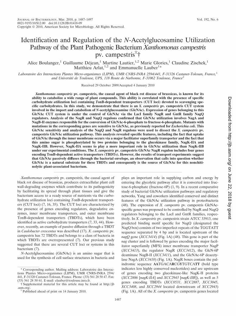
GlcNAc and chitobiose, but not chitin, are carbon and ni-trogen sources for X. campestris pv. campestris. The presencein X. campestris pv. campestris of genes proposed to belong toa GlcNAc utilization pathway suggests that GlcNAc can bemetabolized by X. campestris pv. campestris. Therefore, thegrowth rates of X. campestris pv. campestris cultures in MMEsupplemented with GlcNAc and with other carbon sourceswere compared. After sucrose and glucose, GlcNAc and theGlcNAc dimer chitobiose were among the best carbon sourcesfor X. campestris pv. campestris (Fig. 2A and B). In the pres-ence of the GlcNAc homopolymer chitin, slight growth wasreproducibly observed (Fig. 2B), probably due to the presenceof small amounts of free GlcNAc or chitobiose molecules. Thisresult suggests that X. campestris pv. campestris is not able toefficiently degrade chitin, a suggestion corroborated by theabsence of any obvious chitinase-encoding gene in the genomeof X. campestris pv. campestris strain ATCC 33913 (14, 48).
FIG. 1. X. campestris pv. campestris N-acetylglucosamine (GlcNAc)utilization pathway. (A) Organization of genes in the proposed GlcNAcutilization pathway. NagR boxes are indicated by filled circles, and theNagQ box is indicated by an open circle. (B) GlcNAc is proposed to betransported through the outer membrane by TBDTs and then transportedacross the inner membrane by the MFS transporter NagP. GlcNAc wouldthen be phosphorylated by nagK-II-encoded enzymes. Subsequent metab-olism via the nagA-encoded (GlcNAc-6P deacetylase) and nagB-II-en-coded (GlcN-6P deaminase) enzymes results in fructose 6-phosphate(Fru-6P) (48). MFS, major facilitator superfamily; PP, periplasm; TBDT,TonB-dependent transporter.
GlcNAc is also a nitrogen source for X. campestris pv.campestris, since this bacterium grows in nitrogen-depletedMME (MME without Casamino Acids and NH4SO4 [see Ma-terials and Methods]) in the presence of GlcNAc, whereas nogrowth was observed in the presence of glucose (data notshown).
GlcNAc pathway genes are induced by GlcNAc. The expres-sion of genes located downstream of putative NagR or NagQboxes was measured to assess the relationship of these genesto utilization of GlcNAc. The TBDT gene XCC3046 locateddownstream of the TBDT gene XCC3045 might belong to thesame operon (Fig. 1B) and was therefore included in this
TABLE 1. Plasmids and X. campestris pv. campestris strains used or generated in this study
Strain or plasmid Characteristicsa Locationb Designation Reference
Xanthomonas strainsWild type Wild-type strain; rifampin-resistant derivative
of X. campestris pv. campestris LMG568(� ATCC 33913)
31
nixB::pVO XCC0531::pVO155; Rifr Kmr 736 XP010 7nixC::pVO XCC2944::pVO155; Rifr Kmr 334 XP041 7nixD::pVO XCC2887::pVO155; Rifr Kmr 1558 XP040 7naxA::pVO XCC3045::pVO155; Rifr Kmr 1572 XP044 7naxB::pVO XCC3046::pVO155; Rifr Kmr 702 XP045 7nixA::pVO XCC3408::pVO155; Rifr Kmr 1897 XP059 7nagQ::pVO XCC3414::pVO155; Rifr Kmr 659 XP108 This studynagR::pVO XCC3412::pVO155; Rifr Kmr 519 XP109 This studynagA::pVO XCC3410::pVO155; Rifr Kmr 236 XP110 This studynagK-IIA::pVO XCC2886::pVO155; Rifr Kmr 303 XP111 This study�nagQ �XCC3414; Rifr From 203 to stop XP112 This study�nagR �XCC3412; Rifr From start to stop XP113 This study�nagP �XCC3413; Rifr From start to stop XP114 This study�nagA �XCC3410; Rifr From start to stop XP115 This study�nagB-II �XCC3411; Rifr From start to stop XP116 This study�nagK-IIA �XCC2886; Rifr From start to stop XP117 This study�nagK-IIB �XCC2943; Rifr From start to stop XP118 This study�nagK-IIAB �XCC2886 �XCC2943; Rifr From start to stop XP119 This study�nagA pC-nagA �XCC3410 pC-XCC3410; Rifr Tetr XP120 This study�nagB-II pC-nagB-II �XCC3411 pC-XCC3411; Rifr Tetr XP121 This study�nagB-II pC-nagB-II-nagA �XCC3411 pC-XCC3411-XCC3410; Rifr Tetr XP122 This study�nagK-IIA pC-nagK-IIA �XCC2886 pC-XCC2886; Rifr Tetr XP123 This study�nagK-IIB pC-nagK-IIB �XCC2943 pC-XCC2943; Rifr Tetr XP124 This study�nagK-IIAB pC-nagK-IIA �XCC2886 �XCC2943 pC-XCC2886; Rifr Tetr XP125 This study�nagK-IIAB pC-nagK-IIB �XCC2886 �XCC2943 pC-XCC2943; Rifr Tetr XP126 This study�nagR nagK-IIA::pVO �XCC3412 XCC2886::pVO155; Rifr Kmr XP127 This study�nagQ nagK-IIA::pVO �XCC3414 XCC2886::pVO155; Rifr Kmr XP128 This study�nagP nagA::pVO �XCC3413 XCC3410::pVO155; Rifr Kmr XP129 This study�nagK-IIAB nagA::pVO �XCC2886 �XCC2943 XCC3410::pVO155;
Rifr KmrXP130 This study
�nagP �nagA �XCC3413 �XCC3410; Rifr XP131 This study�nagK-IIAB �nagA �XCC2886 �XCC2943 �XCC3410; Rifr XP132 This study
pFAJ1700 pTR102-derived expression vector containinga multiple-cloning site and transcriptionalterminators in both orientations; Tetr Ampr
15
pSC150 pET-26b(�) derivative vector with a Ptacpromoter sequence; Kmr
13
pCZ917 pFAJ1700 derivative containing 2,094 bp ofpSC150 with lacI, Ptac promoter, and T7terminator; Tetr Ampr
This study
pC-nagA pCZ917-XCC3410; Tetr Kmr From �20 to stop This studypC-nagB-II pCZ917-XCC3411; Tetr Kmr From �19 to stop This studypC-nagB-II-nagA pCZ917-XCC3411-XCC3410; Tetr Kmr From �19 of XCC3411
to stop of XCC3410This study
pC-nagK-IIA pCZ917-XCC2886; Tetr Kmr From �21 to stop This studypC-nagK-IIB pCZ917-XCC2943; Tetr Kmr From �19 to stop This study
a Rif: rifampin; Km: kanamycin; Tet: tetracycline.b Position of insertion, deletion, or X. campestris pv. campestris sequence cloned relative to the putative start codon.
VOL. 192, 2010 GlcNAc UTILIZATION PATHWAY IN XANTHOMONAS 1489
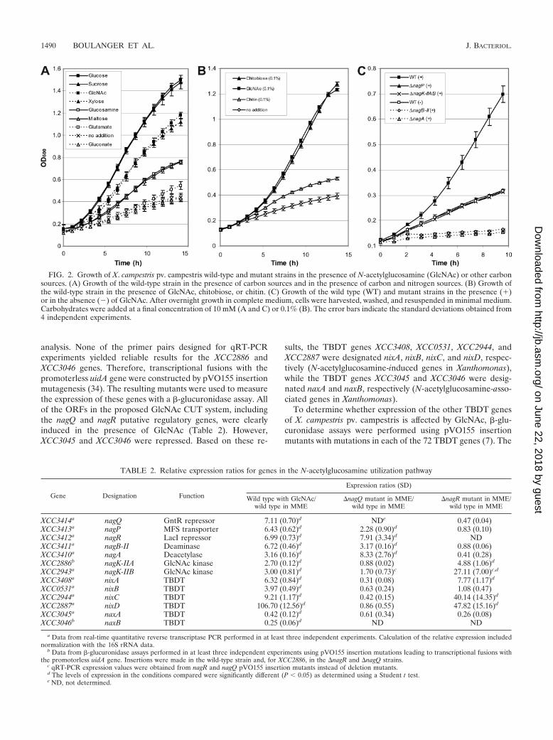
analysis. None of the primer pairs designed for qRT-PCRexperiments yielded reliable results for the XCC2886 andXCC3046 genes. Therefore, transcriptional fusions with thepromoterless uidA gene were constructed by pVO155 insertionmutagenesis (34). The resulting mutants were used to measurethe expression of these genes with a �-glucuronidase assay. Allof the ORFs in the proposed GlcNAc CUT system, includingthe nagQ and nagR putative regulatory genes, were clearlyinduced in the presence of GlcNAc (Table 2). However,XCC3045 and XCC3046 were repressed. Based on these re-
sults, the TBDT genes XCC3408, XCC0531, XCC2944, andXCC2887 were designated nixA, nixB, nixC, and nixD, respec-tively (N-acetylglucosamine-induced genes in Xanthomonas),while the TBDT genes XCC3045 and XCC3046 were desig-nated naxA and naxB, respectively (N-acetylglucosamine-asso-ciated genes in Xanthomonas).
To determine whether expression of the other TBDT genesof X. campestris pv. campestris is affected by GlcNAc, �-glu-curonidase assays were performed using pVO155 insertionmutants with mutations in each of the 72 TBDT genes (7). The
FIG. 2. Growth of X. campestris pv. campestris wild-type and mutant strains in the presence of N-acetylglucosamine (GlcNAc) or other carbonsources. (A) Growth of the wild-type strain in the presence of carbon sources and in the presence of carbon and nitrogen sources. (B) Growth ofthe wild-type strain in the presence of GlcNAc, chitobiose, or chitin. (C) Growth of the wild type (WT) and mutant strains in the presence (�)or in the absence (�) of GlcNAc. After overnight growth in complete medium, cells were harvested, washed, and resuspended in minimal medium.Carbohydrates were added at a final concentration of 10 mM (A and C) or 0.1% (B). The error bars indicate the standard deviations obtained from4 independent experiments.
TABLE 2. Relative expression ratios for genes in the N-acetylglucosamine utilization pathway
a Data from real-time quantitative reverse transcriptase PCR performed in at least three independent experiments. Calculation of the relative expression includednormalization with the 16S rRNA data.
b Data from �-glucuronidase assays performed in at least three independent experiments using pVO155 insertion mutations leading to transcriptional fusions withthe promotorless uidA gene. Insertions were made in the wild-type strain and, for XCC2886, in the �nagR and �nagQ strains.
c qRT-PCR expression values were obtained from nagR and nagQ pVO155 insertion mutants instead of deletion mutants.d The levels of expression in the conditions compared were significantly different (P � 0.05) as determined using a Student t test.e ND, not determined.
nixA, nixB, nixC, and nixD genes were the only TBDT genesinduced by GlcNAc (data not shown). It is worth noting thatthe expression of the nix TBDT genes was not as stronglyinduced in the pVO155 insertion mutant (the induction levelsranged from 1.8-fold for nixB to 36-fold for nixD [data notshown]) as was expected based on the results of qRT-PCR fora wild-type background (for which the induction levels rangedfrom 3.97-fold for nixB to 106.7-fold for nixD [Table 2]).
The expression of GlcNAc-induced genes was then mea-sured after growth in MME supplemented with a range ofGlcNAc concentrations. Representative results obtained withthe nixD::pVO mutant are reported here because this mutantdisplayed one of the highest levels of induction in the presenceof GlcNAc and because its growth was not impaired in MMEsupplemented with GlcNAc (see below). The reporter genewas induced with 5 �M to 20 mM GlcNAc. The maximalinduction (around 30-fold) was observed with 50 �M GlcNAc(data not shown). Induction was also observed with high con-centrations of glucosamine (GlcN), but the maximal inductionwas only 3-fold (data not shown).
NagQ and NagR are GlcNAc pathway-specific regulators.The involvement of two presumptive regulators, NagQ andNagR, was evaluated by comparing nix gene expression in thewild-type strain and nix gene expression in nagQ and nagRmutants in MME without added GlcNAc. Mutants with inser-tions and deletions of these two regulatory genes were con-structed, but deletion mutants were chosen to avoid possiblepolar effects, since both regulatory genes may be expressed aspart of an operon (Fig. 1A).
The levels of expression of the nagP, nagR, nagB-II, andnagA genes were clearly higher in the �nagQ deletion mutantthan in the wild-type strain (Table 2). These genes are down-stream of the nagQ gene, which is itself downstream of theunique putative NagQ box detected in the X. campestris pv.campestris genome (Fig. 1A). This result suggests that NagQregulates its own expression and that the genes from nagQ tonagA form an operon. The expression of the other GlcNAcpathway genes was not significantly affected by deletion ofnagQ.
The expression of NagQ-regulated genes was not affected bydeletion of nagR. The expression of nixA, nixC, nixD, nagK-IIA,and nagK-IIB was derepressed in the �nagR deletion mutantcompared to the expression in the wild-type strain (Table 2).This is in agreement with the presence of putative NagR boxesin the promoter regions of these genes or operons, as deter-mined by Yang and coworkers (48). Surprisingly, the GlcNAc-induced TBDT nixB gene located downstream of a NagR boxseemed not to be regulated by NagR under our conditions(Table 2), suggesting that four of the five putative NagR boxesare functional. This result prompted us to generate a positionweight matrix with the PREDetector program (23) using thefour functional NagR boxes for screening the X. campestris pv.campestris genome. Of the 61 predicted targets, 16 are locatedin intergenic regions (see Table S1 in the supplemental mate-rial). Sequences upstream of nixA, nixB, nixD, nagK-IIA, andnagK-IIB each had strong predicted NagR-binding sites.However, the score obtained for the nixB promoter site wasclose to the scores for weak sites (see Table S1 in thesupplemental material). This low score might explain the
poor NagR regulation of nixB, a gene which is neverthelessinduced by GlcNAc.
A sequence logo was generated by WebLogo (http://weblogo.berkley.edu/; 11) from the alignment of the four putative func-tional NagR boxes, which resulted in discovery of a new NagRbox (GTTGACARCGYTGTCANC). This NagR box differedat positions 1, 2, and 18 from the previously proposed NagRbox (AATGACARCGYTGTCATT) (48).
Together, these results show that NagR and NagQ are func-tional repressors of genes belonging to the GlcNAc CUT sys-tem. Proteins encoded by NagR- and NagQ-regulated genescan be classified into two main categories: transport and me-tabolism of GlcNAc.
Transport of GlcNAc in X. campestris pv. campestris. (i) FreeGlcNAc passively diffuses through the envelope. GlcNAc up-take rates in the X. campestris pv. campestris wild-type strainwere compared after overnight preculture in the presence ofGlcNAc (induced) and after overnight preculture in the pres-ence of xylose (uninduced), a substrate that results in a growthrate similar to that obtained with GlcNAc (Fig. 2A) but doesnot affect the expression of GlcNAc-induced TBDT genes(data not shown). Before transport experiments were per-formed with [14C]GlcNAc, cells were washed to remove non-radiolabeled GlcNAc from the medium. The GlcNAc uptakerates under the two conditions were not significantly different(data not shown), suggesting that GlcNAc import is limited bya GlcNAc-independent transport step.
The initial concentration-dependent [14C]GlcNAc transport,reflecting the dissociation constant (Kd) for GlcNAc uptake,was determined using the previously described rapid dilutionmethod (7, 33). The kinetic values revealed that the uptakerate was low and monophasic (Fig. 3), suggesting either thatthe outer and inner membrane transporters have similar affin-ities for GlcNAc or that transport through the outer membraneis limiting and masks transport through the inner membrane.The deduced Kd (138.9 �M) is more than 100-fold higher thanthe Kd estimated for passive uptake of GlcNAc through the
FIG. 3. Concentration-dependent transport of 14C-labeled N-acetyl-glucosamine (GlcNAc) into X. campestris pv. campestris. Cells weregrown in minimal medium without GlcNAc, and transport was mea-sured for 15 s at the [14C]GlcNAc concentrations indicated.
VOL. 192, 2010 GlcNAc UTILIZATION PATHWAY IN XANTHOMONAS 1491
CC0446 TBDT in C. crescentus (17) and is in a range similar tothe range for Kd values obtained for passive diffusion throughporins (18). Therefore, free GlcNAc uptake through the X.campestris pv. campestris envelope seems to occur via passivediffusion rather than by active uptake, although the X. campes-tris pv. campestris GlcNAc regulon contains at least fourTBDT genes encoding active outer membrane transporters.
A 100-fold excess of unlabeled glucose, galactose, sucrose,mannose, xylose, fructose, or GlcNAc-6P had no effect onradiolabeled GlcNAc uptake (Table 3). With unlabeled Glc-NAc, the concentration for inhibition of the transport rate toone-half of the control rate was estimated to be 193.7 �M,which is in accordance with the Kd deduced from the results ofthe initial concentration-dependent GlcNAc transport assays.Glucosamine and chitobiose both inhibit radioactive GlcNAcuptake as much as unlabeled GlcNAc (Table 3). Inhibitionof GlcNAc uptake by chitobiose could be due either to thechitobiose molecule itself or to degradation of this moleculeto GlcNAc. These competition experiments suggest that GlcNAc,glucosamine, and probably chitobiose are transported acrossthe envelope via the same transporters.
(ii) None of the GlcNAc-induced TBDTs seems to play amajor role in utilization of free GlcNAc. The rates of [14C]GlcNAc uptake in the X. campestris pv. campestris wild-typestrain and in GlcNAc regulon TBDT insertion mutants werecompared, and none of the TBDT mutants exhibited a signif-icant effect in GlcNAc uptake (Table 4). Furthermore, thegrowth rates of strains with mutations in the nix and nax TBDTgenes in the presence of 10 mM GlcNAc were similar to thegrowth rate of the wild-type strain (data not shown). Theabsence of a phenotype for mutants with single mutations inTBDT genes could be due to the redundant functions of thetransporters.
(iii) NagP is the major GlcNAc inner membrane transporterin X. campestris pv. campestris. The nagP gene was deleted totest the putative role of NagP, which belongs to the majorfacilitator superfamily (MFS), in the transport of GlcNAc.Growth of the �nagP strain was impaired on MME containingGlcNAc as the sole carbon source (Fig. 2C), suggesting thatthis transporter could be involved in the uptake of GlcNActhrough the inner membrane. The rate of uptake of radiola-
beled GlcNAc obtained for the �nagP strain was only 1.2% ofthe rate obtained for the wild-type strain (Table 4). In thenagP-complemented strain, GlcNAc transport capacity (Table4) and growth on GlcNAc-containing MME (Fig. 4A) wererestored, confirming that NagP is the major transporter ofGlcNAc across the inner membrane.
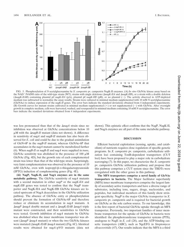
Catabolism of GlcNAc in X. campestris pv. campestris. (i)NagK-IIA and NagK-IIB phosphorylate GlcNAc. In the cyto-plasm, the first step in the X. campestris pv. campestris GlcNAcutilization pathway is phosphorylation of GlcNAc (Fig. 1B).Two genes, nagK-IIA and nagK-IIB, coding for proteins be-longing to the glucokinase family, belong to the GlcNAc regu-lon (Table 2), and their products have been proposed to act asputative GlcNAc kinases in Xanthomonas (48). To test thefunction of these proteins in the phosphorylation of GlcNAc,�nagK-IIA and �nagK-IIB single mutants, as well as a �nagK-IIAB double mutant, were constructed. The GlcNAc kinaseactivity of the �nagK-IIAB double mutant was about 41%of the wild-type activity (Fig. 5A). The GlcNAc kinase activityof the double mutant could have been due to the presence ofresidual ADP or pyruvate in the crude extracts used in theexperiments. Wild-type GlcNAc kinase activity was restoredwhen either nagK-IIA or nagK-IIB was supplied in trans on anexpression plasmid, suggesting that both proteins phosphory-late GlcNAc. However, the activities obtained for each singlemutant did not differ significantly from the wild-type activity(Fig. 5A), suggesting that these two proteins are functionallyredundant.
The growth of the �nagK-IIAB double mutant was clearlyimpaired in GlcNAc-containing minimal medium (Fig. 2C andFig. 5B). The growth of the �nagK-IIA single mutant was alsoaffected, but to a lesser extent, whereas the �nagK-IIB mutantgrew like the wild-type strain (Fig. 5B). Growth of the �nagK-IIAB double mutant in GlcNAc minimal medium was partiallyrestored when nagK-IIA or nagK-IIB was overexpressed intrans on an expression plasmid; however, better complemen-tation was observed with nagK-IIA (Fig. 4B). These resultssuggest that although both GlcNAc kinases are enzymaticallyfunctional, NagK-IIA plays a major role in GlcNAc utilization,whereas NagK-IIB, which is apparently not essential, can alsofunction in this capacity.
(ii) GlcNAc is toxic for nagA and nagB-II mutants. NagAdeacetylase (XCC3410) catalyzes the conversion of GlcNAc-6P
TABLE 3. Inhibition of uptake of 0.5 �M �14CGlcNAc by variouscarbohydrates in X. campestris pv. campestris wild-type
strain after 1 h of incubation
Carbohydrate
% of control uptake (SD) atcarbohydrate concn ofa:
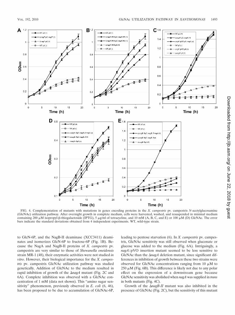
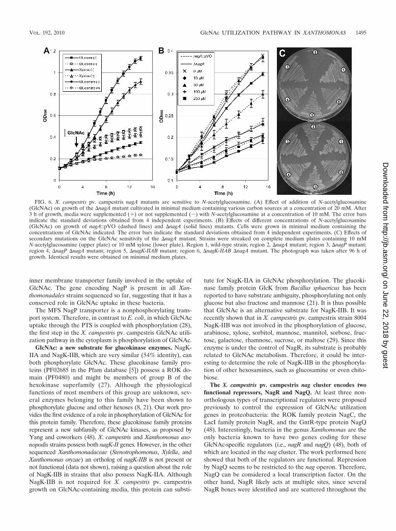
to GlcN-6P, and the NagB-II deaminase (XCC3411) deami-nates and isomerizes GlcN-6P to fructose-6P (Fig. 1B). Be-cause the NagA and NagB-II proteins of X. campestris pv.campestris are very similar to those of Shewanella oneidensisstrain MR-1 (48), their enzymatic activities were not studied invitro. However, their biological importance for the X. campes-tris pv. campestris GlcNAc utilization pathway was studiedgenetically. Addition of GlcNAc to the medium resulted inrapid inhibition of growth of the �nagA mutant (Fig. 2C and6A). Complete inhibition was observed with a GlcNAc con-centration of 1 mM (data not shown). This “amino sugar sen-sitivity” phenomenon, previously observed in E. coli (6, 46),has been proposed to be due to accumulation of GlcNAc-6P,
leading to pentose starvation (6). In X. campestris pv. campes-tris, GlcNAc sensitivity was still observed when gluconate orglucose was added to the medium (Fig. 6A). Intriguingly, anagA::pVO insertion mutant seemed to be less sensitive toGlcNAc than the �nagA deletion mutant, since significant dif-ferences in inhibition of growth between these two strains wereobserved for GlcNAc concentrations ranging from 10 �M to250 �M (Fig. 6B). This difference is likely not due to any polareffect on the expression of a downstream gene becauseGlcNAc sensitivity was abolished when nagA was supplied in transin both mutants (Fig. 4C).
Growth of the �nagB-II mutant was also inhibited in thepresence of GlcNAc (Fig. 2C), but the sensitivity of this mutant
FIG. 4. Complementation of mutants with mutations in genes encoding proteins in the X. campestris pv. campestris N-acetylglucosamine(GlcNAc) utilization pathway. After overnight growth in complete medium, cells were harvested, washed, and resuspended in minimal mediumcontaining 200 �M isopropyl-�-thiogalactoside (IPTG), 5 �g/ml of tetracycline, and 10 mM (A, B, C, and E) or 100 �M (D) GlcNAc. The errorbars indicate the standard deviations obtained from 4 independent experiments. WT, wild-type strain.
VOL. 192, 2010 GlcNAc UTILIZATION PATHWAY IN XANTHOMONAS 1493
was less pronounced than that of the �nagA strain since noinhibition was observed at GlcNAc concentrations below 10�M with the �nagB-II mutant (data not shown). A differencein sensitivity of nagA and nagB-II mutants has also been ob-served for E. coli and could be due to the gradual assimilationof GlcN-6P in the nagB-II mutant, whereas GlcNAc-6P thataccumulates in the nagA mutant cannot be metabolized further(6). When nagB-II or nagB-II and nagA were supplied in trans,GlcNAc sensitivity was abolished in the presence of 100 �MGlcNAc (Fig. 4D), but the growth rate of each complementedstrain was lower than that of the wild-type strain. Surprisingly,very faint complementation was observed in the presence of 10mM GlcNAc, even with isopropyl-�-D-thiogalactopyranoside(IPTG) induction of complementing genes (Fig. 4E).
(iii) NagP, NagK-II, and NagA enzymes are in the samemetabolic pathway. The GlcNAc sensitivity of nagA mutantswith a second mutation in either nagP or the nagK-IIA andnagK-IIB genes was tested to confirm that the NagP trans-porter and NagK-IIA and NagK-IIB GlcNAc kinases are lo-cated upstream of NagA deacetylase in the GlcNAc utilizationpathway. The absence of GlcNAc entry or phosphorylationshould prevent the formation of GlcNAc-6P and thereforereduce or eliminate its accumulation in nagA mutants. A�nagP �nagA double mutant and a �nagK-IIAB �nagA triplemutant were constructed, and their sensitivities to GlcNAcwere tested. Growth inhibition of nagA mutants by GlcNAcwas abolished when the inner membrane transporter was ab-sent (�nagP �nagA mutant) or when the two GlcNAc kinaseswere mutated (�nagK-IIAB �nagA mutant) (Fig. 6C). Identicalresults were obtained for nagA::pVO mutants (data not
shown). This epistatic effect confirms that the NagP, NagK-II,and NagA enzymes are all part of the same metabolic pathway.
DISCUSSION
Efficient bacterial exploitation (sensing, uptake, and catab-olism) of nutrients requires close regulation of specific geneticprograms. In X. campestris pv. campestris, carbohydrate utili-zation loci containing TonB-dependent transporters (CUTloci) have been proposed to play a major role in carbohydratescavenging (7). In this paper, we characterize the X. campestrispv. campestris GlcNAc utilization pathway and propose thatthis pathway comprises a CUT system since the TBDTs werecoregulated with the other genes in this pathway.
The MFS transporters comprise a novel family of GlcNActransporters in bacteria. The Major facilitator superfamily(MFS) inner membrane transporters comprise the largest fam-ily of secondary active transporters and have a diverse range ofsubstrates, including ions, sugars, drugs, nucleosides, andpeptides, but individual members of this family show strin-gent specificity. NagP is the major GlcNAc transporter in X.campestris pv. campestris and is required for bacterial growthon GlcNAc as the sole carbon source. To our knowledge, thisis the first report of bacterial GlcNAc uptake through an MFStransporter. Previously, two important families of inner mem-brane transporters for the uptake of GlcNAc in bacteria wereidentified: the phosphotransferase transporter systems (PTS),analogous to NagE in E. coli (28), and the ATP-binding cas-sette transporters (ABC), such as NgcEFG in Streptomycesolivaceoviridis (47). Our results indicate that the MFS is a third
FIG. 5. Phosphorylation of N-acetylglucosamine by X. campestris pv. campestris NagK-II enzymes. (A) In vitro GlcNAc kinase assay based onthe NAD�/NADH ratio of the wild-type strain (WT), strains with single deletions (�nagK-IIA and �nagK-IIB), or a strain with a double deletion(�nagK-IIAB) containing plasmid pC-nagK-IIA (pA), plasmid pC-nagK-IIB (pB), or no plasmid (�). The activity observed in ATP-depletedmedium was subtracted to normalize the assay results. Strains were cultured in minimal medium supplemented with 10 mM N-acetylglucosamine(GlcNAc) to induce expression of the nagK-II genes. The error bars indicate the standard deviations obtained from 4 independent experiments.(B) Growth curves for mutant strains cultivated in minimal medium supplemented (�) or not supplemented (�) with GlcNAc. After overnightgrowth in complete medium, cells were harvested, washed, and resuspended in minimal medium containing 10 mM N-acetylglucosamine. The errorbars indicate the standard deviations obtained from 4 independent experiments.
inner membrane transporter family involved in the uptake ofGlcNAc. The gene encoding NagP is present in all Xan-thomonadales strains sequenced so far, suggesting that it has aconserved role in GlcNAc uptake in these bacteria.
The MFS NagP transporter is a nonphosphorylating trans-port system. Therefore, in contrast to E. coli, in which GlcNAcuptake through the PTS is coupled with phosphorylation (28),the first step in the X. campestris pv. campestris GlcNAc utili-zation pathway in the cytoplasm is phosphorylation of GlcNAc.
GlcNAc: a new substrate for glucokinase enzymes. NagK-IIA and NagK-IIB, which are very similar (54% identity), canboth phosphorylate GlcNAc. These glucokinase family pro-teins (PF02685 in the Pfam database [5]) possess a ROK do-main (PF0480) and might be members of group B of thehexokinase superfamily (27). Although the physiologicalfunctions of most members of this group are unknown, sev-eral enzymes belonging to this family have been shown tophosphorylate glucose and other hexoses (8, 21). Our work pro-vides the first evidence of a role in phosphorylation of GlcNAc forthis protein family. Therefore, these glucokinase family proteinsrepresent a new subfamily of GlcNAc kinases, as proposed byYang and coworkers (48). X. campestris and Xanthomonas axo-nopodis strains possess both nagK-II genes. However, in the othersequenced Xanthomonadaceae (Stenotrophomonas, Xylella, andXanthomonas oryzae) an ortholog of nagK-IIB is not present ornot functional (data not shown), raising a question about the roleof NagK-IIB in strains that also possess NagK-IIA. AlthoughNagK-IIB is not required for X. campestris pv. campestrisgrowth on GlcNAc-containing media, this protein can substi-
tute for NagK-IIA in GlcNAc phosphorylation. The glucoki-nase family protein GlcK from Bacillus sphaericus has beenreported to have substrate ambiguity, phosphorylating not onlyglucose but also fructose and mannose (21). It is thus possiblethat GlcNAc is an alternative substrate for NagK-IIB. It wasrecently shown that in X. campestris pv. campestris strain 8004NagK-IIB was not involved in the phosphorylation of glucose,arabinose, xylose, sorbitol, mannose, mannitol, sorbose, fruc-tose, galactose, rhamnose, sucrose, or maltose (29). Since thisenzyme is under the control of NagR, its substrate is probablyrelated to GlcNAc metabolism. Therefore, it could be inter-esting to determine the role of NagK-IIB in the phosphoryla-tion of other hexosamines, such as glucosamine or even chito-biose.
The X. campestris pv. campestris nag cluster encodes twofunctional repressors, NagR and NagQ. At least three non-orthologous types of transcriptional regulators were proposedpreviously to control the expression of GlcNAc utilizationgenes in proteobacteria: the ROK family protein NagC, theLacI family protein NagR, and the GntR-type protein NagQ(48). Interestingly, bacteria in the genus Xanthomonas are theonly bacteria known to have two genes coding for theseGlcNAc-specific regulators (i.e., nagR and nagQ) (48), both ofwhich are located in the nag cluster. The work performed hereshowed that both of the regulators are functional. Repressionby NagQ seems to be restricted to the nag operon. Therefore,NagQ can be considered a local transcription factor. On theother hand, NagR likely acts at multiple sites, since severalNagR boxes were identified and are scattered throughout the
FIG. 6. X. campestris pv. campestris nagA mutants are sensitive to N-acetylglucosamine. (A) Effect of addition of N-acetylglucosamine(GlcNAc) on growth of the �nagA mutant cultivated in minimal medium containing various carbon sources at a concentration of 20 mM. After3 h of growth, media were supplemented (�) or not supplemented (�) with N-acetylglucosamine at a concentration of 10 mM. The error barsindicate the standard deviations obtained from 4 independent experiments. (B) Effects of different concentrations of N-acetylglucosamine(GlcNAc) on growth of nagA::pVO (dashed lines) and �nagA (solid lines) mutants. Cells were grown in minimal medium containing theconcentrations of GlcNAc indicated. The error bars indicate the standard deviations obtained from 4 independent experiments. (C) Effects ofsecondary mutations on the GlcNAc sensitivity of the �nagA mutant. Strains were streaked on complete medium plates containing 10 mMN-acetylglucosamine (upper plate) or 10 mM xylose (lower plate). Region 1, wild-type strain; region 2, �nagA mutant; region 3, �nagP mutant;region 4, �nagP �nagA mutant; region 5, �nagK-IIAB mutant; region 6, �nagK-IIAB �nagA mutant. The photograph was taken after 96 h ofgrowth. Identical results were obtained on minimal medium plates.
VOL. 192, 2010 GlcNAc UTILIZATION PATHWAY IN XANTHOMONAS 1495
X. campestris pv. campestris genome. The NagR regulon couldbe broader, and it would be interesting to identify additionaltargets of this repressor in global transcriptomic analyses.
In X. campestris pv. campestris, NagQ represses nagR. Theimportance of this regulatory loop in GlcNAc catabolism is amatter of conjecture. Furthermore, the GlcNAc regulatorynetwork could be much more complicated, and additional reg-ulatory elements could participate in this network. Indeed,nixA, naxA, and nagK-IIA belong to the Clp regulon, a con-served global regulator shown to play a central role in theregulation of virulence factors in X. campestris pv. campestris(22).
The nagQ gene is specific for Xanthomonas strains. Indeed,in the sequenced plant-pathogenic Xylella strains, which havereduced genomes (37), nagQ is partially deleted, suggestingthat this gene has become vestigial and can therefore be lostduring evolution. This gene is also not present in Stenotropho-monas maltophilia, a non-plant-pathogenic species belongingto the Xanthomonadaceae family that includes free-living aswell as endophytic isolates and opportunistic human pathogens(12, 41). Interestingly, in both bacteria, the NagQ box locatedupstream of the nag cluster is replaced by a candidate NagRbox (48).
GlcNAc passively diffuses through the outer membrane. Al-though four X. campestris pv. campestris active outer mem-brane TBDT genes are induced by GlcNAc, three of which areunder the control of NagR, GlcNAc passively diffuses throughthe outer membrane. Furthermore, none of the GlcNAc-in-duced TBDTs was individually required for growth on GlcNAcand for uptake of this molecule. This implies either that severalof these TBDTs allow diffusion of GlcNAc or, alternatively,that GlcNAc can diffuse through the outer membrane via othertransporters, such as porins.
TBDTs, which are well known for their role in iron andvitamin B12 uptake (36), were shown previously to be involvedin the active uptake of carbohydrates, such as maltodextrins inC. crescentus (33) or sucrose in X. campestris pv. campestris (7).In C. crescentus, TBDT CC0446 is essential for growth on thechito-oligosaccharides (GlcNAc)3 and (GlcNAc)5 in a TonB-dependent manner, but transport of GlcNAc apparently occursby passive diffusion through this transporter, a process which isTonB independent (17). Therefore, we propose that X.campestris pv. campestris TBDTs belonging to the GlcNAcregulon are involved in the active transport of complex mole-cules containing GlcNAc, as observed for C. crescentus (17).Consequently, this implies that the source of GlcNAc could bemore complex than monomeric GlcNAc.
What is the source of GlcNAc for X. campestris pv. campes-tris in the environment? There are two principal sources ofGlcNAc in nature: chitin and bacterial cell walls. In E. coli,about 50% of cell wall peptidoglycan is broken down eachgeneration (20, 24). Recent bioinformatic analyses identifiedthe gene coding for the D-Ala-D-Ala aminopeptidase that isresponsible for catabolism of the cell wall precursor D-Ala-D-Ala as a part of the Streptomyces coelicolor DasR regulon (39).This pleiotropic regulator belonging to the GntR family isessential for development and is involved in the regulation ofgenes encoding both the GlcNAc PTS and the GlcN-6P deami-nase NagB (38). Therefore, there is a direct relationship be-tween peptidoglycan recycling and GlcNAc utilization. Al-
though this link was not studied in X. campestris pv. campestris,a role for an X. campestris pv. campestris GlcNAc-inducedTBDT(s) in the active uptake of peptidoglycan degradationproducts can be readily envisaged.
Alternatively, X. campestris pv. campestris could have devel-oped a system to exploit chito-oligosaccharides derived fromfungal and insect chitin degraded by plant chitinases (26) or bychitinolytic bacteria. Occupation of niches in the plant phyllo-sphere by epiphytic, saprophytic, and pathogenic fungi andbacteria and the interactions of these organisms are importantfor the Xanthomonas life cycle (40). In this environment, thegrowth of X. campestris pv. campestris on chitin or its poly-meric subunits relies on other organisms for chitin degrada-tion, as has recently been proposed for the nonchitinolyticaquatic bacterium C. crescentus (17). To exploit chito-oligosac-charides, X. campestris pv. campestris must produce enzymesinvolved in degradation of these compounds. This bacteriumhas one of the largest glycobiomes, as determined using theCAZy database (http://www.cazy.org/) (10). Interestingly,downstream of the nixD TBDT gene (XCC2887), we identifiedseveral putative enzyme-encoding genes which could be in-volved in the degradation of oligosaccharides that containGlcNAc. Among these, XCC2889 codes for a protein be-longing to the GH-18 family, which includes endo-beta-N-acetylglucosaminidase (EC 3.2.1.96), and XCC2890 codes for aprotein belonging to the glycoside hydrolase GH-20 family,which includes the �-hexosaminidases (EC 3.2.1.52). Work isnow under way to further characterize these genes, to deter-mine whether they are part of the GlcNAc CUT system, and todetermine which molecules are targeted by these enzymes.
ACKNOWLEDGMENTS
We are grateful to Scott Soby for critical reading of the manuscript.We also acknowledge Laurent Noel and Servane Baufume for valuablediscussions and critical reading of the manuscript.
A. Boulanger and G. Dejean were supported by a grant from theFrench Ministere de la Recherche et de l’Enseignement Superieur. Wegratefully acknowledge financial support from the Departement Santedes Plantes et Environnement de l’Institut National de la RechercheAgronomique (grant 2007_0441_02) and from the French Agence Na-tionale de la Recherche (grant ANR-08-BLAN-0193-01).
REFERENCES
1. Alvarez-Anorve, L. I., M. L. Calcagno, and J. Plumbridge. 2005. Why doesEscherichia coli grow more slowly on glucosamine than on N-acetyl-glucosamine? Effects of enzyme levels and allosteric activation of GlcN6Pdeaminase (NagB) on growth rates. J. Bacteriol. 187:2974–2982.
2. Angot, A., N. Peeters, E. Lechner, F. Vailleau, C. Baud, L. Gentzbittel, E.Sartorel, P. Genschik, C. Boucher, and S. Genin. 2006. Ralstonia solanacearumrequires F-box-like domain-containing type III effectors to promote diseaseon several host plants. Proc. Natl. Acad. Sci. U. S. A. 103:14620–14625.
3. Arlat, M., C. L. Gough, C. E. Barber, C. Boucher, and M. J. Daniels. 1991.Xanthomonas campestris contains a cluster of hrp genes related to the largerhrp cluster of Pseudomonas solanacearum. Mol. Plant-Microbe Interact.4:593–601.
4. Arlat, M., F. Van Gijsegem, J. C. Huet, J. C. Pernollet, and C. A. Boucher.1994. PopA1, a protein which induces a hypersensitivity-like response onspecific Petunia genotypes, is secreted via the Hrp pathway of Pseudomonassolanacearum. EMBO J. 13:543–553.
5. Bateman, A., L. Coin, R. Durbin, R. D. Finn, V. Hollich, S. Griffiths-Jones,A. Khanna, M. Marshall, S. Moxon, E. L. Sonnhammer, D. J. Studholme, C.Yeats, and S. R. Eddy. 2004. The Pfam protein families database. NucleicAcids Res. 32:D138–D141.
6. Bernheim, N. J., and W. J. Dobrogosz. 1970. Amino sugar sensitivity inEscherichia coli mutants unable to grow on N-acetylglucosamine. J. Bacte-riol. 101:384–391.
7. Blanvillain, S., D. Meyer, A. Boulanger, M. Lautier, C. Guynet, N. Denance,J. Vasse, E. Lauber, and M. Arlat. 2007. Plant carbohydrate scavenging
through tonb-dependent receptors: a feature shared by phytopathogenic andaquatic bacteria. PLoS One 2:e224.
8. Brigham, C. J., and M. H. Malamy. 2005. Characterization of the RokA andHexA broad-substrate-specificity hexokinases from Bacteroides fragilis andtheir role in hexose and N-acetylglucosamine utilization. J. Bacteriol. 187:890–901.
9. Calcagno, M., P. J. Campos, G. Mulliert, and J. Suastegui. 1984. Purifica-tion, molecular and kinetic properties of glucosamine-6-phosphate isomer-ase (deaminase) from Escherichia coli. Biochim. Biophys. Acta 787:165–173.
10. Cantarel, B. L., P. M. Coutinho, C. Rancurel, T. Bernard, V. Lombard, andB. Henrissat. 2009. The Carbohydrate-Active EnZymes database (CAZy):an expert resource for glycogenomics. Nucleic Acids Res. 37:D233–D238.
11. Crooks, G. E., G. Hon, J. M. Chandonia, and S. E. Brenner. 2004. WebLogo:a sequence logo generator. Genome Res. 14:1188–1190.
12. Crossman, L. C., V. C. Gould, J. M. Dow, G. S. Vernikos, A. Okazaki, M.Sebaihia, D. Saunders, C. Arrowsmith, T. Carver, N. Peters, E. Adlem, A.Kerhornou, A. Lord, L. Murphy, K. Seeger, R. Squares, S. Rutter, M. A.Quail, M. A. Rajandream, D. Harris, C. Churcher, S. D. Bentley, J. Parkhill,N. R. Thomson, and M. B. Avison. 2008. The complete genome, comparativeand functional analysis of Stenotrophomonas maltophilia reveals an organ-ism heavily shielded by drug resistance determinants. Genome Biol. 9:R74.
13. Cunnac, S. 2004. Identification a l’echelle genomique des effecteurs depen-dant du systeme de secretion de type III de la bacterie phytopathogeneRalstonia solanacearum. Ph.D. thesis. Universite Toulouse III Paul Sabatier,Toulouse, France.
14. da Silva, A. C. R., J. A. Ferro, F. C. Reinach, C. S. Farah, L. R. Furlan, R. B.Quaggio, C. B. Monteiro-Vitorello, M. A. V. Sluys, N. F. Almeida, L. M. C.Alves, A. M. do Amaral, M. C. Bertolini, L. E. A. Camargo, G. Camarotte, F.Cannavan, J. Cardozo, F. Chambergo, L. P. Ciapina, R. M. B. Cicarelli, L. L.Coutinho, J. R. Cursino-Santos, H. El-Dorry, J. B. Faria, A. J. S. Ferreira,R. C. C. Ferreira, M. I. T. Ferro, E. F. Formighieri, M. C. Franco, C. C.Greggio, A. Gruber, A. M. Katsuyama, L. T. Kishi, R. P. Leite, E. G. M.Lemos, M. V. F. Lemos, E. C. Locali, M. A. Machado, A. M. B. N. Madeira,N. M. Martinez-Rossi, E. C. Martins, J. Meidanis, C. F. M. Menck, C. Y.Miyaki, D. H. Moon, L. M. Moreira, M. T. M. Novo, V. K. Okura, M. C.Oliveira, V. R. Oliveira, H. A. Pereira, A. Rossi, J. A. D. Sena, C. Silva, R. F.de Souza, L. A. F. Spinola, M. A. Takita, R. E. Tamura, E. C. Teixeira,R. I. D. Tezza, M. Trindade dos Santos, D. Truffi, S. M. Tsai, F. F. White,J. C. Setubal, and J. P. Kitajima. 2002. Comparison of the genomes of twoXanthomonas pathogens with differing host specificities. Nature 417:459–463.
15. Dombrecht, B., J. Vanderleyden, and J. Michiels. 2001. Stable RK2-derivedcloning vectors for the analysis of gene expression and gene function ingram-negative bacteria. Mol. Plant-Microbe Interact. 14:426–430.
16. Dow, J. M., and M. J. Daniels. 1994. Pathogenicity determinants and globalregulation of pathogenicity of Xanthomonas campestris pv. campestris. Curr.Top. Microbiol. Immunol. 192:29–41.
17. Eisenbeis, S., S. Lohmiller, M. Valdebenito, S. Leicht, and V. Braun. 2008.NagA-dependent uptake of N-acetyl-glucosamine and N-acetyl-chitin oligo-saccharides across the outer membrane of Caulobacter crescentus. J. Bacte-riol. 190:5230–5238.
18. Freundlieb, S., U. Ehmann, and W. Boos. 1988. Facilitated diffusion ofp-nitrophenyl-alpha-D-maltohexaoside through the outer membrane ofEscherichia coli. Characterization of LamB as a specific and saturable chan-nel for maltooligosaccharides. J. Biol. Chem. 263:314–320.
19. Gonzali, S., L. Pistelli, L. De Bellis, and A. Alpi. 2001. Characterization oftwo Arabidopsis thaliana fructokinases. Plant Sci. 160:1107–1114.
20. Goodell, E. W. 1985. Recycling of murein by Escherichia coli. J. Bacteriol.163:305–310.
21. Han, B., H. Liu, X. Hu, Y. Cai, D. Zheng, and Z. Yuan. 2007. Molecularcharacterization of a glucokinase with broad hexose specificity from Bacillussphaericus strain C3-41. Appl. Environ. Microbiol. 73:3581–3586.
22. He, Y. W., A. Y. Ng, M. Xu, K. Lin, L. H. Wang, Y. H. Dong, and L. H. Zhang.2007. Xanthomonas campestris cell-cell communication involves a putativenucleotide receptor protein Clp and a hierarchical signalling network. Mol.Microbiol. 64:281–292.
23. Hiard, S., R. Maree, S. Colson, P. A. Hoskisson, F. Titgemeyer, G. P. vanWezel, B. Joris, L. Wehenkel, and S. Rigali. 2007. PREDetector: a new toolto identify regulatory elements in bacterial genomes. Biochem. Biophys. Res.Commun. 357:861–864.
24. Jacobs, C., L. J. Huang, E. Bartowsky, S. Normark, and J. T. Park. 1994.Bacterial cell wall recycling provides cytosolic muropeptides as effectors forbeta-lactamase induction. EMBO J. 13:4684–4694.
25. Jefferson, R. A., T. A. Kavanagh, and M. W. Bevan. 1987. GUS fusions:beta-glucuronidase as a sensitive and versatile gene fusion marker in higherplants. EMBO J. 6:3901–3907.
26. Kasprzewska, A. 2003. Plant chitinases—regulation and function. Cell. Mol.Biol. Lett. 8:809–824.
27. Kawai, S., T. Mukai, S. Mori, B. Mikami, and K. Murata. 2005. Hypothesis:structures, evolution, and ancestor of glucose kinases in the hexokinasefamily. J. Biosci. Bioeng. 99:320–330.
28. Lengeler, J. W., K. Jahreis, and U. F. Wehmeier. 1994. Enzymes II of thephosphoenol pyruvate-dependent phosphotransferase systems: their struc-ture and function in carbohydrate transport. Biochim. Biophys. Acta 1188:1–28.
29. Lu, G. T., Z. J. Yang, F. Y. Peng, Y. N. Tan, Y. Q. Tang, J. X. Feng, D. J.Tang, Y. Q. He, and J. L. Tang. 2007. The role of glucose kinase in carbo-hydrate utilization and extracellular polysaccharide production in Xan-thomonas campestris pathovar campestris. Microbiology 153:4284–4294.
30. Marx, C. J., and M. E. Lidstrom. 2002. Broad-host-range cre-lox system forantibiotic marker recycling in gram-negative bacteria. Biotechniques 33:1062–1067.
31. Meyer, D., E. Lauber, D. Roby, M. Arlat, and T. Kroj. 2005. Optimization ofpathogenicity assays to study the Arabidopsis thaliana-Xanthomonas campestrispv. campestris pathosystem. Mol. Plant Pathol. 6:327–333.
32. Morales, C. Q., J. Posada, E. Macneale, D. Franklin, I. Rivas, M. Bravo, J.Minsavage, R. E. Stall, and M. C. Whalen. 2005. Functional analysis of theearly chlorosis factor gene. Mol. Plant-Microbe Interact. 18:477–486.
33. Neugebauer, H., C. Herrmann, W. Kammer, G. Schwarz, A. Nordheim, andV. Braun. 2005. ExbBD-dependent transport of maltodextrins through thenovel MalA protein across the outer membrane of Caulobacter crescentus. J.Bacteriol. 187:8300–8311.
34. Oke, V., and S. R. Long. 1999. Bacterial genes induced within the noduleduring the Rhizobium-legume symbiosis. Mol. Microbiol. 32:837–849.
35. Onsando, J. 1992. Black rot of crucifers. Dis. Veg. Oil Seed Crops 2:243–252.36. Postle, K., and R. J. Kadner. 2003. Touch and go: tying TonB to transport.
Mol. Microbiol. 49:869–882.37. Puhler, A., M. Arlat, A. Becker, M. Gottfert, J. P. Morrissey, and F. O’Gara.
2004. What can bacterial genome research teach us about bacteria-plantinteractions? Curr. Opin. Plant Biol. 7:137–147.
38. Rigali, S., H. Nothaft, E. E. Noens, M. Schlicht, S. Colson, M. Muller, B.Joris, H. K. Koerten, D. A. Hopwood, F. Titgemeyer, and G. P. van Wezel.2006. The sugar phosphotransferase system of Streptomyces coelicolor isregulated by the GntR-family regulator DasR and links N-acetylglucosaminemetabolism to the control of development. Mol. Microbiol. 61:1237–1251.
39. Rigali, S., F. Titgemeyer, S. Barends, S. Mulder, A. W. Thomae, D. A.Hopwood, and G. P. van Wezel. 2008. Feast or famine: the global regulatorDasR links nutrient stress to antibiotic production by Streptomyces. EMBORep. 9:670–675.
40. Rudolph, K. 1993. Infection of the plant by Xanthomonas, p. 193–264. InJ. G. Swings and E. L. Civerolo (ed.), Xanthomonas. Chapman and Hall,London, United Kingdom.
41. Ryan, R. P., S. Monchy, M. Cardinale, S. Taghavi, L. Crossman, M. B.Avison, G. Berg, D. van der Lelie, and J. M. Dow. 2009. The versatility andadaptation of bacteria from the genus Stenotrophomonas. Nat. Rev. Micro-biol. 7:514–525.
42. Sambrook, J., E. F. Fritsch, and T. Maniatis. 1989. Molecular cloning: alaboratory manual, 2nd ed. Cold Spring Harbor Laboratory, Cold SpringHarbor, NY.
43. Schafer, A., A. Tauch, W. Jager, J. Kalinowski, G. Thierbach, and A. Puhler.1994. Small mobilizable multi-purpose cloning vectors derived from theEscherichia coli plasmids pK18 and pK19: selection of defined deletions inthe chromosome of Corynebacterium glutamicum. Gene 145:69–73.
44. Schauer, K., D. A. Rodionov, and H. de Reuse. 2008. New substrates forTonB-dependent transport: do we only see the ‘tip of the iceberg’? TrendsBiochem. Sci. 33:330–338.
45. Turner, P., C. E. Barber, and M. J. Daniels. 1985. Evidence for clusteredpathogenicity genes in Xanthomonas campestris pv. campestris. Mol. Gen.Genet. 199:338–343.
46. White, R. J. 1968. Control of amino sugar metabolism in Escherichia coli andisolation of mutants unable to degrade amino sugars. Biochem. J. 106:847–858.
47. Xiao, X., F. Wang, A. Saito, J. Majka, A. Schlosser, and H. Schrempf. 2002.The novel Streptomyces olivaceoviridis ABC transporter Ngc mediates uptakeof N-acetylglucosamine and N,N-diacetylchitobiose. Mol. Genet. Genomics267:429–439.
48. Yang, C., D. A. Rodionov, X. Li, O. N. Laikova, M. S. Gelfand, O. P.Zagnitko, M. F. Romine, A. Y. Obraztsova, K. H. Nealson, and A. L. Oster-man. 2006. Comparative genomics and experimental characterization ofN-acetylglucosamine utilization pathway of Shewanella oneidensis. J. Biol.Chem. 281:29872–29885.
VOL. 192, 2010 GlcNAc UTILIZATION PATHWAY IN XANTHOMONAS 1497