Identification of Novel Rotihibin Analogues in Streptomycesscabies, Including Discovery of Its Biosynthetic Gene Cluster
Sören Planckaert,a Benoit Deflandre,b Anne-Mare de Vries,c Maarten Ameye,d José C. Martins,c Kris Audenaert,d
Sébastien Rigali,b Bart Devreesea
aLaboratory for Microbiology, Department of Biochemistry and Microbiology, Ghent University, Ghent, BelgiumbInBioS-Centre for Protein Engineering, Institut de Chimie B6a, University of Liège, Liège, BelgiumcNMR and Structure Analysis Group, Ghent University, Ghent, BelgiumdLaboratory of Applied Mycology and Phenomics, Department of Plants and Crops, Ghent University, Ghent, Belgium
ABSTRACT Streptomyces scabies is a phytopathogen associated with common scabdisease. This is mainly attributed to its ability to produce the phytotoxin thaxtomin A,the biosynthesis of which is triggered by cellobiose. During a survey of other metabo-lites released in the presence of cellobiose, we discovered additional compounds in thethaxtomin-containing extract from Streptomyces scabies. Structural analysis by mass spec-trometry (MS) and nuclear magnetic resonance (NMR) revealed that these compoundsare amino acid sequence variants of the TOR (target of rapamycin) kinase (TORK) path-way-inhibitory lipopeptide rotihibin A, and the main compounds were named rotihibinsC and D. In contrast to thaxtomin, the production of rotihibins C and D was also elicitedin the presence of glucose, indicating different regulation of their biosynthesis. Througha combination of shotgun and targeted proteomics, the putative rotihibin biosyntheticgene cluster rth was identified in the publicly available genome of S. scabies 87-22. Thiscluster spans 33 kbp and encodes 2 different nonribosomal peptide synthetases (NRPSs)and 12 additional enzymes. Homologous rth biosynthetic gene clusters were found inother publicly available and complete actinomycete genomes. Rotihibins C and D dis-play herbicidal activity against Lemna minor and Arabidopsis thaliana at low concentra-tions, shown by monitoring the effects on growth and the maximal photochemistry effi-ciency of photosystem II.
IMPORTANCE Rotihibins A and B are plant growth inhibitors acting on the TORK pathway.We report the isolation and characterization of new sequence analogues of rotihibin fromStreptomyces scabies, a major cause of common scab in potato and other tuber and rootvegetables. By combining proteomics data with genomic analysis, we found a cryptic bio-synthetic gene cluster coding for enzyme machinery capable of rotihibin production. Thiswork may lead to the biotechnological production of variants of this lipopeptide to investi-gate the exact mechanism by which it can target the plant TORK pathway in Arabidopsisthaliana. In addition, bioinformatics revealed the existence of other variants in plant-associ-ated Streptomyces strains, both pathogenic and nonpathogenic species, raising new ques-tions about the actual function of this lipopeptide. The discovery of a module in the nonri-bosomal peptide synthetase (NRPS) that incorporates the unusual citrulline residue mayimprove the prediction of peptides encoded by cryptic NRPS gene clusters.
KEYWORDS Streptomyces, TORK, common scab, lipopeptide, nonribosomal peptide,proteomics
S treptomyces scabies (syn. S. scabiei) is a plant-pathogenic bacterium causing com-mon scab disease, resulting in substantial damage to potatoes and other root and
tuber crops, including carrot, radish, beet, parsnip, and turnip. Common scab disease iswidely distributed and seriously diminishes the market value of the crops (1). The
Citation Planckaert S, Deflandre B, de Vries A-M, Ameye M, Martins JC, Audenaert K, Rigali S,Devreese B. 2021. Identification of novelrotihibin analogues in Streptomyces scabies,including discovery of its biosynthetic genecluster. Microbiol Spectr 9:e00571-21. https://doi.org/10.1128/Spectrum.00571-21.
Editor Jeffrey A. Gralnick, University ofMinnesota
disease is characterized by deep-pitted and corky lesions on the root or tuber surface(2). Other Streptomyces species, S. reticuliscabiei, S. cheloniumii, and S. ipomoeae, are re-sponsible for netted scab, russet scab, and soil rot of sweet potato, respectively (3–5).
In the last decades, several studies focused on elucidating the molecular mecha-nisms of virulence of S. scabies. The production of thaxtomin A has been shown to be akey pathogenicity determinant (6). The txtABCDEH gene cluster is responsible for thebiosynthesis of this 4-nitroindol-3-yl-containing 2,5-dioxo-piperazine. These thaxtominbiosynthetic genes are highly conserved across plant-pathogenic streptomycetes andreside on a pathogenicity island that is mobilized in some species (7). Thaxtomin A pri-marily targets expanding host tissue by affecting cellulose synthase complex density,expression of cell wall genes, and cell wall composition (8, 9).
The production of thaxtomin A is strictly controlled, involving several layers of regu-lation. Cellobiose, together with cellotriose, is recognized as the main specific elicitorof thaxtomin A biosynthesis in S. scabies (10). Once imported via the CebEFG-MsiK ABCtransporter (11), these products of cellulose turnover directly target the pathway-spe-cific transcriptional activator TxtR and the cellulose utilization regulator CebR, whichtogether constitute a double-locking system on the txtABCDE gene cluster. Specificinteraction between CebR and cellobiose triggers the release of the repressor from dif-ferent binding sites within the thaxtomin biosynthetic gene cluster (BGC), includingtxtR. This results in the transcriptional activation of txtA and txtB, consequently inducingthaxtomin A production and pathogenicity (12, 13). In addition, several bld global regula-tors directing secondary metabolism or morphological differentiation are involved in theregulation of thaxtomin synthesis (14).
It is believed that plant-pathogenic streptomycetes produce other important phyto-toxins involved in pathogenicity. Using proteomics, we previously demonstrated thatthe levels of enzymes involved in the biosynthesis of other secondary metabolites likeconcanamycin A and coronafacoyl phytotoxins are also dependent on cellobiose lev-els, a finding later confirmed by measuring altered levels of these compounds in thepresence of cellobiose and/or upon cebR deletion (15). Natsume et al. isolated conca-namycins A and B from Japanese S. scabies isolates (16). Recently, they demonstratedroot growth-inhibitory activity, necrosis-inducing activity, and a synergistic effect withthaxtomin A (17). Coronafacoyl phytotoxins contribute to the development of root dis-ease symptoms and cause hypertrophy of potato tuber tissue (18). The causative agentof russet scab, S. cheloniumii, produces FD-891, which induces necrosis of potato tubertissue (19). All these data indicate that multiple secondary metabolites are involved inscab disease. Moreover, thaxtomin A-deficient streptomycetes that are also able tocause scab disease have been isolated. Streptomyces sp. strain GK18 produces borreli-din, which is reported to cause severe deep, black holes on potato tuber slices (20, 21).Similarly, fridamycin E was isolated from an S. turgidiscabies strain from a netted scab lesionin Sweden. This phytotoxin was demonstrated to reduce or even inhibit the sprouting of invitromicrotubers (22).
Driven by the discovery that under thaxtomin production-inducing conditions, i.e.,the addition of cellobiose, multiple proteins potentially involved in secondary metabo-lism were increased in abundance, we further analyzed extracts from the extracellularmedium of Streptomyces scabies RL-34 in order to obtain insight into the metabolitessecreted by this organism when grown in the presence of cellobiose. We report herethe discovery of two new compounds that were characterized to be variants of the lip-opeptide rotihibins A and B, previously discovered by Fukuchi et al. in extracellularextracts of Streptomyces sp. strain 3C02, later designated an S. graminofaciens strain(23, 24). A culture filtrate containing rotihibins A and B inhibits the growth of lettuceseedlings, while purified rotihibin A causes shoot stunting in tobacco seedlings at lowconcentrations (25). Recently, it was demonstrated that rotihibin A acts as a TOR (targetof rapamycin) kinase (TORK) pathway inhibitor (26). Therefore, we assessed the plantgrowth-inhibitory effect of the newly isolated compounds, designated rotihibins C andD, and found a severe effect on growth and photosystem II photochemistry efficiency
in Lemna minor L. and Arabidopsis thaliana L. Heynh. Furthermore, we provide datathat demonstrate that the biosynthetic machinery to produce rotihibins C and D isencoded by a gene cluster covering a 33-kb segment containing 14 open readingframes (ORFs) that is conserved in both pathogenic and nonpathogenic plant-associ-ated Streptomyces species.
RESULTS AND DISCUSSIONRotihibin production by Streptomyces scabies. Cellobiose is known to induce
thaxtomin A production in Streptomyces scabies (10). We postulated that cellobiosealso elicits the production of other virulence factors important for infection of root andtuber crops. When comparing the high-performance liquid chromatography (HPLC)profiles of n-butanol extracts of media from cultures obtained from S. scabies RL-34grown in International Streptomyces Project medium 4 (ISP-4) either supplemented ornot with 0.7% cellobiose, new peaks appeared in the HPLC profile of the extract obtained aftergrowth in the presence of cellobiose (Fig. 1). We focused further analysis on the peaks elutingat 5.52, 5.75, and 6.10min, and the corresponding metabolites were characterized by high-resolution electrospray ionization mass spectrometry (HR-ESI-MS). A distinct ion peak atm/z439.16 [M 1 H]1 could be identified in the spectrum obtained from the fraction eluting at6.10min, accompanied by peaks at m/z 421.15 [M 2 H2O 1 H]1, 461.14 [M 1 Na]1, and477.11 [M 1 K]1 (data not shown). This pattern of m/z values can be attributed to thaxto-min A (theoretical monoisotopic mass, 438.11), confirming that our culture conditionsinduced the production of the main pathogenic determinant of S. scabies.
The ESI spectrum of the fraction eluting at 5.52min displayed a major peak at m/z860.47, while the spectrum of the fraction eluting at 5.75min displayed a peak at m/z874.47, which is also present in the 5.52-min sample (see Fig. S1 in the supplemental
FIG 1 Effect of cellobiose on the secreted metabolome of Streptomyces scabies RL-34. HPLC runs wereperformed to compare n-butanol extracts of Streptomyces scabies RL-34 ISP-4 cultures grown in thepresence of cellobiose (upward chromatogram) to those of cultures grown in the absence of cellobiose(downward chromatogram). mAU, milli-absorbance unit.
material). Both molecular ions were selected for tandem MS (MS/MS) analysis. Putativedaughter b-ions at m/z 742 [M 1 H 2 118]1, 612 [M 1 H 2 118 2 130]1, and 310[M 1 H 2 118 2 130 2 302]1 and y-ions at m/z 551 [M 1 H 2 309]1, 450 [M 1 H 2
309 2 101]1, and 249 [M 1 H 2 309 2 101 2 201]1 were observed in ESI-MS/MSanalyses of the precursor ion at m/z 860.47 (Fig. 2). The bioinformatics tool InsilicoPeptidic Natural Products Dereplicator was used to dereplicate the structurethrough database searching of mass spectra (27). The mass spectrum of rotihibinA, a nonribosomal peptide (NRP) functional as a plant growth regulator inStreptomyces graminofaciens, was found to have the best fit to our data. However,the spectrum of our newly detected compound from S. scabies indicated the pres-ence of a threonine in the NRP backbone instead of a serine (as in rotihibin A)(Fig. 2A) (25). Indeed, the neutral losses of 118, 130, 302, 309, 101, and 201 massunits correspond to the masses of asparaginol (Asn-ol); hydroxy-asparagine (OH-Asn); (allo)-threonine (aThr), 2,4-diamino butyric acid (Dab), and threonine (Thr);2-cis-decenoic acid (cis-DA) and citrulline (Cit); threonine (Thr); and aThr and 2,4-diamino butyric acid, respectively (Fig. 2B). Previously, Halder et al. established achemical synthesis pathway of rotihibin A and structural analogues (26). In theirstructural analogue RotA-D3, the serine residue at position 2 of the amino acidchain was replaced by alanine. This derivative turned out to be more active thanrotihibin A, as shown by the overall more pronounced plant growth retardation inan A. thaliana bioassay (26). Interestingly, our data suggest that S. scabies RL-34would produce a natural rotihibin variant that is modified at the same position.
Fragmentation of the ion at m/z 874.47 displayed the same fragment ions, exceptfor the daughter ions containing the acyl chain (m/z 114), which could be explainedby a longer/branched acyl chain. For further description, we named the compound atm/z 860 rotihibin C and the compound at m/z 874 rotihibin D. For each of the compounds,we also see a component that is 2Da smaller, which we assume to be due to desaturation.We also see a minor component at m/z 844/846, which, according to the MS/MS spectrum(data not shown), is a similar compound with a shorter fatty acyl chain.
Glucose is known to suppress thaxtomin A production in Streptomyces scabies andStreptomyces acidiscabies (28, 29). The effect of glucose, a breakdown product of cello-biose, on the production of the rotihibin analogues was tested. Indeed, the peak elut-ing at 6.1min, corresponding to thaxtomin A, disappeared in the HPLC profile of the S.scabies RL-34 cultures grown in the presence of glucose. In contrast, compounds elut-ing at 5.52 and 5.75min were still present (Fig. 3), and they were confirmed by direct-infusion HR-ESI-MS/MS to be rotihibins C and D (data not shown). This is a clear indica-tion that cellobiose is not the trigger for rotihibin production in S. scabies RL-34 as it isfor thaxtomin A.
Finally, we verified whether rotihibins C and D are also produced by other S. scabiesstrains. We performed similar extraction and purification methods on extracellular me-dium from the hypervirulent S. scabies 87-22 strain (12). The chromatograms and ESIspectra of these components were identical, indicating that this strain is also able toproduce the same compounds (data not shown).
To confirm the structure proposed from the MS data, HPLC-purified rotihibins C andD were characterized using 1H nuclear magnetic resonance (NMR). Superposition ofthe two-dimensional (2D) total correlation spectroscopy (TOCSY) spectra of both com-pounds as depicted in Fig. 4 clearly indicates the presence of identical correlation pat-terns. In addition, analysis of the various spin systems in the TOCSY spectrum showedgood agreement with the amino acid residues expected from the MS/MS analysis ofthe peptide chains of rotihibins C and D. Specifically, it agrees with our suggestion thata threonine in rotihibin C replaces the serine observed in rotihibin A. Since the NMRspectra of purified rotihibins C and D did not reveal structural differences between the peptidechains, the NMR analysis also supports that the 14-Da difference in mass likely results from anextra methylene group for the acyl chain in rotihibin D; however, the spectral overlap betweenthe many CH2 units does not allow us to clearly establish acyl chain length using NMR.
FIG 2 (A) MS/MS spectrum of the metabolite eluting after 5.52min as displayed in Fig. 1. The fragment ions of this compound, designated rotihibin C(metabolite 1), are compared with those from rotihibin A, displaying a difference only in the NRP backbone (Ser!Thr). n.d., not detected. (B) Proposedstructure of rotihibin C compared to the previously identified rotihibin A.
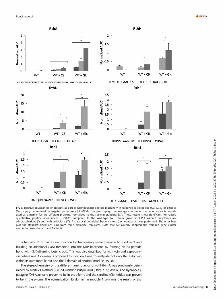
Identification of the biosynthetic gene cluster responsible for rotihibin production.Apart from the thaxtomin biosynthetic gene cluster (BGC), S. scabies also possessesfour other NRP synthase (NRPS)-type BGCs; two of them are cryptic, that is, BGCs forwhich no biomolecule has yet been associated with the genetic material. Proteins asso-ciated with a cryptic NRPS-type BGC (from scab_3221 to scab_3351) (see details below)were found in a previous proteomic experiment (15). We investigated the possible relationshipbetween this BGC and rotihibin production by targeted proteomics using multiple-reactionmonitoring (MRM). The proteotypic peptides of SCAB_3241 (LIDEEPYR and ATGLSDEEFLAR),SCAB_3251 (IPVYLAALGPK and IDVGSAVLQIPAR), SCAB_3281 (VTDEQLAALDLSR andEDPLLTDALAGQR), SCAB_3291 (GQLPEGAWR and LGTADLWLR), SCAB_3301 (LYGGAATDIPHVRand SELAGVFADLLR), and SCAB_3321 (AWIDSDLATPVPVTGER, ADTSGDPTFEELLDR,and GGTVPFAVPAALR) were used to evaluate the protein levels. Figure 5 shows thepositive correlation between the presence of either glucose or cellobiose and the productionof the selected proteins, thereby providing the first evidence that rotihibin production andthis cryptic BGC might be linked. Interestingly, the inactivation of the cellulose utilization reg-ulator cebR had no effect on the production of enzymes for this cluster or even instead hadthe reverse effect for the scab_3221 gene product (15). This indicates that the effect of cello-biose on the expression of the NRPS gene cluster is not dependent on cebR and that there isno coregulation with thaxtomin production.
We then performed a bioinformatic analysis of this gene cluster to investigatewhether it effectively contains the information for enzyme machinery allowing rotihi-bin production. We first analyzed the cryptic gene cluster using antiSMASH (30). Itresides on a 70.1-kb segment of the S. scabies genome with 51 open reading frames(ORFs). The region around the largest NRPS gene (scab_3321) was compared with allpublic genomes using PATRIC (31). This analysis confined the cluster to a 33-kb seg-ment with 14 ORFs (scab_3221 to scab_3351). The presence of a putative integrin pro-tein (SCAB_3201) and a putative transposase (SCAB_3371) at the borders of this clusteris evidence for the possible mobilization of this gene cluster.
FIG 3 Effect of glucose on the secreted metabolome of Streptomyces scabies RL-34. An HPLC run was performedon n-butanol extracts of Streptomyces scabies RL-34 ISP-4 cultures grown in the presence of glucose. mAU, milli-absorbance unit.
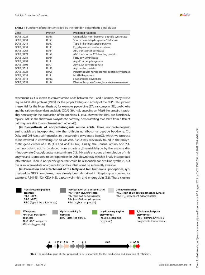
The sequences of these 14 ORFs were individually submitted to a BLAST searchagainst the UniProtKB/Swiss-Prot database, and putative functions derived from thissearch are listed in Table 1 and Fig. 6. A preliminary analysis of these functions allowedus to conclude that this gene cluster contains the necessary information to producethe enzymes needed for the biosynthesis of rotihibins C and D, as outlined below.Therefore, this cluster is further referred to as the rotihibin (rth) gene cluster.
Analysis of the genes involved in the rotihibin BGC. (i) NRPS genes for peptidechain assembly. Two NRPSs, RthA and RthB, are present in this cryptic gene cluster.RthA is predicted to contain 5 modules, while RthB contains a single module for theincorporation of amino acids in a peptide chain (Fig. 7A). Different tools were used topredict their NRPS adenylation domain (A domain) specificity (Table 2), and from theseresults, we propose a biosynthetic path for the production of the rotihibin peptide chain(Fig. 7B). Module 1 of RthA was predicted to be specific for Glu or Ser, depending on the toolused. However, we believe that module 1 is responsible for the incorporation of citrulline, thefirst amino acid of the peptide: the signature sequence for an A domain that could be specificfor citrulline is possibly not included in the prediction tools. The A domain was missing frommodule 2, while (allo)-threonine should be built into the rotihibin backbone. RthB is the bestcandidate to introduce threonine into the rotihibin peptide sequence, as its A domain is pre-dicted to be specific for threonine. This phenomenon was previously described in the endur-acin biosynthetic gene cluster from Streptomyces fungicidicus. Another NRPS activates andtransfers L-allo-threonine to the module with the missing A domain (32). RthD, a type II thio-esterase-like (TEII) enzyme, is believed to assist in the shuttling of the activated aThr betweenthe stand-alone A-T didomain module RthB and the A-less C-T module of RthA, as describedpreviously for WS9326A biosynthesis (33). A serine residue was predicted in module 3, but(2,4)-di-amino butyric acid was observed. The prediction of the two last modules to introduceasparagine residues is in line with the rotihibin structure having two asparagine-like residuesat its C terminus. Module 5 has a reductase (RE) domain. This domain is known to reducethe peptidyl thioester into its corresponding alcohol, which explains the presence of L-aspar-aginol (34). This also explains why rotihibin does not form a cyclic structure like many otherNRPS-derived lipopeptides.
FIG 4 2D 1H-1H TOCSY spectra used for the analysis of the rotihibin C and D structures. The 2D TOCSY spectra of rotihibins C (blue) and D (red) are shownoverlaid, with a slight offset of one with respect to the other to clearly show the identity of the correlation patterns for the peptide chains in bothcompounds. The dashed line identifies the correlation belonging to the threonine residue.
Potentially, RthB has a dual function by transferring L-allo-threonine to module 2 andbuilding an additional L-allo-threonine into the NRP backbone by forming an iso-peptidebond with (2,4)-di-amino butyric acid. This was also described for viomycin and capreomy-cin, where one A domain is proposed to function twice, to acetylate not only the T domainwithin its own module but also the T domain of another module (35, 36).
The stereochemistry of the different amino acids of rotihibin A was previously deter-mined by Marfey’s method (23). 2,4-Diamino butyric acid (Dab), aThr, Asn-ol, and hydroxy-as-paragine (OH-Asn) were proven to be in the L-form, and the citrulline (Cit) residue was provento be in the D-form. The epimerization (E) domain in module 1 confirms the results of this
FIG 5 Relative abundances of proteins as part of nonribosomal peptide machinery in response to cellobiose (CB) (Glc2) or glucose(Glc) supply determined by targeted proteomics (LC-MRM). The plot displays the average area under the curve for each peptideused as a marker for the different proteins, normalized to the spike-in standard BSA. These results show significant normalizedquantitative peptide abundances (P, 0.05) compared to the wild-type (WT) strain grown in ISP-4 without supplementaryoligosaccharides (*) and with cellobiose (**). A statistical two-sided Student t test (homoscedastic) was performed. The error barsplot the standard deviations (SD) from three biological replicates. Note that we already adopted the rotihibin gene clusterannotation (see the text and Table 1).
experiment, as it is known to convert amino acids between the L- and D-isomers. Many NRPSsrequire MbtH-like proteins (MLPs) for the proper folding and activity of the NRPS. This proteinis essential for the biosynthesis of, for example, pyoverdine (37), vancomycin (38), coelichelin,and the calcium-dependent antibiotic (CDA) (39). rthL, encoding an MbtH-like protein, is prob-ably necessary for the production of the rotihibins. Li et al. showed that RthL can functionallyreplace TxtH in the thaxtomin biosynthetic pathway, demonstrating that MLPs from differentpathways are able to complement each other (40).
(ii) Biosynthesis of nonproteinogenic amino acids. Three nonproteinogenicamino acids are incorporated into the rotihibin nonribosomal peptide backbone: Cit,Dab, and OH-Asn. rthM encodes an L-asparagine oxygenase (AsnO), which we proposeto be involved in converting Asn to OH-Asn. AsnO was previously found in the biosyn-thetic gene cluster of CDA (41) and A54145 (42). Finally, the unusual amino acid 2,4-diamino butyric acid is produced from aspartate b-semialdehyde by the enzyme dia-minobutyrate-2-oxoglutarate transaminase (43, 44). rthN encodes a homologue of thisenzyme and is proposed to be responsible for Dab biosynthesis, which is finally incorporatedinto rotihibin. There is no specific gene that could be responsible for citrulline synthesis, butthis is an intermediate of arginine biosynthesis that could be sufficiently available.
(iii) Formation and attachment of the fatty acid tail. Numerous lipopeptides, syn-thesized by NRPS complexes, have already been described in Streptomyces species, forexample, A54145 (42), CDA (45), daptomycin (46), and enduracidin (32). These clusters
FIG 6 The rotihibin gene cluster proposed to be responsible for the production and secretion of rotihibins.
TABLE 1 Functions of proteins encoded by the rotihibin biosynthetic gene cluster
contain genes whose products act together to acylate the first amino acid. Bioinformatic analy-sis of the rth gene cluster revealed the presence of a gene encoding an acyl carrier protein(rthK), which is immediately flanking rthA. This protein is proposed to transfer medium-chainfatty acids to the N-terminal domain of the Cit-incorporating module (Fig. 8). Fatty acyl-AMPligases are enzymes establishing the cross talk between fatty acid synthases and NRPSs or pol-yketide synthetases (PKSs). They initiate the biosynthesis of lipopeptides by the activation of afatty acyl residue and occur with a high incidence in putative lipopeptide NRPS/PKS clusters
Pred Score (%) Pred Score (HMMER bit) Pred Score (bits) Pred ProbRthA1 Glu 0.595 Ser 661.3 Glu 16.5 Ser 0.485 Lipo-D-Cit2 L-allo-Thr3 Arg 0.528 Ser 568.5 Ser 16.9 Ser 0.474 L-Dab4 Asn 0.934 Asn 486.6 Asn 16.5 Asn 0.615 L-Asn-OH5 Asn 0.700 Asn 450.4 Asn 18.1 Orn 0.463 L-Asn
RthB1 Thr 0.955 Thr 651.9 Thr 19.2 Thr 0.469 Thr
aThe specificities of the A domains of the different modules in RthA and RthB were predicted using different software tools. Pred, predicted aminoacid; Prob, probability.
FIG 7 (A) Organization of the NRPSs in the rotihibin gene cluster. RthA contains five modules, while RthB has only one module. C, condensation; A,adenylation; T, thiolation; E, epimerization; RE, reduction. (B) Proposed biosynthetic pathway and modular organization of the NRPS for rotihibinbiosynthesis. RthA is responsible for the NRP backbone. Cis-DA, cis-2-decenoic acid; Cit, citrulline; Dab, (2,4)-di-amino butyric acid; C, condensation domain;A, adenylation domain; T, thiolation; E, epimerization domain; RE, reductase domain; ACP, acyl carrier protein.
(47). RthH is supposed to recruit a fatty acid and transfer it to the acyl carrier protein RthK. Thefatty acid side chain lengths and/or degrees of saturation are the only differences betweenthe rotihibin C and D analogues. The desaturation could be explained by the presence of twoacyl-CoA dehydrogenases (RthI and RthJ), similar to the situation in the ramoplanin biosyn-thetic gene cluster (48). One is expected to introduce the first double bond, while the secondadditional dehydrogenation is possibly not essential. The minor amounts of other lipopeptidesidentified via MS, which differ by only 22Da, could be explained this way (Fig. S1). This phe-nomenon was previously described in acyl-desferrioxamines of Streptomyces coelicolor (49).
(iv) Self-resistance genes. During antibiotic production, transporter and trans-porter-associated proteins are important for the import of effector molecules, self-re-sistance, and guiding/exporting the antibiotic to the extracellular environment. rthFand rthG are predicted to act as the rotihibin-exporting machinery as they encode anABC-type permease and an ABC transporter ATP-binding protein, respectively, a com-bination often found in antibiotic biosynthetic gene clusters. RthF shows 47% similaritywith the daunorubicin/doxorubicin resistance ABC transporter permease protein DrrB,while RthG shows 61% similarity with the daunorubicin/doxorubicin resistance ATP-binding protein DrrA in Streptomyces peucetius. This DrrAB efflux system in S. peucetiushas been shown to be a multidrug transporter with broad specificity (50). RthFG possi-bly has a similar role in the self-resistance mechanism of S. scabies.
(v) Other genes within the rth cluster. The BGC for rotihibin in S. scabies houses anumber of genes not directly associated with rotihibin assembly. The protein productof rthE is predicted to be an F420-dependent oxidoreductase, and rthC encodes a short-chain dehydrogenase/reductase. These two enzymes are, for example, directly involved inthe production of coronafacoyl phytotoxins in S. scabies (51). The impact of the inactivationof these genes on rotihibin biosynthesis has to be studied to reveal their exact role.
(vi) Inactivation of rthB abolishes rotihibin production. To ascertain whether therth cluster is responsible for the production of rotihibins, the scab_3221 gene was disruptedand replaced in S. scabies 87-22 by an apramycin resistance cassette (11, 13, 29). HPLC analysisand targeted metabolomics on n-butanol extracts of the DrthB strain confirmed the absenceof rotihibins (Fig. 9). These data support the evidence that the rth BGC is indeed responsiblefor rotihibin production. Simple complementation of the DrthB strain with an integrative plas-mid harboring an intact copy of rthB with its own promoter did not restore the production ofrotihibins, which could be attributed to polar effects after the removal of the rthB gene and/orthe insertion of the new copy of rthB in a region of the chromosome not optimal for itsexpression.
FIG 8 Proposed activation and transfer of cis-2-decenoic acid (cis-DA).
Biological activity of rotihibins C and D. Since rotihibin A has a plant growth-in-hibitory effect, we tested whether the new rotihibins C and D display similar properties.After 4 days of exposure to concentrations ranging from 0.84 to 84.4mM rotihibin Cand from 0.3 to 157.8mM rotihibin D, the growth of duckweed (Lemna minor L.), espe-cially the fronds, was suppressed compared to that in the control group (a neighboringHPLC fraction that did not display UV absorption) (Fig. 10).
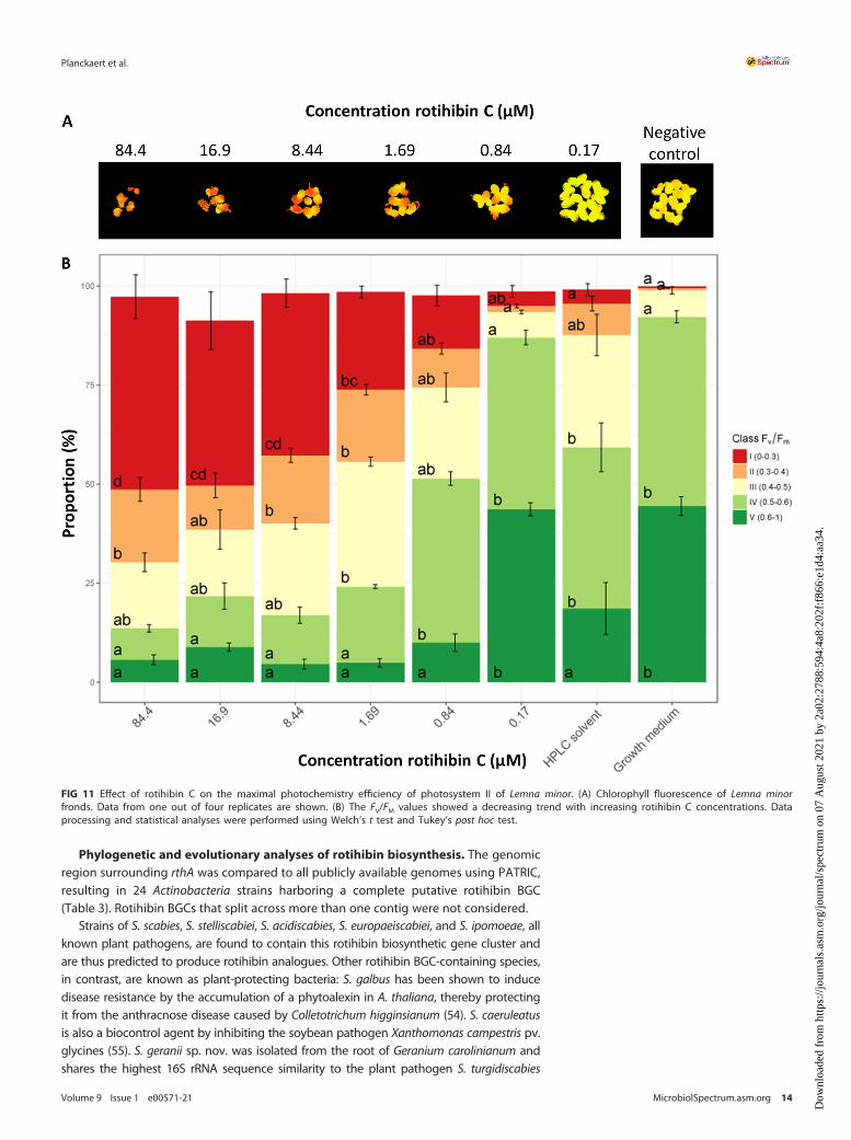
Additionally, we assessed the impact of rotihibins C and D on the photochemistryof photosystem II using the FV/FM ratio as a proxy. In dark-adapted FV/FM measure-ments, the minimal fluorescence (F0) is measured. Next, an intense light flash is used toclose (reduce) all reaction centers and measure the maximum fluorescence, called FM.The difference of FM 2 F0 is the FV value. The FV/FM ratio represents the maximumpotential quantum efficiency of photosystem II. In general, the greater the plant stress,the fewer open reaction centers available, which results in a lowered FV/FM ratio. TheRGB and chlorophyll fluorescence images are depicted in Fig. S2 to S5 in the supplementalmaterial. Using the FV/FM ratio as a proxy, the maximal photochemistry efficiency of photo-system II showed a decreasing trend with increasing rotihibin concentrations in the nutrientmedium, revealing that rotihibins affect the photosynthesis of L. minor (Fig. 11 and 12).
The increased surface area of L. minor treated with 0.17mM rotihibin C compared tothe HPLC solvent indicates a hormetic effect. Hormesis is a dose-response phenom-enon where a low dose of a toxic component promotes plant growth, while at higherdoses, inhibition of growth is observed.
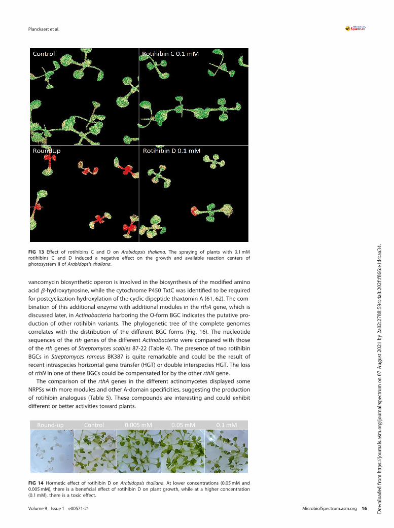
Ordinal analysis 96 h after application also showed an effect on the growth andmaximal photochemistry efficiency of photosystem II (FV/FM) (52) of Arabidopsis thali-ana plants treated with 0.1mM rotihibins C and D (Fig. 13). Here, rotihibins were notadded to the nutrient medium but were sprayed onto the plants; this explains thehigh concentration needed compared to the L. minor bioassay. Most herbicides areapplied as water-based sprays, the easiest way to protect plant products for farmers.The positive control was a commercial glyphosate formulation (RoundUp Turbo) at a molarconcentration of approximately 3 M. At lower concentrations of rotihibin D (0.05mM and0.005mM), we observed growth promotion on Arabidopsis seedlings, which again reflects ahormetic effect (Fig. 14).
FIG 9 Inactivation of the nonribosomal peptide gene rthB. Shown are data from HPLC analysis of rotihibin productionby Streptomyces scabies 87-22 wild-type, mutant, and complementation strains. mAU, milli-absorbance unit.
Recently, Halder et al. chemically synthesized rotihibin A and a number of variants(26). They provide evidence that rotihibin A targets the TOR (target of rapamycin) ki-nase (TORK) pathway, which explains the observed effects on plants. This highly con-served pathway is involved in the regulation of shoot and root development (53).
FIG 10 Surface area measurements of Lemna minor treated with rotihibins. The plant growth-inhibitory activity of rotihibin C (A) and rotihibin D (B) wasanalyzed after 4 days compared to the control. Data are given as means from four replicates 6 standard deviations (SD).
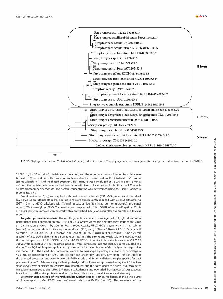
Phylogenetic and evolutionary analyses of rotihibin biosynthesis. The genomicregion surrounding rthA was compared to all publicly available genomes using PATRIC,resulting in 24 Actinobacteria strains harboring a complete putative rotihibin BGC(Table 3). Rotihibin BGCs that split across more than one contig were not considered.
Strains of S. scabies, S. stelliscabiei, S. acidiscabies, S. europaeiscabiei, and S. ipomoeae, allknown plant pathogens, are found to contain this rotihibin biosynthetic gene cluster andare thus predicted to produce rotihibin analogues. Other rotihibin BGC-containing species,in contrast, are known as plant-protecting bacteria: S. galbus has been shown to inducedisease resistance by the accumulation of a phytoalexin in A. thaliana, thereby protectingit from the anthracnose disease caused by Colletotrichum higginsianum (54). S. caeruleatusis also a biocontrol agent by inhibiting the soybean pathogen Xanthomonas campestris pv.glycines (55). S. geranii sp. nov. was isolated from the root of Geranium carolinianum andshares the highest 16S rRNA sequence similarity to the plant pathogen S. turgidiscabies
FIG 11 Effect of rotihibin C on the maximal photochemistry efficiency of photosystem II of Lemna minor. (A) Chlorophyll fluorescence of Lemna minorfronds. Data from one out of four replicates are shown. (B) The FV/FM values showed a decreasing trend with increasing rotihibin C concentrations. Dataprocessing and statistical analyses were performed using Welch’s t test and Tukey’s post hoc test.
ATCC 700248 (56). S. hygroscopicus subsp. jinggangensis strains are known to produce theantibiotic validamycin (57), while S. corchorusii produces butalactin (58). The latter showsbiocontrol and plant growth-promoting activities and potential as a biofertilizer agent forrice plants (59). The actinomycete Lechevalieria aerocolonigenes has been isolated from soilin Japan and is known to produce rebeccamycin with antitumor properties (60). Curiously,the cluster is found only in plant-associated species but is not restricted to plant patho-gens. Rotihibin C and D production was experimentally verified in Streptomyces stelliscabieiNCPPB 4040 (data not shown).
Rotihibin BGCs exist in three different organizations (Table 3), and the gene clustersidentified here were classified as E-form, O-form, and X-form clusters, based on thepresence of two genes: rthE, encoding, a luciferease-like monogygenase (LLM) classF420-dependent oxidoreductase with unknown function, and rthO, which encodes acytochrome P450 hydroxylase (Fig. 15). The cytochrome P450 OxyD from the
FIG 12 Effect of rotihibin D on the maximal photochemistry efficiency of photosystem II of Lemna minor. (A) Chlorophyll fluorescence of Lemna minorfronds. Data from one out of four replicates are shown. (B) The FV/FM values showed a decreasing trend with increasing rotihibin D concentrations. Dataprocessing and statistical analyses were performed using Welch’s t test and Tukey’s post hoc test (B).
vancomycin biosynthetic operon is involved in the biosynthesis of the modified aminoacid b-hydroxytyrosine, while the cytochrome P450 TxtC was identified to be requiredfor postcyclization hydroxylation of the cyclic dipeptide thaxtomin A (61, 62). The com-bination of this additional enzyme with additional modules in the rthA gene, which isdiscussed later, in Actinobacteria harboring the O-form BGC indicates the putative pro-duction of other rotihibin variants. The phylogenetic tree of the complete genomescorrelates with the distribution of the different BGC forms (Fig. 16). The nucleotidesequences of the rth genes of the different Actinobacteria were compared with thoseof the rth genes of Streptomyces scabies 87-22 (Table 4). The presence of two rotihibinBGCs in Streptomyces rameus BK387 is quite remarkable and could be the result ofrecent intraspecies horizontal gene transfer (HGT) or double interspecies HGT. The lossof rthN in one of these BGCs could be compensated for by the other rthN gene.
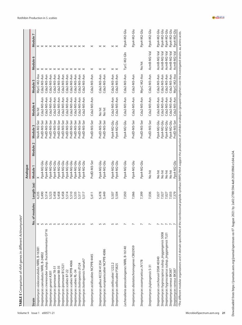
The comparison of the rthA genes in the different actinomycetes displayed someNRPSs with more modules and other A-domain specificities, suggesting the productionof rotihibin analogues (Table 5). These compounds are interesting and could exhibitdifferent or better activities toward plants.
FIG 13 Effect of rotihibins C and D on Arabidopsis thaliana. The spraying of plants with 0.1mMrotihibins C and D induced a negative effect on the growth and available reaction centers ofphotosystem II of Arabidopsis thaliana.
FIG 14 Hormetic effect of rotihibin D on Arabidopsis thaliana. At lower concentrations (0.05mM and0.005mM), there is a beneficial effect of rotihibin D on plant growth, while at a higher concentration(0.1mM), there is a toxic effect.
MATERIALS ANDMETHODSFermentation and analysis of Streptomyces scabies RL-34, 87-22, and mutant strains. Streptomyces
scabies RL-34, S. scabies 87-22, and the DrthB mutant were cultured in a shaking incubator at 250 rpm at28°C for 4 days using borosilicate-baffled flasks with membrane screw caps containing 100ml of ISP-4medium, prepared as follows: 10 g soluble starch (Sigma-Aldrich), 1 g K2HPO4, 1 g MgSO4, 1 g NaCl, 2 g(NH4)2SO4, 2 g CaCO3, 1mg FeSO4, 1mg MnCl2, and 1mg ZnSO4 were dissolved in 1 liter of deionizedwater. For the induction of rotihibin production, wild-type cultures were grown in the presence of 0.7%cellobiose (Sigma-Aldrich) or 0.5% glucose (Merck).
Cultures were centrifuged at 10,000 � g for 10min, and 2ml of each supernatant was transferred to anew tube. The samples were mixed with n-butanol (1:1), vortexed gently for 10min, and centrifuged for15min at 16,000 � g. The aqueous phase of the extract was dried and stored at 20°C until further use.
The dried extract was dissolved in 10mM ammonium formate (NH4FA) (pH 3) and analyzed by HPLC, mon-itoring the absorbance at 220nm. The sample was eluted with solvent A (10mM NH4FA, pH 3) and solvent B(acetonitrile [ACN]) using a 2-to-90% 7.5-min gradient at 1ml/min on a Zorbax Eclipse Plus C18 column (4.6 by100mm, 3.5mm). Fractions were collected every 15 s, corresponding to 250ml. The total area under the curve(AUC) for each metabolite was calculated. Student’s t test (two tailed, homoscedastic) was executed to evaluatethe differential metabolite abundances between the different conditions in a statistical way.
The fractions of interest were loaded into a Triverse Nanomate source (Advion) and, using 1.6 kV and0.3 lb/in2 of pressure, electrosprayed into a Waters Synapt G1 mass spectrometer. Scans typically rangedfrom m/z 100 to 1,200, with a scan time of 1 s, for a total of 2min. For MS/MS experiments, the collisionenergy was optimized to achieve good fragmentation spectra.
Generation of the DrthB (scab_3221; scab_RS01465) deletion mutant in Streptomyces scabies87-22. Deletion of the rthB gene was performed in S. scabies 87-22 using the ReDirect PCR targetingmethod (63) by replacing the genomic copy of the gene with an apramycin resistance cassette: using a
TABLE 3 Actinobacteria harboring a putative rotihibin biosynthetic gene clustera
Organism Source
Genbank genomeassemblyaccession no.
Presence of rotihibin BGC form
E O XStreptomyces scabiei 87-22 GCA_000091305.1Streptomyces scabiei NCPPB 4066 Solanum tuberosum, New
HindIII-EcoRI restriction fragment obtained from pIJ773 (Table 6) as the PCR template, a disruption cas-sette, containing the resistance gene aac(3)IV and oriT for conjugative transfer, was amplified by PCRwith primers BDF33 and BDF34 and gel purified as performed previously for cebR, cebE, msiK, and bglC(11, 13, 29).
In parallel, cosmid 2012 (cos2012), based on Supercos-1 and containing a genomic insert of S. scabies87-22 ranging from positions 341913 to 386384, including rthB, was introduced by electroporation intoEscherichia coli BW25113 carrying plasmid pIJ790 for the arabinose-inducible expression of the l Redrecombination machinery. After culture to an optical density (OD) of 0.4 with arabinose at 10mM toinduce the l Red machinery, 50 ml of washed E. coli BW25113/pIJ790 cos2012 cells was electroporatedwith 100 ng (in 1 ml) of the rthB-targeting disruption cassette, and apramycin-resistant clones wereselected on LB agar plates plus apramycin (50mg/ml) and kanamycin (50mg/ml). The gene replacementby the disruption cassette on the cosmid was confirmed by PCR using primers BDF35 and BDF36(Table 6) and Sanger sequencing. The resulting cos2012D3221 construct was purified using a GeneJETplasmid miniprep kit (Thermo Scientific) (according to the manufacturer’s guidelines) and transferred byintergeneric conjugation with E. coli ET12567/pUZ8002 (ETpUZ) as the donor strain and S. scabies 87-22as the recipient strain. After growing ETpUZ to an OD of 0.4 (50-ml culture in LB plus chloramphenicol[30mg/ml], apramycin [50mg/ml], kanamycin [50mg/ml], and ampicillin [100mg/ml]), cells were washedto remove antibiotics and mixed with S. scabies 87-22 spores for mating. The conjugative mixture wasthen plated on 30-ml petri dishes of soy flour mannitol medium (SFM) (20 g/liter mannitol, 20 g/liter soyflour, and 20 g/liter agar in tap water and autoclaved twice) plus 10mM MgCl2 and overlaid with nali-dixic acid (50mg/ml) and apramycin (40mg/ml). Exconjugants were transferred to ISP-4 agar plates plusnalidixic acid (25mg/ml) and apramycin (50mg/ml) to obtain uniform mutant lines that were then usedto prepare spore stocks. Each S. scabies DrthB mutant was checked for apramycin resistance and kana-mycin sensitivity on ISP-2 agar plates. Genomic DNA was extracted from 48-h liquid cultures in trypticsoy broth (TSB) using a GenElute bacterial genomic DNA kit (Sigma-Aldrich) (according to the manufac-turer’s guidelines) and used as the PCR template for mutation confirmation.
Complementation of the DrthB deletion mutant. rthB, including its upstream (2701 bp) anddownstream (1501 bp) regions, was amplified by PCR with primers BDF35 and BDF36 to obtain a 3,512-bp fragment flanked by two XbaI restriction sites. The PCR product was first cloned into a pJET1.2 vector,which was named pBDF054. Using the XbaI enzyme, rthB and surrounding regions were isolated andcloned into a pAU3-45 (64) integrative vector named pBDF043. Conjugation was performed with S. sca-bies DrthB clones 1 and 2 on SFM (plus 10mM MgCl2) overlaid with apramycin (50mg/ml), nalidixic acid(50mg/ml), and thiostrepton (12.5mg/ml). The integration of the plasmid was confirmed by PCR with pri-mers BDF69 and BDF70.
Structural confirmation by NMR spectroscopy. Both rotihibin analogues were dissolved in 0.6mlof DMSO-d6, and the nuclear magnetic resonance (NMR) spectra were recorded at 25°C on a BrukerAvance II 700-MHz spectrometer equipped with a 5-mm Prodigy TCI N2 cooled cryoprobe. Total corre-lated spectroscopy (TOCSY) spectra were recorded using the standard pulse sequences from the Brukerlibrary.
Protein extraction and sample preparation. S. scabies RL-34 cells were cultivated in three differentgrowth media (ISP-4 medium, ISP-4 medium supplemented with 0.7% cellobiose, and ISP-4 mediumsupplemented with 0.5% glucose) for 96 h (28°C at 250 rpm), with three biological replicates under eachcondition. The cultures were centrifuged at 10,000 � g for 10min. The resulting pellet was washed twotimes and resuspended in 10ml lysis buffer (1� phosphate-buffered saline [PBS], 0.1% SDS, protease in-hibitor cocktail). The homogeneous mycelium suspension was sonicated (20 times with 30 s on and 30 soff at 30%) on ice using a Branson digital sonifier 450 cell disruptor. The homogenate was centrifuged at
FIG 15 Schematic representation of E-, O-, and X-form rotihibin biosynthetic gene clusters.
16,000 � g for 30min at 4°C. Pellets were discarded, and the supernatant was subjected to trichloroace-tic acid (TCA) precipitation. The crude intracellular extract was mixed with a 100% (wt/vol) TCA solution(Sigma-Aldrich) (4:1) and incubated overnight. This mixture was centrifuged at 16,000 � g for 15min at4°C, and the protein pellet was washed two times with ice-cold acetone and solubilized in 2 M urea in50mM ammonium bicarbonate. The protein concentration was determined using the Pierce Coomassieprotein assay kit.
Protein extracts (10mg) were spiked with bovine serum albumin (BSA) (MS-grade protein standard)(0.2 ng/ml) as an internal standard. The proteins were subsequently reduced with 2.5mM dithiothreitol(DTT) (10min at 60°C), alkylated with 7.5mM iodoacetamide (20min at room temperature), and trypsi-nized (1/50) (overnight at 37°C). The reaction was stopped with 1% HCOOH. After centrifugation (30minat 13,200 rpm), the samples were filtered with a prewashed 0.22-mm Costar filter and transferred to cleantubes.
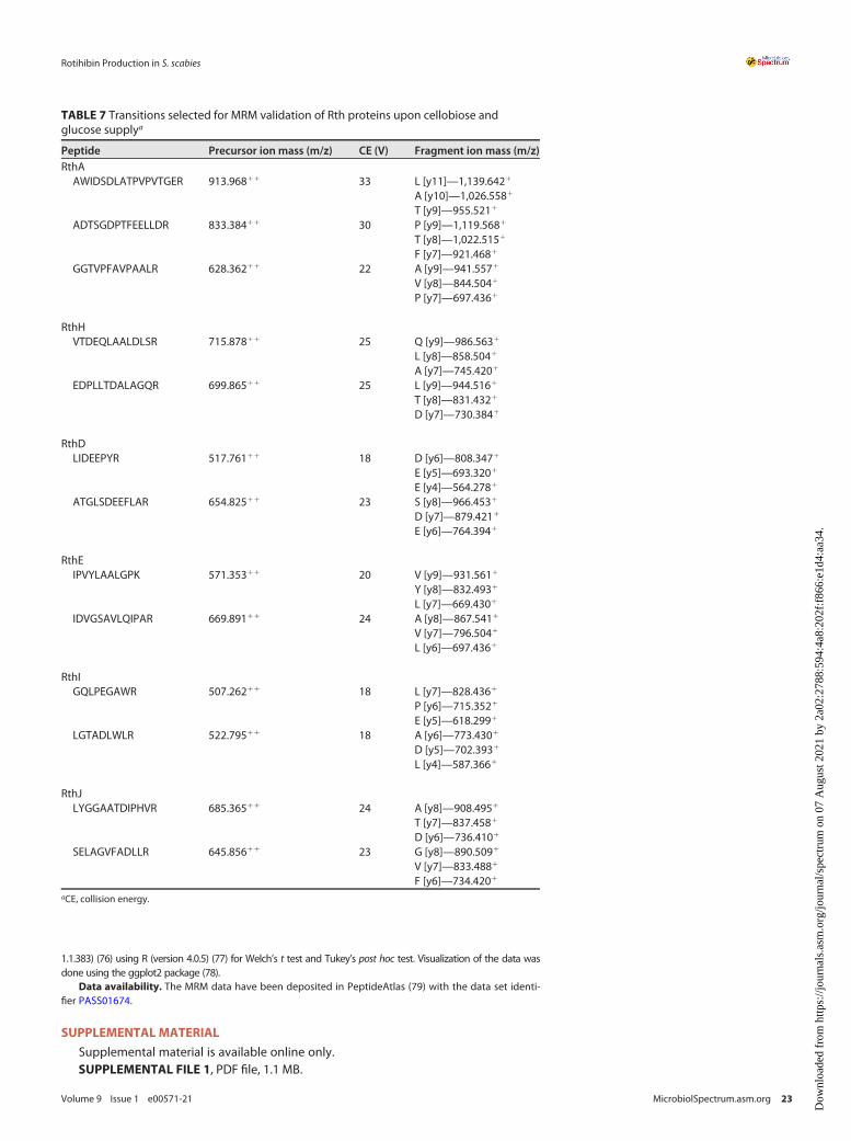
Targeted proteomic analysis. The resulting peptide solutions were injected (0.1mg) onto an ultra-performance liquid chromatography (UPLC) M-Class system where the peptides were trapped for 5min,at 15ml/min, on a 300-mm by 50-mm, 5-mm, 100-Å Acquity UPLC M-Class symmetry C18 trap column(Waters) and separated on the iKey separation device (150mm by 100mm, 1.8mm) (HSS T3; Waters) withsolvent A (0.1% HCOOH in H2O [Biosolve]) and solvent B (0.1% HCOOH in ACN [Biosolve]) using a 20-mingradient of 3 to 50% solvent B at a flow rate of 1ml/min. The strong and weak solutions used to washthe autosampler were 0.1% HCOOH in H2O and 0.1% HCOOH in acetonitrile-water-isopropanol (50:25:25,vol/vol/vol), respectively. The separated peptides were introduced into the IonKey source coupled to aWaters Xevo TQ-S triple-quadrupole mass spectrometer for quantification of the analytes in the positive-ion mode (ESI1). The ESI-MS/MS parameters were as follows: capillary voltage of 3.6 kV, cone voltage of40 V, source temperature of 120°C, and collision gas argon flow rate of 0.19ml/min. The transitions ofthe selected precursor ions were detected in MRM mode at different collision energies specific for eachprecursor (Table 7). Data were acquired using MassLynx 4.1 software and processed in Skyline 3.7. The tran-sition curves were subjected to Savitsky-Golay smoothing, and their area under the curve (AUC) was deter-mined and normalized to the spiked BSA standard. Student’s t test (two tailed, homoscedastic) was executedto evaluate the differential protein abundances between the different conditions in a statistical way.
Bioinformatics analysis of the rotihibin biosynthetic gene cluster. Prediction of the gene clusterof Streptomyces scabies 87-22 was performed using antiSMASH 3.0 (30). The sequence of the
FIG 16 Phylogenetic tree of 23 Actinobacteria analyzed in this study. The phylogenetic tree was generated using the codon tree method in PATRIC.
biosynthetic rotihibin gene cluster was further analyzed and annotated using UniProt and BLAST (65,66). Similar gene clusters were identified in other Actinomycetes strains by PATRIC 3.5.21 analysis (31). Aphylogenetic tree was calculated, from the rthA MUSCLE sequence alignment, using the neighbor-join-ing method with MEGA-X (67). Thaxtomin synthetase A (txtA) was selected as the outgroup.
The module/domain organization of the different NRPSs in the rotihibin gene cluster was predictedvia PRISM (68, 69). The A-domain specificity was predicted via four different software tools: the LSI-basedA-domain function predictor, NRPSsp, the PKS/NRPS Web server, and SEQL-NRPS. The LSI-based predictor useslatent semantic indexing to predict adenylation domain specificities (70). NRPSsp uses hits against hiddenMarkov model (HMM) databases to predict specificities of NRPS adenylation domains (71). The PKS/NRPS Webserver uses BLAST to detect catalytic domains in NRPS and predicts A-domain specificities by comparing signa-tures of A domains with those of known substrates (72). Finally, SEQL-NRPS predicts A-domain specificitiesusing the discriminative classification method sequence learner (SEQL) (73).
Lemna minor L. and Arabidopsis thaliana L. Heynh. bioassay. A sterile 24-well plate was used togrow Lemna minor (duckweed) in 2ml mineral medium [11.1mg CaCl2, 202mg KNO3, 49.6mg MgSO4�7H2O,50.3mg KH2PO4, 27.8mg K2HPO4, 6mg FeSO4�7H2O, 17.4mg K2SO4, 5.72mg H3BO3, 2.82mg MnCl2�4H2O,0.6mg ZnSO4, 10mg Na2-EDTA, 0.008mg CuCl2�H2O, 0.054mg CoCl2�6H2O, and 0.043mg (NH4)6Mo7O24 dis-solved in 1 liter of MilliQ (MQ) water (pH 6.56 0.1)] (74). The plants were incubated in quadruple for 4days in agrowth chamber (16-h light exposure at 22°C). In order to assess the sensitivity of L. minor to rotihibins C andD, a wide concentration range (0.17 to 84.4mM for rotihibin C and 0.3 to 157.6mM for rotihibin D) was tested.After 4days of incubation, the plates were analyzed based on the growth and maximal photochemistry effi-ciency of photosystem II via chlorophyll fluorescence (FV/FM) of the duckweed plants.
Arabidopsis thaliana ecotype Col-0 pregerminated seeds were transferred to petri dishes with half-strength Murashige-Skoog (MS) medium without sucrose (0.22% MS salts and 0.8% plant agar) (75).Nine days after germination, the plants were treated with rotihibins C and D dissolved in water (0.005 to0.1mM) via nebulization on the leaves. After 4 days of treatment, the plates were analyzed based on thegrowth and FV/FM values of the Arabidopsis plants.
Imaging was achieved with an in-house-developed phenotyping platform, in an environment-controlledgrowth chamber, property of the Ghent University Laboratory of Applied Mycology and Phenomics. The platformallows visualization of diverse physiological traits via a multispectral 3CCD camera equipped with 12 interferencefilters in real time, based on specific absorption, reflection, and emission patterns, such as leaf surface, efficiencyof photosynthesis, chlorophyll and anthocyanin contents, and green fluorescent protein (GFP)-tagged organisms.This platform is equipped with a dispenser, which can be fitted with a nozzle to treat the plants with rotihibins orother agrochemicals in a standardized manner. Image data processing was performed using Data Analysis (ver-sion 5.4.6; Phenovation, Wageningen, Netherlands), and statistical analysis was performed in RStudio (version
TABLE 6 Primers and genetic constructs used in this studya
Primer or genetic construct Sequence (59!39) or descriptionApplication,reference, or source
Plasmids or cosmidspIJ773 Template for amplification of the apramycin resistance cassette [aac(3)IV (Aprar) oriT
(RK2) FRT amp (Ampr)]63
cos2012 Supercos-1 (Agilent) derivative containing the genomic insert of S. scabies 87-22from positions 341913–386384 (Ampr Kanr)
Isolde Francis
pIJ790 Contains the l Red recombination under the control of an arabinose-induciblepromoter (paraBAD) (Cmlr)
63
cos2012D3221 Cosmid 2012 derivative containing the apramycin resistance cassette instead of therthB gene
This study
pUZ8002 Nontransmissible plasmid supplying transfer functions for mobilization of oriT-containing vectors from E. coli to Streptomyces spp. (Kanr)
80
pJET1.2/blunt E. coli plasmid used for high-efficiency blunt-end cloning of PCR products (Ampr) Thermo ScientificpBDF054 pJET1.2 derivative containing the 3,512-bp DNA fragment for rthB complementation This studypAU3-45 pSET152 derivative, integrative plasmid with a thiostrepton resistance gene inserted
into the blunted NheI restriction site [lacZa ori (pUC18) aac(3)IV (Aprar) oriT (RK2)attP (wC31) int (wC31) tsr (Thior)]
64
aEngineered restriction sites are indicated in italic and underlined, non-homologous extensions are underlined.
1.1.383) (76) using R (version 4.0.5) (77) for Welch’s t test and Tukey’s post hoc test. Visualization of the data wasdone using the ggplot2 package (78).
Data availability. The MRM data have been deposited in PeptideAtlas (79) with the data set identi-fier PASS01674.
SUPPLEMENTAL MATERIAL
Supplemental material is available online only.SUPPLEMENTAL FILE 1, PDF file, 1.1 MB.
TABLE 7 Transitions selected for MRM validation of Rth proteins upon cellobiose andglucose supplya
Peptide Precursor ion mass (m/z) CE (V) Fragment ion mass (m/z)RthAAWIDSDLATPVPVTGER 913.96811 33 L [y11]—1,139.6421
ACKNOWLEDGMENTSB.D. is supported by grants from the Ghent University research council supporting a
proteomics expertise center. We acknowledge the Hercules initiative for the multispectralimaging platform that was granted (grant number AUGE/15/17).
We are thankful to Isolde Francis at California State University—Bakersfield forproviding cosmid 2012 and helpful discussions.
REFERENCES1. Loria R, Kers J, Joshi M. 2006. Evolution of plant pathogenicity in Streptomy-
ces. Annu Rev Phytopathol 44:469–487. https://doi.org/10.1146/annurev.phyto.44.032905.091147.
2. Lerat S, Simao-Beaunoir AM, Beaulieu C. 2009. Genetic and physiologicaldeterminants of Streptomyces scabies pathogenicity. Mol Plant Pathol10:579–585. https://doi.org/10.1111/j.1364-3703.2009.00561.x.
3. Bouchek-Mechiche K, Gardan L, Normand P, Jouan B. 2000. DNA relat-edness among strains of Streptomyces pathogenic to potato in France:description of three new species, S. europaeiscabiei sp. nov. and S.stelliscabiei sp. nov. associated with common scab, and S. reticulisca-biei sp. nov. associated with netted scab. Int J Syst Evol Microbiol50(Part 1):91–99. https://doi.org/10.1099/00207713-50-1-91.
4. Faucher E, Otrysko B, Paradis �E, Hodge NC, Stall RE, Beaulieu C. 1993. Char-acterization of streptomycetes causing russet scab in Québec. Plant Dis77:1217–1220. https://doi.org/10.1094/PD-77-1217.
5. Guan D, Grau BL, Clark CA, Taylor CM, Loria R, Pettis GS. 2012. Evidencethat thaxtomin C is a pathogenicity determinant of Streptomyces ipo-moeae, the causative agent of Streptomyces soil rot disease of sweetpotato. Mol Plant Microbe Interact 25:393–401. https://doi.org/10.1094/MPMI-03-11-0073.
6. King RR, Lawrence CH, Clark MC, Calhoun LA. 1989. Isolation and char-acterization of phytotoxins associated with Streptomyces scabies. JChem Soc Chem Commun (Camb) 1989:849–850. https://doi.org/10.1039/c39890000849.
7. Loria R, Bignell DR, Moll S, Huguet-Tapia JC, Joshi MV, Johnson EG, SeipkeRF, Gibson DM. 2008. Thaxtomin biosynthesis: the path to plant pathoge-nicity in the genus Streptomyces. Antonie Van Leeuwenhoek 94:3–10.https://doi.org/10.1007/s10482-008-9240-4.
8. Bischoff V, Cookson SJ, Wu S, Scheible WR. 2009. Thaxtomin A affectsCESA-complex density, expression of cell wall genes, cell wall composi-tion, and causes ectopic lignification in Arabidopsis thaliana seedlings. JExp Bot 60:955–965. https://doi.org/10.1093/jxb/ern344.
9. Scheible WR, Fry B, Kochevenko A, Schindelasch D, Zimmerli L, SomervilleS, Loria R, Somerville CR. 2003. An Arabidopsis mutant resistant to thaxto-min A, a cellulose synthesis inhibitor from Streptomyces species. Plant Cell15:1781–1794. https://doi.org/10.1105/tpc.013342.
10. Wach MJ, Krasnoff SB, Loria R, Gibson DM. 2007. Effect of carbohydrateson the production of thaxtomin A by Streptomyces acidiscabies. ArchMicrobiol 188:81–88. https://doi.org/10.1007/s00203-007-0225-x.
11. Jourdan S, Francis IM, Kim MJ, Salazar JJ, Planckaert S, Frere JM, MatagneA, Kerff F, Devreese B, Loria R, Rigali S. 2016. The CebE/MsiK transporter isa doorway to the cello-oligosaccharide-mediated induction of Streptomy-ces scabies pathogenicity. Sci Rep 6:27144. https://doi.org/10.1038/srep27144.
12. Joshi MV, Bignell DR, Johnson EG, Sparks JP, Gibson DM, Loria R. 2007.The AraC/XylS regulator TxtR modulates thaxtomin biosynthesis and viru-lence in Streptomyces scabies. Mol Microbiol 66:633–642. https://doi.org/10.1111/j.1365-2958.2007.05942.x.
13. Francis IM, Jourdan S, Fanara S, Loria R, Rigali S. 2015. The cellobiose sen-sor CebR is the gatekeeper of Streptomyces scabies pathogenicity. mBio6:e02018-14. https://doi.org/10.1128/mBio.02018-14.
14. Bignell DR, Francis IM, Fyans JK, Loria R. 2014. Thaxtomin A productionand virulence are controlled by several bld gene global regulators inStreptomyces scabies. Mol Plant Microbe Interact 27:875–885. https://doi.org/10.1094/MPMI-02-14-0037-R.
15. Planckaert S, Jourdan S, Francis IM, Deflandre B, Rigali S, Devreese B. 2018.Proteomic response to thaxtomin phytotoxin elicitor cellobiose and to dele-tion of cellulose utilization regulator CebR in Streptomyces scabies. J Pro-teome Res 17:3837–3852. https://doi.org/10.1021/acs.jproteome.8b00528.
16. Natsume M, Ryu R, Abe H. 1996. Production of phytotoxins, concanamy-cins A and B by Streptomyces spp. causing potato [Solanum tuberosum]
scab. Ann Phytopathol Soc Jpn 62:411–413. https://doi.org/10.3186/jjphytopath.62.411.
17. Natsume M, Tashiro N, Doi A, Nishi Y, Kawaide H. 2017. Effects of concana-mycins produced by Streptomyces scabies on lesion type of common scabof potato. J Gen Plant Pathol 83:78–82. https://doi.org/10.1007/s10327-017-0696-9.
19. Natsume M, Komiya M, Koyanagi F, Tashiro N, Kawaide H, Abe H. 2005.Phytotoxin produced by Streptomyces sp. causing potato russet scab inJapan. J Gen Plant Pathol 71:364–369. https://doi.org/10.1007/s10327-005-0211-6.
20. Cao Z, Khodakaramian G, Arakawa K, Kinashi H. 2012. Isolation of borreli-din as a phytotoxic compound from a potato pathogenic streptomycesstrain. Biosci Biotechnol Biochem 76:353–357. https://doi.org/10.1271/bbb.110799.
21. Park DH, Kim JS, Kwon SW, Wilson C, Yu YM, Hur JH, Lim CK. 2003. Strepto-myces luridiscabiei sp. nov., Streptomyces puniciscabiei sp. nov. and Strep-tomyces niveiscabiei sp. nov., which cause potato common scab disease inKorea. Int J Syst Evol Microbiol 53:2049–2054. https://doi.org/10.1099/ijs.0.02629-0.
22. Natsume M, Nagagata A, Aittamaa M, Okaniwa N, Somervuo P, Fiedler H-P, Kreuze JF, Rokka V-M, Bång H, Kawaide H, Valkonen JPT. 2018. Phyto-toxin produced by the netted scab pathogen, Streptomyces turgidiscabiesstrain 65, isolated in Sweden. J Gen Plant Pathol 84:108–117. https://doi.org/10.1007/s10327-018-0765-8.
23. Fukuchi N, Furihata K, Takayama S, Isogai A, Suzuki A. 1992. Rotihibin A, anovel plant growth regulator, from Streptomyces sp. Biosci BiotechnolBiochem 56:840–841. https://doi.org/10.1271/bbb.56.840.
24. Fukuchi N, Nakayama J, Takayama S, Isogai A, Suzuki A. 1992. Structuralelucidation of rotihibin B by tandem mass spectrometry. Biosci Biotech-nol Biochem 56:1152–1153. https://doi.org/10.1271/bbb.56.1152.
25. Fukuchi N, Furihata K, Nakayama J, Goudo T, Takayama S, Isogai A, SuzukiA. 1995. Rotihibins, novel plant growth regulators from Streptomyces gra-minofaciens. J Antibiot (Tokyo) 48:1004–1010. https://doi.org/10.7164/antibiotics.48.1004.
26. Halder V, Oeljeklaus J, Heilmann G, Krahn JH, Liu Y, Xiong Y, Schlicht M,Schillinger J, Kracher B, Ehrmann M, Kombrink E, Kaschani F, Kaiser M. 2018.Identification of the natural product rotihibin A as a TOR kinase signaling in-hibitor by unbiased transcriptional profiling. Chemistry 24:12500–12504.https://doi.org/10.1002/chem.201802647.
27. Mohimani H, Gurevich A, Mikheenko A, Garg N, Nothias LF, Ninomiya A,Takada K, Dorrestein PC, Pevzner PA. 2017. Dereplication of peptidic nat-ural products through database search of mass spectra. Nat Chem Biol13:30–37. https://doi.org/10.1038/nchembio.2219.
28. el-Sayed ES. 2000. Production of thaxtomin A by two species of Strepto-myces causing potato scab. Folia Microbiol (Praha) 45:415–422. https://doi.org/10.1007/BF02817614.
29. Jourdan S, Francis IM, Deflandre B, Tenconi E, Riley J, Planckaert S,Tocquin P, Martinet L, Devreese B, Loria R, Rigali S. 2018. Contribution ofthe beta-glucosidase BglC to the onset of the pathogenic lifestyle ofStreptomyces scabies. Mol Plant Pathol 19:1480–1490. https://doi.org/10.1111/mpp.12631.
30. Weber T, Blin K, Duddela S, Krug D, Kim HU, Bruccoleri R, Lee SY,Fischbach MA, Muller R, Wohlleben W, Breitling R, Takano E, Medema MH.2015. antiSMASH 3.0—a comprehensive resource for the genome miningof biosynthetic gene clusters. Nucleic Acids Res 43:W237–W243. https://doi.org/10.1093/nar/gkv437.
31. Wattam AR, Brettin T, Davis JJ, Gerdes S, Kenyon R, Machi D, Mao C, OlsonR, Overbeek R, Pusch GD, Shukla MP, Stevens R, Vonstein V, Warren A, Xia
F, Yoo H. 2018. Assembly, annotation, and comparative genomics in PAT-RIC, the All Bacterial Bioinformatics Resource Center. Methods Mol Biol1704:79–101. https://doi.org/10.1007/978-1-4939-7463-4_4.
33. Kim M-S, Bae M, Jung Y-E, Kim JM, Hwang S, Song MC, Ban YH, Bae ES,Hong S, Lee SK, Cha S-S, Oh D-C, Yoon YJ. 7 May 2021. Unprecedentednoncanonical features of the nonlinear nonribosomal peptide synthetaseassembly line for WS9326A biosynthesis. Angew Chem Int Ed Engl.https://doi.org/10.1002/anie.202103872.
34. Manavalan B, Murugapiran SK, Lee G, Choi S. 2010. Molecular modelingof the reductase domain to elucidate the reaction mechanism of reduc-tion of peptidyl thioester into its corresponding alcohol in non-ribosomalpeptide synthetases. BMC Struct Biol 10:1. https://doi.org/10.1186/1472-6807-10-1.
35. Thomas MG, Chan YA, Ozanick SG. 2003. Deciphering tuberactinomycinbiosynthesis: isolation, sequencing, and annotation of the viomycin bio-synthetic gene cluster. Antimicrob Agents Chemother 47:2823–2830.https://doi.org/10.1128/AAC.47.9.2823-2830.2003.
36. Felnagle EA, Rondon MR, Berti AD, Crosby HA, Thomas MG. 2007. Identifi-cation of the biosynthetic gene cluster and an additional gene for resist-ance to the antituberculosis drug capreomycin. Appl Environ Microbiol73:4162–4170. https://doi.org/10.1128/AEM.00485-07.
37. Drake EJ, Cao J, Qu J, Shah MB, Straubinger RM, Gulick AM. 2007. The 1.8A crystal structure of PA2412, an MbtH-like protein from the pyoverdinecluster of Pseudomonas aeruginosa. J Biol Chem 282:20425–20434.https://doi.org/10.1074/jbc.M611833200.
38. Lee KS, Lee BM, Ryu JH, Kim DH, Kim YH, Lim SK. 2016. Increased vanco-mycin production by overexpression of MbtH-like protein in Amycolatop-sis orientalis KFCC10990P. Lett Appl Microbiol 63:222–228. https://doi.org/10.1111/lam.12617.
40. Li Y, Liu J, Adekunle D, Bown L, Tahlan K, Bignell DRD. 2019. TxtH is a keycomponent of the thaxtomin biosynthetic machinery in the potato commonscab pathogen Streptomyces scabies. Mol Plant Pathol 20:1379–1393. https://doi.org/10.1111/mpp.12843.
41. Neary JM, Powell A, Gordon L, Milne C, Flett F, Wilkinson B, Smith CP,Micklefield J. 2007. An asparagine oxygenase (AsnO) and a 3-hydroxyaspara-ginyl phosphotransferase (HasP) are involved in the biosynthesis of calcium-dependent lipopeptide antibiotics. Microbiology (Reading) 153:768–776.https://doi.org/10.1099/mic.0.2006/002725-0.
42. Miao V, Brost R, Chapple J, She K, Gal MF, Baltz RH. 2006. The lipopeptideantibiotic A54145 biosynthetic gene cluster from Streptomyces fradiae. JInd Microbiol Biotechnol 33:129–140. https://doi.org/10.1007/s10295-005-0028-5.
43. Saum SH, Muller V. 2008. Growth phase-dependent switch in osmolytestrategy in a moderate halophile: ectoine is a minor osmolyte but majorstationary phase solute in Halobacillus halophilus. Environ Microbiol10:716–726. https://doi.org/10.1111/j.1462-2920.2007.01494.x.
44. Vandenende CS, Vlasschaert M, Seah SY. 2004. Functional characteriza-tion of an aminotransferase required for pyoverdine siderophore biosyn-thesis in Pseudomonas aeruginosa PAO1. J Bacteriol 186:5596–5602.https://doi.org/10.1128/JB.186.17.5596-5602.2004.
45. Hojati Z, Milne C, Harvey B, Gordon L, Borg M, Flett F, Wilkinson B,Sidebottom PJ, Rudd BA, Hayes MA, Smith CP, Micklefield J. 2002. Structure,biosynthetic origin, and engineered biosynthesis of calcium-dependent anti-biotics from Streptomyces coelicolor. Chem Biol 9:1175–1187. https://doi.org/10.1016/s1074-5521(02)00252-1.
46. Miao V, Coeffet-Legal MF, Brian P, Brost R, Penn J, Whiting A, Martin S,Ford R, Parr I, Bouchard M, Silva CJ, Wrigley SK, Baltz RH. 2005. Daptomy-cin biosynthesis in Streptomyces roseosporus: cloning and analysis of thegene cluster and revision of peptide stereochemistry. Microbiology(Reading) 151:1507–1523. https://doi.org/10.1099/mic.0.27757-0.
47. Galica T, Hrouzek P, Mares J. 2017. Genome mining reveals high incidenceof putative lipopeptide biosynthesis NRPS/PKS clusters containing fattyacyl-AMP ligase genes in biofilm-forming cyanobacteria. J Phycol53:985–998. https://doi.org/10.1111/jpy.12555.
48. Hoertz AJ, Hamburger JB, Gooden DM, Bednar MM, McCafferty DG. 2012.Studies on the biosynthesis of the lipodepsipeptide antibiotic ramoplanin
A2. Bioorg Med Chem 20:859–865. https://doi.org/10.1016/j.bmc.2011.11.062.
49. Traxler MF, Watrous JD, Alexandrov T, Dorrestein PC, Kolter R. 2013. Inter-species interactions stimulate diversification of the Streptomyces coeli-color secreted metabolome. mBio 4:e00459-13. https://doi.org/10.1128/mBio.00459-13.
50. Li W, Sharma M, Kaur P. 2014. The DrrAB efflux system of Streptomycespeucetius is a multidrug transporter of broad substrate specificity. J BiolChem 289:12633–12646. https://doi.org/10.1074/jbc.M113.536136.
51. Bown L, Altowairish MS, Fyans JK, Bignell DR. 2016. Production of theStreptomyces scabies coronafacoyl phytotoxins involves a novel biosyn-thetic pathway with an F420-dependent oxidoreductase and a short-chain dehydrogenase/reductase. Mol Microbiol 101:122–135. https://doi.org/10.1111/mmi.13378.
52. Baker NR. 2008. Chlorophyll fluorescence: a probe of photosynthesis invivo. Annu Rev Plant Biol 59:89–113. https://doi.org/10.1146/annurev.arplant.59.032607.092759.
53. Li X, Cai W, Liu Y, Li H, Fu L, Liu Z, Xu L, Liu H, Xu T, Xiong Y. 2017. Differen-tial TOR activation and cell proliferation in Arabidopsis root and shootapexes. Proc Natl Acad Sci U S A 114:2765–2770. https://doi.org/10.1073/pnas.1618782114.
54. Shimizu M, Meguro A, Hasegawa S, Nishimura T, Kunoh H. 2006. Disease re-sistance induced by nonantagonistic endophytic Streptomyces spp. on tis-sue-cultured seedlings of rhododendron. J Gen Plant Pathol 72:351–354.https://doi.org/10.1007/s10327-006-0305-9.
55. Mingma R, Pathom-Aree W, Trakulnaleamsai S, Thamchaipenet A,Duangmal K. 2014. Isolation of rhizospheric and roots endophytic actino-mycetes from Leguminosae plant and their activities to inhibit soybeanpathogen, Xanthomonas campestris pv. glycine. World J Microbiol Bio-technol 30:271–280. https://doi.org/10.1007/s11274-013-1451-9.
56. Li X, Lai X, Gan L, Long X, Hou Y, Zhang Y, Tian Y. 2018. Streptomyces gera-nii sp. nov., a novel endophytic actinobacterium isolated from root of Ge-ranium carolinianum L. Int J Syst Evol Microbiol 68:2562–2567. https://doi.org/10.1099/ijsem.0.002876.
57. Yu Y, Bai L, Minagawa K, Jian X, Li L, Li J, Chen S, Cao E, Mahmud T, FlossHG, Zhou X, Deng Z. 2005. Gene cluster responsible for validamycin bio-synthesis in Streptomyces hygroscopicus subsp. jinggangensis 5008. ApplEnviron Microbiol 71:5066–5076. https://doi.org/10.1128/AEM.71.9.5066-5076.2005.
58. Franco CM, Borde UP, Vijayakumar EK, Chatterjee S, Blumbach J, GanguliBN. 1991. Butalactin, a new butanolide antibiotic. Taxonomy, fermenta-tion, isolation and biological activity. J Antibiot (Tokyo) 44:225–231.https://doi.org/10.7164/antibiotics.44.225.
59. Tamreihao K, Ningthoujam DS, Nimaichand S, Singh ES, Reena P, SinghSH, Nongthomba U. 2016. Biocontrol and plant growth promoting activ-ities of a Streptomyces corchorusii strain UCR3-16 and preparation of pow-der formulation for application as biofertilizer agents for rice plant. Micro-biol Res 192:260–270. https://doi.org/10.1016/j.micres.2016.08.005.
60. Nettleton DE, Doyle TW, Krishnan B, Matsumoto GK, Clardy J. 1985. Isola-tion and structure of rebeccamycin—a new antitumor antibiotic fromNocardia aerocoligenes. Tetrahedron Lett 26:4011–4014. https://doi.org/10.1016/S0040-4039(00)89280-1.
61. Cryle MJ, Meinhart A, Schlichting I. 2010. Structural characterization ofOxyD, a cytochrome P450 involved in beta-hydroxytyrosine formation invancomycin biosynthesis. J Biol Chem 285:24562–24574. https://doi.org/10.1074/jbc.M110.131904.
62. Healy FG, Krasnoff SB, Wach M, Gibson DM, Loria R. 2002. Involvement ofa cytochrome P450 monooxygenase in thaxtomin A biosynthesis byStreptomyces acidiscabies. J Bacteriol 184:2019–2029. https://doi.org/10.1128/JB.184.7.2019-2029.2002.
63. Gust B, Challis GL, Fowler K, Kieser T, Chater KF. 2003. PCR-targeted Strepto-myces gene replacement identifies a protein domain needed for biosynthe-sis of the sesquiterpene soil odor geosmin. Proc Natl Acad Sci U S A100:1541–1546. https://doi.org/10.1073/pnas.0337542100.
64. Bignell DR, Tahlan K, Colvin KR, Jensen SE, Leskiw BK. 2005. Expression ofccaR, encoding the positive activator of cephamycin C and clavulanicacid production in Streptomyces clavuligerus, is dependent on bldG. Anti-microb Agents Chemother 49:1529–1541. https://doi.org/10.1128/AAC.49.4.1529-1541.2005.
67. Kumar S, Stecher G, Tamura K. 2016. MEGA7: Molecular Evolutionary Genet-ics Analysis version 7.0 for bigger datasets. Mol Biol Evol 33:1870–1874.https://doi.org/10.1093/molbev/msw054.
68. Skinnider MA, Dejong CA, Rees PN, Johnston CW, Li H, Webster AL, Wyatt MA,Magarvey NA. 2015. Genomes to natural products PRediction Informatics forSecondary Metabolomes (PRISM). Nucleic Acids Res 43:9645–9662. https://doi.org/10.1093/nar/gkv1012.
69. Skinnider MA, Johnston CW, Edgar RE, Dejong CA, Merwin NJ, Rees PN,Magarvey NA. 2016. Genomic charting of ribosomally synthesized naturalproduct chemical space facilitates targeted mining. Proc Natl Acad Sci U S A113:E6343–E6351. https://doi.org/10.1073/pnas.1609014113.
70. Baranasic D, Zucko J, Diminic J, Gacesa R, Long PF, Cullum J, Hranueli D,Starcevic A. 2014. Predicting substrate specificity of adenylation domainsof nonribosomal peptide synthetases and other protein properties bylatent semantic indexing. J Ind Microbiol Biotechnol 41:461–467. https://doi.org/10.1007/s10295-013-1322-2.
71. Prieto C. 2016. Characterization of nonribosomal peptide synthetaseswith NRPSsp. Methods Mol Biol 1401:273–278. https://doi.org/10.1007/978-1-4939-3375-4_17.
72. Bachmann BO, Ravel J. 2009. Chapter 8. Methods for in silico prediction ofmicrobial polyketide and nonribosomal peptide biosynthetic pathwaysfrom DNA sequence data. Methods Enzymol 458:181–217. https://doi.org/10.1016/S0076-6879(09)04808-3.
73. Knudsen M, Sondergaard D, Tofting-Olesen C, Hansen FT, Brodersen DE,Pedersen CN. 2016. Computational discovery of specificity-conferringsites in non-ribosomal peptide synthetases. Bioinformatics 32:325–329.https://doi.org/10.1093/bioinformatics/btv600.
74. Megateli S, Dosnon-Olette R, Trotel-Aziz P, Geffard A, Semsari S,Couderchet M. 2013. Simultaneous effects of two fungicides (copper anddimethomorph) on their phytoremediation using Lemna minor. Ecotoxi-cology 22:683–692. https://doi.org/10.1007/s10646-013-1060-2.
75. Murashige T, Skoog F. 1962. A revised medium for rapid growth and bioassays with tobacco tissue cultures. Physiol Plant 15:473–497. https://doi.org/10.1111/j.1399-3054.1962.tb08052.x.
76. RStudio Team. 2020. RStudio: integrated development for R. RStudio,PBC, Boston, MA.
77. R Core Team. 2013. R: a language and environment for statistical comput-ing. R Foundation for Statistical Computing, Vienna, Austria.
78. Wickham H. 2011. ggplot2. WIREs Comp Stat 3:180–185. https://doi.org/10.1002/wics.147.
79. Desiere F, Deutsch EW, King NL, Nesvizhskii AI, Mallick P, Eng J, Chen S,Eddes J, Loevenich SN, Aebersold R. 2006. The PeptideAtlas project.Nucleic Acids Res 34:D655–D658. https://doi.org/10.1093/nar/gkj040.
80. Kieser T, Bibb MJ, Buttner MJ, Chater KF, Hopwood DA. 2000. Practical Strep-tomyces genetics. John Innes Foundation, Norwich, United Kingdom.