Food Sci. Technol, Campinas, 41(Suppl. 1): 174-182, June 2021 174 174/182 Food Science and Technology OI: D https://doi.org/10.1590/fst.07120 ISSN 0101-2061 (Print) ISSN 1678-457X (Online) 1 Introduction Lactic acid bacteria (LAB) are known to be capable of inhibiting pathogenic and degrading microorganisms, bringing desirable changes in taste and texture leading to different natural antimicrobials production. ese features have encouraged the search for new strains with technological potential (Tulini et al., 2016). On the other hand, LAB give flavor and preserve foods by producing antimicrobial substances such as lactic and acetic acids, hydrogen peroxide, diacetyl, carbon dioxide, ethanol, bacitracin, reuterin and reutericyclin (Aymerich et al., 2000; Messens et al., 2002; Gálvez et al., 2007). Bacteriocins and other metabolites as LAB productions are regarded generally as safe compounds. e other advantage of LAB is their non-toxic effects (Carr et al., 2002; Cotter et al., 2005). A total of 56 LAB were isolated by Jabbari et al. (2017) and 12 of them were identified by using biochemical methods and 11 were identified using molecular method. Antimicrobial activity tests were performed using disc diffusion method and Staph. aureus ATCC 25923 exhibited 15 ± 0.3 mm antimicrobial activity. Macaluso et al. (2016) obtained 699 LAB strains isolated from traditional Sicilian cheese and raw milk. L. monocytogenes ATCC 7644, Staph. aureus, E. coli and S. Enteritidis bacteria were used as indicators for antimicrobial activity. A total of 223 strains were found to inhibit L. monocytogenes growth. It has been reported that adding bacteriocin-producing cultures is a practical and cost-effective method to improve product quality and safety. e main cause of antimicrobial resistance is the inappropriate and excessive use of antibiotics in humans and animals. In recent years, due to increase in the global trade and travel, the spread of antimicrobial resistance has also increased around the world and therefore, antimicrobial resistance became a global public health problem. Most studies show that not only pathogenic bacteria, but also the risk of antibiotic resistance spread in the commensal bacteria such as LAB, play a role as resistance genes reservoir for pathogens (Lukasova & Sustackova, 2003). In particular, some of the Enterococcus bacteria have been found as resistant to certain antibiotics. e spread of resistant is a major risk strains with the food chain (Bertrand et al., 2000; Ammor et al., 2008). Molds are spoilage organisms in different food products. This spoiling moulds cause economic losses worldwide. Food contamination with fungi and mycotoxins poses potential health hazards to consumers (Schnürer & Magnusson, 2005). Preventing the growth of fungi in food remains a major challenge for the food industry. Many physical and chemical methods have been developed that inhibit fungi for years. e use of lactic acid bacteria to control fungal growth appears to be a good alternative (Dalié et al., 2010). Matrix-assisted laser desorption ionization-time of flight mass spectrometry (MALDI-TOF MS) has been recognized recently as a LAB identification technique (Doan et al., 2012). Identification, antibacterial and antifungal effects, antibiotic resistance of some lactic acid bacteria Eda Kılıç KANAK 1 , Suzan Öztürk YILMAZ 1 * a Received 02 Apr., 2020 Accepted 18 June, 2020 1 Department of Food Engineering, Sakarya University, Esentepe Campus, Sakarya, Turkey *Corresponding author: [email protected]Abstract A total of 74 lactic acid bacteria (LAB) isolates were obtained from yoghurt, cheese, raw milk, boza and whey. 36 strains were identified at species levels as Lactococcus lactis (15), Lc. garvieae (8) Lactobacillus plantarum (7), Enterococcus faecium (3), Leuconostoc citreum (2) and Lb.casei (1) by MALDI-TOF MS analysis. e strains were tested for antimicrobial properties using disc diffusion method against Listeria monocytogenes, Staphylococcus aureus, Escherichia coli O157:H7, Cronobacter sakazakii, Bacillus cereus and Salmonella Typhimurium. 18 strains from available samples showed antimicrobial activity. Formation zones were appeared from 7 mm to 19 mm against all bacteria, except B. cereus. Additionally, antibiotic susceptibility of these 18 strains were investigated and strains related to 18 LAB were found resistant despite of 72.2% rifampicin, 53.3% tetracycline and vancomycin, 27.7% to erythromycin and nitrofurantoin. In this study, we investigated antifungal effects of the strains. LAB were screened for antifungal effects by using dual agar overlay against mycotoxigenic Aspergillus candidus, Cladosporium cladosporioides, Cladosporium sphaerospermum, Mucor hiemalis, Ulocladium chartarum, Aspergillus niger and Penicillium expansum. 18 LAB isolates showed antifungal effects. As a result, Enterococcus faecium has antimicrobial and antifungal properties, and therefore, it can be used under various experimental conditions in future studies. Keywords: antibacterial activity; antibiotic resistance; antifungal effects; lactic acid bacteria; MALDI- TOF MS. Practical Application: Identification of new strains with many useful properties of lactic acid bacteria.
Transcript
Food Sci. Technol, Campinas, 41(Suppl. 1): 174-182, June 2021174 174/182
Food Science and Technology
OI: D https://doi.org/10.1590/fst.07120
ISSN 0101-2061 (Print)ISSN 1678-457X (Online)
1 IntroductionLactic acid bacteria (LAB) are known to be capable of
inhibiting pathogenic and degrading microorganisms, bringing desirable changes in taste and texture leading to different natural antimicrobials production. These features have encouraged the search for new strains with technological potential (Tulini et al., 2016). On the other hand, LAB give flavor and preserve foods by producing antimicrobial substances such as lactic and acetic acids, hydrogen peroxide, diacetyl, carbon dioxide, ethanol, bacitracin, reuterin and reutericyclin (Aymerich et al., 2000; Messens et al., 2002; Gálvez et al., 2007). Bacteriocins and other metabolites as LAB productions are regarded generally as safe compounds. The other advantage of LAB is their non-toxic effects (Carr et al., 2002; Cotter et al., 2005). A total of 56 LAB were isolated by Jabbari et al. (2017) and 12 of them were identified by using biochemical methods and 11 were identified using molecular method. Antimicrobial activity tests were performed using disc diffusion method and Staph. aureus ATCC 25923 exhibited 15 ± 0.3 mm antimicrobial activity. Macaluso et al. (2016) obtained 699 LAB strains isolated from traditional Sicilian cheese and raw milk. L. monocytogenes ATCC 7644, Staph. aureus, E. coli and S. Enteritidis bacteria were used as indicators for antimicrobial activity. A total of 223 strains were found to inhibit L. monocytogenes growth. It has been reported that adding bacteriocin-producing cultures is a practical and cost-effective method to improve product quality and safety. The
main cause of antimicrobial resistance is the inappropriate and excessive use of antibiotics in humans and animals.
In recent years, due to increase in the global trade and travel, the spread of antimicrobial resistance has also increased around the world and therefore, antimicrobial resistance became a global public health problem. Most studies show that not only pathogenic bacteria, but also the risk of antibiotic resistance spread in the commensal bacteria such as LAB, play a role as resistance genes reservoir for pathogens (Lukasova & Sustackova, 2003). In particular, some of the Enterococcus bacteria have been found as resistant to certain antibiotics. The spread of resistant is a major risk strains with the food chain (Bertrand et al., 2000; Ammor et al., 2008).
Molds are spoilage organisms in different food products. This spoiling moulds cause economic losses worldwide. Food contamination with fungi and mycotoxins poses potential health hazards to consumers (Schnürer & Magnusson, 2005). Preventing the growth of fungi in food remains a major challenge for the food industry. Many physical and chemical methods have been developed that inhibit fungi for years. The use of lactic acid bacteria to control fungal growth appears to be a good alternative (Dalié et al., 2010).
Matrix-assisted laser desorption ionization-time of flight mass spectrometry (MALDI-TOF MS) has been recognized recently as a LAB identification technique (Doan et al., 2012).
Identification, antibacterial and antifungal effects, antibiotic resistance of some lactic acid bacteria
Eda Kılıç KANAK1 , Suzan Öztürk YILMAZ1*
a
Received 02 Apr., 2020
Accepted 18 June, 20201Department of Food Engineering, Sakarya University, Esentepe Campus, Sakarya, Turkey
AbstractA total of 74 lactic acid bacteria (LAB) isolates were obtained from yoghurt, cheese, raw milk, boza and whey. 36 strains were identified at species levels as Lactococcus lactis (15), Lc. garvieae (8) Lactobacillus plantarum (7), Enterococcus faecium (3), Leuconostoc citreum (2) and Lb.casei (1) by MALDI-TOF MS analysis. The strains were tested for antimicrobial properties using disc diffusion method against Listeria monocytogenes, Staphylococcus aureus, Escherichia coli O157:H7, Cronobacter sakazakii, Bacillus cereus and Salmonella Typhimurium. 18 strains from available samples showed antimicrobial activity. Formation zones were appeared from 7 mm to 19 mm against all bacteria, except B. cereus. Additionally, antibiotic susceptibility of these 18 strains were investigated and strains related to 18 LAB were found resistant despite of 72.2% rifampicin, 53.3% tetracycline and vancomycin, 27.7% to erythromycin and nitrofurantoin. In this study, we investigated antifungal effects of the strains. LAB were screened for antifungal effects by using dual agar overlay against mycotoxigenic Aspergillus candidus, Cladosporium cladosporioides, Cladosporium sphaerospermum, Mucor hiemalis, Ulocladium chartarum, Aspergillus niger and Penicillium expansum. 18 LAB isolates showed antifungal effects. As a result, Enterococcus faecium has antimicrobial and antifungal properties, and therefore, it can be used under various experimental conditions in future studies.
The most distinguished feature of MALDI-TOF MS would be the rapid analysis that can lead to results in minutes. However, the reference database for food-origin LAB is still limited. Due to the limitation of each particular identification method, results need to be cross-checked to complement the limitations and their combination with results from different identification methods (Han et al., 2014).
The aim of this study is to identify antimicrobial and antifungal LAB isolates from cheese, whey, raw milk, boza and yoghurt in order to investigate their antimicrobial, antifungal activities. In addition, the resistance of the isolated bacteria to antibiotics was also investigated.
2 Materials and methods
2.1 Materials
Five samples (yoghurt, cheese, raw milk, boza and whey) were collected in Turkey and stored in sterile sample containers at 4 °C until they were brought to the Food Microbiology Laboratory of the Department of Food Engineering of Sakarya University. L. monocytogenes ATCC 7644, Staph. aureus ATCC 25923, E. coli O157:H7, C. sakazakii ATCC 29544, B. cereus ATCC 10876, and S. Typhimurium ATCC 140828 are supplied from the culture collection of the Department of Food Engineering of the Faculty of Engineering of Sakarya University.
2.2 Lactic acid bacteria isolation from cheese samples
For the LAB isolation from cheese, first 10 gr samples were homogenized with 90 mL sterilized buffered peptone water, and then serial dilutions (10-1 to 10−6) were performed and portions (0.1 mL) from each dilution were plated onto de Man, Rogosa and Sharp (MRS) (Merck, Germany) agar, M17 (Merck, Germany) agar and Kanamycin Esculin Azide Agar (KAA) (Merck, Germany) plates. M17 and MRS plates were incubated at 30 °C for 48 h and KAA plates at 37 °C for 24 to 48 h under anaerobic conditions (5% CO2).
74 individual isolates/colonies from MRS agar, M17 agar and KAA plates were picked randomly and purified three times by sub-culturing onto the appropriate MRS medium. Small, white or pale rectified and smooth-edged colonies were selected for enterococcus isolates with cream-colored and smooth-edged columns for lactobacillus isolates; and white, smooth-edged and bright colonies for lactococcus isolates. The isolates inoculated into MRS Broth or M17 Broth were incubated for 48 h. Stocks were prepared from 800 μL LAB cultured in MRS broth or M17 broth, and 200 μL sterile liquid glycerol (800 μL active isolate + 200 μL glycerol) was mixed in a 1 mL Eppendorf tube and stored at -80 °C (Harrigan & McCance, 1990). Prior to each analysis, the isolates were activated in MRS, M17 and KAA.
2.3 Determination of microbiological and biochemical characterization of lactic acid bacteria
LAB identification was based on morphological, physiological and biochemical properties. Furthermore, 74 pure bacterial
isolates were tested for cell morphology, gram reaction and catalase production. For subsequent studies, only Gram-positive and catalase-negative isolates considered as LAB were considered and 36 isolates were tested for growth at different concentrations of NaCl (4% and 6.5%), different temperatures (30 °C and 45 °C) and pH values (9.2 and 9.6) (Harrigan & McCance 1990; Temiz, 2000; Carr et al., 2002; Halkman, 2005).
• Catalase test: 3% H2O2 was added for bacterial suspension. If there were no air bubbles, the result was interpreted as negative (York et al., 2010);
• Gram staining test: The method was used to test whether LAB were gram-positive. The purple bacteria appearance under the microscope was defined as gram-positive (Akşit et al., 2006);
• Gas production from glucose test of isolates: All isolates were tested for ability to ferment glucose with CO2 production and for β-glucosidase activity. LAB strains were grown for 48 h at 32 °C on modified MRS agar medium. Inoculated plates were incubated at 30 °C for 7 days (Randazzo et al., 2004);
• Temperature test: The strain isolates were plated onto MRS and M17 Broth and then they were incubated at 30 °C and 40 °C for 48 h. Tubes with and without turbidity were considered as positive and negative, respectively;
• pH test: For this purpose, 3.0 mL M17 and MRS Broth media with pH 9.2 were inoculated with the isolates. 1% isolate was added to MRS and M17 Broth media. They were incubated at 30 °C for 7 days. NaOH and HCl (sterile filtered) were used and the media pH was adjusted (Papamanoli et al., 2003; G-Alegría et al., 2004; Salminen & Von Wright, 1993; Holt et al., 1994);
• Salt test: In this test, M17 and MRS agars containing 6.5% and 4% NaCl were used. Cultures were examined for 48 h after incubation at 37 °C. The media with and without growth factors were defined as positive and negative, respectively.
2.4 Identification of bacteria using MALDI-TOF MS Biotyper
After the isolates identification using biochemical methods with pure cultures were also identified using MALDI-TOF MS (Matrix Supported Laser Desorption/Ionization Flight Time Mass Spectrometry, Bruker, Germany) method. Samples were automatically analyzed through a MALDI-TOF mass spectrometer (Bruker, Germany) running Flexcontrol 3.4 software. Mass spectrometer calibration was achieved with the Bruker’s bacterial test standard (Bruker Daltonics), according to Özcan et al. (2016). The identification probability was expressed by a score in a scale ranging from 0 to 3.0. Biotyper logs (scores) below 1.70 do not allow for reliable identification; logs between 1.70 and 1.99 indicate genus level identification; logs between 2.00 and 2.29 imply secure identification at the genus level and probable identification at the species level; and logs higher than 2.30 imply highly probable identification at the species level (Michalak et al., 2018).
Food Sci. Technol, Campinas, 41(Suppl. 1): 174-182, June 2021176 176/182
Identification of new strains with many useful properties of lactic acid bacteria
2.5 Determination of antimicrobial activity of lactic acid bacteria using Kirby-Bauer Disk diffusion method
In total 36 LAB isolates were inoculated on MRS agar. The isolates cell concentration was adjusted to a density of 0.5-0.6 McFarland (107 cfu/mL) using McFarland Biosan 1B. 20 mL sterile MRS broth supplemented with 1% isolate was incubated at 30 °C for 48 h. Following the incubation, the isolates were centrifuged for 45 min at 6000 g at 4 °C. Supernatants were sterilized using sterile membrane filters with a pore diameter of 0.22 μm (Yamato et al., 2003; Campos et al., 2006). Similarly, test microorganisms, L. monocytogenes ATCC 7644, Staph. aureus ATCC 25923, E. coli O157: H7, C. sakazakii ATCC 29544, B. cereus and S. Typhimurium ATCC 140828 were inoculated into TSA and incubated at 37 °C for 24 h. The cell concentration was set at 107 cfu/mL and the test microorganisms were spread on TSA. Subsequently, 15 μL supernatants were deposited onto the discs. The petri dishes were incubated for 24 h at the temperature appropriate for each indicator pathogen. After 24 h, zones of inhibition were recorded in mm around the discs in each plate (Yamato et al., 2003; Campos et al., 2006).
2.6 Determination of antibiotic resistance of lactic acid bacteria
Antibiotic susceptibility of the isolated strains was examined with the agar disc-diffusion method. The bacterial strains were grown for 24 h at 30 °C in MRS broth, and then 200 μL of each culture were applied to MRS agar plates. Antibiotic discs (diameter = 6 mm, Oxoid Ltd, Basingstoke, UK) were placed on the plates. Eight antibiotics were used for the test: vancomycin (VA, 30 mg), chloramphenicol (C, 30 mg), rifampicin (RA, 5 mg), tetracycline (TE, 30 mg), erythromycin (E, 15 mg), nitrofurantoin (F, 300 mg), gentamicin (CN, 10 mg) and ciprofloxacin (CIP, 5 mg) paper discs were used. Bacterial strains were evaluated according to the NCCLS document M2-A9 criteria.
2.7 Determination of antifungal effects of lactic acid bacteria
Antifungal effects of the isolated strains were examined using the streaking or overlay methods (Ström et al., 2002; Magnusson & Schnürer, 2001). Each lactic acid bacteria strain was inoculated in lines of 2 cm on 15 mL of MRS agar plates and incubated in anaerobic condition for 48 h at 30 °C. The plates were then, overlaid with 10 mL of PDA (0.8% w/w agar) containing 106 spores/mL of each strains of A. candidus, C. cladosporioides, C. sphaerospermum, M. hiemalis, U. chartarum, A. niger and P. expansum. The plates were examined for the formation of inhibition zones around the bacterial colonies. This assay was performed in duplicate.
3 Results and discussion
3.1 Identification of lactic acid bacteria
LABs were biochemically classified according to Bergey’s Manual of Systematic Bacteriology published in 1984. Table 1 shows the results of the biochemical tests performed to identify isolates. All isolates in the table are Gram (+) and catalase (-). Due to low sensitivity to biochemical identification, it is better to identify strains at the genus level on the basis of the biochemical method
(Dimitonova et al., 2008; Freitas et al., 2008). The experiment was carried out in three parallel directions. All isolates in the table are gram-positive and catalase-negative.
Morphological, temperature, pH and salt tests were performed on these isolates. Microscopic examination revealed that the vast majority of isolates were coccus (72.2%), whereas 25% isolates exhibited positive growth at 45 °C. 94.4% and %75 yielded positive results at 9.2 and 9.6 pH, respectively. Among all 30.5% exhibited weak positive growth, while 22.2% with positive growth at a salt concentration of 4%. On the other hand, 2.7% exhibited weak growth, while 11.1% yielded positive results at a salt concentration of 6.5%. Glucose-free gas formation was observed in none of the isolates, indicating that isolates were homofermentative.
Table 1 indicates the identification results at the species level using MALDI-TOF method. L. lactis (15), Lc. garvieae (8) Lb. plantarum (7), Entrococcus faecium (3), Leu. citreum (2) and Lb. casei (1) were identified by means of MALDI-TOF MS. Lc. lactis was determined as dominant in whey and yoghurt. MALDI-TOF M is a new technology for LAB identification.
The comparative results of MALDI-TOF MS and biochemical identification methods showed that although some strains had the same characteristics, they were different bacteria identificationed at the molecular level. This was also consistent with the literature suggesting that biochemical identification methods fail to identify bacteria accurately. Fguiri et al. (2015) used biochemical methods to identify Lc. lactis, Lb. pentosus, Lb. plantarum, Lb. brevis and Pediococcus pentosaceus through the molecular methods to identify E. faecium. They reported that molecular analysis was the most reliable method for identification.
In recent years, there has been an increase in the use of MALDI-TOF MS to identify bacteria due to its advantages such as speed, cost effectiveness, robustness and accuracy (Pavlovic et al., 2013). MALDI-TOF MS appears to be a promising alternative to biochemical and to even molecular biological methods for bacteria identification (Dec et al., 2014; Vithanage et al., 2014). Dusková et al. (2012) reported that MALDI-TOF MS (93%) demonstrated higher success rates in the identification of lactobacillus species than polymerase chain reaction (PCR) (77%). In some cases, MALDI-TOF MS allows bacteria identification at subspecies levels (Carbonnelle et al., 2011). It can be concluded that MALDI-TOF MS is an affordable, sustainable and robust method.
3.2 Antimicrobial activities of lactic acid bacteria
This study investigated the antimicrobial activity of 36 LAB isolated from raw milk, cheeses, whey, boza and yoghurt. During the study 18 isolates exhibited antimicrobial activity (Table 2). The supernatant results were 3 isolates from raw milk, 5 isolates from cheese, 9 isolate from boza and 1 isolate from yoghurt were found to have antibacterial properties. The isolates exhibited the highest antibacterial activity against E. coli O157:H7 (19 mm) and S. Typhimurium ATCC 140828 (13 mm), L. monocytogenes ATCC 7644 (14 mm) and C. sakazakii ATCC 29544 (17 mm).
About 50% of LAB exhibited antimicrobial activity against 6 pathogens. The inhibition of antimicrobial active substances produced by different isolates of the same strain against pathogens appeared to be different from each other, which may be due to different metabolites produced by the subspecies of the isolates. Lc. garvieae isolate (A77) exhibited very good antimicrobial activity against L. monocytogenes ATCC 7644, E. coli O157:H7, C. sakazakii ATCC 2954, S. Typhimurium ATCC 140828 and Staph. aureus ATCC 25923. Lc. lactis isolate (B3A) exhibited high antimicrobial activity (17 mm) against C. sakazakii ATCC 29544. Lb. plantarum isolate (AS5) indicated the highest antimicrobial activity against E. coli O157:H7. Lb. plantarum isolate (G5S) also exhibited very good antimicrobial activity against L. monocytogenes ATCC 7644, C. sakazakii ATCC 2954 and Staph. aureus ATCC 25923. None of the isolates showed antimicrobial activity against B. cereus. The isolated LAB inhibited the pathogenic strains
successfully, indicating that the addition of LAB in commercial food products can provide effective protection against infections caused by these pathogens.
3.3 Antibiotic resistance of lactic acid bacteria
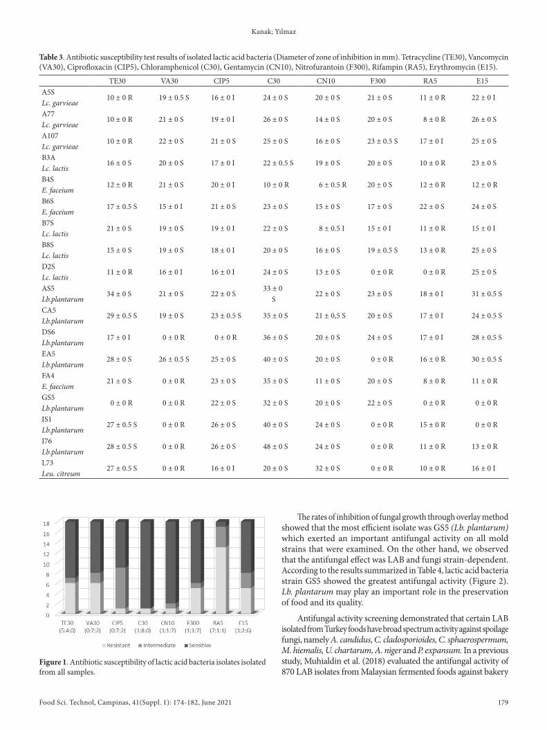
Although LAB has been “generally recognized as safe” (GRAS), it has been shown that these bacteria can exchange genes to enhance their survival in antibiotic-containing environments and are able to transfer them among bacteria of different genera in the intestine, both commensal and pathogenic species. Hence, absence of antibiotic resistance is considered as a preliminary stage for the selection of potential probiotic strains. The results of the antibiotic susceptibility tests carried out in selected strains are shown in Table 3. Of the 18 LAB isolates tested, 13 were resistant to rifampicin, 6 resistant to tetracycline and vancomycin, 5 resistant to erythromycin and
Table 1. Biochemical identification results of isolates isolated from samples.
Sample Code Morphology test 45 °C 4% NaCl 6.5% NaCl pH 9.2 pH 9.6 Biochemical results MALDI-TOF results
-: negative reaction; +: positive reaction; w: weak reaction; *could not be determined.
Food Sci. Technol, Campinas, 41(Suppl. 1): 174-182, June 2021178 178/182
Identification of new strains with many useful properties of lactic acid bacteria
nitrofurantoin, 1 resistant to gentamycin, chloramphenicole and ciprofloxacin. Figure 1 shows the results of antibiotic susceptibility testing of LABs.
The three numbers in parentheses under each column indicate the number of sensitive, moderate and resistant isolates displayed in different colors. Over whole 72.2% of the strains were found to be resistant to rifampicin, 53.3% of the strains resistant to tetracycline and vancomycin, 27.7% of the strains resistant to erythromycin and nitrofurantoin. These results support the hypothesis that foodborne bacteria may be one of the sources of antibiotic resistance genes.
3.4 Antifungal effects of lactic acid bacteria against fungi
In this study, we hypothesized that LAB could inhibit the development of fungi isolated from the same substrates.
In the present study, a screening for antifungal activity against mold strains was done for 36 lactic acid bacteria strains that were isolated. Initial screening for the antifungal activity of LAB isolates against the spoilage fungi showed that out of 36 isolates, only 17 (47.2%) inhibited the growth of the fungi indicator strain. The antifungal activity of the isolates ranged from weak to strong. The plates were examined for clear zones of inhibition around the bacterial streaks, and the area of the zones was scored as follows: -, no suppression; (+) weak inhibition (İnhibition zone ≤ 0.5 mm); (++) moderate inhibition (İnhibition zone 0.6- 1.4 mm) (+++) İnhibition zone 1.5- 2.4 mm. The diameter of the clear zone around the two LAB lines varied from as low as 10 mm up to 70 mm. The MRS control plate containing fungal spores without LAB showed increased growth after 48 h at 30 °C, and the plates were completely covered by the fungi.
Table 2. Antimicrobial activity of lactic acid bacteria against pathogens (Diameter of zone of inhibition in mm).
Sample Code E. coli O157:H7 Staph. aureus ATCC 25923
The rates of inhibition of fungal growth through overlay method showed that the most efficient isolate was GS5 (Lb. plantarum) which exerted an important antifungal activity on all mold strains that were examined. On the other hand, we observed that the antifungal effect was LAB and fungi strain-dependent. According to the results summarized in Table 4, lactic acid bacteria strain GS5 showed the greatest antifungal activity (Figure 2). Lb. plantarum may play an important role in the preservation of food and its quality.
Antifungal activity screening demonstrated that certain LAB isolated from Turkey foods have broad spectrum activity against spoilage fungi, namely A. candidus, C. cladosporioides, C. sphaerospermum, M. hiemalis, U. chartarum, A. niger and P. expansum. In a previous study, Muhialdin et al. (2018) evaluated the antifungal activity of 870 LAB isolates from Malaysian fermented foods against bakery
Table 3. Antibiotic susceptibility test results of isolated lactic acid bacteria (Diameter of zone of inhibition in mm). Tetracycline (TE30), Vancomycin (VA30), Ciprofloxacin (CIP5), Chloramphenicol (C30), Gentamycin (CN10), Nitrofurantoin (F300), Rifampin (RA5), Erythromycin (E15).
TE30 VA30 CIP5 C30 CN10 F300 RA5 E15A5S
10 ± 0 R 19 ± 0.5 S 16 ± 0 I 24 ± 0 S 20 ± 0 S 21 ± 0 S 11 ± 0 R 22 ± 0 ILc. garvieaeA77
10 ± 0 R 21 ± 0 S 19 ± 0 I 26 ± 0 S 14 ± 0 S 20 ± 0 S 8 ± 0 R 26 ± 0 SLc. garvieaeA107
10 ± 0 R 22 ± 0 S 21 ± 0 S 25 ± 0 S 16 ± 0 S 23 ± 0.5 S 17 ± 0 I 25 ± 0 SLc. garvieaeB3A
16 ± 0 S 20 ± 0 S 17 ± 0 I 22 ± 0.5 S 19 ± 0 S 20 ± 0 S 10 ± 0 R 23 ± 0 SLc. lactisB4S
12 ± 0 R 21 ± 0 S 20 ± 0 I 10 ± 0 R 6 ± 0.5 R 20 ± 0 S 12 ± 0 R 12 ± 0 RE. faceiumB6S
17 ± 0.5 S 15 ± 0 I 21 ± 0 S 23 ± 0 S 15 ± 0 S 17 ± 0 S 22 ± 0 S 24 ± 0 SE. faceiumB7S
21 ± 0 S 19 ± 0 S 19 ± 0 I 22 ± 0 S 8 ± 0.5 I 15 ± 0 I 11 ± 0 R 15 ± 0 ILc. lactisB8S
15 ± 0 S 19 ± 0 S 18 ± 0 I 20 ± 0 S 16 ± 0 S 19 ± 0.5 S 13 ± 0 R 25 ± 0 SLc. lactisD2S
11 ± 0 R 16 ± 0 I 16 ± 0 I 24 ± 0 S 13 ± 0 S 0 ± 0 R 0 ± 0 R 25 ± 0 SLc. lactisAS5
34 ± 0 S 21 ± 0 S 22 ± 0 S33 ± 0
22 ± 0 S 23 ± 0 S 18 ± 0 I 31 ± 0.5 SLb.plantarum SCA5
29 ± 0.5 S 19 ± 0 S 23 ± 0.5 S 35 ± 0 S 21 ± 0,5 S 20 ± 0 S 17 ± 0 I 24 ± 0.5 SLb.plantarumDS6
17 ± 0 I 0 ± 0 R 0 ± 0 R 36 ± 0 S 20 ± 0 S 24 ± 0 S 17 ± 0 I 28 ± 0.5 SLb.plantarumEA5
28 ± 0 S 26 ± 0.5 S 25 ± 0 S 40 ± 0 S 20 ± 0 S 0 ± 0 R 16 ± 0 R 30 ± 0.5 SLb.plantarumFA4
21 ± 0 S 0 ± 0 R 23 ± 0 S 35 ± 0 S 11 ± 0 S 20 ± 0 S 8 ± 0 R 11 ± 0 RE. faeciumGS5
0 ± 0 R 0 ± 0 R 22 ± 0 S 32 ± 0 S 20 ± 0 S 22 ± 0 S 0 ± 0 R 0 ± 0 RLb.plantarumIS1
27 ± 0.5 S 0 ± 0 R 26 ± 0 S 40 ± 0 S 24 ± 0 S 0 ± 0 R 15 ± 0 R 0 ± 0 RLb.plantarumI76
28 ± 0.5 S 0 ± 0 R 26 ± 0 S 48 ± 0 S 24 ± 0 S 0 ± 0 R 11 ± 0 R 13 ± 0 RLb.plantarumL73
27 ± 0.5 S 0 ± 0 R 16 ± 0 I 20 ± 0 S 32 ± 0 S 0 ± 0 R 10 ± 0 R 16 ± 0 ILeu. citreum
Figure 1. Antibiotic susceptibility of lactic acid bacteria isolates isolated from all samples.
Food Sci. Technol, Campinas, 41(Suppl. 1): 174-182, June 2021180 180/182
Identification of new strains with many useful properties of lactic acid bacteria
spoilage fungi, namely A. niger, A. flavus MD3, P. roqueforti MD4, E. rubrum MD5, M. sitophila MD6, and R. nigricans MD8; and LAB isolates showed activity against selected fungi.
As a result, E. faecium has antimicrobial and antifungal properties. According to Rehaiem et al. (2014), some active enterococci strains have been suggested as safe candidates due to the growing interest for the usage of probiotics, along with the currently most commonly used strains of Lactobacillus and Bifidobacterium. E. faecium can be used under various experimental conditions in future studies. In this sense, several scientists reported that E. faecium is considered a healthy agent used as a natural starter culture.
4 ConclusionConsumers negative attitudes towards the use of chemical
preservatives in food products have resulted in an increase in the
number of studies on the possible use of LABs as biopreservatives against pathogenic bacteria. Foods can serve as a source of beneficial and various LABs for consumers. Results show that foods contain various antimicrobial LABs that can be used as food additives. The LABs contain components that inhibit pathogen development, suggesting that they can replace chemical additives and provide attractive and diverse food products. Results also show that MALDI-TOF MS is substantially faster, more cost-effective and yields more accurate results than biochemical tests for LAB identification. In recent years, human and animal resistance genes and the spread of bacterial resistance have been put forward by many studies led by the food chain. For this reason, LAB usage in foods or as starters must be monitored continuously against the risk of antibiotic resistance and the use of antibiotics should also be controlled. Several studies have recently reported the isolation and identification of LAB strains with antifungal effect, and these finding are of interest due to the important role of LAB in the bio-preservation of processed foods.
AcknowledgementsWe are most grateful to Sölen Dincer for her assistance
with MALDI-TOF MS.
ReferencesAkşit, F., Akgün, Y., & Kiraz, N. (2006). Genel mikrobiyoloji ve immünoloji
(8th ed., pp. 2-4). Eskişehir, Turkey: Anadolu University Press.Ammor, M. S., Flórez, A. B., van Hoek, A. H. A. M., de los Reyes-
Gavilán, C. G., Aarts, H. J. M., Margolles, A., & Mayo, B. (2008). Molecular characterization of intrinsic and acquired antibiotic resistance in lactic acid bacteria and bifidobacteria. Journal of Molecular Microbiology and Biotechnology, 14(1-3), 6-15. http://dx.doi.org/10.1159/000106077. PMid:17957105.
Aymerich, M. T., Garriga, M., Monfort, J. M., Nes, I., & Hugas, M. (2000). Bacteriocin-producing lactobacilli in Spanish-style fermented sausages: characterization of bacteriocins. Food Microbiology, 17(1), 33-45. http://dx.doi.org/10.1006/fmic.1999.0275.
Bertrand, X., Mulin, B., Viel, J. F., Thouverez, M., & Talon, D. (2000). Common pfge patterns in antibiotic-resistant E. faecalis from humans and cheeses. Food Microbiology, 17(5), 543-551. http://dx.doi.org/10.1006/fmic.2000.0345.
Figure 2. GS5 (Lb. plantarum) etc. A. niger showing clear zones of fungal inhibition by the overlay agar method.
Table 4. Inhibition of molds in a dual-culture overlay system.
Activity was scored as follows: -, no suppression; +, weak suppression around the streaks; ++, strong suppression, with detectable clear zones around the streaks; +++, very strong suppression, with large, clear zones around the streaks.
Campos, C. A., Rodríguez, Ó., Calo-Mata, P., Prado, M., & Barros-Velázquez, J. (2006). Preliminary characterization of bacteriocins from Lactococcus lactis, Enterococcus faecium and Enterococcus mundtii strains isolated from turbot (Psetta maxima). Food Research International, 39(3), 356-364. http://dx.doi.org/10.1016/j.foodres.2005.08.008.
Carbonnelle, E., Mesquita, C., Bille, E., Day, N., Dauphin, B., Beretti, J.-L., Ferroni, A., Gutmann, L., & Nassif, X. (2011). MALDI-TOF mass spectrometry tools for bacterial identification in clinical microbiology laboratory. Clinical Biochemistry, 44(1), 104-109. http://dx.doi.org/10.1016/j.clinbiochem.2010.06.017. PMid:20620134.
Carr, F. J., Chill, D., & Maida, N. (2002). The lactic acid bacteria: a literature survey. Critical Reviews in Microbiology, 28(4), 281-370. http://dx.doi.org/10.1080/1040-840291046759. PMid:12546196.
Cotter, P. D., Hill, C., & Ross, R. P. (2005). Bacteriocins: developing innate immunity for food. Nature Reviews. Microbiology, 3(10), 777-788. http://dx.doi.org/10.1038/nrmicro1273. PMid:16205711.
Dalié, D. K. D., Deschamps, A. M., & Richard-Forget, F. (2010). Lactic acid bacteria potential for control of mould growth and mycotoxins: a review. Food Control, 21(4), 370-380. http://dx.doi.org/10.1016/j.foodcont.2009.07.011.
Dec, M., Urban-Chmiel, R., Gnat, S., Puchalski, A., & Wernicki, A. (2014). Identification of Lactobacillus strains of goose origin using MALDI-TOF mass spectrometry and 16S–23S rDNA intergenic spacer PCR analysis. Research in Microbiology, 165(3), 190-201. http://dx.doi.org/10.1016/j.resmic.2014.02.003. PMid:24607713.
Dimitonova, S. P., Bakalov, B. V., Aleksandrova-Georgieva, R. N., & Danova, S. T. (2008). Phenotypic and molecular identification of lactobacilli isolated from vaginal secretions. Journal of Microbiology, Immunology, and Infection, 41(6), 469-477. PMid:19255690.
Doan, N. T. L., Van Hoorde, K., Cnockaert, M., De Brandt, E., Aerts, M., Le Thanh, B., & Vandamme, P. (2012). Validation of MALDI‐TOF MS for rapid classification and identification of lactic acid bacteria, with a focus on isolates from traditional fermented foods in Northern Vietnam. Letters in Applied Microbiology, 55(4), 265-273. http://dx.doi.org/10.1111/j.1472-765X.2012.03287.x. PMid:22774847.
Dušková, M., Šedo, O., Kšicová, K., Zdráhal, Z., & Karpíšková, R. (2012). Identification of lactobacilli isolated from food by genotypic methods and MALDI-TOF MS. International Journal of Food Microbiology, 159(2), 107-114. http://dx.doi.org/10.1016/j.ijfoodmicro.2012.07.029. PMid:23072695.
Fguiri, I., Ziadi, M., Atigui, M., Arroum, S., & Khorchani, T. (2015). Biochemical and molecular identification of lactic acid bacteria isolated from camel milk in Tunisia. Emirates Journal of Food and Agriculture, 27(9), 716. http://dx.doi.org/10.9755/ejfa.2015.04.114.
Freitas, D. B., Reis, M. P., Lima-Bittencourt, C. I., Costa, P. S., Assis, P. S., Chartone-Souza, E., & Nascimento, A. M. (2008). Genotypic and phenotypic diversity of Bacillus spp. isolated from steel plant waste. BMC Research Notes, 1(1), 92. http://dx.doi.org/10.1186/1756-0500-1-92. PMid:18928552.
G-Alegría, E., López, I., Ruiz, J. I., Sáenz, J., Fernández, E., Zarazaga, M., Dizy, M., Torres, C., & Ruiz-Larrea, F. (2004). High tolerance of wild Lactobacillus plantarum and Oenococcus oeni strains to lyophilisation and stress environmental conditions of acid pH and ethanol. FEMS Microbiology Letters, 230(1), 53-61. http://dx.doi.org/10.1016/S0378-1097(03)00854-1. PMid:14734166.
Gálvez, A., Abriouel, H., López, R. L., & Ben Omar, N. (2007). Bacteriocin-based strategies for food biopreservation. International Journal of Food Microbiology, 120(1-2), 51-70. http://dx.doi.org/10.1016/j.ijfoodmicro.2007.06.001. PMid:17614151.
Halkman, A. K. (2005). Gıda mikrobiyolojisi uygulamaları. Ankara: Başak Press.
Han, S.-K., Hong, Y., Kwak, H.-L., Kim, E.-S., Kim, M.-J., Shrivastav, A., Oh, M.-H., & Kim, H.-Y. (2014). Identification of lactic acid bacteria in pork meat and pork meat products using SDS‐PAGE, 16 S rRNA gene sequencing and MALDI‐TOF mass spectrometry. Journal of Food Safety, 34(3), 224-232. http://dx.doi.org/10.1111/jfs.12117.
Harrigan, W. F., & McCance, M. E. (1990). Laboratory methods in food and dairy microbiology (8th ed.). London: Academic Press.
Holt, J. G., Krieg, N. R., Sneath, P. H. H., Staley, J. T., & Williams, S. T. (1994). Bergey’s manual of determinative bacteriology (9th ed.). Baltimore; William & Wilkins.
Jabbari, V., Khiabani, M. S., Mokarram, R. R., Hassanzadeh, A. M., Ahmadi, E., Gharenaghadeh, S., Karimi, N., & Kafil, H. S. (2017). Lactobacillus plantarum as a probiotic potential from kouzeh cheese (traditional Iranian cheese) and its antimicrobial activity. Probiotics and Antimicrobial Proteins, 9(2), 189-193. http://dx.doi.org/10.1007/s12602-017-9255-0. PMid:28155128.
Lukasova, J., & Sustackova, A. (2003). Enterococci and antibiotic resistance. Journal of the University of Veterinary and Pharmaceutical Sciences in Brno, 72(2), 315-323.
Macaluso, G., Fiorenza, G., Gaglio, R., Mancuso, I., & Scatassa, M. L. (2016). In vitro evaluation of bacteriocin-like inhibitory substances produced by lactic acid bacteria isolated during traditional Sicilian cheese making. Italian Journal of Food Safety, 5(1), 5503. http://dx.doi.org/10.4081/ijfs.2016.5503. PMid:27800430.
Magnusson, J., & Schnürer, J. (2001). Lactobacillus coryniformis subsp.coryniformis strain Si3 produces a broad-spectrum proteinaceous antifungal compound. Applied and Environmental Microbiology, 67(1), 1-5. http://dx.doi.org/10.1128/AEM.67.1.1-5.2001. PMid:11133421.
Messens, W., Neysens, P., Vansieleghem, W., Vanderhoeven, J., & De Vuyst, L. (2002). Modeling growth and bacteriocin production by Lactobacillus amylovorus DCE 471 in response to temperature and pH values used for sourdough fermentations. Applied and Environmental Microbiology, 68(3), 1431-1435. http://dx.doi.org/10.1128/AEM.68.3.1431-1435.2002. PMid:11872497.
Michalak, M., Gustaw, K., Waśko, A., & Polak-Berecka, M. (2018). Composition of lactic acid bacteria during spontaneous curly kale (Brassica oleracea var. sabellica) fermentation. Microbiological Research, 206, 121-130. http://dx.doi.org/10.1016/j.micres.2017.09.011. PMid:29146249.
Muhialdin, B. J., Hassan, Z., & Saari, N. (2018). In vitro antifungal activity of lactic acid bacteria low molecular peptides against spoilage fungi of bakery products. Annals of Microbiology, 68(9), 557-567. http://dx.doi.org/10.1007/s13213-018-1363-x.
Özcan, N., Ezin, Ö., Akpolat, N., Bozdağ, H., Mete, M., & Gül, K. (2016). Klinik örneklerde saptanan Candida türlerinin MALDI-TOF MS ile tiplendirilmesi. Dicle Medikal Journal, 43(3), 390-394.
Papamanoli, E., Tzanetakis, N., Litopoulou-Tzanetaki, E., & Kotzekidou, P. (2003). Characterization of lactic acid bacteria isolated from a Greek dry-fermented sausage in respect of their technological and probiotic properties. Meat Science, 65(2), 859-867. http://dx.doi.org/10.1016/S0309-1740(02)00292-9. PMid:22063449.
Pavlovic, M., Huber, I., Konrad, R., & Busch, U. (2013). Application of MALDI-TOF MS for the identification of food borne bacteria. The Open Microbiology Journal, 7(1), 135-141. http://dx.doi.org/10.2174/1874285801307010135. PMid:24358065.
Food Sci. Technol, Campinas, 41(Suppl. 1): 174-182, June 2021182 182/182
Identification of new strains with many useful properties of lactic acid bacteria
Randazzo, C. L., Restuccia, C., Romano, A. D., & Caggia, C. (2004). Lactobacillus casei, dominant species in naturally fermented Sicilian green olives. International Journal of Food Microbiology, 90(1), 9-14. http://dx.doi.org/10.1016/S0168-1605(03)00159-4. PMid:14672826.
Rehaiem, A., Belgacem, Z. B., Edalatian, M. R., Martínez, B., Rodríguez, A., Manai, M., & Guerra, N. P. (2014). Assessment of potential probiotic properties and multiple bacteriocin encoding-genes of the technological performing strain E. faecium MMRA. Food Control, 37, 343-350. http://dx.doi.org/10.1016/j.foodcont.2013.09.044.
Salminen, S., & Von Wright, A. (1993). Lactic acid bacteria: microbiology and functional aspects. New York: Marcel Dekker.
Schnürer, J., & Magnusson, J. (2005). Antifungal lactic acid bacteria as biopreservatives. Trends in Food Science & Technology, 16(1-3), 70-78. http://dx.doi.org/10.1016/j.tifs.2004.02.014.
Ström, K., Sjögren, J., Broberg, A., & Schnürer, J. (2002). Lb. plantarum MiLAB 393 produces the antifungal cyclic dipeptides cyclo (L-Phe-L-Pro) and cyclo (L-Phe-trans-4-OH-L-Pro) and 3-phenyllactic acid. Applied and Environmental Microbiology, 68(9), 4322-4327. http://dx.doi.org/10.1128/AEM.68.9.4322-4327.2002. PMid:12200282.
Temiz, A. (2000). Genel mikrobiyoloji uygulama teknikleri (3rd ed.). Ankara: Hatipoğlu.
Tulini, F. L., Hymery, N., Haertlé, T., Le Blay, G., & De Martinis, E. C. (2016). Screening for antimicrobial and proteolytic activities of lactic acid bacteria isolated from cow, buffalo and goat milk and cheeses marketed in the southeast region of Brazil. The Journal of Dairy Research, 83(1), 115-124. http://dx.doi.org/10.1017/S0022029915000606. PMid:26608755.
Vithanage, N. R., Yeager, T. R., Jadhav, S. R., Palombo, E. A., & Datta, N. (2014). Comparison of identification systems for psychrotrophic bacteria isolated from raw bovine milk. International Journal of Food Microbiology, 189, 26-38. http://dx.doi.org/10.1016/j.ijfoodmicro.2014.07.023. PMid:25113043.
Yamato, M., Ozaki, K., & Ota, F. (2003). Partial purification and characterization of the bacteriocin produced by Lactobacillus acidophilus YIT 0154. Microbiological Research, 158(2), 169-172. http://dx.doi.org/10.1078/0944-5013-00190. PMid:12906390.
York, M. K., Traylor, M. M., Hardy, J., & Henry, M. (2010). Biochemical tests for the identification of aerobic bacteria. Washington: Clinical Microbiology Procedures Handbook.