Vol. 42, No. 3 JOURNAL OF VIROLOGY, June 1982, p. 1017-1028 0022-538X/82/061017-12$02.00/0 Identification of the Initiation Site of Poliovirus Polyprotein Synthesis ANDREW J. DORNER,l* LYDIA F. DORNER,1 GLENN R. LARSEN,1t ECKARD WIMMER,1 AND CARL W. ANDERSON2 Department of Microbiology, School of Medicine, State University of New York at Stony Brook, Stony Brook, New York 11794,1 and Department of Biology, Brookhaven National Laboratory, Upton, New York 119732 Received 28 December 1981/Accepted 16 February 1982 The complete nucleotide sequence of poliovirus RNA has a long open reading frame capable of encoding the precursor polyprotein NCVPOO. The first AUG codon in this reading frame is located 743 nucleotides from the 5' end of the RNA and is preceded by eight AUG codons in all three reading frames. Because all proteins that map at the amino terminus of the polyprotein (P1-la, VPO, and VP4) are blocked at their amino termini and previous studies of ribosome binding have been inconclusive, direct identification of the initiation site of protein synthesis was difficult. We separated and identified all of the tryptic peptides of capsid protein VP4 and correlated these peptides with the amino acid sequence predicted to follow the AUG codon at nucleotide 743. Our data indicate that VP4 begins with a blocked glycine that is encoded immediately after the AUG codon at nucleotide 743. An S1 nuclease analysis of poliovirus mRNA failed to reveal a splice in the 5' region. We concluded that synthesis of the poliovirus polyprotein is initiated at nucleotide 743, the first AUG codon in the long open reading frame. Poliovirus-specific protein synthesis has been studied for several decades and has been used as a model of translation of eucaryotic mRNA (5, 21). The poliovirus genome is a single-stranded RNA of positive polarity (1). After infection, the poliovirus RNA directs the synthesis of a large polyprotein, NCVPOO, which has been suggest- ed to contain most, if not all, of the translatable information of the virus. This polyprotein is cleaved as a nascent chain into the primary precursor proteins P1-la, P2-3b, and P3-tb, which correspond to three distinct classes of poliovirus proteins (9, 10, 30) (Fig. IA). Pacta- mycin mapping and salt shock studies (25, 31, 33) have indicated that the three main protein classes are encoded on the RNA in the order (from the 5' end to the 3' end): Pl-la-P2-3b-P3- lb. The primary precursors are processed pro- teolytically to produce all of the known poliovi- rus-specific proteins. P1-la, contained at the amino terminus of the polyprotein, is the precur- sor to the four poliovirus capsid proteins that map in the order VP4-VP2-VP3-VP1 (23). Cleav- age of P1-la occurs in distinct steps. This pro- tein is cleaved initially to VP3, VP1, and VPO. In turn, VPO is cleaved to VP4 and VP2. Cleavage of VPO is intimately associated with morphogen- t Present address: Genetics Institute, Boston, MA 02115. esis and probably occurs upon association of vRNA with procapsid structures (24). The genomic RNA (vRNA) has the small virus-encoded protein VPg linked to the 5' ter- minus (17) and is polyadenylated at the 3' termi- nus (37). The virus-specific RNA associated with polyribosomes (mRNA) lacks VPg and terminates with a 5'-uridylate (6, 8, 19). The complete nucleotide sequence of the poliovirus genome has been determined recently (11, 22). This sequence analysis has revealed a long open reading frame that encompasses 89% of the viral genome and is capable of encoding the precursor polyprotein NCVPOO. A partial amino acid se- quence analysis of 13 viral proteins has shown that all of these proteins may be derived by processing of the polyprotein and has confirmed the genetic map of the poliovirus genome (5a, 11, 14a, 27, 28). The nucleotide assignments used in this paper are from a "consensus" sequence for poliovirus type 1 [Mahoney] derived from the previously published sequences of Mahoney strain virus [11, 22], a sequence analysis of cloned cDNA segments derived from Mahoney virus [S. van der Werf, personal communication], and a se- quence analysis of Sabin vaccine strain virus [A. Nomoto, personal communication]. This se- quence is available through the MOLGEN pro- 1017 Downloaded from https://journals.asm.org/journal/jvi on 21 February 2022 by 221.124.192.234.
Transcript
Vol. 42, No. 3JOURNAL OF VIROLOGY, June 1982, p. 1017-10280022-538X/82/061017-12$02.00/0
Identification of the Initiation Site of Poliovirus PolyproteinSynthesis
ANDREW J. DORNER,l* LYDIA F. DORNER,1 GLENN R. LARSEN,1t ECKARD WIMMER,1 ANDCARL W. ANDERSON2
Department of Microbiology, School of Medicine, State University ofNew York at Stony Brook, StonyBrook, New York 11794,1 and Department ofBiology, Brookhaven National Laboratory, Upton, New York
119732
Received 28 December 1981/Accepted 16 February 1982
The complete nucleotide sequence of poliovirus RNA has a long open readingframe capable of encoding the precursor polyprotein NCVPOO. The first AUGcodon in this reading frame is located 743 nucleotides from the 5' end of the RNAand is preceded by eight AUG codons in all three reading frames. Because allproteins that map at the amino terminus of the polyprotein (P1-la, VPO, and VP4)are blocked at their amino termini and previous studies of ribosome binding havebeen inconclusive, direct identification of the initiation site of protein synthesiswas difficult. We separated and identified all of the tryptic peptides of capsidprotein VP4 and correlated these peptides with the amino acid sequence predictedto follow the AUG codon at nucleotide 743. Our data indicate that VP4 beginswith a blocked glycine that is encoded immediately after the AUG codon atnucleotide 743. An S1 nuclease analysis of poliovirus mRNA failed to reveal asplice in the 5' region. We concluded that synthesis of the poliovirus polyproteinis initiated at nucleotide 743, the first AUG codon in the long open reading frame.
Poliovirus-specific protein synthesis has beenstudied for several decades and has been used asa model of translation of eucaryotic mRNA (5,21). The poliovirus genome is a single-strandedRNA of positive polarity (1). After infection, thepoliovirus RNA directs the synthesis of a largepolyprotein, NCVPOO, which has been suggest-ed to contain most, if not all, of the translatableinformation of the virus. This polyprotein iscleaved as a nascent chain into the primaryprecursor proteins P1-la, P2-3b, and P3-tb,which correspond to three distinct classes ofpoliovirus proteins (9, 10, 30) (Fig. IA). Pacta-mycin mapping and salt shock studies (25, 31,33) have indicated that the three main proteinclasses are encoded on the RNA in the order(from the 5' end to the 3' end): Pl-la-P2-3b-P3-lb. The primary precursors are processed pro-teolytically to produce all of the known poliovi-rus-specific proteins. P1-la, contained at theamino terminus of the polyprotein, is the precur-sor to the four poliovirus capsid proteins thatmap in the order VP4-VP2-VP3-VP1 (23). Cleav-age of P1-la occurs in distinct steps. This pro-tein is cleaved initially to VP3, VP1, and VPO. Inturn, VPO is cleaved to VP4 and VP2. Cleavageof VPO is intimately associated with morphogen-
t Present address: Genetics Institute, Boston, MA 02115.
esis and probably occurs upon association ofvRNA with procapsid structures (24).The genomic RNA (vRNA) has the small
virus-encoded protein VPg linked to the 5' ter-minus (17) and is polyadenylated at the 3' termi-nus (37). The virus-specific RNA associatedwith polyribosomes (mRNA) lacks VPg andterminates with a 5'-uridylate (6, 8, 19). Thecomplete nucleotide sequence of the poliovirusgenome has been determined recently (11, 22).This sequence analysis has revealed a long openreading frame that encompasses 89% of the viralgenome and is capable of encoding the precursorpolyprotein NCVPOO. A partial amino acid se-quence analysis of 13 viral proteins has shownthat all of these proteins may be derived byprocessing of the polyprotein and has confirmedthe genetic map of the poliovirus genome (5a,11, 14a, 27, 28).The nucleotide assignments used in this paper
are from a "consensus" sequence for poliovirustype 1 [Mahoney] derived from the previouslypublished sequences of Mahoney strain virus[11, 22], a sequence analysis of cloned cDNAsegments derived from Mahoney virus [S. vander Werf, personal communication], and a se-quence analysis of Sabin vaccine strain virus [A.Nomoto, personal communication]. This se-quence is available through the MOLGEN pro-
1017
Dow
nloa
ded
from
http
s://j
ourn
als.
asm
.org
/jour
nal/j
vi o
n 21
Feb
ruar
y 20
22 b
y 22
1.12
4.19
2.23
4.
1018 DORNER ET AL.
Pi P2
3386
NCVP00
la (97) lb (84)- 3b (65)
. VPO(37)VP3(27)
VPI (33)Y.?4(7.4)
VP2 (30)
NCVPOO*pu
Frame 3. VPg-pU..
N200 N400N0 t N1
N600 N800 N 1000
FIG. 1. Genomic organization of poliovirus. (A) The three regions of the poliovirus polyprotein are delineatedabove the heavy horizontal line representing genomic RNA. A simplified processing scheme detailing the capsidprotein region is presented. The numbers at the ends of the vertical lines indicate the nucleotide assignments onthe genomic RNA for initiation (nucleotide 743) and termination (nucleotide 7,369) of polyprotein synthesis andfor the primary protein cleavage sites (nucleotides 3,386 and 5,111). The numbers in parentheses are themolecular weights (x10-3) calculated from the predicted amino acid sequence. The solid circles indicateexperimentally determined blocked amino termini of processed proteins. The unprocessed polyprotein(NCVPOO) was not examined directly; the amino terminus of this protein is indicated by an open circle. poly(A),Polyadenylic acid. (B) Positions of AUG codons and termination codons within the S'-terminal 1,000 nucleotides of thepoliovirus genome. The numbers on the left indicate the three reading frames beginning with the first, second, orthird nucleotide of the RNA. The vertical lines represent termination codons. The solid boxes represent AUGcodons, and the open boxes indicate open translation regions following an AUG codon. The long open readingframe (nucleotides 743 through 7,369) present in the second reading frame is labeled NCVPOO. The numbersbelow the vertical arrows indicate distances from the 5' end (in nucleotides). The nucleotide assignments arefrom a "consensus" sequence (see text).
ject of the Stanford University SUMEX-AIMfacility. Differences between previously pub-lished sequences and the "consensus" sequencedo not affect the conclusions reached in thispaper.
Unexpectedly, an analysis of the poliovirusRNA sequence showed that the first AUG co-don in the long open reading frame is located 743nucleotides from the 5' end of the RNA and ispreceded by eight other AUG codons whichoccur in all three reading frames. Translationinitiated at the 5'-proximal AUG codon at nucle-otide 185 could produce a protein of 4,720 dal-tons; the eighth AUG codon, located at nucleo-tide 586, is followed by an open reading frame of64 codons, which overlaps the beginning of thelong open reading frame. The six other AUGcodons are followed shortly by in-phase termi-
nation codons (Fig. 1B). The existence of thesepotential initiation codons has raised questionsconcerning the exact location of the initiationsite for synthesis of the poliovirus polyprotein.
Nucleotide sequence analysis of poliovirusRNA alone did not identify the initiation site ofpolyprotein synthesis. The possibility of se-quencing errors or of an RNA splicing eventnecessitated an analysis of viral mRNA andprotein sequences. Because all of the proteinswhich map at the amino terminus of the poliovi-rus polyprotein (P1-la, VPO, VP4) carry blockedamino termini, direct protein sequencing has notbeen possible. Ribosome binding experimentshave also failed to produce the initiation sitesequence (Dorner and Wimmer, unpublisheddata; E. Ehrenfeld, personal communication).We located the initiation site for synthesis of the
A743
VPg-pU.
P3
5111 7369
nnl I(Al
BFrame 1. VPg-pl
Frame 2. VPg-
J. VIROL.
.1fti 1.
Dow
nloa
ded
from
http
s://j
ourn
als.
asm
.org
/jour
nal/j
vi o
n 21
Feb
ruar
y 20
22 b
y 22
1.12
4.19
2.23
4.
POLIOVIRUS PROTEIN SYNTHESIS INITIATION SITE 1019
poliovirus polyprotein by mapping the trypticpeptides of VP4 within the genomic 5' codingsequence. Our strategy involved identifying allof the tryptic peptides of VP4 and determiningthe composition of the amino-terminal trypticpeptide. We found no evidence of a splice withinthe 5' region of poliovirus mRNA. Together, theprotein and RNA analyses demonstrate thatpoliovirus polyprotein synthesis is initiated atthe first AUG codon in the long open readingframe of the poliovirus genomic sequence.
MATERIAL AND METHODSPreparation of radiolabeled poliovirus proteins. Sus-
pension cultures ofHeLa S3 cells (5 x 10⁶ cells per ml)were infected with poliovirus type 1 (Mahoney) at amultiplicity of 50 PFU/cell. Infected cultures werelabeled with radioactive amino acids (AmershamCorp.; ICN) at concentrations of 0.04 to 0.06 mCi/mlfor 5 h beginning 2 h postinfection. Harvested cellswere lysed by three rounds offreeze-thawing in reticu-locyte standard buffer containing Mg2+ (0.01 M NaCl,0.01 M Tris, pH 7.4, 1.5 mM MgCl2), and the cellnuclei were pelleted. The supernatant containing po-liovirus virions was made 2 mM in EDTA and 0.1% insodium dodecyl sulfate (SDS) and applied to a 15% to30%o sucrose gradient (0.1 M NaCl, 0.01 M Tris, pH7.4, 1 mM EDTA, 0.5% SDS). Centrifugation was at16,000 x g for 16 h. Gradient-purified virus waspelleted, suspended in protein gel sample buffer, andsubjected to preparative SDS-polyacrylamide gel elec-trophoresis (5a). The band corresponding to VP4 wasexcised from the gel and eluted electrophoretically(27). P1-la and VPO were purified from infected cellextracts as described previously (27).
Trypsin digestion of VP4. Apomyoglobin (1 mg) wasadded as a carrier to the gel-purified VP4. SDS remov-al was accomplished by three 30-min extractions withice-cold acetone-acetic acid-triethylamine (90:5:5), fol-lowed by two washes with ice-cold acetone (7). Afterdrying, the protein precipitate was incubated on ice for1.5 h with 0.5 ml of performic acid (19:1 mixture of88% formic acid and 30%o hydrogen peroxide kept atroom temperature for 2 h). The oxidized protein waslyophilized three times. Then the protein was suspend-ed in 0.4 ml of1% ammonium bicarbonate (pH 8.5) andincubated with 10 ,ug of tolylsulfonyl phenylalanylchloromethyl ketone-treated trypsin (Worthington Di-agnostics) for 2 h at 37°C. An additional 20 ,ug oftrypsin was added, and the incubation was continuedfor 2 h. Then 10 ,ug of trypsin was added again, and theincubation was continued for a final 2 h (total incuba-tion time, 6 h). The reaction mixture was then lyophi-lized. The digested protein was dissolved in 250 ,u of20%o formic acid for high-performance liquid chroma-tography or small volumes of 15% NH40H or 20%pyridine for application to cellulose thin-layer plates.
Two-dimensional tryptic peptide mapping. Separa-tion in the first dimension was by ascending chroma-tography on cellulose-coated thin-layer plates (20 by20 cm; EM Reagents) in n-butanol-pyridine-aceticacid-water (30:20:6:24) until the solvent frontwas 2 cm from the top of the plate. Plates were driedovernight at room temperature. Electrophoresis in thesecond dimension was for 30 to 40 min at 1,000 V (40to 50 mA) in pH 3.5 buffer containing acetic acid,
pyridine, and water (10:1:89). Dried plates weresprayed with En3Hance (New England Nuclear Corp.)and exposed at -70°C to Kodak XAR-5 film.Amino acid analysis. A 100-sLg portion of electropho-
retically purified VP4 was dialyzed extensively againstdistilled water to remove SDS and other low-molecu-lar-weight components. The sample was divided intofour equal portions, three of which were hydrolyzed in0.3 ml of 6 N HCl at 115°C for 24, 48, or 72 h inevacuated, sealed borosilicate tubes. After hydrolysis,each sample was analyzed for amino acid content witha single-column analyzer. A standard amino acid mix-ture was analyzed before and after the set of experi-mental samples, and this standard was used to calcu-late the amount of each amino acid in eachexperimental sample. To convert the data to residueequivalents, the values for six stable amino acids(alanine, asparagine-aspartic acid, glutamine-glutamicacid, leucine, phenylalanine, valine) were added to-gether and divided by the total number of residues ofthese six amino acids predicted by the sequencebetween nucleotides 746 and 949. Division by thesefactors (24 h, 1.725; 48 h, 1.688; 72 h, 1.685) gave thenumber of residues of each amino acid present in VP4.The values for threonine and serine for each hydroly-sis time were plotted and extrapolated to zero time inorder to correct for the destruction of these residues.No other amino acid showed a significant trend withhydrolysis time, and so the values for all three hydro-lysis times were averaged.High-performance, reverse-phase (C18) liquid chro-
matography. A mixture of radiolabeled tryptic pep-tides of VP4 dissolved in 250 Il of20% formic acid wasapplied to a LiChromosorb RP-18,10 p.m column, 250by 4.6 mm; EM Reagents). The column was developedwith a discontinuous linear gradient made from 20 mMsodium phosphate (pH 5.8) (buffer A) and 100%o aceto-nitrile (buffer B). The flow rate was 2.0 ml/min, and1.0-ml fractions were collected. A portion of eachfraction was counted with Aquasol II (New EnglandNuclear Corp.) by using a Searle Mark III scintillationcounter.Amino acid sequence analysis. Sequence analyses of
purified radiolabeled proteins or tryptic peptides wereperformed essentially as described previously (27).S1 nuclease analysis. [3H]uridine-labeled poliovirus
vRNA and mRNA were purified essentially as de-scribed previously (15, 19). pPV1-366, a pBR322 deriv-ative containing a 3.05-kilobase insertion completelyspanning the 5' end of the poliovirus genome, wasobtained from S. van der Werf (34). EcoRI (NewEngland Biolabs) cuts this plasmid at a single siteoutside the poliovirus-specific sequences and was usedto linearize pPV1-366. Hybridization of radiolabeledpoliovirus vRNA and mRNA with linearized pPV1-366was performed in 10-pl reaction mixtures containing80% formamide, 0.4 M NaCl, 0.01 M PIPES [pipera-zine-N,N'-bis(2-ethanesulfonic acid)] (pH 6.4), and0.001 M EDTA (2). A poliovirus-specific DNA-RNAsingle-stranded sequence ratio of 3:1 was used forhybridization. The samples were heated for 10 min at60°C and then incubated for 2.5 h at 55.5°C. Thehybridization reaction was terminated by adding 8 to10 volumes of ice-cold 0.2 M NaCl-0.05 M sodiumacetate (pH 4.5)-0.001 M zinc sulfate. S1 nuclease(Bethesda Research Laboratories) was added (concen-tration range, 50 to 1,000 U/ml), and the samples were
VOL. 42, 1982
Dow
nloa
ded
from
http
s://j
ourn
als.
asm
.org
/jour
nal/j
vi o
n 21
Feb
ruar
y 20
22 b
y 22
1.12
4.19
2.23
4.
1020 DORNER ET AL.
incubated for 45 min at 37°C. Then 10 ,ig of yeasttRNA and 2.5 volumes of chilled 100o ethanol wereadded, and the nucleic acids were precipitated at-20°C overnight. After centrifugation, the dried nucle-ic acid pellet was dissolved in water, adjusted to 10%glycerol, 0.01% bromophenol blue, 0.04 M Tris-ace-tate (pH 8.1), and 0.002 M EDTA, and subjected toelectrophoresis in 0.7% agarose gels in 0.04 M Tris-acetate (pH 8.1)-0.002 M EDTA. The agarose gelswere treated with En3Hance (New England NuclearCorp.), dried, and exposed at -70°C to Kodak XAR-5film.
RESULTSPl-la, VPO, and VP4 have blocked amino
termini. Pactamycin and salt shock mappingstudies, molecular weight considerations, andthe known genomic sequence of poliovirus sug-gest that NCVPOO, P1-la, VPO, and VP4 sharethe same amino-terminal sequence. To provethis hypothesis, we attempted to determine theamino-terminal sequences of P1-la, VPO, andVP4 by direct radiochemical sequence analysis.VP4 labeled with [3H]tyrosine, [3H]valine, or
[3H]glutamine was purified as described aboveand subjected to automated Edman degradation.No significant yield of radioactivity was ob-tained in any of the first 21 degradation cycles(data not shown). An analysis of the carrierapomyoglobin from the same experiments indi-cated that the sequencer was functioning with arepetitive yield of over 95%. The amino acidsequence shown in Table 1 indicates that if VP4has the amino-terminal sequence predicted forinitiation of polyprotein synthesis at the AUGcodon at nucleotide 743, tyrosine, valine, andglutamine residues would be present within thefirst 20 amino acids. Proteins purified and elutedby the method which we used for VP4 have
rarely failed to yield at least 30% of the theoreti-cally expected radioactivity unless the aminoterminus was blocked (Anderson, in GeneticEngineering Principles and Methods, vol. 4, inpress). As described previously (14a), automat-ed Edman degradation of a mixture of [3H]tyro-sine-labeled tryptic peptides of VP4 gave a pat-tern of tyrosine residues consistent with thepredicted tryptic peptide 3 (Table 1). A sequenceanalysis of a high-performance liquid chroma-tography-purified, [3H]histidine-labeled trypticpeptide indicated that there was a histidine resi-due at position four, which was consistent withthe tryptic peptide 2 of VP4 (data not shown).This data established the correct reading frameand ruled out the possibility of extensive amino-terminal processing of VP4. We concluded thatVP4 must have a blocked amino terminus.An analysis of [3H]valine-labeled VPO and
[3H]lysine-labeled P1-la isolated from poliovi-rus-infected cells also failed to yield significantradioactivity in any residue during automatedEdman degradation (data not shown). Theseresults indicate that VPO and P1-la also haveblocked amino termini. We have failed in at-tempts to synthesize unblocked P1-la in vitroby preventing protein acetylation (20). The rea-son for this is unknown; our data do not excludethe possibility that acetylation of the nascentpolyprotein is the blocking mechanism.
Reverse-phase chromatography of VP4 trypticpeptides and identification of the carboxy-termi-nal peptide. Table 1 shows the expected trypticpeptides of VP4, as predicted from the RNAsequence for a protein mapping to the left ofVP2and beginning at the first AUG codon (nucleo-tide 743) in the long open reading frame ofpoliovirus vRNA. This sequence contains 68
TABLE 1. Predicted tryptic peptides of VP4Labeling pattern of tryptic peptidesb
Peptide Sequence' Gln Ser Ala Val Gly Lys Tyr Ile Met
1 B-Gly-Ala-Gln-Val-Ser-Ser-Gln-Lys X X X X X X2 Val-Gly-Ala-His-Glu-Asn-Ser-Asn-Arg' X X X X3 Ala-Tyr-Gly-Gly-Ser-Thr-Ile-Asn-Tyr-Thr-Thr-Ile-Asn- X X X X X
Tyr-Tyr-Argd4 Asp-Ser-Ala-Ser-Asn-Ala-Ala-Ser-Lys X X X5 Gln-Asp-Phe-Ser-Gln-Asp-Pro-Ser-Lys X X X6 Phe-Thr-Glu-Pro-Ile-Lys X X7 Asp-Val-Leu-Ile-Lys X X X8 Thr-Ala-Pro-Met-Leu-Asn' X Xa Tryptic peptide sequences were predicted from the RNA sequence between nucleotide 746 (Gly) and
nucleotide 949 (Asn). B, Blocked amino terminus.b The amino acids listed were used to radiolabel VP4. X, Presence of the amino acid in the tryptic peptide
sequence.c The histidine residue was confirmed by automated Edman degradation.d The tyrosine residues were confirmed by automated Edman degradation.I The alanine and methionine residues were confirmed by automated Edman degradation.
J. VIROL.
Dow
nloa
ded
from
http
s://j
ourn
als.
asm
.org
/jour
nal/j
vi o
n 21
Feb
ruar
y 20
22 b
y 22
1.12
4.19
2.23
4.
POLIOVIRUS PROTEIN SYNTHESIS INITIATION SITE 1021
POLIOVIRUS VP4 TRYPTIC PEPTIDES
% ACETONITRILErlOO -
M I0 4 -280
0
260
U.
0
zo A44- A
cr
ECa.
2-
20 60 100 140 180FRACTION NUMBER (l.Oml)
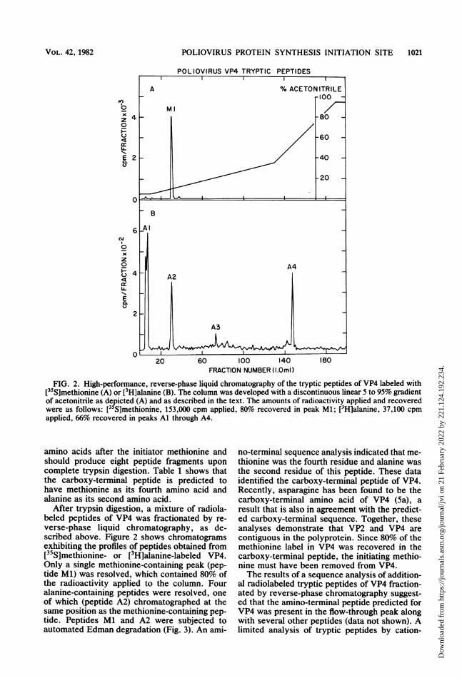
FIG. 2. High-performance, reverse-phase liquid chromatography of the tryptic peptides of VP4 labeled with[355]methionine (A) or [3H]alanine (B). The column was developed with a discontinuous linear 5 to 95% gradientof acetonitrile as depicted (A) and as described in the text. The amounts of radioactivity applied and recoveredwere as follows: [35S]methionine, 153,000 cpm applied, 80% recovered in peak Ml; [3H]alanine, 37,100 cpmapplied, 66% recovered in peaks Al through A4.
amino acids after the initiator methionine andshould produce eight peptide fragments uponcomplete trypsin digestion. Table 1 shows thatthe carboxy-terminal peptide is predicted tohave methionine as its fourth amino acid andalanine as its second amino acid.
After trypsin digestion, a mixture of radiola-beled peptides of VP4 was fractionated by re-verse-phase liquid chromatography, as de-scribed above. Figure 2 shows chromatogramsexhibiting the profiles of peptides obtained from[35S]methionine- or [3H]alanine-labeled VP4.Only a single methionine-containing peak (pep-tide Ml) was resolved, which contained 80% ofthe radioactivity applied to the column. Fouralanine-containing peptides were resolved, oneof which (peptide A2) chromatographed at thesame position as the methionine-containing pep-tide. Peptides Ml and A2 were subjected toautomated Edman degradation (Fig. 3). An ami-
no-terminal sequence analysis indicated that me-thionine was the fourth residue and alanine wasthe second residue of this peptide. These dataidentified the carboxy-terminal peptide of VP4.Recently, asparagine has been found to be thecarboxy-terminal amino acid of VP4 (5a), aresult that is also in agreement with the predict-ed carboxy-terminal sequence. Together, theseanalyses demonstrate that VP2 and VP4 arecontiguous in the polyprotein. Since 80% of themethionine label in VP4 was recovered in thecarboxy-terminal peptide, the initiating methio-nine must have been removed from VP4.The results of a sequence analysis of addition-
al radiolabeled tryptic peptides of VP4 fraction-ated by reverse-phase chromatography suggest-ed that the amino-terminal peptide predicted forVP4 was present in the flow-through peak alongwith several other peptides (data not shown). Alimited analysis of tryptic peptides by cation-
VOL. 42, 1982
Dow
nloa
ded
from
http
s://j
ourn
als.
asm
.org
/jour
nal/j
vi o
n 21
Feb
ruar
y 20
22 b
y 22
1.12
4.19
2.23
4.
1022 DORNER ET AL.
POLIOVIRUS VP4
2
0
inI0
w
E0.
2
0
RESIDUE
FIG. 3. Partial sequence analysis of the VP4 trypticpeptide corresponding to the carboxy terminus. Thesingle [35S]methionine-labeled peptide (Ml) and thecorresponding [3H]alanine-labeled peptide (A2) (seeFig. 2) were subjected to automated Edman degrada-tion. Sequencing was carried out for 11 cycles after aninitial cycle (cycle 0) in which the addition of thecleavage reagent (heptafluorobutyric acid) was omit-ted. The total radioactivity released in the amino acidfraction is plotted as a function of the number of
exchange chromatography also indicated thatthe amino-terminal peptide was likely to befound in the flow-through peak. Consequently,alternative methods of peptide analysis wereused.Amino acid analysis of VP4. Before proceeding
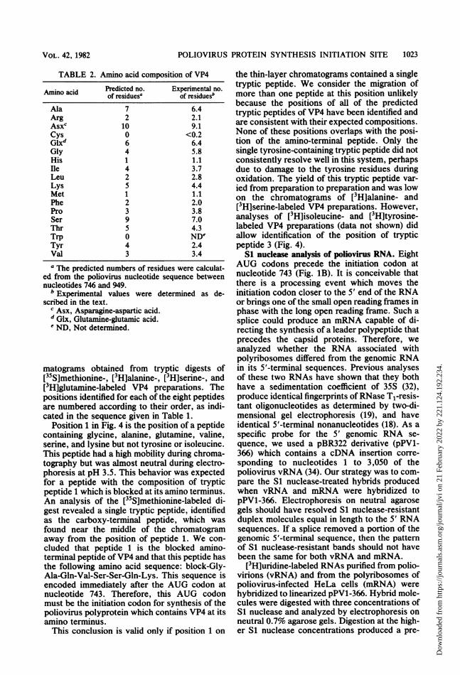
with additional tryptic peptide analyses, we de-termined the amino acid composition of VP4 andcompared it with the composition predictedfrom the genomic sequence. A previously deter-mined amino acid composition for VP4 (36)agrees only moderately well with the predictedcomposition. Our analysis (Table 2) indicatedthat VP4 should contain one methionine residue,one histidine residue, and no cysteine residue, inagreement with the RNA sequence and ourtryptic peptide analysis. The results of the aminoacid analysis for most other residues agreed withthe composition predicted from the 68-aminoacid sequence that begins with the glycine fol-lowing the methionine residue at nucleotide 743.The amount of glycine determined by our
analysis was 1.8 residues more than the predict-ed value. However, glycine is frequently foundas a contaminant in gel-purified protein and canalso be derived from nonprotein contaminantsduring acid hydrolysis. The values found foralanine, glutamine-glutamic acid, and valineagree well with the predicted values. Theseamino acids are predicted to be in the amino-terminal tryptic peptide. Serine, also predictedto be in the amino-terminal tryptic peptide, wasfound at a lower level than predicted. Serine isunstable during acid hydrolysis, and extrapola-tion of its value from the three hydrolysis timesmay not have corrected for its loss. Thus, ouramino acid analysis of VP4 supported the se-quence shown in Table 1 but was not sufficientto establish the composition of the amino-termi-nal peptide.
Two-dimensional thin-layer analysis of the ami-no terminus of VP4. To determine the composi-tion of the amino terminus of VP4, radiolabeledVP4 was digested with trypsin, and the resultingpeptide mixture was analyzed by two-dimen-sional thin-layer techniques, as described above.Table 1 shows the labeling pattern for eachpredicted tryptic peptide with the radioactiveamino acids used in this study. Our choice oflabels allowed the identification of the chro-matographic positions of all eight predicted tryp-tic peptides of VP4. Figure 4 shows the chro-
Edman degradation cycles. A total of 2,300 cpm of[3H]alanine-labeled peptide (A) and 3,800 cpm of[35S]methionine-labeled peptide (B) were applied tothe sequencer. The predicted sequence of the carboxy-terminal tryptic peptide is given at the top of the figurein the single-letter amino acid code.
J. VIROL.
Dow
nloa
ded
from
http
s://j
ourn
als.
asm
.org
/jour
nal/j
vi o
n 21
Feb
ruar
y 20
22 b
y 22
1.12
4.19
2.23
4.
POLIOVIRUS PROTEIN SYNTHESIS INITIATION SITE 1023
TABLE 2. Amino acid composition of VP4
Amino acid Predicted no. Experimental no.of residues' of residuesb
ed from the poliovirus nucleotide sequence betweennucleotides 746 and 949.
b Experimental values were determined as de-scribed in the text.
c Asx, Asparagine-aspartic acid.d Glx, Glutamine-glutamic acid.' ND, Not determined.
matograms obtained from tryptic digests of[35S]methionine-, [3H]alanine-, [3H]serine-, and[3H]glutamine-labeled VP4 preparations. Thepositions identified for each of the eight peptidesare numbered according to their order, as indi-cated in the sequence given in Table 1.
Position 1 in Fig. 4 is the position of a peptidecontaining glycine, alanine, glutamine, valine,serine, and lysine but not tyrosine or isoleucine.This peptide had a high mobility during chroma-tography but was almost neutral during electro-phoresis at pH 3.5. This behavior was expectedfor a peptide with the composition of trypticpeptide 1 which is blocked at its amino terminus.An analysis of the [35S]methionine-labeled di-gest revealed a single tryptic peptide, identifiedas the carboxy-terminal peptide, which wasfound near the middle of the chromatogramaway from the position of peptide 1. We con-cluded that peptide 1 is the blocked amino-terminal peptide ofVP4 and that this peptide hasthe following amino acid sequence: block-Gly-Ala-Gln-Val-Ser-Ser-Gln-Lys. This sequence isencoded immediately after the AUG codon atnucleotide 743. Therefore, this AUG codonmust be the initiation codon for synthesis of thepoliovirus polyprotein which contains VP4 at itsamino terminus.This conclusion is valid only if position 1 on
the thin-layer chromatograms contained a singletryptic peptide. We consider the migration ofmore than one peptide at this position unlikelybecause the positions of all of the predictedtryptic peptides of VP4 have been identified andare consistent with their expected compositions.None of these positions overlaps with the posi-tion of the amino-terminal peptide. Only thesingle tyrosine-containing tryptic peptide did notconsistently resolve well in this system, perhapsdue to damage to the tyrosine residues duringoxidation. The yield of this tryptic peptide var-ied from preparation to preparation and was lowon the chromatograms of [3H]alanine- and[3H]serine-labeled VP4 preparations. However,analyses of [3H]isoleucine- and [3H]tyrosine-labeled VP4 preparations (data not shown) didallow identification of the position of trypticpeptide 3 (Fig. 4).
Si nuclease analysis of poliovirus RNA. EightAUG codons precede the initiation codon atnucleotide 743 (Fig. 1B). It is conceivable thatthere is a processing event which moves theinitiation codon closer to the 5' end of the RNAor brings one of the small open reading frames inphase with the long open reading frame. Such asplice could produce an mRNA capable of di-recting the synthesis of a leader polypeptide thatprecedes the capsid proteins. Therefore, weanalyzed whether the RNA associated withpolyribosomes differed from the genomic RNAin its 5'-terminal sequences. Previous analysesof these two RNAs have shown that they bothhave a sedimentation coefficient of 35S (32),produce identical fingerprints of RNase Tl-resis-tant oligonucleotides as determined by two-di-mensional gel electrophoresis (19), and haveidentical 5'-terminal nonanucleotides (18). As aspecific probe for the 5' genomic RNA se-quence, we used a pBR322 derivative (pPV1-366) which contains a cDNA insertion corre-sponding to nucleotides 1 to 3,050 of thepoliovirus vRNA (34). Our strategy was to com-pare the S1 nuclease-treated hybrids producedwhen vRNA and mRNA were hybridized topPV1-366. Electrophoresis on neutral agarosegels should have resolved Si nuclease-resistantduplex molecules equal in length to the 5' RNAsequences. If a splice removed a portion of thegenomic 5'-terminal sequence, then the patternof Si nuclease-resistant bands should not havebeen the same for both vRNA and mRNA.
[3H]uridine-labeled RNAs purified from polio-virions (vRNA) and from the polyribosomes ofpoliovirus-infected HeLa cells (mRNA) werehybridized to linearized pPV1-366. Hybrid mole-cules were digested with three concentrations ofS1 nuclease and analyzed by electrophoresis onneutral 0.7% agarose gels. Digestion at the high-er Si nuclease concentrations produced a pre-
VOL. 42, 1982
Dow
nloa
ded
from
http
s://j
ourn
als.
asm
.org
/jour
nal/j
vi o
n 21
Feb
ruar
y 20
22 b
y 22
1.12
4.19
2.23
4.
1024 DORNER ET AL.
A [35s]-Met[3H]-Ala B
311%
89
2f."
4%
t
C [3H] -Ser
2
FIG. 4. Two-dimensional mapping of the tryptic peptides of VP4. Radiolabeled VP4 was digested with trypsinas described in the text, and the digestion products were spotted at the origin (arrow). First-dimension ascendingchromatography was from bottom to top. Second-dimension electrophoresis was in the horizontal direction; thecathode was at the right. The numbers to the left of the spots correspond to the peptide numbers in Table 1. (A)[3H]alanine-labeled VP4. The broken circles indicate the positions of tryptic peptides not labeled with[3H]alanine. (B) [35S]methionine-labeled VP4. (C) [3H]serine-labeled VP4. (D) [3H]glutamine-labeled VP4.
I%3
C41
ID
t
II
dominant band of approximately 3,000 basepairs when vRNA (Fig. 5, lanes 10 and 11) ormRNA (Fig. 5, lanes 7 and 8) was hybridizedwith pPV1-366. The size of this fragment isconsistent with the length of the poliovirus-specific insertion (3,050 base pairs). Minor Sinuclease-resistant bands of 2,300, 1,800, and 800base pairs were also present in these lanes.These bands may have been due to cutting atadenine-thymine-rich regions of the duplex mol-ecules at high Si nuclease concentrations, aphenomenon reported by other workers (16).
The positions of several adenine-thymine-richregions in poliovirus vRNA are consistent withthis explanation. Digestion at the lowest Sinuclease concentration (Fig. 5, lanes 6 and 9) didnot produce this pattern of minor bands, and thepredominant Si nuclease-resistant band wasslightly larger and more diffuse than that ob-served at higher Si nuclease concentrations.This pattern is consistent with less completedigestion. Hybridization of mRNA or vRNAwith pBR322 followed by Si nuclease digestiondid not produce any Si nuclease-resistant bands
J. VIROL.
[3H]-Gln
Dow
nloa
ded
from
http
s://j
ourn
als.
asm
.org
/jour
nal/j
vi o
n 21
Feb
ruar
y 20
22 b
y 22
1.12
4.19
2.23
4.
POLIOVIRUS PROTEIN SYNTHESIS INITIATION SITE
'7-23.72
- 9.46
- 6.67
e.",_zg _ ,^. ..... . i
v_
I:
:..
0 - 4.26
S - 2.25- 1.96
I - 0.59
2 3 4 5 6 7 8 9 10 11 12 13 14 15 16FIG. 5. S1 nuclease analysis of [3H]uridine-labeled poliovirus RNA hybridized to a 5'-specific DNA probe
(pPV1-366). The products of hybridization were digested with varying concentrations of S1 nuclease and wereresolved by neutral agarose gel electrophoresis as described in the text. The autoradiogram of the gel is shown.Lanes 3 and 14 contained HindIII restriction digests of 32P-labeled X DNA, which were used as chain lengthmarkers. The numbers on the right indicate the lengths (in kilobase pairs) of the X DNA fragnents. Lane 1,mRNA; lane 2, vRNA; lane 4, mRNA hybridized to pPV1-366; lane 5, mRNA subjected to hybridizationconditions and digested with 50 U of S1 nuclease per ml; lanes 6 through 8, mRNA hybridized to pPV1-366 anddigested with 50, 500, and 1,000 U of S1 nuclease per ml, respectively; lanes 9 through 11, vRNA hybridized topPV1-366 and digested with 50, 500, and 1,000 U of S1 nuclease per ml, respectively; lane 12, vRNA subjected tohybridization conditions and digested with 50 U of S1 nuclease per ml; lane 13, vRNA hybridized to pPV1-366;lane 15, mRNA hybridized to pBR322 and digested with 50 U of S1 nuclease per ml; lane 16, vRNA hybridized topBR322 and digested with 50 U of S1 nuclease per ml.
(Fig. 5, lanes 15 and 16), nor did digestion ofmRNA or vRNA alone (Fig. 5, lanes 5 and 12).Thus, our analysis detected no difference be-tween S1 nuclease-treated vRNA-DNA hybridsand Si nuclease-treated mRNA-DNA hybrids.We estimate that a size difference of 50 basepairs would have been detected by our methods.A small splice that produced a denaturation loopsubstantially resistant to Si nuclease digestionmight not have been observed. However, thispossibility seems unlikely since sufficient Sinuclease was used to cut apparently duplexedmolecules at regions rich in adenine and thy-mine. A similar Si nuclease analysis in which13H]thymidine-labeled pPV1-366 and alkalineagarose gel electrophoresis were used also de-
tected no difference between vRNA and mRNA(data not shown). Inspection of the 5'-terminal1,000 nucleotides of poliovirus vRNA failed toreveal good consensus donor or acceptor splicesites (29). We concluded that mRNA and vRNAdo not differ except for the presence or absenceof VPg linked to the 5' end.
DISCUSSIONWe identified the AUG codon at nucleotide
743, the first AUG codon in the long openreading frame of poliovirus RNA, as the initia-tion codon for the synthesis of polyproteinNCVPOO. This was accomplished by first locat-ing the encoding site for the amino terminus ofVP4, the protein encoded near the 5' end of
VOL . 42, 1982 1025
Dow
nloa
ded
from
http
s://j
ourn
als.
asm
.org
/jour
nal/j
vi o
n 21
Feb
ruar
y 20
22 b
y 22
1.12
4.19
2.23
4.
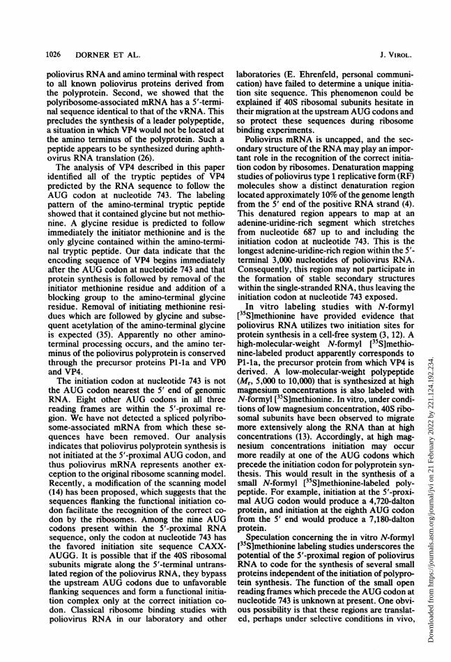
1026 DORNER ET AL.
poliovirus RNA and amino terminal with respectto all known poliovirus proteins derived fromthe polyprotein. Second, we showed that thepolyribosome-associated mRNA has a 5'-termi-nal sequence identical to that of the vRNA. Thisprecludes the synthesis of a leader polypeptide,a situation in which VP4 would not be located atthe amino terminus of the polyprotein. Such apeptide appears to be synthesized during aphth-ovirus RNA translation (26).The analysis of VP4 described in this paper
identified all of the tryptic peptides of VP4predicted by the RNA sequence to follow theAUG codon at nucleotide 743. The labelingpattern of the amino-terminal tryptic peptideshowed that it contained glycine but not methio-nine. A glycine residue is predicted to followimmediately the initiator methionine and is theonly glycine contained within the amino-termi-nal tryptic peptide. Our data indicate that theencoding sequence of VP4 begins immediatelyafter the AUG codon at nucleotide 743 and thatprotein synthesis is followed by removal of theinitiator methionine residue and addition of ablocking group to the amino-terminal glycineresidue. Removal of initiating methionine resi-dues which are followed by glycine and subse-quent acetylation of the amino-terminal glycineis expected (35). Apparently no other amino-terminal processing occurs, and the amino ter-minus of the poliovirus polyprotein is conservedthrough the precursor proteins P1-la and VP0and VP4.The initiation codon at nucleotide 743 is not
the AUG codon nearest the 5' end of genomicRNA. Eight other AUG codons in all threereading frames are within the 5'-proximal re-gion. We have not detected a spliced polyribo-some-associated mRNA from which these se-quences have been removed. Our analysisindicates that poliovirus polyprotein synthesis isnot initiated at the 5'-proximal AUG codon, andthus poliovirus mRNA represents another ex-ception to the original ribosome scanning model.Recently, a modification of the scanning model(14) has been proposed, which suggests that thesequences flanking the functional initiation co-don facilitate the recognition of the correct co-don by the ribosomes. Among the nine AUGcodons present within the 5'-proximal RNAsequence, only the codon at nucleotide 743 hasthe favored initiation site sequence CAXX-AUGG. It is possible that if the 40S ribosomalsubunits migrate along the 5'-terminal untrans-lated region of the poliovirus RNA, they bypassthe upstream AUG codons due to unfavorableflanking sequences and form a functional initia-tion complex only at the correct initiation co-don. Classical ribosome binding studies withpoliovirus RNA in our laboratory and other
laboratories (E. Ehrenfeld, personal communi-cation) have failed to determine a unique initia-tion site sequence. This phenomenon could beexplained if 40S ribosomal subunits hesitate intheir migration at the upstream AUG codons andso protect these sequences during ribosomebinding experiments.
Poliovirus mRNA is uncapped, and the sec-ondary structure of the RNA may play an impor-tant role in the recognition of the correct initia-tion codon by ribosomes. Denaturation mappingstudies of poliovirus type 1 replicative form (RF)molecules show a distinct denaturation regionlocated approximately 10% of the genome lengthfrom the 5' end of the positive RNA strand (4).This denatured region appears to map at anadenine-uridine-rich segment which stretchesfrom nucleotide 687 up to and including theinitiation codon at nucleotide 743. This is thelongest adenine-uridine-rich region within the 5'-terminal 3,000 nucleotides of poliovirus RNA.Consequently, this region may not participate inthe formation of stable secondary structureswithin the single-stranded RNA, thus leaving theinitiation codon at nucleotide 743 exposed.In vitro labeling studies with N-formyl
[35S]methionine have provided evidence thatpoliovirus RNA utilizes two initiation sites forprotein synthesis in a cell-free system (3, 12). Ahigh-molecular-weight N-formyl [35S]methio-nine-labeled product apparently corresponds toP1-la, the precursor protein from which VP4 isderived. A low-molecular-weight polypeptide(Mr, 5,000 to 10,000) that is synthesized at highmagnesium concentrations is also labeled withN-formyl [35S]methionine. In vitro, under condi-tions of low magnesium concentration, 40S ribo-somal subunits have been observed to migratemore extensively along the RNA than at highconcentrations (13). Accordingly, at high mag-nesium concentrations initiation may occurmore readily at one of the AUG codons whichprecede the initiation codon for polyprotein syn-thesis. This would result in the synthesis of asmall N-formyl [35S]methionine-labeled poly-peptide. For example, initiation at the 5'-proxi-mal AUG codon would produce a 4,720-daltonprotein, and initiation at the eighth AUG codonfrom the 5' end would produce a 7,180-daltonprotein.
Speculation concerning the in vitro N-formyl[35S]methionine labeling studies underscores thepotential of the 5'-proximal region of poliovirusRNA to code for the synthesis of several smallproteins independent of the initiation of polypro-tein synthesis. The function of the small openreading frames which precede the AUG codon atnucleotide 743 is unknown at present. One obvi-ous possibility is that these regions are translat-ed, perhaps under selective conditions in vivo,
J. VIROL.
Dow
nloa
ded
from
http
s://j
ourn
als.
asm
.org
/jour
nal/j
vi o
n 21
Feb
ruar
y 20
22 b
y 22
1.12
4.19
2.23
4.
POLIOVIRUS PROTEIN SYNTHESIS INITIATION SITE 1027
and that the polypeptides have escaped detec-tion. An intriguing idea is that interactions of the40S ribosomal subunits with the AUG codonspreceding the polyprotein initiation codon mayfunction as an attenuation mechanism to balanceprotein and RNA syntheses during the initialstages of poliovirus infection.
Alternatively, the long S'-proximal sequenceof plus-strand RNA may be essential only forRNA synthesis or for packaging of the vRNAduring morphogenesis. In this case, the protein-encoding potential of this sequence is not theprimary factor in its evolution, and the long 5'-proximal region may interfere with efficient pro-tein synthesis. Removal of these sequences maymake the poliovirus RNA a better messenger butrender it noninfectious. However, removal of 5'genomic sequences by splicing in vivo may notbe possible because poliovirus replicates in thecytoplasm and so may not have access to thenuclear splicing mechanisms. Resolution ofthese and other speculations awaits the develop-ment of defined poliovirus mutants by recombi-nant DNA methodology.
ACKNOWLEDGMENTSWe thank P. G. Rothberg and B. Semler for many helpful
discussions, M. Hardy and N. Alonzo for excellent technicalassistance, J. Demian, J. Schirmer, and A. Newlin for thepreparation of artwork, K. Thompson for assistance in com-puting data, and S. Studier for typing the manuscript. Comput-er services were provided by the Stanford MOLGEN projectof the National Institutes of Health SUMEX-AIM facility andby Brookhaven National Laboratory.This work was supported by Public Health Service grants
AI-15122 and CA-28146 from the National Institutes of Healthand by the United States Department of Energy.
LITERATURE CITED1. Baltimore, D. 1971. Expression of animal virus genomes.
Bacteriol. Rev. 35:235-241.2. Berk, A. J., and P. A. Sharp. 1978. Spliced early mRNAs
of simian virus 40. Proc. Natl. Acad. Sci. U.S.A. 75:1274-1278.
3. Celma, J. L., and E. Ehrenfeld. 1975. Translation ofpoliovirus RNA in vitro: detection of two different initia-tion sites. J. Mol. Biol. 98:761-780.
4. Cumakov, 1. M., G. Y. Lipskaya, and V. I. Agol. 1979.Comparative studies on the genomes of some picornavi-ruses: denaturation mapping of replicative form RNA andelectron microscopy of heteroduplex RNA. Virology92:259-270.
5. Darnell, J. E., and L. Levintow. 1960. Poliovirus protein:source of amino acids and time course of synthesis. J.Biol. Chem. 235:74-77.
5a.Eminli, E. A., M. Elzinga, and E. Whmmer. 1982. Carboxy-terminal analysis of poliovirus proteins: the termination ofpoliovirus RNA translation and the location of uniquepoliovirus polyprotein cleavage sites. J. Virol. 42:194-199.
6. Fernandez-Munoz, R., and J. E. Darneil. 1976. Structuraldifference between the 5' termini of viral and cellularmRNA in poliovirus-infected cells: possible basis for theinhibition of host protein synthesis. J. Virol. 18:719-726.
7. Henderson, L. E., S. Oroszlan, and W. Konlpberg. 1979.A micromethod for complete removal of dodecyl sulfatefrom proteins by ion-pair extraction. Anal. Biochem.
93:153-157.8. Hewlett, M. J., J. K. Rose, and D. Baltimore. 1976. 5'-
Terminal structure of poliovirus polyribosomal RNA ispUp. Proc. NatI. Acad. Sci. U.S.A. 73:327-330.
9. Holland, J. J., and E. D. Klehn. 1968. Specific cleavage ofviral proteins as steps in the synthesis and maturation ofenteroviruses. Proc. Natl. Acad. Sci. U.S.A. 60:1015-1022.
10. Jacobson, M. F., and D. BaltImore. 1968. Polypeptidecleavages in the formation of poliovirus proteins. Proc.Natl. Acad. Sci. U.S.A. 61:77-84.
11. Kitamura, N., B. L. Semler, P. G. Rothberg, G. R. Larsen,C. J. Adler, A. J. Dorner, E. A. Emini, R. Hanecak, J. J.Lee, S. van der Werf, C. W. Anderson, and E. Wimmer.1981. Primary structure, gene organization, and polypep-tide expression of poliovirus RNA. Nature (London)291:547-553.
12. Knauert, F., and E. Ehrenfeld. 1979. Translation of polio-virus RNA in vitro: studies on n-formylmethionine-la-beled polypeptides initiated in cell-free extracts preparedfrom poliovirus infected HeLa cells. Virology 93:537-546.
13. Kozak, M. 1979. Migration of 40S ribosomal subunits on amessenger RNA when initiation is perturbed by loweringmagnesium or adding drugs. J. Biol. Chem. 254:4731-4738.
14. Kozak, M. 1981. Possible role of flanking nucleotides inrecognition of the AUG initiator codon by eukaryoticribosomes. Nucleic Acids Res. 9:5233-5252.
14a.Larsen, G. R., C. W. Anderson, A. J. Dorner, B. L.Semler, and E. Wimmer. 1982. Cleavage sites within thepoliovirus capsid protein precursors. J. Virol. 41:340-344.
15. Larsen, G. R., B. L. Semler, and E. Wimmer. 1981. Stablehairpin structure within the 5'-terminal 85 nucleotides ofpoliovirus RNA. J. Virol. 37:328-335.
16. Mller, K. G., and B. Sollner-Webb. 1981. Transcription ofmouse rRNA genes by RNA polymerase 1: in vitro and invivo initiation and processing sites. Cell 27:165-174.
17. Nomoto, A., B. Detjen, R. Pozzattl, and E. Wimmer. 1977.The location of the polio genome protein in viral RNAsand its implication for RNA synthesis. Nature (London)268:208-213.
18. Nomoto, A., N. Kitamura, F. Golini, and E. Wimmer.1977. The 5'-terminal structures of poliovirion RNA andpoliovirus mRNA differ only in the genome-linked proteinVPg. Proc. Natl. Acad. Sci. U.S.A. 74:5345-5349.
19. Nomoto, A., Y. F. Lee, and E. Wimmer. 1976. The 5' endof poliovirus mRNA is not capped with m7G(5')ppp(5')Np. Proc. Natl. Acad. Sci. U.S.A. 73:375-380.
20. Palmiter, R. D. 1977. Prevention of NH2-terminal acetyla-tion of proteins synthesized in cell-free systems. J. Biol.Chem. 252:8781-8783.
21. Penman, S., K. Scherrer, Y. Becker, and J. E. DarneU.1963. Polyribosomes in normal and poliovirus-infectedHeLa cells and their relationship to messenger RNA.Proc. Natl. Acad. Sci. U.S.A. 49:654-661.
22. RacanleUo, V. R., and D. Baltimore. 1981. Molecularcloning of poliovirus cDNA and determination of thecomplete nucleotide sequence of the viral genome. Proc.Natl. Acad. Sci. U.S.A. 78:4887-4891.
23. Rekosh, D. 1972. Gene order of the poliovirus capsidproteins. J. Virol. 9:479-487.
24. Rueckert, R. R. 1976. On the structure and morphogenesisof picornaviruses, p. 131-213. In H. Fraenkel-Conrat andR. R. Wagner (ed.), Comprehensive virology, vol. 6.Plenum Press, New York.
25. Saborio, J. L., S. S. Pong, and G. Koch. 1974. Selectiveand reversible inhibition of initiation of protein synthesisin mammalian cells. J. Mol. Biol. 85:195-211.
26. Sanar, D. V., D. N. Black, D. J. Rowlands, T. J. R.Harris, and F. Brown. 1980. Location of the initiation sitefor protein synthesis on foot-and-mouth disease virusRNA by in vitro translation of defined fragments of RNA.J. Virol. 33:59-68.
27. Semler, B. L., C. W. Anderson, N. Kitamura, P. G.Rothberg, W. L. Wishart, and E. Wimmer. 1981. Poliovi-
VOL . 42, 1982
Dow
nloa
ded
from
http
s://j
ourn
als.
asm
.org
/jour
nal/j
vi o
n 21
Feb
ruar
y 20
22 b
y 22
1.12
4.19
2.23
4.
1028 DORNER ET AL.
rus replication proteins: RNA sequence encoding P3-lband the sites of proteolytic processing. Proc. Natl. Acad.Sci. U.S.A. 78:3464-3468.
28. Semler, B. L., R. Hanecak, C. W. Anderson, and E.Wimmer. 1981. Cleavage sites in the polypeptide precur-sors of poliovirus protein P2-X. Virology 114:589-594.
29. Sharp, P. 1981. Speculations on RNA splicing. Cell23:643-646.
30. Summers, D. F., and J. V. Maizel. 1968. Evidence forlarge precursor proteins in poliovirus synthesis. Proc.Natl. Acad. Sci. U.S.A. 59:966-971.
31. Summers, D. F., and J. V. Maizel. 1971. Determination ofthe gene sequence of poliovirus with pactamycin. Proc.Natl. Acad. Sci. U.S.A. 68:2852-2856.
32. Summers, D. F., J. V. Maizel, and J. E. Darnell. 1967. Thedecrease in size and synthetic activity of poliovirus poly-
somes late in the infectious cycle. Virology 31:427-435.33. Taber, R., D. Rekosh, and D. Baltimore. 1971. Effect of
pactamycin on synthesis of poliovirus proteins: a methodfor genetic mapping. J. Virol. 8:395 401.
34. van der Werf, S., F. Bregegere, H. Kopecka, N. Kitamura,P. G. Rothberg, P. Kourilsky, E. Wimmer, and M. Girard.1981. Molecular cloning of the genome of poliovirus type1. Proc. Natl. Acad. Sci. U.S.A. 78:5983-5987.
35. Wold, F. 1981. In vivo chemical modification of proteins.Annu. Rev. Biochem. 50:783-814.
36. Wouters, M., and J. Vandekerckhove. 1976. Amino acidcomposition of the poliovirus capsid polypeptides isolatedas fluorescamine conjugates. J. Gen. Virol. 33:529-533.
37. Yogo, Y., and E. Wimmer. 1972. Polyadenylic acid at the3'-terminus of poliovirus RNA. Proc. Natl. Acad. Sci.U.S.A. 69:1877-1882.