1Department of Neurology, Second Affiliated Hospital, Zhejiang University School of Medicine, Hangzhou, China; 2Department ofNeurology, Zhejiang Hospital, Hangzhou, China; 3Department of Neurobiology, Institute of Neuroscience, Zhejiang UniversitySchool of Medicine, Hangzhou, China
Purpose: FERM domain containing protein 7 (FRMD7) mutations are associated with X-linked idiopathic congenitalnystagmus (ICN). The purpose of this study is to identify a novel splice variant of FRMD7 (FRMD7-S) in both humansand mice with a shortened exon 4 relative to the original form of FRMD7 (FRMD7-FL),and to detect the role of FRMD7-FL and FRMD7-S in the process of neuronal development.Methods: The splice variant of FRMD7 was identified by PCR. Expression levels of hFRMD7-FL and hFRMD7-Stranscripts in developing human fetal brain were tested by RT–PCR, and expression levels in the human pluripotentembryonic carcinoma NTera 2/cl.D1 (NTERA-2; NT2) cell line with all-trans retinoic acid (ATRA) or bonemorphogenetic protein-2 (BMP-2) treatment were tested by real-time qPCR. hemaglutinin (HA)-tagged recombinantplasmids DNA encoding hFRMD7-FL and Myc-tagged recombinant plasmids DNA encoding hFRMD7-S were used totransiently transfect the human NT2 cells. Further, immunofluorescence experiments were performed to determine theco-localization of the two fusion proteins. Finally, using co-immunoprecipitation analyses, we demonstrated that FRMD7-FL and FRMD7-S interacted with each other.Results: A novel splice variant of FRMD7 (FRMD7-S) with a shortened exon 4 relative to the original form of FRMD7(FRMD7-FL) was identified from the cDNA of the human NT2 cell line and mouse fetal brain. The FRMD7 transcriptsshowed similar tissue distributions and were upregulated following all trans retinoic acid (ATRA)-induced differentiationof NT2 cells. FRMD7-FL and FRMD7-S co-localized and co-immunoprecipitated with each other. Further,overexpression of FRMD7-FL in NT2 cells resulted in altered neurite development and upregulation of FRMD7-S.Conclusions: Although the significance of the 45 bp deletion remains unknown, our observations suggest that theFRMD7 isoforms may play a significant role during neuronal differentiation and development.
Idiopathic congenital nystagmus (ICN) is an oculomotordisorder characterized by involuntary horizontal oscillationsof the eyes that presents at birth or appears in the first monthsof life, but does not usually worsen over time. ICN is distinctfrom other ocular disorders in which nystagmus is acquiredlater in life (e.g., cataracts, glaucoma, albinism) or isaccompanied by eye, brain, or other health abnormalities [1].The prevalence of ICN is estimated to be 24 per 10,000 [2],and although some techniques can improve vision (e.g.,glasses, contact lenses, eye muscle surgery), nystagmus isusually permanent and cannot be corrected or cured [3].
Previous studies have speculated that ICN represents aprimary defect in the brain regions involved in ocular motorcontrol [4], although the precise pathogenic mechanismsunderlying ICN are currently unknown. Mutations in the
Correspondence to: Baorong Zhang, M.D., Department ofNeurology, Second Affiliated Hospital, College of Medicine,Zhejiang University, Hangzhou 310009, People’s Republic of China;Phone: +86-571-87784752; FAX: +86-571-87784750; email:[email protected]
human FERM domain containing protein 7 (FRMD7) gene(NM_194277), which encodes the FERM domain containingprotein 7 that is a member of the FERM family, are associatedwith X-linked ICN [5]. Approximately 50% of X-linkedpedigrees and 5% of sporadic ICN cases have been linked toFRMD7 mutations, and more than 35 FRMD7 mutations havebeen reported worldwide in families with X-linked ICN fromvarious ethnic backgrounds [1,6,7].
The 298 amino acid FERM domain was originallyidentified in protein 4.1 [8], and subsequent studies reportedthe structural, transport, and membrane-localizing functionsof this domain [9-11]. Notably, the hFRMD7-FL protein ishighly homologous to FARP1 (FERM, RhoGEF, andpleckstrin domain protein 1) and FARP2 proteins, which areknown to play significant roles in neuronal development.FARP1 is necessary and sufficient for promoting lateral motorcolumn dendritic growth, and FARP2 is a key moleculeinvolved in the response of neuronal growth cones to class-3semaphorins [12,13]. Moreover, recent studies havedemonstrated that inhibition of FRMD7 expression in mouse
Molecular Vision 2011; 17:2986-2996 <http://www.molvis.org/molvis/v17/a322>Received 30 April 2011 | Accepted 8 November 2011 | Published 17 November 2011
neuroblastoma cell line (NEURO2A) during neuronaldifferentiation is associated with significant delays in neuritegrowth and disrupted F-actin/G-actin dynamics [14]. In amouse model, FRMD7 expression levels were low in all adulttissue samples, whereas FRMD7 expression levels werehigher in embryos and underwent a sharp increase atembryonic day 18 in brain tissue [15]. These findings provideevidence that FRMD7 plays a critical role during neuronalmorphogenesis, in synapse function, and in neurite growth,but further studies will be required to uncover the precisemechanisms associated with FRMD7 function.
The NT2 cell line, which is a human embryoniccarcinoma cell line, differentiates into post-mitotic neuron-like cells following treatment with all trans retinoic acid(ATRA) [16,17] and into non-neural epithelial cell lineagesfollowing exposure to bone morphogenetic protein 2 (BMP-2)[18,19]. Therefore, NT2 cells provide an ideal model systemthat mimics normal neuronal differentiation in the brainaccording to many established criteria.
Previous studies have revealed that alternative splicingoccurs much more frequently in the brain relative to othertissues [20,21], and alternative splicing is increasinglyrecognized for its role in neurologic disease [22,23]. In thecurrent study, we have identified and characterized a novelFRMD7 splice variant in humans and mice, hFRMD7-S(GenBank FJ717411) and mFRMD7-S, respectively; thesplice variant contains a 45 bp truncation in the fourth exonand produces an altered form of the FERM domain. Herein,we investigate the expression patterns, role in neuritedevelopment, subcellular localization, and interactionsassociated with the FRMD7-S splice variant relative to theoriginal FRMD7-FL isoform in NT2 cells.
METHODSCell culture and ATRA/BMP-2-induced differentiation:Human NT2 cells (Cell Culture Center of Peking UnionMedical College, Peking, China) were maintained in DMEM/F12 medium supplemented with 10% fetal bovine serum, 100U/ml penicillin, and 100 μg/ml streptomycin. Prior to theinduction of differentiation, the NT2 cells were incubated in25 cm2 cell culture flasks in 4 ml of medium in a 37 °C, highhumidity, 5% CO2 incubator. The cells were trypsinized andsub-cultured at a ratio of 1:3 twice weekly. For the time courseanalyses, the NT2 cells were seeded on a 3.0×10⁶/10 cm2 plateand treated the following day with 10 μM all-trans retinoicacid (ATRA) dissolved in DMSO [16,17] or with 50 ng/mlbone morphogenetic protein-2 (BMP-2) dissolved in normalsaline [18,19]. The cells were collected at 12, 24, 36, 48, and72 h after treatment for the early time points. For the later timepoints (5, 8, and 12 days), the cells were reseeded every threedays in the continued presence of ATRA or BMP-2 andcollected on the corresponding days.Collection of human and mouse fetal tissue: Freshly isolated16-week post-conception (wpc) aborted human fetal tissues
were collected from the Women's Hospital School OfMedicine Zhejiang University. The experimental procedureswere approved by the Human Ethical Committee of theZhejiang University. Mouse fetal brain tissue was isolatedfrom 18-day mouse fetuses. A scissors was used to produce1–3 mm3 pieces of tissue, which were snap frozen in cryogenicvials in liquid nitrogen and stored at −80 °C. Total RNA fromeach sample was prepared for subsequent RT–PCRexperiments.
RNA isolation, reverse transcription PCR, and quantitativereal-time PCR analysis: Total RNA was isolated from humanand mouse fetal tissues and NT2 cells using TRIZOL reagent(Invitrogen, San Diego, CA), according to the manufacturer’sinstructions. Real-time quantitative PCR (RT-qPCR) assayswere performed using a Light CyclerTM real-time PCRthermocycler (Roche, Shanghai, China). Briefly, 1.5 μg oftotal RNA was reverse transcribed using oligo (dT) 12–18with M-MLV Reverse Transcriptase (Promega, Madison, WI)according to the manufacturer's instructions. Relative levelsof gene expression were analyzed using the SYBR® PremixEx Taq™ (TaKaRa, Dalian, China) using the recommendedPCR conditions. The sequences of the RT-qPCR primers areshown in Table 1. The relative mRNA levels of target geneswere calculated relative to glyceraldehyde-3-phosphatedehydrogenase (GAPDH) using the (=2-△△C
T) method [24].
cDNA clone and plasmid construction: Human FRMD7-FLand FRMD7-S genes were isolated from NT2 cell cDNA(synthesized from the total RNA of natural NT2 cells) by PCRamplification using the following primers: 5′-ATG CTA CATTTA AAA GTG CAG TTT-3′ and 5′-TTA AGC TAA AAAGTA ATT ACA TGG T-3′. The mouse FRMD7-FL andFRMD7-S genes were cloned from 18-day fetal mouse braincDNA using the following primers: 5′-GTA TGG ATC CATGCT CCA TTT AAA AGT G-3′ and 5′-AGC TAA GAA ATAATT GCA TGG CTT TAG C-3′. PCR was performed in25 μl of a reaction mixture containing DNA polymerase fromThermococcus kodakaraensis (KOD) DNA polymerasebuffer and 1.25 U KOD DNA polymerase (Toyobo, Osaka,Japan). Subsequent cloning of PCR products into the pEASY-Blunt vector (TransGen Biotech, Beijing, China) wasperformed using the pEASY-Blunt Cloning Kit forSequencing (TransGen Biotech). Sequence analyses wereperformed by the Invitrogen Company (Shanghai, China). Togenerate eukaryotic expression vectors for hFRMD7 splicevariants, cloned RT–PCR fragments in pEASY-Blunt wereused as the template. The hFRMD7-FL cDNA was sub-clonedinto pcDNA3.1(+) in frame with an NH2-terminal HA tag(hFRMD7-FL-HA) and pEGFP-N1 (pEGFP-hFRMD7-FL)with the pAf/pAr and pCf/pCr primer sets, respectively (Table1). The hFRMD7-S gene was subcloned into pcDNA3.1(+)with an NH2-terminal Myc tag (hFRMD7-S-Myc) andplasmid pDsRed-N1 (pDsRed-hFRMD7-S) using the pBf/pBr and pDf/pDr primer sets, respectively (Table 1). NheI and
XhoI were chosen as the cloning sites for all three vectors, andthe sequences of all of the expression cassettes were verified.
Cell transfection and immunofluorescence microscopy: Fortransient transfection experiments, the NT2 cells were placedon glass coverslips or cell culture flasks until they reachedapproximately 60% confluency and were transfected withFuGENE HD transfection reagent (Roche) at a ratio of 1 μgof DNA to 3 μl of FuGENE HD per coverslip. The cells wereprocessed for immunofluorescence at 48 h post-transfection.For stable overexpression experiments, G418 (1.2 μg/ mlmedium; Invitrogen) antibiotic selection was performed for 4weeks. For the immunofluorescence experiments, the induced
or transfected NT2 cells were washed with phosphate-buffered saline (PBS) and fixed for 15 min at roomtemperature in 4% paraformaldehyde (PFA) in PBS. Thecoverslips were washed twice with ice-cold PBS, and the cellswere permeabilized with PBS containing 0.2% Triton X-100for 10 min at 4 °C. After washing the cells in PBS three timesfor 5 min (each wash), a blocking solution containing PBS and10% normal goat serum was applied for 1 h at roomtemperature. Afterwards, primary antibodies (monoclonalanti-Myc or anti-HA or antibodies; Abmart Inc., Shanghai,China) diluted in a blocking solution were applied overnightat 4 °C or for 1 h at room temperature. In the double-labeling
Figure 1. Cloning of a novel FRMD7 isoform and tissue distribution of the two splice variants. A: Components of the FERM domain and genestructure of hFRMD7-FL. B and C: Sequence comparison between hFRMD7-FL/mFRMD7-FL (B) and hFRMD7-S/mFRMD7-S (C) showingthe deletion of 45 bp in the 5′ end of exon 4 in the hFRMD7-S/mFRMD7-S. D: Agarose gel electrophoresis of the RT–PCR products to confirmtheir size and the identification of a single PCR product. E: Expression levels of hFRMD7-FL and hFRMD7-S transcripts in selected humanfetal tissues. The primer sets used in D and E: p1f/p1r, p2f/p2r and p3f/p3r.
experiments, the cultures were incubated simultaneously withtwo primary antibodies. After three washes in PBS, thesecondary antibodies (Dylight 488, Dylight 594; JacksonImmunoResearch Laboratories, Inc. West Grove, PA) werediluted 1:250 in a blocking solution and added for 1 h at roomtemperature in the dark. The secondary antibody solution wasdecanted and washed three times with PBS for 5 min each inthe dark. Finally, the cells were incubated for 5 min with0.1 μg/ml DAPI as a nuclear counterstain. After three washes,the coverslips were mounted with a drop of AntifadeMounting Medium (Beyotime Institute of Biotechnology,Suzhou, China) and stored in the dark at −20 °C or 4 °C. Thecells were viewed using a Nikon eclipse inverted fluorescencemicroscope equipped with a Nikon digital camera (NikonCorporation, Tokyo, Japan). Image overlays and contrastenhancement were performed using Adobe Photoshop CS2(Adobe Systems Inc. San Jose, CA) software.Co-immunoprecipitation and immunoblot analyses: For co-immunoprecipitation (co-IP) experiments, NT2 cells weretransiently co-transfected with 2 μg of each of the hFRMD7-FL-HA and hFRMD7-S-Myc vectors. At 48 h post-transfection, the total protein been extracted from the cells byusing M-PER® Mammalian Protein Extraction Reagent(Thermo Fisher Scientific Inc., Rockford, IL) following themanufacturer's instructions. Then the ProFound™ Co-Immunoprecipitation Kit (Thermo Fisher Scientific Inc.) wasused for co-IP assays according to the manufacturer'sinstructions. Control immunoprecipitations were performedin the presence of non-specific immunoglobulins.
Statistical analyses: All experiments were done in triplicate,and the data are represented as mean±SEM. Statisticalsignificance between groups was determined using theKolmogorov–Smirnov test and Student’s t-test, asimplemented by SPSS13.0 (SPSS Inc., Chicago, IL). Resultswere considered to be statistically significant when p<0.05.
RESULTSIdentification of a novel FRMD7 splice variant and tissuedistribution of two FRMD7 transcripts: In RT–PCRexperiments to clone the hFRMD7-FL gene from NT2 cells,two PCR products were produced. Cloning and sequenceanalysis of the PCR products indicated that they correspondedto two distinct FRMD7 mRNAs: the expected originalhFRMD7-FL mRNA and another unexpected mRNA notdescribed previously, which is referred to herein as hFRMD7-S. This novel transcript, which lacks a 45 bp segment in the 5′region of exon 4, results in the fusion of the 3′ end of exon 3to the 5′ end of truncated exon 4; therefore, hFRMD7-Sencodes a putative protein of 699 amino acids (Figure 1A,B).We then performed RT–PCR to confirm the presence ofmFRMD7-S in 18-day-old fetal mice (Figure 1C). Theexpression of hFRMD7-FL and hFRMD7-S transcripts wasexamined in tissues from 16-wpc human fetal tissue usingsequence-specific primers (Figure 1D). We detected highexpression of hFRMD7-FL in the cerebellum and moderateexpression in the cerebral cortex, optic nerve, kidney, testis,and gastric wall. We detected high expression of hFRMD7-Sin the cerebellum and moderate expression in the kidney,testis, and gastric wall. However, hFRMD7-S was not
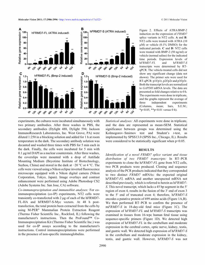
Figure 2. Effects of ATRA/BMP-2induction on the expression of FRMD7splice variants in NT2 cells. A and B:NT2 cells were treated with ATRA (10µM) or vehicle (0.1% DMSO) for theindicated periods. C and D: NT2 cellswere treated with BMP-2 (50 ng/ml) orvehicle (normal saline) for the indicatedtime periods. Expression levels ofhFRMD7-FL and hFRMD7-Stranscripts were determined by RT-qPCR. The vehicle-treated cells did notshow any significant change (data notshown). The primer sets were used forRT-qPCR: p1f/p1r, p2f/p2r and p3f/p3r.Both the transcript levels are normalizedto GAPDH mRNA levels. The data arepresented as fold changes relative to 0 h.The experiments were done in triplicate,and the graphs represent the average ofthree independent experiments(Columns, mean; bars, S.E.M.;*p<0.05, **p<0.01 versus 0 h).
expressed in the optic nerve or cerebral cortex. NeitherhFRMD7-FL nor hFRMD7-S were expressed in the medullaoblongata, pons, or liver. Taken together, these resultsrevealed that hFRMD7-S exhibited a similar but limited tissuedistribution compared to hFRMD7-FL (Figure 1E).
Levels of both hFRMD7 transcripts significantly increased inATRA-induced differentiating NT2 cells: As shown in Figure2A,B, the levels of hFRMD7-FL and hFRMD7-S mRNAexpression in NT2 cells started to increase at 12 h after ATRAtreatment and reached a maximal increase at 8 days (81 fold
Figure 3. Effects of hFRMD7-FL or hFRMD7-S overexpression on neurite outgrowth. NT2 cells stably transfected with hFRMD7-FL-HA(A), hFRMD7-S-Myc (B), or the empty pcDNA3.1(+) vector (C) were treated with ATRA for 5 days. The cells were fixed and immunostainedwith monoclonal anti-Myc or anti-HA antibodies and a DyLight-488 conjugated secondary antibody. Class III β-tubulin was immunostainedusing a monoclonal TuJ1 antibody and DyLight-594 secondary antibody. Magnification: 200×.
TABLE 2. NEURITE OUTGROWTH OF NT2 CELLS TREATED WITH 10 μM RETINOIC ACID.
Norm Control hFRMD7-S hFRMD7-FLPercentage of cells with neurites* 46.2±2.1 47.5±1.3 (ns) 77.8±1.6Average neurite length** (μm) 42.7±2.6 43.6±2.1 (ns) 72.3±1.1
Cells were treated with 10 μm retinoic acid, 10% FCS/DMEM-F12 for 5 days, and neurite parameters were quantified. Data are mean±SEM of three independent experiments. At least 250 cells per transfection were counted. *Cells that possess at least one outgrowth of more than one-half the cell body diameter in length. **Neurite length from cell body to distal tip. (ns) stands for Not Significant.
for hFRMD7-FL and 96 fold for hFRMD7-S relative to the 0h time point; Figure 2A,B). The control group did not exhibitany change relative to the 0 h time point. Compared to ATRA,BMP-2 had a repressive effect on hFRMD7-FL and hFRMD7-S expression. After 24 h of BMP-2 treatment, the levels ofhFRMD7-FL and hFRMD7-S transcripts started to declinerelative to the 0 h time point, and continued to decrease after36 h; after 8 days, the expression levels of hFRMD7-FL andhFRMD7-S transcripts began to increase (Figure 2C,D).hFRMD7-FL and hFRMD7-S co-localized with class III β-tubulin and exhibited differential effects on neuritedevelopment: Stable NT2 cell lines overexpressing hFRMD7-
FL-HA or hFRMD7-S-Myc were established for use inexperiments to study their role during differentiation. Weexamined the co-localization of class III β-tubulin, which isnot expressed at detectable levels in neural progenitor cells,with the two FRMD7 isoforms by indirectimmunofluorescence. Both isoforms exhibited strong co-localization with neuronal class III β-tubulin in differentiatingNT2 cells induced with ATRA for 5 days (Figure 3A,B).Having established that the hFRMD7-FL or hFRMD7-Soverexpressed cell lines, we next sought to find how thischange would affect neurite development in differentiatedNT2 cells. Notably, following ATRA treatment for 5 days, a
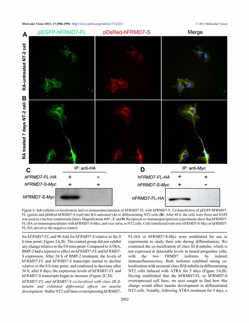
Figure 4. Sub-cellular co-localization and co-immunoprecipitation of hFRMD7-FL with hFRMD7-S. Co-transfection of pEGFP-hFRMD7-FL (green) and pDsRed-hFRMD7-S (red) into RA-untreated (A) or differentiating NT2 cells (B). After 48 h, the cells were fixed and DAPIwas used as a nuclear counterstain (blue). Magnification 400×. C and D: Reciprocal co-immunoprecipitation experiments show that hFRMD7-FL-HA co-immunoprecipitates with hFRMD7-S-Myc, and vice versa, in NT2 cells. Cells transfected with only hFRMD7-S-Myc or hFRMD7-FL-HA served as the negative control.
significant increase in the length and number of neurites wasobserved in NT2 cells stably overexpressing hFRMD7-FL-HA NT2 (Figure 3A,C and Table 2; p<0.05). However, thedegree of neurite branching did not differ between the controland hFRMD7-S overexpressing NT2 cells (Figure 3B,C andTable 2; p>0.05).hFRMD7-FL co-localizes and interacts with hFRMD7-S inNT2 cells: To investigate the subcellular co-localization ofFRMD7 isoforms and to differentiate between the localizationof endogenous FRMD7 and exogenously expressed FRMD7proteins, we used expression vectors to express hFRMD7-FLor hFRMD7-S differentially tagged with green (EGFP) or red(DsRed) fluorescent protein, respectively. Following transientco-transfection with the fusion constructs encoding pEGFP-hFRMD7-FL and pDsRed-hFRMD7-S, untreated NT2 cells(without ATRA) and NT2 cells treated with ATRA for 7 dayswere subjected to fluorescence microscopy. In theseexperiments, EGFP-hFRMD7-FL and DsRed-hFRMD7-Sexhibited a high degree of co-localization (Figure 4); bothisoforms were primarily localized in cytoplasm andperinuclear regions in undifferentiated NT2 cells (Figure 4A).Similarly, in NT2 cells induced with ATRA for 7 days, EGFP-hFRMD7-FL and DsRed-hFRMD7-S co-localizedextensively in the cytoplasm, perinuclear regions, and neuriteprocesses. Notably, the distribution of EGFP-hFRMD7-FLwas more abundant in the distal terminals of the neuritesrelative to that of DsRed-hFRMD7-S (Figure 4B).
To test for protein–protein interactions, we performed co-IP experiments. We co-expressed HA and Myc NH2-terminally tagged versions of hFRMD7-FL (hFRMD7-FL-HA) and hFRMD7-S (hFRMD7-S-Myc), respectively, inNT2 cells. At 48 h post-transfection, total cell lysates were
extracted, and hFRMD7-FL-HA was immunoprecipitatedusing anti-HA antibodies, followed by immunoblot analysiswith anti-Myc antibodies. The results showed that hFRMD7-FL-HA co-immunoprecipitated with hFRMD7-S-Myc(Figure 4C). This finding was confirmed by reciprocalimmunoprecipitation experiments, which showed thathFRMD7-S-Myc co-immunoprecipitated with hFRMD7-FL-HA (Figure 4D). The results of these experiments may suggestthat hFRMD7-FL and hFRMD7-S either interact directly orare associated with the same protein complex in NT2 cells.hFRMD7-FL protein stimulates transcription of hFRMD7-S:Alternative splicing often impacts the transcription andsubsequent translation of the major protein. Therefore, weperformed experiments to determine whether overexpressionof one FRMD7 splice variant was associated with alteredtranscription of the other splice variant. Overexpression ofhFRMD7-FL or hFRMD7-S was confirmed by RT–PCR andimmunoblot analyses (data not shown). We found that thetranscription levels of hFRMD7-S exhibited a time-dependentincrease in cells overexpressing hFRMD7-FL relative tocontrol cells, with an average of 4.8 fold higher hFRMD7-Sexpression levels at 72 h (Figure 5A); in contrast, hFRMD7-FL transcription levels did not change significantly in NT2cells overexpressing hFRMD7-S (Figure 5B).
DISCUSSIONThe human FRMD7 gene spans a region of 2,145 bp andcomprises 12 exons. Until now, no FRMD7 mRNA variantshave been described. Herein, we report the discovery of anovel transcript, hFRMD7-S, from the human FRMD7 genegenerated by alternative splicing. An increased understandingof the hFRMD7-S variant relative to the original hFRMD7-
Figure 5. Overexpression of hFRMD7-FL stimulates hFRMD7-S transcription.Expression levels of hFRMD7-FL orhFRMD7-S transcripts were monitoredby RT-qPCR in transfected NT2 cells atthe indicated time points. Cellstransfected with the empty vector servedas the negative control (MOCK group).A: Expression levels of hFRMD7-S inNT2 cells overexpressing hFRMD7-FL. B: Expression levels of hFRMD7-FL in NT2 cells overexpressinghFRMD7-S. The primer sets were usedfor RT-qPCR: p1f/p1r, p2f/p2r and p3f/p3r. Both the transcript levels arenormalized to GAPDH mRNA levels.The data are presented as the foldchanges relative to MOCK group. All ofthe experiments were done in triplicate,and the graph represents the average(columns, mean; bars, S.E.M.; *p<0.05,**p<0.01 versus MOCK group).
FL transcript may help clarify the role of FRMD7 splicevariants in neuronal development and the pathogenesis ofICN.
We found that the hFRMD7-FL and hFRMD7-Stranscripts exhibit a similar tissue distribution, with highestlevels in the cerebellum; neither hFRMD7-FL nor hFRMD7-S were expressed in the medulla oblongata or pons. Theseresults corroborate previous studies in mouse models [15].Further, our previous studies demonstrated that hFRMD7-FL expression is ubiquitous during fetal brain development,but is restricted to regions of eye movement in adults [25]. Wemay suppose that the FRMD7 transcripts may carry outcerebellum-associated functions (e.g., vestibulo-cerebellarsystem regulation of normal eye movement). Nevertheless, inthe previous work, using immunohistochemistry testing,FRMD7-FL was observed to be highly expressed in the ponsand medulla oblongata. Since the size differences in thecorresponding mRNAs and protein isoforms are very small(i.e., 2,145 bp for hFRMD7-FL versus 2,100 bp for hFRMD7-S), neither northern blot nor immunoblot analyses wereperformed to determine endogenous hFRMD7-FL andhFRMD7-S protein expression. The reason for the differencesof expression distribution between RNA level and proteinlevel of FRMD7-FL are worth exploring in future studies.
Recent studies suggest that ATRA-induceddifferentiation is followed by the upregulation of hFRMD7-FL mRNA and protein expression levels in NEURO2A cells[14]. In the current study, we found that ATRA-inducedneuronal differentiation of NT2 cells resulted in elevatedexpression of both hFRMD7-FL and hFRMD7-S transcripts,whereas BMP-2 treatment of NT2 cells had a repressive effecton hFRMD7-FL and hFRMD7-S expression. Further, a recentstudy demonstrated that downregulation of hFRMD7-FL inNEURO2A cells resulted in altered neurite development[14]. In the current study, we found that hFRMD7-FLoverexpression leads to a significant increase in neurite lengthin differentiating NT2 cells; however, hFRMD7-Soverexpression had no effect on this process.
NT2 cells undergo programmed terminal differentiationalong a neuronal pathway, resulting in the accumulation ofneuronal cells with neurite outgrowths and extendedprocesses within a period of 8 days. Thus, we speculate thatboth hFRMD7-FL and hFRMD7-S are involved in the processof neurite outgrowth extension. Since hFRMD7-FL andhFRMD7-S share highly similar sequences, their differenteffect on ATRA-induced NT2 differentiation is of particularinterest. Although it is unclear what underlies thesedifferences, our findings suggest that the FRMD7 isoforms areinvolved in the formation and growth of neurites duringneuronal differentiation, and also provide evidence that thehFRMD7-S isoform may play a minor role during this process.
A previous study showed that FARP1 and FARP2, whichshare homology with FRMD7, are involved in signal
transduction between the plasma membrane and cytoskeleton,and that the hFRMD7-FL protein co-localizes with actin inNEURO2A cells [12-14,26,27]. In the current study, we foundthat hFRMD7-FL and hFRMD7-S splice variants also exhibitstrong co-localization with class III β-tubulin, which isconsidered to be one of the earliest neuron-associatedcytoskeletal marker proteins during mammalian development[28,29]. These results give evidence that the FRMD7 isoformscarry out functions associated with microtubules andcytoskeleton-associated proteins or at least play a role in arelated molecular pathway.
We found that the hFRMD7-FL isoform co-localized toa high extent with the hFRMD7-S isoform in undifferentiatedand differentiating NT2 cells, although the hFRMD7-FLisoform exhibited a more abundant distribution in the distalterminals of axon branches relative to the hFRMD7-Sisoform. These differences may be due to different levels ofprotein translation efficiency. Further, the results of ourreciprocal co-IP experiments also suggest that hFRMD7-FLand hFRMD7-S interact in NT2 cells. Taken together, theseresults uncovered a putative protein complex formed by thetwo FRMD7 isoforms; however, further studies will berequired to confirm this finding.
Notably, we also found that hFRMD7-FL overexpressionled to the stimulation of hFRMD7-S gene expression in NT2cells, whereas overexpression of hFRMD7-S showed nosignificant effect on hFRMD7-FL expression. These resultsindicate that hFRMD7-FL might be involved in hFRMD7-Sregulation. Considering their different effects on ATRA-induced NT2 differentiation, we speculate that hFRMD7-Splays a less important role of biologic functions. Although themechanism(s) by which FRMD7-FL regulates hFRMD7-Sexpression remains elusive, we speculate that the alteredFERM domain in hFRMD7-S may explain its inability toregulate hFRMD7-FL expression.
In conclusion, we identified a novel FRMD7 variantgenerated by alternative mRNA splicing that results in thedeletion of 45 base pairs in exon 4. This is the first splicevariant of FRMD7 to be reported. The two isoforms share asimilar tissue distribution, and they co-localize and interactwith each other in NT2 cells. However, the hFRMD7-Sisoform showed a slightly limited tissue distribution and alimited function in altering the length and number of neuritesin differentiating NT2 cells relative to the hFRMD7-FLisoform. Taken together, although the significance of the 45bp deletion remains unknown, our observations suggest thatthe FRMD7 isoforms may play a significant role duringneuronal differentiation and development. Therefore,although additional studies will be required to further confirmthe association between the two transcripts, the currentfindings may enhance our understanding of their precisefunctional roles in the pathogenesis of ICN.
ACKNOWLEDGMENTSThis work was supported by grants from the National NaturalScience Foundation of China [81070903 to B.Z.]. We thankthe parents who donated the abortions. We also thank allmedical staff at the Women's Hospital School Of MedicineZhejiang University for their dedication in this study.
REFERENCES1. Thomas S, Proudlock FA, Sarvananthan N, Roberts EO, Awan
M, McLean R, Surendran M, Kumar AS, Farooq SJ, Degg C,Gale RP, Reinecke RD, Woodruff G, Langmann A, LindnerS, Jain S, Tarpey P, Raymond FL, Gottlob I. Phenotypicalcharacteristics of idiopathic infantile nystagmus with andwithout mutations in FRMD7. Brain 2008; 131:1259-67.[PMID: 18372314]
2. Sarvananthan N, Surendran M, Roberts E, Jain S, Thomas S,Shah N, Proudlock FA, Thompson JR, McLean RJ, Degg C,Woodruff G, Gottlob I. The prevalence of nystagmus: theLeicestershire nystagmus survey. Invest Ophthalmol Vis Sci2009; 50:5201-6. [PMID: 19458336]
3. Lee J. Surgical management of nystagmus. J R Soc Med 2002;95:238-41. [PMID: 11983764]
4. Jacobs JB, Dell’Osso LF. Congenital nystagmus, hypothesesfor its genesis and complex waveforms within a behavioralocular motor system model. J Vis 2004; 4:604-25. [PMID:15330705]
5. Tarpey P, Thomas S, Sarvananthan N, Mallya U, Lisgo S,Talbot CJ, Roberts EO, Awan M, Surendran M, McLean RJ,Reinecke RD, Langmann A, Lindner S, Koch M, Jain S,Woodruff G, Gale RP, Degg C, Droutsas K, Asproudis I,Zubcov AA, Pieh C, Veal CD, Machado RD, Backhouse OC,Baumber L, Constantinescu CS, Brodsky MC, Hunter DG,Hertle RW, Read RJ, Edkins S, O'Meara S, Parker A, StevensC, Teague J, Wooster R, Futreal PA, Trembath RC, StrattonMR, Raymond FL, Gottlob I. Mutations in FRMD7, a newlyidentified member of the FERM family, cause X-linkedidiopathic congenital nystagmus. Nat Genet 2006;38:1242-4. [PMID: 17013395]
6. Self J, Lotery A. A review of the molecular genetics ofcongenital idiopathic nystagmus (CIN). Ophthalmic Genet2007; 28:187-91. [PMID: 18161616]
7. Zhang B, Liu Z, Zhao G, Xie X, Yin X, Hu Z, Xu S, Li Q, SongF, Tian J, Luo W, Ding M, Yin J, Xia K, Xia J. Novelmutations of the FRMD7 gene in X-linked congenital motornystagmus. Mol Vis 2007; 13:1674-9. [PMID: 17893669]
8. Leto TL, Marchesi VT. A structural model of humanerythrocyte protein 4.1. J Biol Chem 1984; 259:4603-8.[PMID: 6707022]
9. Bretscher A, Reczek D, Berryman M. Ezrin: a protein requiringconformational activation to link microfilaments to theplasma membrane in the assembly of cell surface structures.J Cell Sci 1997; 110:3011-8. [PMID: 9365271]
10. Tepass U. FERM proteins in animal morphogenesis. Curr Opingenet Dev 2009; 19:357-67. [PMID: 19596566]
11. Kubo T, Yamashita T, Yamaguchi A, Sumimoto H, HosokawaK, Tohyama M. A novel FERM domain including guaninenucleotide exchange factor is involved in Rac signaling andregulates neurite remodeling. J Neurosci 2002; 22:8504-13.[PMID: 12351724]
12. Zhuang B, Su YS, Sockanathan S. FARP1 promotes thedendritic growth of spinal motor neuron subtypes throughtransmembrane Semaphorin6A and PlexinA4 signaling.Neuron 2009; 61:359-72. [PMID: 19217374]
13. Toyofuku T, Yoshida J, Sugimoto T, Zhang H, Kumanogoh A,Hori M, Kikutani H. FARP2 triggers signals for Sema3A-mediated axonal repulsion. Nat Neurosci 2005; 8:1712-9.[PMID: 16286926]
14. Betts-Henderson J, Bartesaghi S, Crosier M, Lindsay S, ChenHL, Salomoni P, Gottlob I, Nicotera P. The nystagmus-associated FRMD7 gene regulates neuronal outgrowth anddevelopment. Hum Mol Genet 2010; 19:342-51. [PMID:19892780]
16. Andrews PW. Retinoic acid induces neuronal differentiation ofa cloned human embryonal carcinoma cell line in vitro. DevBiol 1984; 103:285-93. [PMID: 6144603]
17. Pleasure SJ, Page C, Lee VM-Y. Pure, postmitotic, polarizedhuman neurons derived from NTera 2 cells provide a systemfor expressing exogenous proteins in terminally differentiatedneurons. J Neurosci 1992; 12:1802-15. [PMID: 1578271]
18. Chadalavada RS, Houldsworth J, Olshen AB, Bosl GJ, StuderL, Chaganti RS. Transcriptional program of bonemorphogenetic protein-2-induced epithelial and smoothmuscle differentiation of pluripotent human embryonalcarcinoma cells. Funct Integr Genomics 2005; 5:59-69.[PMID: 15690164]
19. Caricasole A. Ward-van, Oostwaard, D., Zeinstra, L., van, den,Eijnden-van, Raaij, A., Mummery, C. Bone morphogeneticproteins (BMPs) induce epithelial differentiation of NT2D1human embryonal carcinoma cells. Int J Dev Biol 2000;44:443-50. [PMID: 11032177]
20. Lopez AJ. Alternative splicing of pre-mRNA: developmentalconsequences and mechanisms of regulation. Annu RevGenet 1998; 32:279-305. [PMID: 9928482]
21. Blencowe BJ. Alternative splicing: new insights from globalanalyses. Cell 2006; 126:37-47. [PMID: 16839875]
22. Dredge BK, Polydorides AD, Darnell RB. The splice of life:alternative splicing and neurological disease. Nat RevNeurosci 2001; 2:43-50. [PMID: 11253358]
24. Livak KJ, Schmittgen TD. Analysis of Relative GeneExpression Data Using Real-Time Quantitative PCR and the2-△△CT Method. Methods 2001; 25:402-8. [PMID:11846609]
25. Pu J, Li YZ, Liu ZR, Yan YP, Tian J, Chen S, Zhang BR.Expression and localization of FRMD7 in human fetal brain,and a role for F-actin. Mol Vis 2011; 17:591-7. [PMID:21386928]
26. Zhuang B, Su YS, Sockanathan S. FARP1 promotes thedendritic growth of spinal motor neuron subtypes throughtransmembrane Semaphorin6A and PlexinA4 signaling.Neuron 2009; 61:359-72. [PMID: 19217374]
27. Nikolic M. The role of Rho GTPases and associated kinases inregulating neurite outgrowth. Int J Biochem Cell Biol 2002;34:731-45. [PMID: 11950591]
Articles are provided courtesy of Emory University and the Zhongshan Ophthalmic Center, Sun Yat-sen University, P.R. China.The print version of this article was created on 14 November 2011. This reflects all typographical corrections and errata to thearticle through that date. Details of any changes may be found in the online version of the article.