Page 1
IDENTIFICATION OF LONG NON-CODING RNAS
THAT REGULATE APOPTOSIS IN HUMAN
A Thesis Submitted to
the Graduate School of Engineering and Sciences of
İzmir Institute of Technology
in Partial Fulfillment of the Requirements for the Degree of
MASTER OF SCIENCE
in Molecular Biology and Genetics
by
Ulvi AHMADOV
December 2015
İzmir
Page 2
We approve the thesis of Ulvi AHMADOV
Examining Committee Members
__________________________________
Assoc. Prof. Dr. Bünyamin AKGÜL
Department of Molecular Biology and Genetics,İzmir Institute of Technology
__________________________________
Prof. Dr. Semra KOÇTÜRK
Department of Medical Biochemistry, Dokuz Eylul University
__________________________________
Assist. Prof. Dr. Ayten NALBANT
Department of Molecular Biology and Genetics, İzmir Institute of Technology
24 December 2015
__________________________________
Assoc. Prof. Dr. Bünyamin AKGÜL
Supervisor, Department of Molecular Biology and Genetics,
İzmir Institute of Technology
________________________________
Prof. Dr. Ahmet KOÇ
Head of Department of Molecular
Biology and Genetics
______________________________
Prof. Dr. Bilge KARAÇALI
Dean of Graduate School of
Engineering and Sciences
Page 3
ACKNOWLEDGEMENTS
First of all, I would like to indicate my deepest regards and thanks to my
supervisor Assoc. Prof. Dr. Bünyamin AKGÜL for his encouragement, understanding,
guidance, and excellent support during my graduate studies. I want to indicate my
regards and thanks to TUBITAK (Scientific and Technological Research Council of
Turkey) due to their support and fund (Project No: 113Z371).
I would like to thank Assist. Prof. Dr. Ayten NALBANT and Assoc. Prof. Dr.
Jens ALLMER for their assistance, suggestions and support during my study.
Furthermore, kind thanks to Prof. Dr. Yusuf BARAN, Prof. Dr. Volkan
SEYRANTEPE, Assoc. Prof. Dr. Alper ARSLANOĞLU and Prof. Dr. Ahmet KOÇ to
let me use their laboratory and materials during my study.
I want to thank the committee members, Prof. Dr. Semra KOÇTÜRK and Prof.
Dr. Kemal KORKMAZ due to their support and time for my thesis.
I am grateful to Caner BAĞCI due to his help in Bioinformatics analysis as a
colleague and as a very best friend. Likewise, much thanks to other my colleagues,
Osama SWEEF, Ramazan YILDIZ, M. Caner YARIMÇAM, Günel Alizade, İlayda
AYDINLI and Seminay GÜLER for their extra interest and help in dealing with
experiments. I am also thankful to Biotechnology and Bioengineering Central Research
specialists Özgür AKIN and Dane RUSÇUKLU for their sincere help and kindness
during studies.
I want to declare my deepest gratitude to my most beloved and dearest person in
my life – to my sweetheart Franziska MARKERT AHMADOV due to her moral and
technical support during my thesis. Behind every successful man there is a woman must
mean this.
I am also grateful to my family for their infinite love, motivation,
encouragement and support throughout my life.
Page 4
iv
ABSTRACT
IDENFICATION OF LONG NON-CODING RNAS THAT REGULATE APOPTOSIS IN HUMAN
Apoptosis is essential for cellular homeostasis and normal development. Aberrant
apoptosis (too much or too less) is associated with many important diseases such as
autoimmune diseases and cancer. Studies have led to the identification of a number of
proteins and microRNAs involved in the regulation of apoptosis. However, the role of
long non-coding RNAs (lncRNAs) is still unclear. In this study, two cancer therapeutics
drugs, cisplatin and doxorubicin, and two ligands, Fas mAb and TNF-alpha, were used
in identification of differentially expressed pathway-drug specific and/or global
lncRNAs in apoptotic HeLa cells. Following dose-kinetics experiments the level of
apoptosis was measured by Flow Cytometry and was further verified by Fluorescence
Microscopy and Western Blotting via measurement of Caspase 3, 8 and 9 protein levels.
Three replicates of total RNAs (control and drug/ligand-treated cells) were sent to deep-
sequencing using the Illumina platform. The resulting reads matched to the human
genome greater than 95%. Under our experimental setting, treatments with cisplatin,
doxorubicin, Fas mAb and TNF-alpha led to the differential expression of 1644, 506,
584 and 807 lncRNAs, respectively (2-fold or higher, P < 0.01). Two of identified
lncRNAs common for all inducers was in antisense position to TRAIL-R2 receptor and
FasR associated factor which play directly in apoptosis. Results suggest that many
lncRNAs are differentially expressed upon treatment with the indicated agents.
Functional characterization of candidates might provide an interesting insight into
regulation of apoptosis.
Keywords: apoptosis, long non-coding RNA, deep sequencing
Page 5
v
ÖZET
İNSANDA APOPTOZU DÜZENLEYEN UZUN KODLAMAYAN RNA’LARIN BELİRLENMESİ
Hücre içi homeostazinin sağlanması açısından çok önemli olan apoptoz normal
gelişimin yanı sıra otoimmun ve kanser gibi önemli hastalıklarla da bağlantılıdır.
Biyokimyasal ve genetik analizler sonucu apoptozun kontrol mekanizmasında görev
alan bir dizi protein ve mikroRNA’lar belirlenmiştir. Post-genomik çağdaki son
çalışmalar genomda bir dönem ‘çöp’ DNA olarak belirlenen bölgelerden çok sayıda
uzun kodlanmayan RNA’ların (ukmRNA) keşfine yol açmıştır. Bu çalışmada,
apoptozun tetiklendiği HeLa hücrelerinde farklı ifade edilen ukmRNA’ların
belirlenebilmesi için iki anti-kanser ilaç, sisplatin ve doksorubisin, ve iki ligant,
TNFalpha ve Fas monoklonal antikoru, kullanılmıştır. Doz - ve zaman - kinetik
deneylerini müteakip apoptoz seviyesi akış sitometresiyle ölçülmüş ve floresan
mikroskopuyla sonuçlar teyit edilmiştir. Apoptozun tetiklendiğini doğrulamak için
biyokimyasal olarak kaspaz 3, 8 ve 9 proteinlerinin seviyeleri ölçüldü. İllumina
platformunu kullanarak derin sekans analizi yapabilmek için kontrol ve ilaç ile muamele
edilmiş hücrelerden üçer replika toplam RNA örnekleri elde edildi. Sekans sırasında
örnekler insan genomu ile %95 eşleşmiştir. Doksorubisin, sisplatin, TNFalpha ve Fas
monoklonal antikoru muamelesi hücrelerde sırası ile 1644, 506, 584 and 807 adet
ukmRNA’nın farklı ifade edilmesine neden olmuştur (an ez 2 kat, P < 0.05). Tüm ilaç
muamelerinde ortak olarak faklı ifade edilen ukmRNA’lardan ikisi apoptozda önemli
rol oynayan TRAIL-R2 reseptör ve FasR reseptöre bağlı öğe 1’ye (FAF1) antisens
olarak bulunmuştur. Deneysel şartlarımız çerçevesinde sonuçlar ukmRNA’ların
yukarıda belirtilmiş ilaçla muamele sırasında farklı ifade edildiğini göstermektedir.
Adayların fonksiyonel karakterizasyonu ukmRNA’ların apoptozdaki rollerinin
moleküler düzeyde anlaşılmasına yardımcı olacaktır.
Anahtar Kelimeler: apoptoz, ukmRNA, derin sekanslama
Page 6
vi
To the idol of my life – to my Grandfather
Yusif AHMADOV
Page 7
vii
TABLE OF CONTENTS
LIST OF FIGURES ......................................................................................................... ix
LIST OF TABLES ............................................................................................................ x
CHAPTER 1. INTRODUCTION ..................................................................................... 1
1.1. Apoptosis ............................................................................................. 1
1.2. Mechanism of Apoptosis ...................................................................... 2
1.2.1. Caspase-dependent Mechanism ……………………………2
1.2.1.1. Extrinsic Pathway.................................................................. 3
1.2.1.2. Intrinsic Pathway ................................................................... 4
1.2.1.3. Execution Pathway ................................................................ 6
1.2.1.4. Induction of Apoptosis via Drugs and Ligands .................... 7
1.2.1.5. Perforine/Granzyme Pathway ............................................... 9
1.2.2. Caspase-independent Mechanism ................................................... 9
1.3. Long Non-Coding RNA ..................................................................... 10
1.3.1. Classification of lncRNAs ............................................................ 12
1.3.2. Functional roles of lncRNAs ........................................................ 14
1.3.3. LncRNAs: Act of Mechanism ...................................................... 15
1.3.4. Long-Coding RNAs in Apoptosis ................................................ 17
1.4. Aim ……… ........................................................................................ 17
CHAPTER 2. MATERIALS AND METHODS ............................................................ 18
2.1. Cell Culturing ..................................................................................... 18
2.2. Measurement of Apoptosis ................................................................. 19
2.3. Total Protein Purification ................................................................... 19
2.4. Western Blotting ................................................................................. 20
2.5. Total RNA Isolation and RNA-Seq .................................................... 21
2.6. Bioinformatics Analyses..................................................................... 22
2.7. Flow Chart of Overall Approach ........................................................ 23
Page 8
viii
CHAPTER 3. RESULTS ................................................................................................ 24
3.1. Drug Dose-kinetics ............................................................................. 24
3.2. Western Blotting ................................................................................. 28
3.3. Total RNA Isolation and RNA-Seq Quality Control.......................... 29
3.4. Bioinformatics Analysis – lncRNA candidates .................................. 30
CHAPTER 4. DISCUSSION .......................................................................................... 35
CHAPTER 5. CONCLUSION ....................................................................................... 40
REFERENCES ............................................................................................................... 41
Page 9
ix
LIST OF FIGURES
Figures Page
Figure 1.1. Schematic Representation of Apoptosis ......................................................... 3
Figure 1.2. Genomic contexts of lncRNAs ..................................................................... 12
Figure 2.1. Flow Chart of Overall Approach .................................................................. 23
Figure 3.1. Cisplatin Dose-Kinetics ................................................................................ 24
Figure 3.2. Doxorubicin Dose-Kinetics .......................................................................... 25
Figure 3.3. Anti-Fas mAb Dose-Kinetics ....................................................................... 26
Figure 3.4. TNF-alpha Dose-Kinetics ............................................................................. 27
Figure 3.5. Western Blotting Analysis of All Agents ..................................................... 28
Figure 3.6. RNA Gel and Quality Check (QC) Analysis of Total RNAs ....................... 29
Figure 3.7. Heatmap Analysis of Differentially Expressed lncRNAs ............................ 31
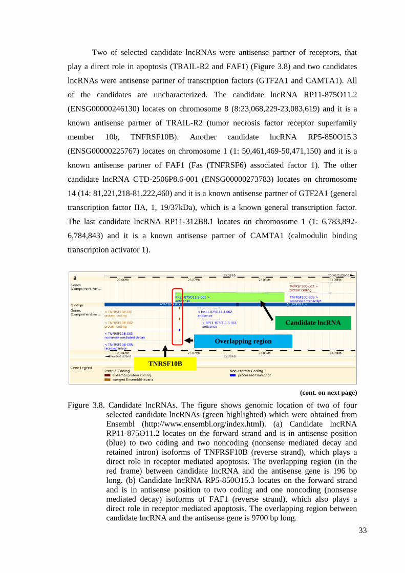
Figure 3.8. Candidate lncRNAs ...................................................................................... 33
Page 10
x
LIST OF TABLES
Tables ........................................................................................... Page
Table 3.1. Library Preparation of RNA Samples ........................................................ 31
Table 3.2. Types of Identified lncRNAs ..................................................................... 32
Table 3.3. List of Top 20 Common Upregulated and Downregulated lncRNAs ....... 33
Page 11
1
CHAPTER 1
INTRODUCTION
1.1. Apoptosis
The balance between cell death and survival of normal cells is mediated by
programmed cell death (PCD) which leads to cell death in pathological events once it is
mediated by an intracellular program as well as playing key roles in concluding
decisions of cancer cell fate (Hanahan and Weinberg 2011; Laubenbacher et al. 2009).
There are three main forms of PCD: apoptosis, autophagy and programmed necrosis.
These three forms are easily distinguished due to their morphological differences (Tan
et al. 2009; Bialik et al. 2010). Apoptosis is referred as type I PCD and it was first
described by Kerr et al. (1972). Apoptosis is crucial for normal development, aging and
a part of homeostatic mechanism to preserve cells in tissue as well as a defence
mechanism in case of immune response or cellular damage due to disease or harmful
agents (Norbury and Hickson 2001). Inappropriate apoptosis, either too much or low,
can cause some serious problems like neurodegenerative diseases, ischemic damage,
autoimmune disorders and many types of cancer (Elmore et al. 2007)
Several studies identified various morphological changes during death of cells,
apoptosis. During the early stage of apoptosis, cell shrinkage (smaller cell size,
condensation of cytoplasm and tight packaging of organelles) and pyknosis (chromatin
condensation) occurs which can be easily observed by light microscope (Kerr et al.
1972). Next, extensive plasma membrane blebbing takes place which is then followed
by karyorrhexis (nuclear fragmentation) and budding, separation of cell fragments into
apoptotic bodies that consist of cytoplasm with tightly packed organelles which can be
with or without a nuclear fragment. These bodies have intact membrane and organelles.
They are phagocytosed by macrophages, parenchymal or neoplastic cells which then
degraded within phagolysosomes (Savill and Fadok, 2000; Kurosaka et al, 2003). In
addition to morphological changes, apoptosis has some biochemical changes taking
place as well: chromosomal DNA is cleaved into internucleosomal fragments, extensive
Page 12
2
protein cross-linking, and phosphatidylserine are externalized and some proteolytic
cleavage of a number of intracellular substrates (Cohen et al. 1994, Martin et al. 1995).
1.2. Mechanism of Apoptosis
1.2.1. Caspase-dependent Mechanism
The known mechanisms of apoptosis are mediated by energy-dependent cascade
of molecular events and they are highly complex. Up to date, two main and one
additional pathway have been identified. One of the main pathways is extrinsic pathway
which is mediated by death receptor pathway. The other main pathway is intrinsic
pathway and it is also called as mitochondrial pathway. The additional pathway is
mediated by perforin/granzyme pathway via granzyme A or granzyme B. Despite
differences among stimuli for those pathways, extrinsic, intrinsic and granzyme B
pathways converge on the same terminal-execution pathway: cleavage of caspase 3
which results in fragmentation of DNA, cytoskeletal and nuclear protein degradation,
cross-linking of proteins, apoptotic body formation and expression of ligands for
phagocytosis (Igney and Krammer, 2002).
Caspases (cysteine-aspartic proteases or cysteine-dependent aspartate-directed
proteases) are a member of cysteine protein family function in apoptotic and
inflammatory signaling pathways and they are present in most cells. They are inactive
proenzyme until they are cleaved. Once a procaspase is cleaved, it gets activated and
this leads to the activation of other procaspases. Thus once caspases are activated, a
signaling caspase cascade occurs which serves as amplification of apoptotic signals and
ends with irreversible rapid cell death. Up to date, fourteen caspases have been
identified and categorized into three major classes: apoptotic initiator caspases (caspase-
2, -8, -9 and -10), executioner (effector) caspases (caspase-3, -6 and -7) and
inflammatory caspases (caspase-1, -4 and -5) (Cohen 1997; Rai et al. 2005). Caspase-
11, -12 and -14 play role in apoptosis under specific conditions and tissues. Caspase-13
is a bovine gene (Hu et al. 1998; Nakagawa et al., 2000, Koenig et al., 2001; Kang et
al., 2002).
Page 13
3
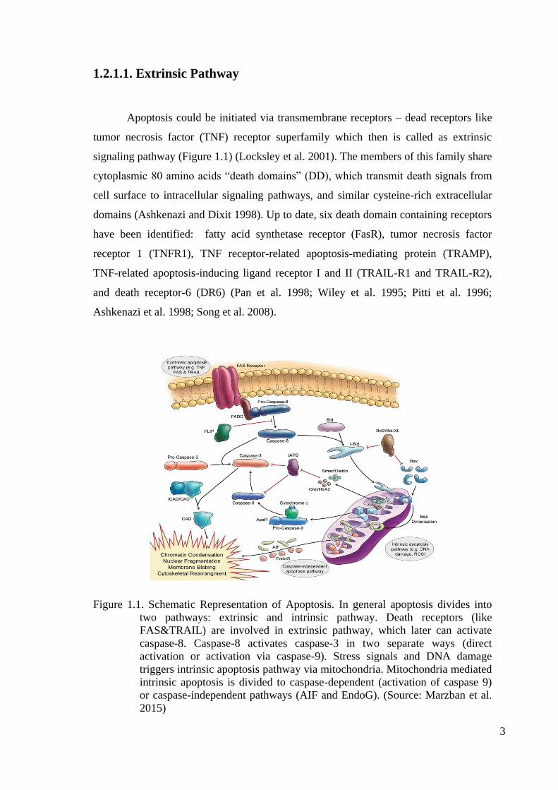
1.2.1.1. Extrinsic Pathway
Apoptosis could be initiated via transmembrane receptors – dead receptors like
tumor necrosis factor (TNF) receptor superfamily which then is called as extrinsic
signaling pathway (Figure 1.1) (Locksley et al. 2001). The members of this family share
cytoplasmic 80 amino acids “death domains” (DD), which transmit death signals from
cell surface to intracellular signaling pathways, and similar cysteine-rich extracellular
domains (Ashkenazi and Dixit 1998). Up to date, six death domain containing receptors
have been identified: fatty acid synthetase receptor (FasR), tumor necrosis factor
receptor 1 (TNFR1), TNF receptor-related apoptosis-mediating protein (TRAMP),
TNF-related apoptosis-inducing ligand receptor I and II (TRAIL-R1 and TRAIL-R2),
and death receptor-6 (DR6) (Pan et al. 1998; Wiley et al. 1995; Pitti et al. 1996;
Ashkenazi et al. 1998; Song et al. 2008).
Figure 1.1. Schematic Representation of Apoptosis. In general apoptosis divides into
two pathways: extrinsic and intrinsic pathway. Death receptors (like
FAS&TRAIL) are involved in extrinsic pathway, which later can activate
caspase-8. Caspase-8 activates caspase-3 in two separate ways (direct
activation or activation via caspase-9). Stress signals and DNA damage
triggers intrinsic apoptosis pathway via mitochondria. Mitochondria mediated
intrinsic apoptosis is divided to caspase-dependent (activation of caspase 9)
or caspase-independent pathways (AIF and EndoG). (Source: Marzban et al.
2015)
Page 14
4
The best characterized ligand/receptor models are between FasL and FasR, and
tumor TNF-α and TNFR1. Fas and TNF receptors cluster at the cell surface and binding
of trimeric Fas and TNF ligands to the corresponding receptors results recruitment of
adaptor proteins having death effector domains (DED): FADD (Fas-associated death
domain) recruits to FasR upon FasL binding; TRADD (TNF receptor-associated death
domain) is recruited to the TNFR1 due to TNF-α binding. TRADD itself mediates
recruitment of FADD and RIP (Receptor-interacting protein) and a death-inducing
signaling complex (DISC) is formed after association of procascase-8 with FADD via
dimerization of the death effector domain (Hsu et al. 1995; Kelliher et al. 1998; Wajant
2002). Formation of complex results with autocatalytic activation of procaspase-8 and
trigger of apoptotic execution pathway (Kischkel et al. 1995). However, the pathway
can be inhibited by c-FLIP (FLICE-inhibitory protein) binding to FADD and caspase-8
and turn them inactive (Kataoka et al. 1998; Scaffidi 1999).
1.2.1.2. Intrinsic Pathway
The intrinsic pathway of apoptosis arises from various non-receptor mediated
stimuli which produce intracellular signals and mitochondrial-initiated series of events
(Figure 1.1). Those stimuli-initiated signals act directly on targets either in a positive or
a negative fashion. The stimuli acting in a positive mode happens due to presence of
free radicals, radiation, toxins, viral infections, and hypoxia and so on. Negative mode
of acting happens after failure of suppressing death pathways and subsequent activation
of apoptosis due to absence of growth factors, cytokines and hormones (Saelens et al.
2004).
Both modes of action give rise to permeabilization of the outer mitochondrial
membrane: an opening of the mitochondrial permeability transition (MPT) pore due to
changes in the inner mitochondrial membrane, loss of the mitochondrial transmembrane
potential. Upon formation of MPT, a series of pro-apoptotic protein members of the Bcl
family are released which are normally sequestered in the space between the inner and
outer mitochondrial membranes (Saelens et al. 2004; Green and Kroemer 2004).
Proapoptotic Bcl family members involve cytochrome c, Smac/DIABLO, Omi/HtrA2,
AIF, CAD and endonuclease G, and they trigger the apoptosis by caspase-dependent or
caspase-independent death effectors (Saelens et al. 2004). Cytochrome c,
Page 15
5
Smac/DIABLO and Omi/HtrA2 function in a caspase-dependent fashion. Released
cytochrome c binds and activates Apaf-1 and procaspase-9, and they altogether form an
“apoptosome” which leads to activation of caspase 9. On the other hand,
Smac/DIABLO and HtrA2/Omi are stated to induce apoptosis by inhibiting inhibitors of
apoptosis proteins (IAP) activity (Chinnaiyan 1999; van Loo et al. 2002a; Hill et al.
2004; Schimmer 2004).
Bcl-2 family of proteins play very important role in the control and regulation of
mitochondria-derived apoptotic events and these proteins are regulated by p53, the
tumor suppressor protein (Schuler and Green 2001; Cory and Adams 2002). The family
members can be either pro-apoptotic or anti-apoptotic and they regulate mitochondrial
membrane permeability. There are several well-known anti-apoptotic BCL-2 family
members like Bcl-2, Bcl-x, Bcl-XL, Bcl-XS, Bcl-w and BAG; and some pro-apoptotic
proteins like Bcl-10, Bax, Bak, Bid, Bad, Bim, Bik, and Blk. Presence of these proteins
determine between apoptosis and survival through regulation of cytochrome c release
from the mitochondria via alteration of mitochondrial membrane permeability (Schuler
and Green 2001). Interestingly, there is enough evidence suggesting “cross-talk”
between the extrinsic pathway and the intrinsic pathway via mitochondrial damage by
the caspase-8 cleavage of Bid after induction of extrinsic pathway of apoptosis (Li et al.
1998; Esposti, 2002; Igney and Krammer 2002).
Either singly or doubly phosphorylated pro-apoptotic Bad (BCL2 antagonist of
cell death) protein on Ser-112 and Ser-136 binds to 14-3-3, a member of a family of
multifunctional phosphoserine binding molecules, and it is trapped within the cytosol.
However, unphosphorylated Bad translocates to the mitochondria and mediates release
of cytochrome C (Zha, et al. 1996). Another mechanism for induction of apoptosis is
neutralization of anti-apoptotic Bcl-Xl or Bcl-2 activity, inhibition of cytochrome C
release, via forming heterodimer with Bad (Yang et al., 1995). There is another protein
else, Aven, that binds to Bcl-Xl and Apaf-1 and inhibits procaspase 9 activation (Chau
et al. 2000).
Other pro-apoptotic members of Bcl2 family Puma and Noxa play roles in p53-
mediated apoptosis. Overexpression of Puma in vitro results with up-regulation and
conformational change of BAX, which translocates to the mitochondria and mediates
cytochrome c release and reduction in the mitochondrial membrane potential (Liu et al.
2003). Noxa itself can localize to the mitochondria and activate caspase-9 via
interaction with anti-apoptotic Bcl-2 family members (Oda et al. 2000). p53-dependent
Page 16
6
Puma and Noxa arise by genotoxic damage or oncogene activation like the Myc
oncoprotein which can induce apoptosis through both p53-dependent and -independent
mechanisms (Meyer et al. 2006).
1.2.1.3. Execution Pathway
The executioner or effector caspases are responsible for the final pathway of
apoptosis – execution pathway which is the end point of both the extrinsic and intrinsic
pathways. Activated execution caspases, caspase-3, caspase-6, and caspase-7, activate
cytoplasmic endonuclease and proteases: cleaving cytokeratins, PARP, alpha fodrin, the
nuclear protein NuMA and so on. As a result, they govern degradation of nuclear
material, and the nuclear and cytoskeletal proteins leading the morphological and
biochemical changes in apoptotic cells – hallmarks of apoptosis (Slee et al. 2001).
Among execution caspases, caspase 3 has the most critical role in execution
phase of apoptosis and initiator caspases, caspase-8, caspase-9, or caspase-10, are
responsible for its activation. The role of caspase 3 in apoptosis is cleaving the inhibitor
of endonuclease CAD, ICAD, and cause release of CAD that degrades chromosomal
DNA within the nuclei and causes chromatin condensation (Sakahira et al. 1998). In
addition to ICAD, caspase 3 has another target, gelsolin, which has very role in actin
nucleation and signal transduction. Caspase 3 cleaves gelsolin that ultimately causes
disruption of the cytoskeleton, intracellular transport, cell division, and signal
transduction (Kothakota et al. 1997)
The following phase after caspase cascade is phagocytic uptake of apoptotic
cells which is characterized with phospholipid asymmetry and externalization of
phosphatidylserine by scramblase on the cell surface (Wang et al. 2003).
Phosphotidylserine on the cell surface governs early uptake and disposal of apoptotic
cells via noninflammatory phagocytic recognition. The inflammatory response is not
induced due to any release of cellular components after process of early and efficient
uptake (Fadok et al. 2001).
Page 17
7
1.2.1.4. Induction of Apoptosis via Drugs and Ligands
In this study, two anti-cancer drugs, doxorubicin and cisplatin, and two ligands,
TNFalpha and Fas mAb, were used to induce cell death. Doxorubicin is one of the first
anti-cancer drugs and has been used in chemotherapy in many different cancer types
over 30 years. The ability to overcome rapid cell division and slow down cancer
progression make it one of the most potent of the FDA (Food and Drug
Administration)-approved chemotherapeutic drugs (Carvalho et al. 2009). It is known to
intercalate DNA, bind to DNA-associated enzymes like topoisomerase enzymes I and
II, and target various molecular targets to produce a range of cytotoxic effects – anti-
proliferation and DNA damage (Buchholz et al. 2002). The apoptosis pathway is
induced once cell fails to repair lesions on DNA and cell cycle is inhibited. However,
studies have shown that doxorubicin can result in autophagy and necrosis (Tavar et al.
2012). Cytoprotective mechanism can induce autophagy by poly (ADP-ribose)
polymerase-1 (PARP-1) activation as response to DNA damage. PARP-1
activation/deactivation decides whether the cell will undergo autophagy or necrosis
(Minotti et al. 2004). Thus, doxorubicin mediated cell death type is strongly dependent
on the concentration of drug, treatment duration and cell-cancer type. The effect of
doxorubicin on HeLa cells is conditional – depending on concentration. Apoptosis is
induced by only a particular concentration of doxorubicin; induction of necrosis is a
higher probability (Tomoki and Robertson 2004).
The doxorubicin mediated apoptosis pathway is induced via AMPK activation
(Shaw et al. 2004) or activation of various signals to alter the Bcl-2/Bax ratio (Leung
and Wang 1999) which results in downstream activation of different caspases –
induction of apoptosis. Downstream targets of AMPK are c-JunN-terminal kinase
(JNK), p53 and mTORC1 (Meisse et al. 2002; Cao et al. 2008; Gwinn et al. 2008). Bcl-
2/Bax ratio is affected due to p53-independent down-regulation of Bcl-2 mRNA levels.
There is also a conflict about doxorubicin-mediated apoptosis. Some groups claim about
existence of the Fas/Fas ligand apoptosis pathways; however, some groups have shown
contradictory results (McGahon et al. 1998; Adams and Cory 1998).
Cisplatin has also been used as a chemotherapy drug over 30 years. Cisplatin is a
platinum based drug. Chemotherapy with cisplatin often comes along with toxic side
effects and tumor resistance, which leads to secondary malignancies (Chen et al. 2009).
Page 18
8
Cytotoxicity of cisplatin depends on cell type, duration of treatment and drug
concentration. Cisplatin damages tumor cells via activation of various signal
transduction pathways. Cellular interaction of cisplatin includes reactive oxygen species
(ROS), DNA, mitochondria, TNF, p53, caspases, calcium signaling and multidrug
resistant proteins (Florea and Büsselberg 2011). Cisplatin-mediated inhibition of DNA
synthesis, failure in DNA repair and cell cycle arrest lead to apoptosis (Desoize and.
Madoulet 2002). Generation of ROS and interaction with DNA might introduce DNA
damages and cytotoxicity which then results with cell death – death type depends on
(cancer) cell type. (Che et al. 2010; Brozovic et al. 2010). Interestingly, cisplatin
activates extrinsic apoptotic pathway via activating the tumor necrosis factor-related
apoptosis-inducing ligand (TRAIL) receptor-mediated signal-transduction pathway.
TRAIL receptor, DR4 and/or DR5, aggregation leads to death-inducing signaling
complex (DISC) formation and results caspase activation. However, caspase 8
activation is mitochondria-dependent (Shamimi-Noori et al. 2008).
Both extrinsic and intrinsic pathways are induced in cisplatin mediated apoptosis
in HeLa cells (Sui et al. 2015). Although cisplatin is known to induce mitochondria-
mediated apoptosis, still interaction between cisplatin and mitochondria is less known.
In several specific cases it was shown that cisplatin leads to mitochondrial
depolarization, cytochrome c release, translocation of Bax and tBid to mitochondria and
decrease in Bcl-2 expression, which results induction of apoptosis (Muscella et al.
2008). p53 and Protein kinase C (PKC) δ play role in cisplatin mediated cell death. It
was shown that PKCδ is a positive regulator of cisplatin-induced cell death and in
cooperation with p53, they mediates caspase-3 dependent apoptosis (Karger et al.
2005). In addition, cisplatin was shown to have effect on post-translational
modifications like histone methylation and acetylation (Wang or Lippard 2004).
TNF-alpha and Fas ligands are commercially available for research and
development projects. Monoclonal antibodies of both ligands are available as well,
which exert the same function – bind to the ligand-specific death receptors and induces
apoptotic extrinsic pathway. In type I cells, which have enough caspase 8 to activate
effector/executioner caspases, TNF-alpha and FasL can induce executioner caspases
directly. However, in type II cells like HeLa, caspase 8 amount is insufficient in caspase
3 activation, thus proapoptotic signals mediate release of cytochrome c from the
mitochondria, activation of caspase 9 and caspase 3– induction of intrinsic apoptotic
pathway (Kuwana et al. 1998). TNF-alpha cannot induce apoptosis alone in HeLa cells,
Page 19
9
however, in the presence of metabolic inhibitors like cycloheximide (CHX) TNF-alpha
can induce apoptosis (Miura et al. 1995). Several studies have stated that many FasR
expressing tumor cells are completely insensitive to FasR-induced apoptosis (O'Connell
et al. 1996). HeLa cells are not completely insensitive; however, sensitivity increases
with the cellular stress due to ionizing agents and etc. (Park et al. 2003)
1.2.1.5. Perforine/Granzyme Pathway
The sensitized CD8+cells, cytotoxic T lymphocytes (CTLs), kill antigen-bearing
cells by T-cell mediated cytotoxicity via extrinsic pathway, predominantly the
FasL/FasR interaction to induce apoptosis (Brunner et al. 2003). However, in addition
to extrinsic pathway, there is a novel pathway to show cytotoxic effects on virus-
infected and tumor cells that is used by these cytotoxic cells. This pathway involves
secretion of perforin, a transmembrane pore-forming molecule, with following release
of cytoplasmic granules, the serine proteases granzyme A and granzyme B, through the
pore and into the target cell (Trapani and Smyth 2002; Pardo et al., 2004).
Granzyme B can activate pro-caspase 10 via cleaving it at aspartate residue and
can cleave ICAD either to induce apoptosis (Sakahira et al. 1998). Although it is shown
that granzyme B can use intrinsic pathway via cleaving Bid and inducing cytochrome c
release to amplify death signal (Barry and Bleackley 2002; Russell and Ley 2002),
granzyme B can directly activate caspase 3 via bypassing the upstream signaling
pathways to induce the execution phase of apoptosis directly (Goping et al. 2003).
Granzyme A is inducing caspase-independent meachanism to induce apoptosis
(Martinvalet et al. 2005).
1.2.2. Caspase-independent Mechanism
There is another group of mitochondrial released pro-apoptotic proteins, such as
AIF, endonuclease G and CAD that act in a caspase-independent manner. They are
released as a late event during apoptosis - after the cell has committed to die.
Endonuclease G and AIF translocate to the nucleus and they work in a caspase-
independent way (Susin et al., 2000; Li et al. 2001). Endonuclease G causes
oligonucleosomal DNA fragmentation while AIF causes DNA fragmentation into ∼50–
Page 20
10
300 kb pieces. They cause “stage I” peripheral nuclear chromatin condensation (Joza et
al. 2001). The released CAD translocates in nucleus where it leads to oligonucleosomal
DNA fragmentation, advanced chromatin condensation and afterward “stage II”
condensation (Enari et al. 1998, Susin et al. 2000).
The granzyme A pathway activates apoptosis in a caspase-independent manner
via single strand DNA breaks (Martinvalet et al. 2005). The single stranded DNA break
is mediated by the DNase NM23-H1, a tumor suppressor gene product, which has an
important role in preventing cancer by inducting apoptosis in tumor cells (Fan et al.,
2003). The granzyme A cleaves inhibitor of NM23-H1, a nucleosome assembly protein
SET, thus releasing NM23-H1 to induce apoptosis via DNA degradation. The SET
complex proteins, SET, Ape1, pp32, and HMG2, work together and they have vital
roles in chromatin structure and DNA repair via protecting chromatin and DNA
structure. Thus, granzyme A induces apoptosis by inhibiting the functions of the SET
complex proteins (Lieberman and Fan 2003).
1.3. Long Non-Coding RNA
The scientists discovered in 1950s that the C-value - amount of DNA in the
haploid genome has little correlation with organism size or developmental complexity
(Mirsky and Ris 1951; Thomas 1971; Gall 1981). This meant that many less developed
animals can have a bigger genome than more developed animals, including humans.
The “C-value paradox” was partially solved after discovery of noncoding portion of
genome, which is much more than protein coding portion in eukaryotes (Lewin 1980).
The noncoding portion was then called “junk DNA” due to presence of transposons,
pseudogenes, and simple repeats. It is estimated that total transposons, pseudogenes,
and simple repeats are about 50–70% of the human genome (de Koning et al. 2011).
Today the contradiction in genome size is no longer a paradox, but became more a “C-
value enigma” (Gregory 2001). Morphologically similar and phylogenetically close
species can have different genome size and thus noncoding content may indicate
correlation between noncoding content and complexity (Ricroch et al. 2005; Taft et al.
2007).
Although noncoding sequences were called “junk”, it received interest from
1970s to the present. And researchers even in 1970s started to speculate that more
Page 21
11
portion of the genome is transcribed from repetitive and heterochromatic regions, as
well as nonrepetitive regions other than coding sequences and known rRNAs and
tRNAs. Those transcribed RNAs were named as “heterogeneous nuclear RNAs”
(hnRNAs) and it was shown that 50% of them are restricted to the nucleus and they do
not contain coding sequences (Holmes et al. 1972; Pierpont and Yunis 1977; Lewin
1980, Chap. 25). Discovery of introns was shown to account for a small portion of
noncoding sequences (Berget et al. 1977; Chow et al. 1977).
Whole-genome technologies in the late 1990s and early 2000s helped to
estimate the scale of “pervasive transcription”. As much as 70–90% of human genome
is transcribed at some point during development (Okazaki et al. 2002; Rinn et al. 2003;
Bertone et al. 2004; Ota et al. 2004; Carninci et al. 2005; Birney et al. 2007; Kapranov
et al. 2010; Mercer et al. 2011; Djebali et al. 2012). RNA sequencing analyses suggest
that alternative splicing and/or extensions of known protein-coding genes may account
for such amount of pervasive transcription (He et al. 2008; Mortazavi et al. 2008; Sultan
et al. 2008; van Bakel et al. 2010, 2011).
The new findings support noncoding transcription in intergenic regions with a
correlation with chromatin signatures, histone modifications or transcription factor
binding at loci and dependence of expression level of those noncoding on these
transcription factors (Guttman et al. 2009, 2011; van Bakel et al. 2010; Encode Project
Consortium 2012). Although many novel, conserved long noncoding RNAs (lncRNAs)
have been identified, the number of reported lncRNAs are only a few thousand, which
is not enough to explain the “C-value enigma” - 70–90% of the genome. Although the
idea of “transcriptional noise” (Hüttenhofer et al. 2005) is still more powerful in the
field, even in the early 1990s several lncRNAs involved in epigenetic regulation, H19
(Brannan et al. 1990) and Xist, were discovered (Brockdorff et al. 1992; Brown et al.
1992). Calculations have shown that up to 90% of Pol II transcription can initiate non-
specifically and this transcription can be spurious (Struhl 2007). During transcription
there is a tendency to fluctuate away from “legitimate” transcripts – there are leaky
expressions of neighboring regions (Ebisuya et al. 2008)
Page 22
12
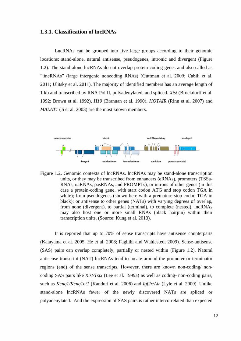
1.3.1. Classification of lncRNAs
LncRNAs can be grouped into five large groups according to their genomic
locations: stand-alone, natural antisense, pseudogenes, intronic and divergent (Figure
1.2). The stand-alone lncRNAs do not overlap protein-coding genes and also called as
“lincRNAs” (large intergenic noncoding RNAs) (Guttman et al. 2009; Cabili et al.
2011; Ulitsky et al. 2011). The majority of identified members has an average length of
1 kb and transcribed by RNA Pol II, polyadenylated, and spliced. Xist (Brockdorff et al.
1992; Brown et al. 1992), H19 (Brannan et al. 1990), HOTAIR (Rinn et al. 2007) and
MALAT1 (Ji et al. 2003) are the most known members.
Figure 1.2. Genomic contexts of lncRNAs. lncRNAs may be stand-alone transcription
units, or they may be transcribed from enhancers (eRNAs), promoters (TSSa-
RNAs, uaRNAs, pasRNAs, and PROMPTs), or introns of other genes (in this
case a protein-coding gene, with start codon ATG and stop codon TGA in
white); from pseudogenes (shown here with a premature stop codon TGA in
black); or antisense to other genes (NATs) with varying degrees of overlap,
from none (divergent), to partial (terminal), to complete (nested). lncRNAs
may also host one or more small RNAs (black hairpin) within their
transcription units. (Source: Kung et al. 2013).
It is reported that up to 70% of sense transcripts have antisense counterparts
(Katayama et al. 2005; He et al. 2008; Faghihi and Wahlestedt 2009). Sense-antisense
(SAS) pairs can overlap completely, partially or nested within (Figure 1.2). Natural
antisense transcript (NAT) lncRNAs tend to locate around the promoter or terminator
regions (end) of the sense transcripts. However, there are known non-coding/ non-
coding SAS pairs like Xist/Tsix (Lee et al. 1999a) as well as coding- non-coding pairs,
such as Kcnq1/Kcnq1ot1 (Kanduri et al. 2006) and Igf2r/Air (Lyle et al. 2000). Unlike
stand-alone lncRNAs fewer of the newly discovered NATs are spliced or
polyadenylated. And the expression of SAS pairs is rather intercorrelated than expected
Page 23
13
by chance alone, however, whether all have any biological function or not still remains
to be investigated (Kung et al. 2013).
Pseudogenes are the “relics” of coding genes that have lost their coding potential
due to nonsense, frameshift, and other mutations (Balakirev and Ayala 2003; Pink et al.
2011). They are extra copies of genes by tandem duplication or retrotransposition and
they are no longer transcribed, dead. However, a small portion (2-20%) is transcribed
and rarely translated. It is thought that expressed pseudogenes are on the way to die,
complete pseudogenization, or dead pseudogenes have gained new functions (Harrison
et al. 2005; Bekpen et al. 2009). Interestingly, some transcribed pseudogenes have been
shown to regulate expression of their ancestral coding genes. Xist is believed to be
formed by the pseudogenization of Lnx3 (a protein-coding gene) and integration of
different transposon-derived repeat elements (Duret et al. 2006; Elisaphenko et al.
2008).
It was known that small ncRNAs such as snoRNAs and miRNAs are transcribed
from introns. Recent studies show similar transcription of lncRNAs from annotated
genes (Louro et al. 2009; Rearick et al. 2011). Although few of them have been studied
detailed, many of them were differently expressed in various conditions and in cancer
(Guil et al. 2012).
There are plenty of short transcripts (20 to 2500 nt) transcribed from
neighborhood in the vicinity around transcription start sites in both sense and antisense
directions (Buratowski 2008; Core et al. 2008; He et al. 2008; Preker et al. 2008; Seila
et al. 2008). Transcription start site-associated (TSSa-)RNAs are the shortest transcripts
among them and believed to be degradation products or they are processed from the
longer upstream antisense (ua)RNAs or promoter upstream transcripts (PROMPTs).
They are usually capped and polyadenylated, have low copy number (0.1 copy per cell),
and are exposed to rapid degradation by exosomes. A subgroup called promoter-
associated short (pas)RNAs were shown to interact with epigenetic factors such as
Polycomb proteins. It is still unclear whether these transcripts are transcriptional by-
products, whether they help maintain open chromatin, or whether they all play a
regulatory role like (pas)RNAs (Kanhere et al. 2010). In addition to promoters, short
bidirectional transcripts are also shown to be transcribed from enhancers. However, up
to date no known biological function has been associated with them (Kim et al. 2010;
Wang et al. 2011a).
Page 24
14
1.3.2. Functional roles of lncRNAs
Currently, our understanding about functional roles of lncRNAs is very limited
although some lncRNAs were functionally characterized. A very limited number of
lncRNAs has shown important roles in various processes and studies showed that
differential expressed lncRNAs are associated with developmental processes and
disease states. However, a majority of lncRNAs require further investigation (Kung et
al. 2013).
Currently, the best-studied biological function for lncRNAs is epigenetic
regulation of allelic expression. Some of them play role in the processes of dosage
compensation and genomic imprinting. The 17-kb X (inactive)-specific transcript (Xist)
is highly expressed from a cluster of lncRNA loci, the X-inactivation centre (Xic), in
inactive X (Xi) chromosome which coats the X chromosome, forms an “Xist cloud” and
acts as a scaffold for the recruitment of silencing factors (Polycomb repressive complex
2 (PRC2) and etc.) during X chromosome inactivation (XIC) (Lyon 1961; Brown et al.
1991; Brown et al. 1992; Clemson et al. 1996; Zhao et al. 2008; Lee 2011).
Another important role of lncRNAs is in genomic imprinting, a phenomenon
when a gene is expressed monoallelically compared to its parent of origin (Edwards and
Ferguson-Smith 2007; Wan and Bartolomei 2008). There are specific genome loci,
imprinting control regions, like in XIC where many lncRNAs are expressed. Both
protein coding and lncRNAs are reciprocally expressed from many of such regions and
lncRNAs may control the imprinted expression of neighbouring coding genes by
recruiting epigenetic factors (Nagano et al. 2008; Pandey et al. 2008; Zhao et al. 2010).
Other than epigenetic regulation, lncRNAs play role during other aspects of
development, from the control of pluripotency to lineage specification. Pluripotency
transcription factors (e.g., Oct4, Sox2, and Nanog) are regulated by lncRNAs (Hawkins
and Morris 2010; Ng et al. 2011). A number of lncRNAs (HOTAIR, HOTTIP and
Mistral) are encoded within Hox genes, which are important for anterior–posterior
pattern formation, regulate expression of Hox genes (either the host or a distant cluster)
(Pearson et al. 2005; Rinn et al. 2007; Bertani et al. 2011; Wang et al. 2011b).
Numerous lncRNAs are associated with several diseases, especially cancer
(Gutschner and Diederichs 2012). PCAT-1, ANRIL, HOTAIR and MALAT1 lncRNAs
are upregulated in several cancer cell types and contribute to cancer progression (Ji et
Page 25
15
al. 2003; Gupta et al. 2010; Kotake et al. 2011; Lin et al. 2011; Prensner et al. 2011).
There are several lncRNAs that play role in DNA damage and eventually in apoptosis,
lincRNA-p21 and PANDA, upregulated by p53 upon DNA damage (Huarte et al. 2010;
Hung et al. 2011).
1.3.3. LncRNAs: Act of Mechanism
There are very few lncRNAs characterized in mechanistic details although we
know many of them. According to current knowledge, they are categorized into some
groups. However, in future, due to new discoveries in the field, we can have additional
groups and themes about them. One of the major themes of lncRNAs is playing role in
epigenetics as recruiters, tethers, and scaffolds. They mediate recruitment of protein
factors for regulation of chromatin states via acting cis, acting on neighbor genes in the
periphery of their site of synthesis; or acting in trans, acting on distant genes in the
same or even in another chromosome (Campos and Reiberg 2009). Chromatin-
modifying complexes, such as PRC2, are shown to interact with massive number of
lncRNAs (Khalil et al. 2009; Kanhere et al. 2010; Zhao et al. 2010; Guil et al. 2012).
Due to some features of lncRNAs, they are excellent candidates for cis-acting
tethers, but still trans-action is not defined yet. In X chromosome inactivation (XCI),
tethering Xist RNA to the Xic is an example for tethering (Jeon and Lee 2011). Some
other epigenetic complexes, other than PRC2, may interact with lncRNAs and some
lncRNAs act as scaffold where multiple proteins can assemble (Yap et al. 2010; Kotake
et al. 2011). Beside the epigenetic complexes, lncRNAs may recruit transcription
factors to activate certain genes in cis (Bertani et al. 2011; Wang et al. 2011b). Long
ncRNAs may modulate DNA methylation at CpG dinucleatides during epigenetic
regulation for the stable repression of genes (Law and Jacobsen 2010). DNA
methylation of ribosomal (r)DNA, which some remain always silenced by
heterochromatic histone marks and DNA methylation, is also directed by certain
lncRNAs (McStay and Grummt 2008).
Regulation of gene expression by lncRNAs can be directly affecting the process
of transcription. They can act as decoys for TFs or competing for TF binding, and even
affect the cellular localization of TFs (Willingham et al. 2005; Hung et al. 2011). Long
ncRNAs can act as transcriptional coregulators and they recruit regulators which in turn
Page 26
16
carry out their function on downstream targets by recruiting additional factors (Lanz et
al. 1999, 2002). In addition to TFs, lncRNAs can affect gene expression by directly
interfering with Pol II activity by preventing formation of preinitiation complexes via
DNA:RNA triplex formation on promoter or binding with general transcription factors
(Yakovchuk et al. 2009; Martianov et al. 2007).
Long ncRNAs play role as key regulators of nuclear compartments - “nuclear
bodies” that exert important functions (Mao et al. 2011b). Long ncRNAs are linked to
the function and structure of the members of nucleolus, paraspeckles and other nuclear
compartments (Zhang et al. 2007; Chen and Carmichael 2009). Certain lncRNAs
(MALAT1 or NEAT2) mediate proper localization of splicing factors to nuclear
speckles and thus may have role in alternative splicing of certain mRNA precursors
(Bernard et al. 2010; Tripathi et al. 2010). Thus, there is a complex interaction among
lncRNAs, cell-signaling pathways, chromatin-modifying factors, and nuclear bodies in
regulating gene expression.
Several studies have shown diverse functions of lncRNAs in mRNA processing,
stability and translation including alternative splicing. Natural antisense (NAT)
lncRNAs may affect alternative splicing of overlapping transcripts by forming RNA
duplexes that inhibit splicing which is a type of post-transcriptional regulation (Beltran
et al. 2008; Annilo et al. 2009). NATs, mainly produced from the 3’-UTR, may be
involved in stability of its antisense by recruiting factors that lead to stabilization or
destabilization of the transcripts (Kim et al. 2005, 2007; Barreau et al. 2006). LncRNAs,
specifically NATs, may even play role in translational regulation of their targets,
specifically on their sense mRNAs, by competing for binding to the certain translation
initiation factors (Ebralidze et al. 2008).
It is not surprising that long non-coding RNAs are intertwined with small non-
coding RNAs. Certain lncRNAs were shown to interfere with miRNA-mediated mRNA
destabilization by masking miRNA-binding sites or competing for the miRNAs
themselves (Faghihi et al. 2010, Wang et al. 2010). Some lncRNAs have miRNA-
binding sites in their 3’-UTRs which can serve as “sponge” to keep miRNAs away from
their mRNA targets (Franco-Zorrilla et al. 2007). On the other hand, lncRNAs may
themselves be host genes for small RNAs, such as miRNA and snoRNAs (Smith and
Steitz 1998; Cai and Cullen 2007; Keniry et al. 2012).
Page 27
17
1.3.4. Long-Coding RNAs in Apoptosis
As stated above, certain lncRNAs are differentially expressed in several diseases
and cancer. Some of them are playing role in apoptosis as well. There are known
negative and stimulatory regulators of apoptosis - anti- and pro-apoptotic lncRNAs.
lncRNAs like PCGEM1 (Fu et al. 2006), LincRNA-EPS (Paralkar et al. 2011), PANDA
(Puvvula et al. 2014), AFAP1-AS1 (Wu et al. 2013), SPRY4-IT1 (Khaitan et al. 2011),
PlncRNA-1 (Cui et al. 2013) and HOXA-AS2 (Zhao et al. 2013) are anti-apoptotic.
They are upregulated in cancer cells and often play a role in tumor survival and
progression. On the other hand, certain lncRNAs, such as lincRNA-p21 (Wu et al.
2014), GAS5 (Pickard et al. 2013), ncRNA CCND1 (Wang et al. 2008a), MEG3 (Zhang
et al. 2014), INXS (DeOcesano-Pereira et al. 2014), LOC401317 (Gong et al. 2014), are
pro-apoptotic and are down-regulated in certain cancer types.
Although numerous lncRNAs playing role in apoptosis are known, however,
there is not any systematic study intended to identify the total number of lncRNAs
playing role in apoptosis. The current known anti- and pro-apoptotic lncRNAs were
discovered under specific conditions. Pathway-specific or master regulators require a
systematic approach to demonstrate lncRNA function in apoptosis.
1.4. Aim
The aim of the project is to identify differentially expressed lncRNAs via deep
sequencing under apoptotic conditions in HeLa cells.
Page 28
18
CHAPTER 2
MATERIALS AND METHODS
2.1. Cell Culturing
HeLa cells were obtained from DSMZ GmbH and were cultured in RPMI 1640
(with L-Glutamine, Gibco) in a humidified incubator with 5% CO2 in air at 37°C. The
cell culture medium was supplemented with 10% inactivated fetal bovine serum (FBS)
(Gibco) and 1% penicillin-streptomycin (Gibco). HeLa cells were seeded every two
days with 1/3 or 1/4 (2.0 – 2.5 x 10⁶ cells).
Drug treatments were performed using 6-well plate (Sarsted). HeLa cells were
seeded 0.3 x 10⁶ cells/well density. Overnight grown HeLa cells in 6-well plates were
incubated with agents in time- and dose-kinetics experiments. The entire drug screening
experiments were performed at least three times and the results were analyzed with
student’s t-test to show whether changes statistically significant.
Cisplatin (SantaCruz) was freshly prepared in DMSO as 83.2 mM stock in every
drug screening experiment due to its chemical instability. Cisplatin concentration
varying 2 µM up to 320 µM were screened for 4 hours up to 24 hours. Subsequent
experiments were set to 80 µM for 16 hours. Due to toxic effect of DMSO, one more
control was set as DMSO control.
Doxorubicin (Cell Signaling) was dissolved in DNase and RNase free water and
was prepared as 5mM stock, aliquoted and stored at -20 °C. Doxorubicin concentration
varying 0.0625 µM up to 32 µM were screened for 4 hours up to 24 hours. Subsequent
experiments were set to 4 µM and 4 hours.
Fas mAb (Cell Signaling) concentration ranging from 0.125 µg/ml up to 2 µg/ml
for 4 hours up to 24 hours were tested and subsequent experiments were set up for 0.5
µg/ml and 16 hours.
TNF-alpha ligands (Millipore) were dissolved in DNase and RNase free water
and prepared 100 ng stock, was aliquoted and stored at -20 °C. Cycloheximide (CHX)
(Applichem) was coupled with TNF-alpha due to type II cell feature of HeLa. Less
cytotoxic concentration of cycloheximide was determined by screening cycloheximide
Page 29
19
concentration ranging 5 µg to 80 µg for 4 to 24 hours. TNF-alpha coupled with
cycloheximide with different concentrations, 1 ng/ml up to 125 ng/ml for 4 hours up to
24 hours were screened and subsequent experiments were set to 125 ng/ml TNF-alpha
with 10 µg CHX for 8 hours. In addition to negative untreated control, TNF-alpha and
cycloheximide alone were used as negative controls as well.
2.2. Measurement of Apoptosis
Time- and dose-kinetics were carried out for all drugs and were analyzed with
Flow Cytometry. Annexin V and 7AAD (BD) were used in detection of apoptosis and
all experiments/doses were repeated at least 3 times. Annexin V was diluted 1:5 with
PBS and 7AAD was diluted 1:10 with PBS as well. Drug treated and untreated cells
were harvested with Trypsin-EDTA (Gibco, 0.25%) and washed twice with ice-cold
PBS. After removal of PBS from last wash, cells were suspended in 200-300 µl annexin
binding buffer (BD) and 50 µl of each cell suspension was added into eppendorf.
Further, 5 µl from Annexin V and 7AAD were added into eppendorf as well, after 15
min incubation in dark, cells were suspended again in 200 µl PBS prior to analysis with
Flow Cytometry (Applied Biosystems or BD FACS). Cells with Annexin V signal were
considered to be at the early stage of apoptosis. Cells with both Annexin V and 7AAD
signal were considered to be at the late onset of apoptosis. Dead cells were only 7AAD
positive and live cells were both Annexin V and 7AAD negative.
The efficiency of fluorescent labeling was further verified with Fluorescence
Microscope by the help of same markers, Annexin V and 7AAD. The same procedure
for Flow Cytometry was followed, however, cells were analyzed under microscope.
After incubation in the dark, 10 µl of cell suspension was spread on clean chamber and
covered with cover slide and analyzed with Fluorescence Microscope (Filter 2 and 4,
Olympus IX70)
2.3. Total Protein Purification
Total protein extracts were prepared by using RIPA lysis buffer (Cell Signaling).
Drug treated and untreated cells were harvested with Trypsin-EDTA and washed twice
with ice-cold PBS. Cells were lysed with RIPA (50 µl per 106 cells) , Protease Inhibitor
Page 30
20
Cocktail (100X) (SantaCruz) was added immediately after and cell lysates were kept on
ice up to 20 min with vortexing every 5 min. Lysates were centrifuged for 10 minutes at
14.000 rpm at 4 °C. Supernatants aliquoted into two or three eppendorf tubes and stored
at -80 °C.
In order to determine protein concentration, Bradford Assay was used. Standard
curve standard was drawn with 40 µl of different BSA (bovine serum albumin)
concentrations ranging between 20 and 200 µg/ml in Bradford reagent (0.01% (w/v)
Coomassie Brilliant Blue G-250, 4.7% (w/v) ethanol, 8.5% (w/v) phosphoric acid) in
order to relate protein concentration with absorbance (595 nm). Equation obtained from
standard curve was used to estimate protein concentration from absorbance reads of
samples. Protein samples were diluted 1:10, 4 µl diluted with RIPA to 40 µl and added
into 1.5 ml of Bradford reagent into cuvettes. After 5 min dark incubation, OD of each
sample was detected immediately with spectrophotometer.
2.4. Western Blotting
The potential effect of all drug treatments on caspase activation was determined
biochemically by western blotting. Caspase 3, 8 and 9, and β-Actin monoclonal (mouse)
antibodies were purchased from Cell Signaling. Caspase 3 activation shows induction of
apoptosis; specifically, caspase 8 activation indicates induction of extrinsic apoptotic
pathway while caspase 9 activation indicates induction of intrinsic pathway.
Protein amount was fixed to 20 µg per well and protein samples with protein
loading dye was heated for 5 minutes at 90 °C. Protein samples were run in two 15%
separating and 5% stacking SDS gel [dH20, separating/stacking buffer, 30%
Acrylamide (Sigma), 10% SDS (Applichem), TEMED (Sigma), 10% APS
(Applichem)] vertically for two hours at 100V in running buffer (25 mM Tris, 192 mM
Glycine, 1% SDS (w/v)).
One of the gels was used to stain with Coomassie Blue solution [1 mg/ml
Coomasie Blue (Sigma), 10% acetic acid (v/v), 30% methanol (v/v)] in order to check
proper running and verify presence of proteins. Incubation of gel at room temperature
with Coomassie Blue for an hour followed incubation with Coomassie Destaining
solution [10% acetic acid (v/v) and 30% methanol (v/v)] for an hour in order to
visualize total protein bands. On the other side, the other gel was run for blotting; gel
Page 31
21
was placed between Whatman paper and methanol activated PVDF membrane
(Millipore). Proteins were transferred to PVDF membrane at 30V overnight. Transfer of
proteins onto PVDF membrane was tested with Ponceau S [0.1% (w/v) Ponceau S in
5% (v/v) acetic acid] staining for 30 min and destaining with ultrapure water for 30 min.
Membrane was blocked with blocking buffer (1X TBS, 0.05% Tween20, 0.5%
non-fat dry milk) for an hour at room temperature. Caspase 8 and 9, and β-Actin
primary antibodies were diluted 1:5000 whereas Caspase 3 mAb was diluted 1:7000 in
wash buffer (1X TBS, 0.05% Tween20) including 10% blocking buffer and incubated
for an hour at room temperature with gentle shaking with shaker. After primary
antibody incubation, membrane was washed with wash buffer 5 times for 45 min and
incubated with gentle shaking for an hour with HRP-conjugated anti-mouse secondary
antibody (Cell Signaling) with 1:20000 ratio in wash buffer including 10% blocking
buffer. The wash step was repeated after the addition of secondary antibody as well.
The membrane was then prepared for visualization via adding SuperSignal West Femto
Maximum Sensitivity Substrate (Thermo Scientific) on membrane; 500 µl enhanced
chemiluminescent substrate for HRP and 500 µl enhancer solution were mixed and
spread on membrane, and incubated for 2 minutes. Chemiluminescence from
membranes was visualized with VersaDoc MP 4000 Molecular Digital Imaging System
(BioRad) in Biotechnology and Bioengineering Research and Application Center at
Izmir Institute of Technology or Fusion SL (PEQLAB).
2.5. Total RNA Isolation and RNA-Seq
Cells were grown overnight in 75cm2 Flasks prior to treatment. Drug treated and
untreated cells were harvested with Trypsin-EDTA and washed twice ice cold PBS.
After the complete removal of PBS from the last wash step, 1 ml TRIzol (Life
Technologies) was used to dissolve each pellet and cell lysate was stored at -80 °C.
RNA isolation from the cell lysate was performed within one week and the protocol
from manufacturer was followed.
Frozen cell lysates were thawed and was incubated for 5 minutes at room
temperature to facilitate complete dissociation of the nucleoprotein complex. 0.2 ml of
RNase free chloroform (Sigma) was added per 1 ml of TRIzol for homogenization.
After vigorous shaking by hand for 15 seconds, tubes were incubated for 2-3 min at
Page 32
22
room temperature (RT). Centrifugation of samples at 12,000 × g for 15 minutes at 4°C
led phase separation; aqueous phase was pipetted out into new eppendorf by angling the
tube at 45° without disturbing middle and down phase. 0.5 mL of 100% RNase free
isopropanol (Sigma) was added per 1 mL of TRIzol for homogenization and incubated
at RT for 10 minutes. Further, samples were centrifuged at 12,000 × g for 10 minutes at
4°C and supernatant was removed after. Pellet was washed with 1 mL of 75% RNase
free ethanol (Sigma) per 1 mL of TRIzol in the initial homogenization. After a short
vortex, samples were centrifuged at 7500 × g for 5 minutes at 4°C and supernatant wash
was discarded. Pellet RNA samples were dried in air for 5-10 minutes, were dissolved
with DNase and RNase free water, aliquoted and kept at -80 °C.
Initial RNA quality control was checked by NanoDrop (Thermo Scientific) and
by running on 1% agarose gel. 1 µl from each RNA sample was used in NanoDrop and
260/280 and 260/230 ratios were obtained for initial RNA purity and quality (for “pure”
RNA 260/280 ratio is ~2, 260/230 ratio is ~2.0-2.2). 1 µg RNA was mixed with gel
loading dye (2X), heated for 2 min at 85 °C and directly was kept on ice for next 2 min.
RNA was run in TBE buffer (Tris-borate-EDTA buffer, 1M Tris base, 1M Boric acid
and 0.02M EDTA) for 30 min at 100V. Gels were visualized with AlphaImager (The
AlphaImager High Performance Gel Documentation and Image Analysis System,
Model IS-2200) for 5 to 15 sec with UV light filter.
Total RNAs from three replicates of selected doses from four drugs with control
untreated cells were sent for deep sequencing by Fasteris SA (Switzerland) using
Illumina Platform. Totally 5 µg from each RNA sample was sent for initial Quality
Check (QC) and RNA-Seq was performed using a specific method based on
identification of long non-coding RNAs.
2.6. Bioinformatics Analyses
The output from RNA-Seq was analysed by Allmer lab (collaboration). Firstly,
the output (fastq) files were subjected to Quality Control via FasQC. Then, adaptor and
quality trimming was done via cutadapt and Sickle tools, respectively. The output was
then mapped to the human GRCh38 genome as a reference. Further, read counting was
done via using HTSeq Count, and normalization of reads, RPKM (Reads per kilo base
Page 33
23
per million) values and detection differentially expressed genes were obtained via
DESeq2 tool.
Stringent filters were set to detect meaningful expression differences: two-fold
and upper, and P<0.01. Candidates were chosen from top 20 up- and down-regulated
lncRNAs that were commonly differentially expressed upon treatment with four agents.
2.7. Flow Chart of Overall Approach
Figure 2.1. Flow Chart of Overall Approach. The figure illustrates the flow of the
project.
Page 34
24
CHAPTER 3
RESULTS
3.1. Drug Dose-kinetics
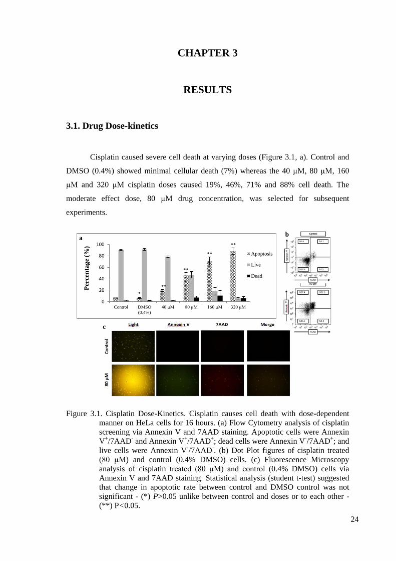
Cisplatin caused severe cell death at varying doses (Figure 3.1, a). Control and
DMSO (0.4%) showed minimal cellular death (7%) whereas the 40 µM, 80 µM, 160
µM and 320 µM cisplatin doses caused 19%, 46%, 71% and 88% cell death. The
moderate effect dose, 80 µM drug concentration, was selected for subsequent
experiments.
Figure 3.1. Cisplatin Dose-Kinetics. Cisplatin causes cell death with dose-dependent
manner on HeLa cells for 16 hours. (a) Flow Cytometry analysis of cisplatin
screening via Annexin V and 7AAD staining. Apoptotic cells were Annexin
V+/7AAD
- and Annexin V
+/7AAD
+; dead cells were Annexin V
-/7AAD
+; and
live cells were Annexin V-/7AAD
-. (b) Dot Plot figures of cisplatin treated
(80 µM) and control (0.4% DMSO) cells. (c) Fluorescence Microscopy
analysis of cisplatin treated (80 µM) and control (0.4% DMSO) cells via
Annexin V and 7AAD staining. Statistical analysis (student t-test) suggested
that change in apoptotic rate between control and DMSO control was not
significant - (*) P>0.05 unlike between control and doses or to each other -
(**) P<0.05.
b
a
b c-1
c-2
a
c
0
20
40
60
80
100
Control DMSO
(0.4%)
40 µM 80 µM 160 µM 320 µM
Per
cen
tag
e (%
)
Apoptosis
Live
Dead
b
*
**
**
**
**
a
Page 35
25
Doxorubicin treatment of HeLa was challenging due to its unique way of death
induction. Cells were shifting directly from live cell quadrant (Annexin V-/7AAD
-) to
double positive late apoptosis quadrant (Annexin V+/7AAD
+). At lower concentration
of doxorubicin (below 1 µM), cells could not be gated properly due to shifting and
overflow of population to other quadrants. At a drug concentration above 1 µM, cells
became 98% double positive and hence showed no significant difference. The
subsequent experiments were performed at 4 µM due to its clear shift to double positive
quadrant (Figure 3.2, b-2).
Figure 3.2. Doxorubicin Dose-Kinetics. Doxorubicin causes severe cell death with dose-
independent manner between 1 µM and 32 µM for 4 hours on HeLa cells. In
smaller doses, below 1 µM , cells could not be gated properly due to shifting
and overflow of population to other quadrants Thus, subsequent experiments
were set to 4 µM due to its clear shift between quadrants. (a) Flow Cytometry
analysis of doxorubicin screening via Annexin V and 7AAD staining.
Apoptotic cells were Annexin V+/7AAD
- and Annexin V
+/7AAD
+; dead cells
were Annexin V-/7AAD
+; and live cells were Annexin V
-/7AAD
-. (b) Dot
Plot figures from Flow Cytometry analysis of control and doxorubicin treated
(4 µM) cells. (c) Fluorescence Microscopy analysis of doxorubicin treated (4
µM) and control cells via Annexin V and 7AAD staining. Statistical analysis
(student t-test) suggested the change in apoptotic/cell death rate between
control and 1 µM was significant (*) P<0.01, however, not significant (**)
P>0.05 among doses from 1 µM to 32 µM.
0
20
40
60
80
100
Control 1 uM 2 uM 4 uM 8 uM 16 uM 32 uM
Per
cen
tag
e (%
)
Apoptosis
Live
Dead
a
c
* ** ** ** ** **
b
Page 36
26
Anti-Fas treatment of HeLa cells resulted in dose-independent mild cell death
due to limited expression of FasR (Figure 3.3). Doses varying from 0.125 µg/ml to 2
µg/ml showed very slight increase, from 16% to 23%, thus 0.5 µg/ml was selected for
16 hours in subsequent experiments.
Figure 3.3. Anti-Fas mAb Dose-Kinetics. Anti-Fas mAb causes mild cell death with
dose-independent manner between 0.125 µg/ml and 2 µg/ml concentrations
for 16 hours on HeLa cells due to lower expression of FasR. Subsequent
experiments were set to 0.5 µg/ml concentration. (a) Flow Cytometry analysis
of Anti-Fas mAb screening via Annexin V and 7AAD staining. Apoptotic
cells were Annexin V+/7AAD
- and Annexin V
+/7AAD
+; dead cells were
Annexin V-/7AAD
+; and live cells were Annexin V
-/7AAD
-. (b). Dot Plot
figures from Flow Cytometry analysis of anti-Fas treated (0.5 µg/ml) and
control cells. (c) Fluorescence Microscopy analysis of anti-Fas treated (0.5
µg/ml) and control cells via Annexin V and 7AAD staining. Statistical
analysis (student t-test) suggests that the change in apoptotic rate between
control and the 0.125 µg/ml is significant, (*) P<0.05, except the change
between 0.25 µg/ml and 1 and 2 µg/ml, (***) P<0.05, the rest changes
between doses are not significant, (**) P->0.05.
a
c
b
0102030405060708090
100
Control 0.125µg/ml
0.25µg/ml
0.5µg/ml
1 µg/ml 2 µg/ml
Per
cen
tag
e (%
)
Apoptosis
Live
Dead* *** ** ** **
a
Page 37
27
TNF-alpha alone had no effect on HeLa cells and coupling with
cycloheximide sensitized HeLa cells to TNF-alpha. Cycloheximide concentration
was adjusted to 10 µg/ml and TNF-alpha dose kinetics was done with varying
doses of TNF-alpha (Figure 3.4). Compared to control, increase in drug
concentration led to a slight increase in cell death; 1 ng/ml, 5 ng/ml, 25 ng/ml and
125 ng/ml showed 10%, 25%, 29% and 36%, respectively. 125 ng/ml was chosen
due to moderate and effective efficacy on cell death.
Figure 3.4. TNF-alpha Dose-Kinetics. TNF-alpha alone had no effect on HeLa cells and
coupling with cycloheximide sensitized HeLa cells to TNF-alpha. TNF-alpha
with cycloheximide (CHX, 10 µg/ml) causes moderate death with dose-
dependent manner between 1 ng/ml and 125 ng/ml for 8 hours on HeLa cells.
Subsequent experiments were set to 125 ng/ml due to its moderate cell death.
Note that cycloheximide and TNF-alpha alone were chosen as negative
controls and they did cause cell death compare to control. (a) Flow Cytometry
analysis of TNF-alpha screening via Annexin V and 7AAD staining.
Apoptotic cells were Annexin V+/7AAD
- and Annexin V
+/7AAD
+; dead cells
were Annexin V-/7AAD
+; and live cells were Annexin V
-/7AAD
-. (b) Dot
Plot figures from Flow Cytometry analysis of TNF-alpha treated (125 ng/ml)
and control (10 µg/ml CHX) cells. (c) Fluorescence Microscopy analysis of
TNF-alpha treated (125 ng/ml) and control (10 µg/ml cycloheximide, CHX)
cells via Annexin V and 7AAD staining. Statistical analysis (student t-test)
suggests that the changes in apoptotic rate between control and TNF-alpha
only, cycloheximide (CHX) and 1ng TNF-alpha-CHX were not significant as
well as the change among 5ng TNF-alpha-CHX, 25ng TNF-alpha-CHX and
125ng TNF-alpha-CHX, (*) P>0.05. However, the change between control
and 5, 25 and 125ng TNF-alpha-CHX is significant, (**) P<0.05.
c
0,0
20,0
40,0
60,0
80,0
100,0
Control TNFα
(100ng/ml)
CHX
(10ug/ml)CHX
+TNFα
(1ng/ml)
CHX
+TNFα
(5ng/ml)
CHX
+TNFα
(25ng/ml)
CHX
+TNFα
(125ng/ml)
Per
cen
tag
e (%
)
Apoptosis
Live
Dead
a
* * *
** **
**
b
Page 38
28
3.2. Western Blotting
The western blotting analysis was performed to investigate the effect of each
drug dose on initiator (8 and 9) and effector (3) caspase at specific times. According to
our result and our experimental settings, 4 hours doxorubicin treatment (4 µM) showed
very slight cleavage of effector caspase 8 and 9 caspases, but not executioner caspase 3.
On the other hand, cisplatin (80 µM, CP, 16 hours), TNF-alpha (125 ng/ml, TNF, 8
hours) and anti-Fas (0.5 µg/ml, 16 hours) treatments led to the cleavage of caspase 8.
Caspase 9 cleavage was detected upon TNF-alpha and anti-Fas treatment, but not
cisplatin. Anti-Fas treatment dose and timing was sufficient to detect caspase 3
cleavage, however we could not detect active caspase 3 at given time and dose for
cisplatin and doxorubicin. Neither procaspase 3 nor active caspase 3 could be detected
upon TNF-alpha treatment under our experimental settings.
Figure 3.5. Western Blotting Analysis of All Agents. Western Blotting analysis was
further performed for showing relationship between caspases and chosen dose
and time for each drug [doxorubicin (4 µM, Dox, 4 hours), cisplatin (80 µM,
CP, 16 hours), TNF-alpha (125 ng/ml, TNF, 8 hours) and anti-Fas (0.5 µg/ml,
16 hours)]. Activation of initiator caspases, procaspase-9 cleavage in
activation of intrinsic pathway and procaspase-8 cleavage in activation of
extrinsic pathway, and executor caspase, procaspase 3 cleavage, indicate
overall induction of apoptosis. Under our experimental design, doxorubicin
treatment led slight cleavage of activate effector caspases, but not executioner
caspase. Cisplatin, TNF-alpha and anti-Fas treatments led cleavage of
procaspase 8 and detection of active caspase 8 fragment, suggesting induction
of apoptotic extrinsic pathway. TNF-alpha and anti-Fas treatments led
cleavage of procaspase 9 and detection of active caspase 9 fragment,
suggesting induction of apoptotic intrinsic pathway. However, neither
procaspase 9 nor active caspase 9 detected upon cisplatin under our
experimental setting. Caspase 3 activation was detected only upon anti-Fas
treatment and neither procaspase 3 nor caspase 3 was detected upon TNF-
alpha treatment. β-Actin was used as loading control in comparison of protein
concentration in each well.
Page 39
29
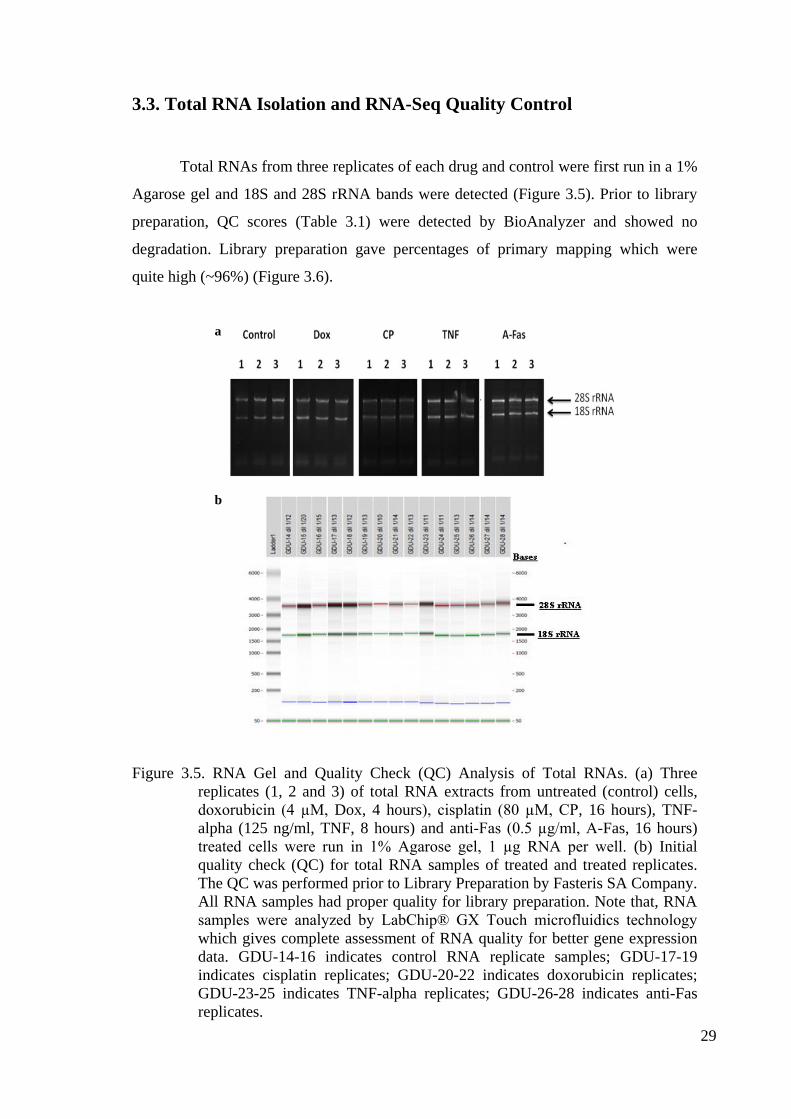
3.3. Total RNA Isolation and RNA-Seq Quality Control
Total RNAs from three replicates of each drug and control were first run in a 1%
Agarose gel and 18S and 28S rRNA bands were detected (Figure 3.5). Prior to library
preparation, QC scores (Table 3.1) were detected by BioAnalyzer and showed no
degradation. Library preparation gave percentages of primary mapping which were
quite high (~96%) (Figure 3.6).
Figure 3.5. RNA Gel and Quality Check (QC) Analysis of Total RNAs. (a) Three
replicates (1, 2 and 3) of total RNA extracts from untreated (control) cells,
doxorubicin (4 µM, Dox, 4 hours), cisplatin (80 µM, CP, 16 hours), TNF-
alpha (125 ng/ml, TNF, 8 hours) and anti-Fas (0.5 µg/ml, A-Fas, 16 hours)
treated cells were run in 1% Agarose gel, 1 µg RNA per well. (b) Initial
quality check (QC) for total RNA samples of treated and treated replicates.
The QC was performed prior to Library Preparation by Fasteris SA Company.
All RNA samples had proper quality for library preparation. Note that, RNA
samples were analyzed by LabChip® GX Touch microfluidics technology
which gives complete assessment of RNA quality for better gene expression
data. GDU-14-16 indicates control RNA replicate samples; GDU-17-19
indicates cisplatin replicates; GDU-20-22 indicates doxorubicin replicates;
GDU-23-25 indicates TNF-alpha replicates; GDU-26-28 indicates anti-Fas
replicates.
b
a
Page 40
30
Table 3.1. Library Preparation of RNA Samples. Library of total RNA replicates of
control and -treated cells were prepared. The read and mapping results
indicated in the table. The percentage of primary mapping was around 96%
percent which is good.
Library RNA Quality
Score
Reads Primary
Mappings
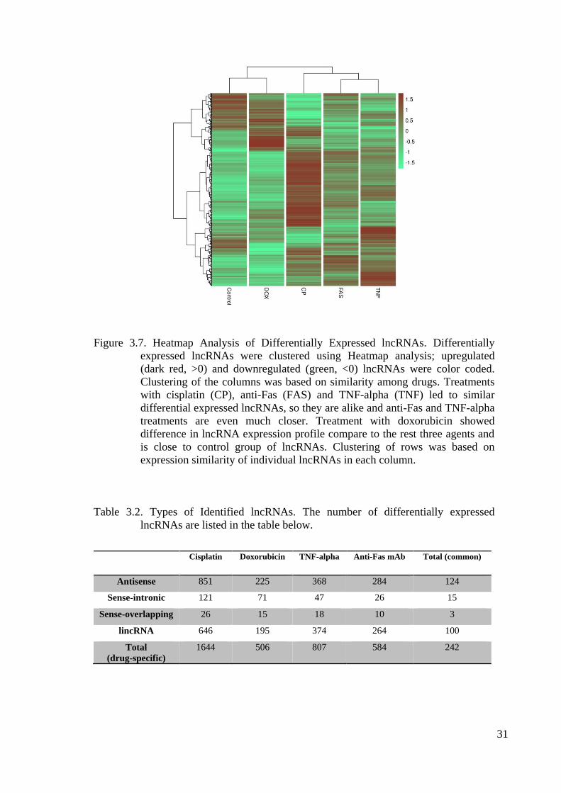
% of Primary
Mappings
Alternate
mappings
Control (1) 9.3 19,478,665 19,066,837 97.89% 5,031,257
Control (2) 9.2 13,977,569 13,636,817 97.56% 3,451,273
Control (3) 9.4 13,989,348 13,640,586 97.51% 3,476,351
Cisplatin (1) 9.0 16,695,258 15,975,390 95.69% 6,082,994
Cisplatin (2) 9.3 18,871,780 17,888,988 94.79% 7,554,017
Cisplatin (3) 8.9 16,922,803 16,051,158 94.85% 6,122,212
Doxorubicin (1) 7.7 13,255,959 12,888,311 97.23% 3,920,319
Doxorubicin (2) 8.8 13,635,427 13,293,528 97.49% 3,732,917
Doxorubicin (3) 7.9 15,485,199 15,148,239 97.82% 4,883,967
TNF-alpha (1) 8.9 15,720,221 15,243,443 96.97% 4,475,529
TNF-alpha (2) 8.3 19,665,744 18,901,417 96.11% 6,027,820
TNF-alpha (3) 8.6 20,958,445 20,121,618 96.01% 6,417,161
anti-Fas (1) 9.3 22,066,898 21,090,702 95.58% 7,061,540
anti-Fas (2) 8.6 19,332,230 18,460,235 95.49% 5,994,549
anti-Fas (3) 9.1 18,825,067 18,129,644 96.31% 6,015,316
3.4. Bioinformatics Analysis – lncRNA candidates
The bioinformatics analysis identified 24 antisense lncRNAs, 5 sense-intronic
lncRNAs and 17 lincRNAs that were significantly up- or down-regulated. Cisplatin,
doxorubicin, anti-Fas and TNF-alpha treatment triggered differential expression of drug
specific 1644, 506, 584 and 807 lncRNAs, respectively (2-fold and upper, P<0.01)
(Table 3.2). The HeatMap analysis of identified lncRNAs illustrated the similarity
among expressional profiles of each drug. As a result, TNF-alpha and anti-Fas treatment
showed a higher similarity and they together were similar to cisplatin. However,
doxorubicin showed different lncRNA expression profile and was closer to the profile
of control cells (figure 3.7). Top 20 up- and down-regulated lncRNAs common for all
drugs were listed and among them four candidates were selected (Table 3.3).
Page 41
31
Figure 3.7. Heatmap Analysis of Differentially Expressed lncRNAs. Differentially
expressed lncRNAs were clustered using Heatmap analysis; upregulated
(dark red, >0) and downregulated (green, <0) lncRNAs were color coded.
Clustering of the columns was based on similarity among drugs. Treatments
with cisplatin (CP), anti-Fas (FAS) and TNF-alpha (TNF) led to similar
differential expressed lncRNAs, so they are alike and anti-Fas and TNF-alpha
treatments are even much closer. Treatment with doxorubicin showed
difference in lncRNA expression profile compare to the rest three agents and
is close to control group of lncRNAs. Clustering of rows was based on
expression similarity of individual lncRNAs in each column.
Table 3.2. Types of Identified lncRNAs. The number of differentially expressed
lncRNAs are listed in the table below.
Cisplatin Doxorubicin TNF-alpha Anti-Fas mAb Total (common)
Antisense 851 225 368 284 124
Sense-intronic 121 71 47 26 15
Sense-overlapping 26 15 18 10 3
lincRNA 646 195 374 264 100
Total
(drug-specific)
1644 506 807 584 242
Page 42
32
Table 3.3. List of Top 20 Common Upregulated and Downregulated lncRNAs.
lncRNAs that were significantly up- and down-regulated upon treatment with
all agents is listed below with their antisense/sense partner coding genes. The
majority top 20 upregulated (a) and downregulated (b) lncRNAs are antisense
lncRNAs and their counter genes are protein coding genes playing role in
different cellular events and pathways. The lncRNAs in red boxes were
chosen as candidates due to their potential to regulate apoptosis.
a
b
Page 43
33
Two of selected candidate lncRNAs were antisense partner of receptors, that
play a direct role in apoptosis (TRAIL-R2 and FAF1) (Figure 3.8) and two candidates
lncRNAs were antisense partner of transcription factors (GTF2A1 and CAMTA1). All
of the candidates are uncharacterized. The candidate lncRNA RP11-875O11.2
(ENSG00000246130) locates on chromosome 8 (8:23,068,229-23,083,619) and it is a
known antisense partner of TRAIL-R2 (tumor necrosis factor receptor superfamily
member 10b, TNFRSF10B). Another candidate lncRNA RP5-850O15.3
(ENSG00000225767) locates on chromosome 1 (1: 50,461,469-50,471,150) and it is a
known antisense partner of FAF1 (Fas (TNFRSF6) associated factor 1). The other
candidate lncRNA CTD-2506P8.6-001 (ENSG00000273783) locates on chromosome
14 (14: 81,221,218-81,222,460) and it is a known antisense partner of GTF2A1 (general
transcription factor IIA, 1, 19/37kDa), which is a known general transcription factor.
The last candidate lncRNA RP11-312B8.1 locates on chromosome 1 (1: 6,783,892-
6,784,843) and it is a known antisense partner of CAMTA1 (calmodulin binding
transcription activator 1).
(cont. on next page)
Figure 3.8. Candidate lncRNAs. The figure shows genomic location of two of four
selected candidate lncRNAs (green highlighted) which were obtained from
Ensembl (http://www.ensembl.org/index.html). (a) Candidate lncRNA
RP11-875O11.2 locates on the forward strand and is in antisense position
(blue) to two coding and two noncoding (nonsense mediated decay and
retained intron) isoforms of TNFRSF10B (reverse strand), which plays a
direct role in receptor mediated apoptosis. The overlapping region (in the
red frame) between candidate lncRNA and the antisense gene is 196 bp
long. (b) Candidate lncRNA RP5-850O15.3 locates on the forward strand
and is in antisense position to two coding and one noncoding (nonsense
mediated decay) isoforms of FAF1 (reverse strand), which also plays a
direct role in receptor mediated apoptosis. The overlapping region between
candidate lncRNA and the antisense gene is 9700 bp long.
a
TNRSF10B
Overlapping region
Candidate lncRNA
Page 44
34
Figure 3.8. (cont.)
b
FAF1
Candidate lncRNA
Overlapping region
Page 45
35
CHAPTER 4
DISCUSSION
The dose kinetics of cisplatin showed a significant increase in parallel to
increasing in drug concentration. The reference dose prior to experiment was obtained
from Wang et al. (2010) (30 μM for 24 hours). Under our experimental setup and
reagents, 80 μM cisplatin was selected due to its moderate affect for 16 hours according
to Flow Cytometry analysis. Compared to the reference dose (30 μ), we needed to
increase concentration drug dose to find an optimum dose during drug dose kinetics. On
the other hand, it should be noted that the experiment setting of apoptosis detection was
different - Annexin V and 7AAD staining vs. DAPI staining.
Doxorubicin treatment of HeLa cells was challenging due to its unique way of
drug response. Treated cells were shifting directly from live cell quadrant (Annexin V-
/7AAD
-) to double positive quadrant (Annexin V
+/7AAD
+), thus in a smaller doses
overflow to other quadrants was observed. However, a drug concentration of 4 μM
showed a clear shift of cell to double positive quadrant. The reference dose and time
was obtained from Bien et al. (2010) (1 μM and 24 hours). However, we decided to
increase the doxorubicin dose and decrease exposure time. Considering the difference in
detection method of cell death between Bien et al. and us, doxorubicin dose was chosen
high enough to see clear shifting of population despite the risk of causing necrosis. In
such case, doxorubicin may be used as another control group; however, possible
presence of necrosis should be detected by performing extra experiments – detection
necrotic markers (like necrostatins), which was not in our experimental settings.
Anti-Fas mAb treatment showed similar apoptotic rates with our doses and the
maximum apoptotic rate (~20%) due to having slight expression of FasR as mentioned
in Chapter 1. The reference dose and time was 0.1 μg/ml for 24 hours (Holmstorm et al.
1999). Due to similarity among doses, we chose 0.5 μg/ml, the middle dose which had
~20% apoptosis in 16 hours.
TNF-alpha alone did not induce cell death in HeLa cells, which is expected as
indicated in Chapter 1. Coupling with cycloheximide, TNF-alpha caused cell death in a
dose-dependent manner. Our reference dose and time was 100 ng/ml and 6 hours (Wang
Page 46
36
et al. 2008b). Although, the same apoptotic rate (35%) could be achieved by treating
with 125 ng/ml for 8 hours, this could be explained by the difference in measurement
technique of apoptotic rate.
Fluorescent Microscopy analysis of each chosen drug dose showed similar
results (qualitatively) by using same stains and the same protocol. All the control cells
were not stained any of dyes (Annexin V and 7AAD). However, the cisplatin treatment
caused numerous single or double stained cells. Doxorubicin treated cells showed
double stained pure population, just like in the Flow Cytometry results. Fas caused very
few single (majority was Annexin V positive) stained cells. Treatment with TNF-alpha
resulted with unstained and numerous single stained cells.
Western Blotting experiment gave us the state of mind of cells in a given drug
dose and time with respect to apoptosis. As expected, cisplatin showed caspase 8
activation; however neither procaspase 9 nor active caspase 9 could be detected under
our experimental settings, suggesting another time kinetics (less than 16 hours) to show
procaspase 9 and active fragment. Similar time kinetics (more than 16 hours) should be
performed to show cleavage of procaspase 3 and detection of active fragment.
Doxorubicin treatment led to detection of tiny active fragments of caspase 8 and
9, but not caspase 3, thus presumably no or too less apoptosis and existence of another
type of cell death (like necrosis). Considering together with Flow Cytometry results,
doxorubicin dose (4 μM) might be too much or exposure time (4 hours) might be less.
Additional time kinetics (more than 4 hours) should be done to show cleavage of
caspases. In case of no detected even after time kinetics, then smaller doses should be
tested.
TNF-alpha treatment showed activation of caspase 8 and 9, which was expected
and mentioned in Chapter 1. However, under our experimental settings - chosen time
(8hours) and dose (125 ng/ml), neither procaspase nor active caspase 3 could be
detected due to timing or too much drug. In our case, additional time kinetics (less than
8 hours) should be tested to show procaspase 3 and active caspase 3.
Anti-Fas treatment showed activation of both effector caspases and the
executioner caspase. Compare to other selected drug doses, anti-Fas treatment showed
the least apoptotic rate according to Flow Cytometry analysis, however, it showed the
action of effector and executioner caspases. As it is mentioned in Chapter 1, anti-Fas
binds to and activates CD95/Fas receptor, which play role activation of extrinsic
apoptotic pathway. Unlike anti-Fas, doxorubicin, cisplatin and TNF-alpha induce
Page 47
37
several pathways and can cause cytotoxicity. Thus, treatment with them might have
additional effects.
Deep sequencing method was based on detection of lncRNAs, because not all
lncRNAs have poly A tail. On the other hand, RNA Quality scores were very good and
library preparation showed very high percentage of primary mapping.
Bioinformatics analysis by Allmer lab gave us to direct them and analyze the
output together unlike paid services. Using the latest programs and articles and using
very stringent filters was powerful side of the bioinformatics results.
The bioinformatics results show that lncRNAs are differentially expressed upon
treatment with cisplatin, doxorubicin, anti-Fas and TNF-alpha. Heatmap analysis of
differentially expressed lncRNAs showed similar fashion of drug screening and western
blotting analysis: doxorubicin treated cells had a unique profile and were close to
control, not other agents. On the other hand, anti-Fas and TNF-alpha treatments had
close profiles and their profiles were close profile upon cisplatin treatment. Induction of
extrinsic apoptotic pathway upon cisplatin treatment might contribute the similarity
with TNF-alpha and anti-Fas treatment with respect to apoptosis. In addition, the
expressional similarities of individual lncRNAs in each raw showed expressional
difference between control (being alive) and treatments (dying cells): some lncRNAs
were upregulated only in control and vice versa.
There are known pro- and anti-apoptotic lncRNAs that are differentially
expressed and play role in apoptosis as indicated in Chapter 1 and literature. According
to our data, there are several well-known lncRNAs differentially expressed as well. One
of the well-known lncRNA is GAS5, and it was significantly upregulated upon cisplatin
and anti-Fas treatment, but not upon doxorubicin and TNF-alpha treatment. Such a
result is not surprising, because, expression of lncRNAs, including GAS5 (Ozgur et al.
2013), is cell type and drug/ligand/inducer dependent in different cases, including
apoptosis due to difference in molecular mechanism of specific lncRNA. More
interesting, newly discovered and uncharacterized antisense lncRNA of GAS5, GAS5-
AS1, is upregulated upon cisplatin, TNF-alpha and anti-Fas treatment as well, however,
it is significantly downregulated upon doxorubicin treatment. The expression of GAS5-
AS1 seems independent from GAS5 expression and still the impact of its transcription
on GAS5 expression is unknown (Pickard and Williams 2015).
Expression of pro-apoptotic INXS, as indicated in Chapter 1, was elevated upon
cisplatin, TNF-alpha and anti-Fas treatment, however, it is significantly downregulated
Page 48
38
upon doxorubicin treatment. In a number of tumor types, INXS is downregulated and
upon activation of intrinsic apoptotic pathway in those tumor cells, it is elevated
(DeOcesano-Pereira et al. 2014). However, the effect of doxorubicin on INXS
expression is currently unknown and downregulation of INXS in apoptotic HeLa cells
may serve shifting BCL-XS/BCL-XL ratio to anti-apoptotic BCL-XL splice variant and
thus blocking intrinsic apoptotic pathway.
Most agents, especially anti-cancer drugs, work different and influence various
cellular processes and signaling pathways. There was a clear difference in cell the death
mechanism among agents, especially between doxorubicin and the rest agents,
according to Flow Cytometry and Western Blotting analysis. The same trend was
observed in lncRNA expression levels as well, according to Heatmap Analysis, such as
in Linc-RoR (Grammatikakis et al. 2014), HOTAIR and AFAP1-AS1 expression.
Consistent with literature, in our data, Linc-RoR expression is elevated; HOTAIR and
AFAP1-AS1 expressions are decreased upon cisplatin, TNF-alpha and anti-Fas
treatment, however, their expression level did not change significantly upon
doxorubicin treatment. On the other hand, anti-apoptotic LUST lncRNA (Rintala-Maki
et al. 2009) is significantly decreased upon only doxorubicin treatment. Furthermore,
pro-apoptotic HOXA-AS2 expression is elevated upon treatment with all agents, which
is expected during apoptosis.
Expression of uncharacterized MIR7-3HG lncRNA, which is a miR7-3 host
gene that plays role in apoptosis, is elevated just in TNF-alpha and anti-Fas mediated
cell death and may serve as an important insight about additional molecular mechanism
of TNF-alpha and anti-Fas mediated cell death. Likewise, several lncRNAs like ANRIL
(Huang et al. 2015) and PVT1 (Ding et al. 2015) expression levels vary among agents as
well according to our data. Anti-apoptotic ANRIL expression level decreased upon only
cisplatin and anti-Fas treatment; anti-apoptotic PVT1 expression decreased due to
cisplatin and doxorubicin treatment, however, its level significantly increased
unexpectedly upon TNF-alpha treatment.
Identified lncRNAs, especially antisense and sense intronic/overlapping
lncRNAs, may serve us very important information about regulation of molecular
mechanism behind apoptosis. The selected candidates are common in all agents and
have top scores among differentially expressed lncRNAs. Thus, they can be very
powerful candidates and may have a great impact on apoptotic phenotype. To be
precise, possible regulation of apoptosis via TNFRSF10B (tumor necrosis factor
Page 49
39
receptor superfamily, member 10) antisense and FAF1 (Fas (TNFRSF6) associated
factor 1) antisense lncRNAs would not be a surprise and even that is what we have
expected. TNFRSF10B and FAF1 directly play role in apoptosis and targeting those key
apoptotic markers may have major impacts on downstream events and thus having a
global effect on cell fate.
On the other hand, GTF2A1 (general transcription factor IIA, 1) antisense and
CAMTA1 (calmodulin binding transcription activator 1) antisense candidate lncRNAs
may provide unique information about transcriptional regulation of pro-apoptotic and or
anti-apoptotic genes, because, GTF2A1 gene play role in transcriptional regulation
(Kobayashi et al. 1995) and is hypermethylated in ovarian cancer (Huang et al. 2009).
CAMTA1 gene is reported play role in apoptosis (Juhlin et al. 2015), acts as tumor
suppressor (Henrich et al. 2011) and is deleted in adrenomedullary tumors (Edstrom et
al. 2002).
Page 50
40
CHAPTER 5
CONCLUSION
In this study, identification and functional characterization of differentially
expressed lncRNAs during apoptosis was performed via deep sequencing method. After
a successful drug screening experiments of each drug, Fluorescence Microscopy and
Western Blotting analysis were further done for verification of selected doses and the
results support each other.
Further, RNA samples from samples passed Quality Control and Library
preparation of RNA samples was successful as well. Mapping of primary reads to
human genome was very high. Bioinformatics analysis was done by Allmer lab
(IzTech) and the thresholds and approaches in detection of differentially expressed
genes were very stringent as well.
In conclusion, a systematic and comprehensive approach led to identification of
differentially expressed 1644, 506, 584 and 807 lncRNAs upon treatments with
cisplatin, doxorubicin, Fas mAb and TNF-alpha, respectively (2-fold or higher, P <
0.01). Among them 124 antisense lncRNAs, 15 sense-intronic lncRNAs, 3 sense-
overlapping lncRNAs and 100 lincRNAs were significantly and commonly up- or
down-regulated upon induction of cell death via cisplatin, doxorubicin, anti-Fas mAb
and TNF-alpha. Interestingly, top scored differentially expressed antisense and sense
intronic/overlapping lncRNAs in each drug were similar. Those lncRNAs might have
functional role(s) and might play role in cell death, including apoptosis as regulator of
apoptosis. Antisense and sense intronic/overlapping lncRNAs might regulate their
antisense/sense positioned protein coding genes unlike lincRNAs as indicated in
Chapter 1. According to our data, two of them were positioned as antisense to death
receptors (TRAIL-R2 and FasR) which play role directly in apoptosis as key regulators.
As future direction, the candidates should be functionally characterized by
knockdown and/or overexpression approach.
Page 51
41
REFERENCES
Adams J. M., Cory S. (1998). The Bcl-2 protein family: arbiters of cell survival.
Science 281: 1322–1325.
Annilo T., Kepp K., Laan M. (2009). Natural antisense transcript of natriuretic peptide
precursor A (NPPA): structural organization and modulation of NPPA expression.
BMC Mol. Biol. 10: 81.
Balakirev E. S., Ayala F. J. (2003). Pseudogenes: Are they “junk” or functional DNA?
Annu. Rev. Genet. 37: 123–151.
Barreau C., Paillard L., Osborne H. B. (2006). AU-rich elements and associated factors:
Are there unifying principles? Nucleic Acids Res. 33: 7138–7150.
Barry M., Bleackley R. C. (2002). Cytotoxic T lymphocytes: all roads lead to death.
Nat. Rev. Immunol. 2: 401–9.
Bekpen C., Margues-Bonet T., Alkan C., Antonacci F., Leogrande M. B. et al. (2009).
Death and resurrection of the human IRGM gene. PLoS Genet. 5: e1000403.
Beltran M., Puig I., Peña C., García J. M., Alvarez A. B. et al. (2008). A natural
antisense transcript regulates Zeb2/Sip1 gene expression during Snail1-induced
epithelial-mesenchymal transition. Genes. Dev. 22: 756–769.
Bernard D., Prasanth K. V., Tripathi V., Colasse S., Nakamura T. et al. (2010). A long
nuclear-retained non-coding RNA regulates synaptogenesis by modulating gene
expression. EMBO J. 29: 3082–3093.
Bertani S., Sauer S., Bolotin E., Sauer F. (2011). The noncoding RNA Mistral activates
Hoxa6 and Hoxa7 expression and stem cell differentiation by recruiting MLL1 to
chromatin. Mol. Cell 43: 1040–1046.
Bertone P., Stolc V., Royce T. E., Rozowsky J. S., Urban A. E. et al. (2004). Global
identification of human transcribed sequences with genome tiling arrays. Science
306: 2242–2246.
Bialik S., Zalckvar E., Ber Y., Rubinstein A. D., Kimchi A. (2010) Systems biology
analysis of programmed cell death. Trends Biochem. Sci. 35: 556–564.
Bien S., Rimmbach C., Neumann H., Niessen J., Reimer E., Ritter C. A., Rosskopf D.,
Cinatl J., Michaelis M., Schroeder H. W. S., Kroemer H. K. (2010). Doxorubicin-
induced cell death requires cathepsin B in HeLa cells. Biochem. Pharm., 80:
1466–1477
Birney E., Stamatoyannopoulos J. A., Dutta A., Guigó R., Gingeras T. R. et al. (2007).
Identification and analysis of functional elements in 1% of the human genome by
the ENCODE pilot project. Nature 447: 799–816.
Page 52
42
Brannan C. I., Dees E. C., Ingram R. S., Tilghman S. M. (1990). The product of the H19
gene may function as an RNA. Mol. Cell. Biol. 20: 28–36.
Brockdorff N., Ashworth A., Kay G. F., McCabe V. M., Norris D .P. et al. (1992). The
product of the mouse Xist gene is a 15 kb inactive X-specific transcript containing
no conserved ORF and located in the nucleus. Cell 71: 515-526.
Brown C. J., Hendrich B. D., Rupert J. L., Lafrenière R. G., Xing Y.et al. (1992). The
human XIST gene: analysis of a 17 kb inactive X-specific RNA that contains
conserved repeats and is highly localized within the nucleus. Cell 71: 527–542.
Brown C. J., Lafrenière R. G., Powers V. E., Sebastio G., Ballabio A. et al. (1991).
Localization of the X inactivation centre on the human X chromosome in Xq13.
Nature 349: 82–84.
Brozovic A., Ambriović-Ristov A., Osmak M. (2010). The relationship between
cisplatin-induced reactive oxygen species, glutathione, and BCL-2 and resistance
to cisplatin. Crit. Rev. Toxicol. 40: 347–359.
Brunner T., Wasem C., Torgler R., Cima I., Jakob S., Corazza N. (2003). Fas
(CD95/Apo-1) ligand regulation in T cell homeostasis, cell-mediated cytotoxicity
and immune pathology. Semin. Immunol. 15: 167–76.
Buchholz T. A. et al. (2002). Global gene expression changes during neoadjuvant
chemotherapy for human breast cancer. Cancer J. 8: 461–468.
Buratowski S. (2008). Gene expression—Where to start? Science 322: 1804–1805.
Cabili M. N., Trapnell C., Goff L., Koziol M., Tazon-Vega B. et al. (2011). Integrative
annotation of human large intergenic noncoding RNAs reveals global properties
and specific subclasses. Genes Dev. 25: 1915–1927.
Cai X., Cullen B. R. (2007). The imprinted H19 noncoding RNA is a primary
microRNA precursor. RNA 13: 313–316.
Campos E. I., Reiberg D. (2009). Histones: annotating chromatin. Annu. Rev. Genet.
43: 559–599.
Cao C. et al. (2008). AMP-activated protein kinase contributes to UV and H2O2-
induced apoptosis in human skin keratinocytes. J. Biol. Chem. 283: 28897–28908.
Carninci P., Kasukawa T., Katayama S., Gough J., Frith M. C. et al. (2005). The
transcriptional landscape of the mammalian genome. Science 309: 1559–1563.
Carvalho C. et al. (2009). Doxorubicin: the good, the bad and the ugly effect. Curr.
Med. Chem. 16: 3267–3285.
Chau B. N., Cheng E. H., Kerr D. A., Hardwick J. M. (2000). Aven, a novel inhibitor of
caspase activation, binds Bcl-xL and Apaf-1. Mol. Cell 6: 31–40.
Page 53
43
Che C.M., Siu F.M. (2010). Metal complexes in medicine with a focus on enzyme
inhibition. Curr. Opin. Chem. Biol. 14: 255–261.
Chen D., Milacic V., Frezza M., Dou Q.P. (2009). Metal complexes, their cellular
targets and potential for cancer therapy. Curr. Pharm. Des. 15: 777–791.
Chen L. L., Carmichael G. G. (2009). Altered nuclear retention of mRNAs containing
inverted repeats in human embryonic stem cells: functional role of a nuclear
noncoding RNA. Mol. Cell 35: 467–478.
Chinnaiyan A. M. (1999). The apoptosome: heart and soul of the cell death machine.
Neoplasia 1: 5–15.
Clemson C. M., McNeil J. A., Willard H. F., Lawrence J. B. (1996). XIST RNA paints
the inactive X chromosome at interphase: evidence for a novel RNA involved in
nuclear/chromosome structure. J. Cell Biol. 132: 259–275.
Cohen G. M., Sun X. M., Fearnhead H., MacFarlane M., Brown D. G., Snowden R. T.
et al. (1994) Formation of large molecular weight fragments of DNA is a key
committed step of apoptosis in thymocytes. J. Immunol. 153: 507–516.
Cohen G. M. (1997). Caspases: the executioners of apoptosis. Biochem J. 326: 1–16.
Core L. J., Waterfall J. J., Lis J. T. (2008). Nascent RNA sequencing reveals widespread
pausing and divergent initiation at human promoters. Science 322: 1845–1858.
Cory S., Adams J. M. (2002). The Bcl2 family: regulators of the cellular life-or-death
switch. Nat. Rev. Cancer 2: 647–56.
Cui Z., Ren S., Lu J. et al. (2013). The prostate cancer-up-regulated long noncoding
RNA PlncRNA-1modulates apoptosis and proliferation through reciprocal
regulation of androgen receptor. Urologic Oncology, 31: 1117–1123.
de Koning A. P., Gu W., Castoe T. A., Batzer M. A., Pollock D. D. (2011). Repetitive
elements may comprise over two-thirds of the human genome. PLoS Genet. 7:
E1002384.
DeOcesano-Pereira C., Amaral M. S., Parreira K. S., Ayupe A. C., Jacysyn J. F.,
Amarante-Mendes G. P., Reis E. M., Verjovski-Almeida S. (2014). Long non-
coding RNA INXS is a critical mediator of BCL-XS induced apoptosis. Nucleic
Acids Res. 42:8343-55.
Desoize, Madoulet B. C. (2002). Particular aspects of platinum compounds used at
present in cancer treatment. Crit. Rev. Oncol. Hematol. 42: 317–325.
Ding C., Yang Z., Lv Z., Du C., Xiao H., Peng C., Cheng S., Xie H., Zhou L., Wu J.,
Zheng S. (2015). Long non-coding RNA PVT1 is associated with tumor
progression and predicts recurrence in hepatocellular carcinoma patients. Oncol.
Lett. 9: 955-963.
Page 54
44
Djebali S., Davis C. A., Merkel A., Dobin A., Lassmann T.et al. (2012). Landscape of
transcription in human cells. Nature 489: 101–108.
Duret L., Chureau C., Samain S., Weissenbach J., Avner P. (2006). The Xist RNA gene
evolved in eutherians by pseudogenization of a protein-coding gene. Science 312:
1653–1655.
Ebisuya M., Yamamoto T., Nakajima M., Nishida E. (2008). Ripples from neighbouring
transcription. Nat. Cell Biol. 10: 1106–1113.
Ebralidze A. K., Guibal F. C., Steidl U., Zhang P., Lee S. et al. (2008). PU.1 expression
is modulated by the balance of functional sense and antisense RNAs regulated by
a shared cisregulatory element. Genes Dev. 22: 2085–2092.
Edstrom E.E., Nord B., Carling T., Juhlin C., Backdahl M., Hoog A., Larsson C. (2002).
Loss of heterozygosity on the short arm of chromosome 1 in pheochromocytoma
and abdominal paraganglioma. World J. Surg. 26: 965–71.
Edwards C. A., Ferguson-Smith A. C. (2007). Mechanisms regulating imprinted genes
in clusters. Curr. Opin. Cell Biol. 19: 281–289.
Elisaphenko E. A., Kolesnikov N. N., Shevchenko A. I., Rogozin I. B., Nesterova T. B.
(2008). A dual origin of the Xist gene from a protein-coding gene and a set of
transposable elements. PLoS ONE 3: e2521.
Enari M., Sakahira H., Yokoyama H., Okawa K., Iwamatsu A., Nagata S. (1998). A
caspase-activated DNase that degrades DNA during apoptosis, and its inhibitor
ICAD. Nature 391: 43–50.
ENCODE Project Consortium (2012). An integrated encyclopedia of DNA elements in
the human genome. Nature 489: 57–74.
Esposti M. D. (2002). The roles of Bid. Apoptosis 7: 433–40.
Fadok V. A., de Cathelineau A., Daleke D. L., Henson P. M., Bratton D. L. (2001).
Loss of phospholipid asymmetry and surface exposure of phosphatidylserine is
required for phagocytosis of apoptotic cells by macrophages and fibroblasts. J.
Biol. Chem. 276: 1071–7.
Faghihi M. A., Wahlestedt C. (2009). Regulatory roles of natural antisense transcripts.
Nat. Rev. Mol. Cell Biol. 10: 637–643.
Faghihi M. A., Modarresi F., Khalil A. M, Wood D. E., Sahagan B. G. et al. (2008).
Expression of a noncoding RNA is elevated in Alzheimer’s disease and drives
rapid feed-forward regulation of beta-secretase. Nat. Med. 14: 723–730.
Fan Z., Beresford P. J., Oh D. Y., Zhang D., Lieberman J. (2003). Tumor suppressor
NM23-H1 is a granzyme A-activated DNase during CTLmediated apoptosis, and
the nucleosome assembly protein SET is its inhibitor. Cell 112: 659–72.
Page 55
45
Florea A.M., Büsselberg D. (2011). Cisplatin as an Anti-Tumor Drug: Cellular
Mechanisms of Activity, Drug Resistance and Induced Side Effects. Cancers 3:
1351-1371.
Franco-Zorrilla J. M., Valli A., Todesco M., Mateos I., Puga M. I. et al. (2007). Target
mimicry provides a new mechanism for regulation of microRNA activity. Nat.
Genet. 39: 1033–1037.
Fu X., Ravindranath L., Tran N., Petrovics G., Srivastava S. (2006). Regulation of
apoptosis by a prostate-specific and prostate cancer-associated noncoding gene,
PCGEM1. DNA Cell Biol. 25(3):135-41.
Gall J. G. (1981). Chromosome structure and the C-value paradox. J. Cell Biol. 91: 3s–
14s.
Gong Z., Zhang S., Zeng Z., Wu H., Yang Q., Xiong F., Shi L., Yang J., Zhang W.,
Zhou Y., Zeng Y., Li X., Xiang B., Peng S., Zhou M., Li X., Tan M., Li Y., Xiong
W., Li G. (2014). LOC401317, a p53-regulated long non-coding RNA, inhibits
cell proliferation and induces apoptosis in the nasopharyngeal carcinoma cell line
HNE2. PLoS One 9(11):e110674.
Goping I. S., Barry M., Liston P., Sawchuk T., Constantinescu G., Michalak K. M.,
Shostak I., Roberts D. L., Hunter A. M.,Korneluk R., Bleackley R. C. (2003).
Granzyme B-induced apoptosis requires both direct caspase activation and relief
of caspase inhibition. Immunity 18: 355– 65.
Grammatikakis I., Panda A.C., Abdelmohsen K., Gorospe M. (2014). Long noncoding
RNAs (lncRNAs) and the molecular hallmarks of aging. Aging, 6(12): 992–1009.
Green D.R., Kroemer G. (2004). Science 305: 626–629.
Gregory, T. R.(2001). Coincidence, coevolution, or causation? DNA content, cell size,
and the C-value enigma. Biol. Rev. Camb. Philos. Soc. 76: 65–101.
Guenther M.G., Levine S.S., Boyer L.A., Jaenisch R., Young R.A. (2007). A chromatin
landmark and transcription initiation at most promoters in human cells. Cell 130:
77–88.
Guil S., Soler M., Portela A., Carrère J., Fonalleras E. et al. (2012). Intronic RNAs
mediate EZH2 regulation of epigenetic targets. Nat. Struct. Mol. Biol. 19: 664–
670.
Gupta R. A., Shah N., Wang K. C., Kim J., Horlings H. M. et al. (2010). Long non-
coding RNA HOTAIR reprograms chromatin state to promote cancer metastasis.
Nature 464: 1071–1076.
Gutschner T., Diederichs S. (2012). The hallmarks of cancer: a long non-coding RNA
point of view. RNA Biol. 9: 703–719.
Page 56
46
Guttman M., Amit M., Garber M., French C., Lin M. F. et al. (2009). Chromatin
signature reveals over a thousand highly conserved large non-coding RNAs in
mammals. Nature 458: 223–227.
Guttman M., Donaghey J., Carey B. W., Garberv, Grenier J. K. et al. (2011). lincRNAs
act in the circuitry controlling pluripotency and differentiation. Nature 477: 295–
300.
Gwinn D. M. et al. (2008). AMPK phosphorylation of raptor mediates a metabolic
checkpoint. Mol. Cell. 30: 214–226.
Hanahan D., Weinberg R. A. (2011). Hallmarks of cancer: the next generation. Cell
144: 646–674.
Harrison P. M., Zheng D., Zhang Z., Carriero N., Gerstein M. (2005). Transcribed
processed pseudogenes in the human genome: an intermediate form of expressed
retrosequence lacking protein-coding ability. Nucleic Acids Res. 33: 2374–2383.
Hawkins P. G., Morris K. V. (2010). Transcriptional regulation of Oct4 by a long non-
coding RNA antisense to Oct4-pseudogene 5. Transcription 1: 165–175.
He Y., Vogelstein B., Velculescu V. E., Papadopoulos N., Kinzler K. W. (2008). The
antisense transcriptomes of human cells. Science 322: 1855–1857.
Henrich K.O., Bauer T., Schulte J., Ehemann V., Deubzer H., Gogolin S., Muth D.,
Fischer M., Benner A., König R., Schwab M., Westermann F. (2011). CAMTA1,
a 1p36 tumor suppressor candidate, inhibits growth and activates differentiation
programs in neuroblastoma cells. Cancer Res. 71(8): 3142-51
Hill M. M., Adrain C., Duriez P. J., Creagh E. M., Martin S. J. (2004). Analysis of the
composition, assembly kinetics and activity of native Apaf-1 apoptosomes. Embo
J. 23: 2134–45.
Holmes D. S., Mayfield J. E., Sander G., Bonner J. (1972). Chromosomal RNA: its
properties. Science 177: 72–74.
Hsu H., Xiong J., Goeddel D. V. (1995). The TNF receptor 1-associated protein
TRADD signals cell death and NF-kappa B activation. Cell 81: 495–504.
Hu S., Snipas S. J., Vincenz C., Salvesen G., Dixit V. M. (Dec 1998). "Caspase-14 is a
novel developmentally regulated protease". J. Biol. Chem. 273(45): 29648–53
Huang M.D., Chen W.M., Qi F.Z., Xia R., Sun M., Xu T.P., Yin L., Zhang E.B., De W.,
Shu Y.Q. (2015). Long non-coding RNA ANRIL is upregulated in hepatocellular
carcinoma and regulates cell apoptosis by epigenetic silencing of KLF2. J.
Hematol. Oncol. 14: 8:50.
Huang Y., Jansen R., Fabbri E., Potter D., Liyanarachchi S., Chan M. et al. (2009).
Identification of candidate epigenetic biomarkers for ovarian cancer detection.
Oncol. Rep. 22: 853–861.
Page 57
47
Huarte M., Guttman M., Feldser D., Garber M., Koziol M. J. et al. (2010). A large
intergenic noncoding RNA induced by p53 mediates global gene repression in the
p53 response. Cell 142: 409–419.
Hung, T., Wang Y., Lin M. F., Koegel A. K., Kotake Y. et al. (2011). Extensive and
coordinated transcription of noncoding RNAs within cell-cycle promoters. Nat.
Genet. 43: 621–629.
Hüttenhofer A., Schattner P., Polacek N. (2005). Non-coding RNAs: Hope or hype?
Trends Genet. 21: 289–297.
Igney, F. H., Krammer P. H. (2002). Death and anti-death: tumour resistance to
apoptosis. Nat. Rev. Cancer 2: 277–88.
Jeon Y., Lee J. T. (2011). YY1 tethers Xist RNA to the inactive X nucleation center.
Cell 146: 119–133.
Ji P., Diederichs S., Wang W., Böing S., Metzger R. et al. (2003). MALAT-1, a novel
noncoding RNA, and thymosin beta4 predict metastasis and survival in early-
stage non-small cell lung cancer. Oncogene 22: 8031–8041.
Joza N., Susin S. A., Daugas E., Stanford W. L., Cho S. K., Li C. Y., Sasaki T., Elia A.
J., Cheng H. Y., Ravagnan L., Ferri K. F., Zamzami N., Wakeham A., Hakem R.,
Yoshida H., Kong Y. Y., Mak T. W., Zuniga-Pflucker J. C., Kroemer G.,
Penninger J. M. (2001). Essential role of the mitochondrial apoptosis-inducing
factor in programmed cell death. Nature 410: 549–54.
Juhlin C.C., Stenman A., Haglund F., Clark V.E., Brown T.C., Baranoski J., Bilguvar
K., Goh G., Welander J., Svahn F., Rubinstein J.C., Caramuta S., Yasuno K.,
Günel M., Backdahl M., Gimm O., Soderkvist P., Prasad M.L., Korah R., Lifton
R.P., Carling T. (2015). Whole-exome sequencing defines the mutational
landscape of pheochromocytoma and identifies KMT2D as a recurrently mutated
gene. Genes Chrom. Cancer 54(9): 542-54.
Kanduri C., Thakur N., Pandey R. R. (2006). The length of the transcript encoded from
the Kcnq1ot1 antisense promoter determines the degree of silencing. EMBO J. 25:
2096–2106.
Kang S. J., Wang S., Kuida K., Yuan J. (2002). Distinct downstream pathways of
caspase-11 in regulating apoptosis and cytokine maturation during septic shock
response. Cell Death Differ. 9: 1115–25.
Kanhere A., Viiri K., Araújo C. C., Rasaiyaah J., Bouwman R. D. et al. (2010). Short
RNAs are transcribed from repressed polycomb target genes and interact with
polycomb repressive complex-2. Mol. Cell 38: 675–688.
Kapranov P., St Laurent G., Raz T., Ozsolak F., Reynolds C. P. et al. (2010). The
majority of total nuclear-encoded non-ribosomal RNA in a human cell is ’dark
matter’ un-annotated RNA. BMC Biol. 8: 149.
Page 58
48
Karger, A. G., Basel I. Y., Mishima K., Azuma N., Tsuchida A., Takagi Y., Aoki T.,
Saito I. (2005). Overexpression of protein kinase Cdelta enhances cisplatin-
induced cytotoxicity correlated with p53 in gastric cancer cell line. Pathobiology
72: 152–159.
Kataoka T., Schroter M., Hahne M., Schneider P., Irmler M., Thome M., Froelich C. J.,
Tschopp J. (1998). FLIP prevents apoptosis induced by death receptors but not by
perforin/granzyme B, chemotherapeutic drugs, and gamma irradiation. J.
Immunol. 161: 3936–42.
Katayama S., Tomaru Y., Kasukawa T., Waki K., Nakanishi M. (2005). Antisense
transcription in the mammalian transcriptome. Science 309: 1564–1566.
Kelliher M. A., Grimm S., Ishida Y., Kuo F., Stanger B. Z., Leder P. (1998). The death
domain kinase RIP mediates the TNF-induced NF-kappaB signal. Immunity 8:
297–303.
Keniry A., Oxley D., Monnier P., Kyba M., Dandolo L. et al. (2012). The H19 lincRNA
is a developmental reservoir of miR-675 that suppresses growth and Igf1r. Nat.
Cell Biol. 14: 859–865.
Kerr J. F. R., Wyllie A. H., Currie A. R. (1972) Apoptosis: a basic biological
phenomenon with wide-ranging implications in tissue kinetics. Br. J. Cancer 26:
239–257.
Khaitan D., Dinger M. E., Mazar J.et al. (2011). The melanomaupregulated long
noncoding RNA SPRY4-IT1 modulates apoptosis and invasion. Cancer Research
71(11): 3852– 3862.
Khalil A. M., Guttman M., Huarte M., Garber M., Raj A. et al. (2009). Many human
large intergenic noncoding RNAs associate with chromatin-modifying complexes
and affect gene expression. Proc. Natl. Acad. Sci. USA 106: 11667–11672.
Kim T. K., Hemberg M., Gray J. M., Costa A. M., Bear D. M. et al. (2010). Widespread
transcription at neuronal activity-regulated enhancers. Nature 465: 182–187.
Kim Y. K., Furic L., Desgroseillers L., Maquat L. E. (2005). Mammalian Staufen1
recruits Upf1 to specific mRNA 3'UTRs so as to elicit mRNA decay. Cell 120:
195–208.
Kim,Y. K., Furic L., Parisien M., Major F., Desgroseillers L. et al. (2007). Staufen1
regulates diverse classes of mammalian transcripts. EMBO J. 26: 2670–2681.
Kischkel F. C., Hellbardt S., Behrmann I., Germer M., Pawlita M., Krammer P. H.,
Peter M. E. (1995). Cytotoxicity-dependent APO-1 (Fas/CD95)- associated
proteins form a death-inducing signaling complex (DISC) with the receptor.
EMBO J. 14: 5579–88.
Page 59
49
Kobayashi N., Boyer T.G. and Berk A.J. (1995). A class of activation domains interacts
directly with TFIIA and stimulates TFIIATFIID-promoter complex assembly.
Mol. Cell. Biol., 15: 6465-6473.
Koenig U., Eckhart L., Tschachler E. (2001). Evidence that caspase-13 is not a human
but a bovine gene. Biochem. Biophys. Res. Commun. 285:1150
Kotake Y., Nakagawa T., Kitagawa K., Suzuki S., Liu N.et al. (2011). Long non-coding
RNA ANRIL is required for the PRC2 recruitment to and silencing of
p15(INK4B) tumor suppressor gene. Oncogene 30: 1956–1962.
Kung J. T. Y., Colognori D., Jeannie T. L. (2013). Long Noncoding RNAs: Past,
Present, and Future. Genetics 193: 651–669
Kurosaka K., Takahashi M., Watanabe N., Kobayashi Y. (2003). Silent cleanup of very
early apoptotic cells by macrophages. J. Immunol. 171: 4672–9.
Kuwana T., Smith J. J., Muzio M., Dixit V., Newmeyer D. D., Kornbluth S. (1998).
Apoptosis induction by caspase-8 is amplified through the mitochondrial release
of cytochrome c. J. Biol. Chem. 273: 16589-16594.
Lanz R. B., Razani B., Goldberg A. D., O’Malley B. W. (2002). Distinct RNA motifs
are important for coactivation of steroid hormone receptors by steroid receptor
RNA activator (SRA). Proc. Natl. Acad. Sci. USA 99: 16081–16086.
Laubenbacher R, Hower V, Jarrah A, Torti SV, Shulaev V, Mendes P et al. (2009). A
systems biology view of cancer. Biochimica et Biophysica Acta 1796: 129–139.
Lee J. T. (2011). Gracefully ageing at 50, X-chromosome inactivation becomes a
paradigm for RNA and chromatin control. Nat. Rev. Mol. Cell Biol. 12: 815–826.
Lee J. T., Davidow L. S., Warshawsky D. (1999a). Tsix, a gene antisense to Xist at the
X-inactivation centre. Nat. Genet. 21: 400–404.
Leung K. L., Wang Y. T. (1999). Differential effects of chemotherapeutic agents on the
Bcl-2/Bax apoptosis pathway in human breast cancer cell line MCF-7. Basic
Cancer Res. Treat. 55: 73–83.
Lewin B.(1980). Gene Expression, 2: Eucaryotic Chromosomes. John Wiley & Sons,
New York.
Li H., Zhu H., Xu C. J., Yuan J. (1998). Cleavage of BID by caspase-8 mediates the
mitochondrial damage in the Fas pathway of apoptosis. Cell 94: 491–501.
Li L. Y., Luo X., Wang X. (2001). Endonuclease G is an apoptotic DNase when
released from mitochondria. Nature 412: 95–9.
Lieberman J., Fan Z. (2003). Nuclear war: the granzyme A-bomb. Curr. Opin.
Immunol. 15: 553–9.
Page 60
50
Lin R., Roychowdhury-Saha M., Black C., Watt A. T., Marcusson E. G. et al. (2011).
Control of RNA processing by a large non-coding RNA over-expressed in
carcinomas. FEBS Lett. 585: 671–676.
Liu F. T., Newland A. C., Jia L. (2003). Bax conformational change is a crucial step for
PUMA-mediated apoptosis in human leukemia. Biochem. Biophys. Res.
Commun. 310: 956–62.
Locksley R. M., Killeen N., Lenardo M. J. (2001). The TNF and TNF receptor
superfamilies: integrating mammalian biology. Cell 104: 487– 501.
Louro R., Smirnova A. S., Verjovski-Almeida S. (2009). Long intronic noncoding RNA
transcription: Expression noise or expression\ choice? Genomics 93: 291–298.
Lyle R., Watanabe D., te Vruchte D., Lerchner W., Smrzka O. W. et al. (2000). The
imprinted antisense RNA at the Igf2r locus overlaps but does not imprint Mas1.
Nat. Genet. 25: 19–21.
Lyon, M. F. (1961). Gene action in the X-chromosome of the mouse (Mus musculus
L.). Nature 190: 372–373.
Mao Y. S., Zhang B., Spector D. L. (2011). Biogenesis and function of nuclear bodies.
Trends Genet. 27: 295–306.
Martianov J., Ramadass A., Serra Barros A., Chow N., Akoulitchev A. (2007).
Repression of the human dihydrofolate reductase gene by a non-coding interfering
transcript. Nature 445: 666–670.
Martin S. J., Green D. R. (1995). Protease activation during apoptosis: death by a
thousand cuts? Cell 82: 349–352.
Martinvalet D., Zhu P., Lieberman J. (2005). GranzymeAinduces caspaseindependent
mitochondrial damage, a required first step for apoptosis. Immunity 22: 355–70.
McGahon A. J. et al. (1998). Chemotherapeutic drug-induced apoptosis in human
leukaemic cells is independent of the Fas (APO-1/CD95) receptor/ligand system.
Br. J. Haematol. 101: 539– 547.
McStay B., Grummt I. (2008). The epigenetics of rRNA genes: from molecular to
chromosome biology. Annu. Rev. Cell Dev. Biol. 24: 131–157.
Meisse D. et al. (2002). Sustained activation of AMP-activated protein kinase induces c-
Jun N-terminal kinase activation and apoptosis in liver cells. FEBS Lett. 526: 38–
42.
Mercer T. R., Gerhardt D. J., Dinger M. E., Crawford J., Trapnell C. et al. (2011).
Targeted RNA sequencing reveals the deep complexity of the human
transcriptome. Nat. Biotechnol. 30: 99–104
Page 61
51
Meyer N., Kim S. S., Penn L. Z. (2006). The Oscar-worthy role of Myc in apoptosis.
Semin. Cancer Biol. 16: 275–87.
Minotti G. et al. (2004). Anthracyclines: molecular advances and pharmacologic
developments in antitumor activity and cardiotoxicity. Pharmacol Rev. 56: 185–
229.
Mirsky A. E., Ris H. (1951). The desoxyribonucleic acid content of animal cells and its
evolutionary significance. J. Gen. Physiol. 34: 451–462.
Miura M., Friedlander R. M., Yuan J. (1995). Tumor necrosis factor-induced apoptosis
is mediated by a CrmA-sensitive cell death pathway. Proc. Natl. Acad. Sci. USA,
92: 8318-8322.
Mortazavi A., Williams B. A., McCue K., Schaeffer L., Wold B. (2008). Mapping and
quantifying mammalian transcriptomes by RNA-Seq. Nat. Methods 5: 621–628.
Muscella A., Calabriso N., Fanizzi F.P., De Pascali S.A., Urso L., Ciccarese A., Migoni
D., Marsigliante S. (2008). Pt[(O,O’-acac)(gamma-acac)(DMS)], a new Pt
compound exerting fast cytotoxicity inMCF-7 breast cancer cells via the
mitochondrial apoptotic pathway. Br. J. Pharmacol. 153: 34–49.
Nagano T., Mitchell J. A., Sanz L. A., Pauler F. M., Ferguson- Smith A. C. et al.
(2008). The Air noncoding RNA epigenetically silences transcription by targeting
G9a to chromatin. Science 322: 1717–1720.
Nakagawa T., Zhu H., Morishima N., Li E., Xu J., Yankner B. A., Yuan J. (2000).
Caspase-12 mediates endoplasmicreticulum-specific apoptosis and cytotoxicity by
amyloid-beta. Nature 403: 98–103.
Ng S. Y., Johnson A. C., Stanton L. W. (2011). Human long noncoding RNAs promote
pluripotency and neuronal differentiation by association with chromatin modifiers
and transcription factors. EMBO J. 31: 522–533.
Norbury C. J., Hickson I. D. (2001). Cellular responses to DNA damage. Annu Rev
Pharmacol Toxicol. 41: 367–401.
O'Connell J., O'Sullivan G. C., Collins J. K., Shanahan F. (1996). The Fas
counterattack: Fas-mediated T cell killing by colon cancer cells expressing Fas
ligand. J. Exp. Med. 184(3): 1075-82.
Oda E., Ohki R., Murasawa H., Nemoto J., Shibue T., Yamashita T., Tokino T.,
Taniguchi T., Tanaka N. (2000). Noxa, a BH3-only member of the Bcl-2 family
and candidate mediator of p53-induced apoptosis. Science 288: 1053–8.
Okazaki Y., Furuno M., Kasukawa T., Adachi J., Bono H.et al. (2002). Analysis of the
mouse transcriptome based on functional annotation of 60,770 full-length cDNAs.
Nature 420: 563–573.
Page 62
52
Ota T., Suzuki Y., Nishikawa T., Otsuki T., Sugiyama T. et al. (2004). Complete
sequencing and characterization of 21,243 full-length human cDNAs. Nat. Genet.
36: 40–45.
Ozgur E., Mert U., Isin M., Okutan M., Dalay N., Gezer U. (2013). Differential
expression of long non-coding RNAs during genotoxic stress-induced apoptosis in
HeLa and MCF-7 cells. Clin. and Exp. Med. 13: 119-126.
Pan G., Bauer J. H., Haridas V., Wang S., Liu D., Yu G., Vincenz C., Aggarwal B. B.,
Ni J., Dixit V. M. (1998). Identification and functional characterization of DR6, a
novel death domain-containing TNF receptor. FEBS Lett. 431 (3): 351–356.
Pandey R. R., Mondal T., Mohammad F., Enroth S., Redrup L. et al. (2008). Kcnq1ot1
antisense noncoding RNA mediates lineagespecific transcriptional silencing
through chromatin-level regulation. Mol. Cell 32: 232–246.
Paralkar V. R., Weiss M. J. (2011). A new 'Linc' between noncoding RNAs and blood
development. Genes Dev. 25(24): 2555-8.
Pardo J., Bosque A., Brehm R., Wallich R, Naval J., Müllbacher A., Anel A., Simon M.
M. (2004). Apoptotic pathways are selectively activated by granzyme A and/or
granzyme B in CTL-mediated target cell lysis. J. Cell Biol. 167(3): 457–468.
Park I. C., Woo S. H., Park M. J., Lee H. C., Lee S. J., Hong Y. J., Lee S. H., Hong S.
I., Rhee C. H. (2003). Ionizing radiation and nitric oxide donor sensitize Fas-
induced apoptosis via up-regulation of Fas in human cervical cancer cells. Oncol.
Rep. 10(3): 629-33.
Pearson J. C., Lemons D., McGinnis W. (2005). Modulating Hox gene functions during
animal body patterning. Nat. Rev. Genet. 6: 893–904.
Pickard M. R. Williams G. T. (2015). Molecular and Cellular Mechanisms of Action of
Tumour Suppressor GAS5 LncRNA. Genes 6: 484-499.
Pickard M .R., Mourtada-Maarabouni M., Williams G. T. (2013). Long non-coding
RNA GAS5 regulates apoptosis in prostate cancer cell lines. Biochim. Biophys.
Acta. 1832(10): 1613-23.
Pierpont M. E., Yunis J. J. (1977). Localization of chromosomal RNA in humn G-
banded metaphase chromosomes. Exp. Cell Res. 106: 303–308.
Pink R. C., Wicks K., Caley D. P., Punch E. K., Jacobs L. et al. (2011). Pseudogenes:
Pseudo-functional or key regulators in health and disease? RNA 17: 792–798.
Pitti R. M., Marsters S. A., Ruppert S., Donahue C. J., Moore A., Ashkenazi A. (1996).
Induction of apoptosis by Apo-2 ligand, a new member of the tumor necrosis
factor cytokine family. J. Biol. Chem. 271 (22): 12687–90.
Page 63
53
Preker P., Nielsen J., Kammler S., Lykke-Adersen S., Christensen M. S. et al. (2008).
RNA exosome depletion reveals transcription upstream of active human
promoters. Science 322: 1851–1854.
Prensner J. R., Iyer M. K., Balbin O. A., Dhanasekaran S. M., Cao Q. et al. (2011).
Transcriptome sequencing across a prostate cancer cohort identifies PCAT-1, an
unannotated lincRNA implicated in disease progression. Nat. Biotechnol. 29:
742–749.
Puvvula P. K., Desetty R. D., Pineau P., Marchio A., Moon A., Dejean A., Bischof O.
(2014). Long noncoding RNA PANDA and scaffold-attachment-factor SAFA
control senescence entry and exit. Nat. Commun. 19: 5:5323.
Rai N. K., Tripathi K., Sharma D., Shukla V K. (2005). Apoptosis: a basic physiologic
process in wound healing. Int. J. Low. Extrem. Wounds. 4: 138–44.
Rearick D., Prakash A., McSweeny A., Shepard S. S., Fedorova L. et al. (2011). Critical
association of ncRNA with introns. Nucleic Acids Res. 39: 2357–2366.
Ricroch A., Yockteng R., Brown S. C., Nadot S. (2005). Evolution of genome size
across some cultivated Allium species. Genome 48: 511–520.
Rinn J. L., Kertesz M., Wang J. K., Squazzo S. L., Xu X. et al. (2007). Functional
demarcation of active and silent chromatin domains in human HOX loci by
noncoding RNAs. Cell 129: 1311–1323.
Rinn J. L., Euskirchen G., Bertone P., Martone R., Luscombe N. M. (2003). The
transcriptional activity of human chromosome 22. Genes Dev. 17: 529–540.
Rintala-Maki N. D., Sutherland L. C. (2009). Identification and characterisation of a
novel antisense non-coding RNA from the RBM5 gene locus. Gene 445(1-2): 7-
16.
Russell J. H., Ley, T. J. (2002). Lymphocyte-mediated cytotoxicity. Annu Rev Immunol
20: 323–70.
Saelens X., Festjens N., Vande Walle L., van Gurp M., van Loo G., Vandenabeele P.
(2004). Toxic proteins released from mitochondria in cell death. Oncogene 23:
2861–74.
Sakahira H., Enari M., Nagata S. (1998). Cleavage of CAD inhibitor in CAD activation
and DNA degradation during apoptosis. Nature 391: 96–9.
Savill J., Fadok V. (2000). Corpse clearance defines the meaning of cell death. Nature
407: 784–8.
Scaffidi C., Schmitz I., Krammer P. H., Peter M. E. (1999). The role of c-FLIP in
modulation of CD95-induced apoptosis. J. Biol. Chem. 274: 1541–8.
Page 64
54
Schimmer A. D. (2004). Inhibitor of apoptosis proteins: translating basic knowledge
into clinical practice. Cancer Res. 64: 7183–90.
Schuler M., Green D. R. (2001). Mechanisms of p53-dependent apoptosis. Biochem Soc
Trans 29: 684–8.
Seila A. C., Calabrese J. M., Levine S. S., Yeo G. W., Rahl P. B. et al. (2008).
Divergent transcription from active promoters. Science 322: 1849–1851.
Shamimi-Noori S., Yeow W. S., Ziauddin M. F., Xin H., Tran T. L., Xie J., Loehfelm
A., Patel P., Yang J., Schrump D. S., Fang B. L., Nguyen D. M. (2008). Cisplatin
enhances the antitumor effect of tumor necrosis factor-related apoptosis-inducing
ligand gene therapy via recruitment of the mitochondria-dependent death
signaling pathway. Cancer Gene Ther. 15: 356–370.
Shaw R. J. et al. (2004). The tumor suppressor LKB1kinase directly activates
AMPactivated kinase and regulates apoptosis in response to energy stress. Proc.
Natl. Acad. Sci. USA 101: 3329–3335.
Slee E. A., Adrain C., Martin S. J. (2001). Executioner caspase-3, -6, and -7 perform
distinct, non-redundant roles during the demolition phase of apoptosis. J. Biol.
Chem. 276: 7320–6.
Smith C. M., Steitz J. A. (1998). Classification of gas5 as a multi-small-nucleolar-RNA
(snoRNA) host gene and a member of the 59-terminal oligopyrimidine gene
family reveals common features of snoRNA host genes. Mol. Cell. Biol. 18:
6897–6909.
Song J. J., Lee Y. J. (2008). Differential cleavage of Mst1 by caspase-7/-3 is responsible
for TRAIL-induced activation of the MAPK superfamily. Cell. Signal. 20 (5):
892–906.
Struhl K. (2007). Transcriptional noise and the fidelity of initiation by RNA polymerase
II. Nat. Struct. Mol. Biol. 14: 103–105.
Sui Y., Yang Y., Wang J. et al. (2015). Lysophosphatidic Acid Inhibits Apoptosis
Induced by Cisplatin in Cervical Cancer Cells. BioMed. Research International
598386.
Sultan M., Schulz M. H., Richard H., Magen A., Klingenhoff A.et al. (2008). A global
view of gene activity and alternative splicing by deep sequencing of the human
transcriptome. Science 321: 956–960.
Susan E. (2007). Apoptosis: A Review of Programmed Cell Death. Toxicol. Pathol.
35(4): 495–516.
Susin, S. A., Daugas E., Ravagnan L., Samejima K., Zamzami N., Loeffler M.,
Costantini P., Ferri K. F., Irinopoulou T., Prevost M. C., Brothers G., Mak T.W.,
Penninger J., Earnshaw W. C., Kroemer G. (2000). Two distinct pathways leading
to nuclear apoptosis. J. Exp. Med. 192: 571–80.
Page 65
55
Tacar O., Sriamornsak P. Dass C. R. (2012). Doxorubicin: an update on anticancer
molecular action, toxicity and novel drug delivery systems. Journal of Pharmacy
and Pharmacology 65: 157–170.
Taft R. J., Pheasant M., Mattick J. S. (2007). The relationship between non-protein-
coding DNA and eukaryotic complexity. Bioessays 29: 288–299.
Tan M. L., Ooi J. P., Ismail N., Moad A I., Muhammad T. S. (2009). Programmed cell
death pathways and current antitumor targets. Pharmacol. Res. 26: 1547–1560.
Thomas C. A. (1971). The genetic organization of chromosomes. Annu. Rev. Genet. 5:
237–256.
Trapani J. A., Smyth M. J. (2002). Functional significance of the perforin/granzyme cell
death pathway. Nat Rev Immunol 2: 735–47.
Tripathi V., Ellis J. D., Shen Z., Song D. Y., Pan Q. et al. (2010). The nuclear-retained
noncoding RNA MALAT1 regulates alternative splicing by modulating SR
splicing factor phosphorylation. Mol. Cell 39: 925–938.
Ulitsky I., Shkumatava A., Jan C. H., Sive H., D. P. Bartel (2011). Conserved function
of lincRNAs in vertebrate embryonic development despite rapid sequence
evolution. Cell 147: 1537–1550.
van Bakel H., Nislow C., Blencowe B. J., Hughes T. R. (2010). Most “dark matter”
transcripts are associated with known genes. PLoS Biol. 8: e1000371.
van Bakel H., Nislow C., Blencowe B. J., Hughes T. R. (2011). Response to “The
reality of pervasive transcription.” PLoS Biol. 9: e1001102.
van Loo G., van Gurp M., Depuydt B., Srinivasula S. M., Rodriguez I., Alnemri E. S.,
Gevaert K., Vandekerckhove J., Declercq W., Vandenabeele P. (2002a). The
serine protease Omi/HtrA2 is released from mitochondria during apoptosis. Omi
interacts with caspase-inhibitor XIAP and induces enhanced caspase activity. Cell
Death Differ 9: 20–6.
Wajant H. (2002). The Fas signaling pathway: more than a paradigm. Science 296:
1635–6.
Wan L. B., Bartolomei M. S. (2008). Regulation of imprinting in clusters: noncoding
RNAs vs. insulators. Adv. Genet. 61: 207–223.
Wang K. C. Chang H. Y. (2011). Molecular mechanisms of long noncoding RNAs. Mol
Cell 43(6): 904–914.
Wang X., Arai S., Song X., Reichart D., Du K., Pascual G., Tempst P., Rosenfeld M.
G., Glass C. K., Kurokawa R. (2008a). Induced ncRNAs allosterically modify
RNA-binding proteins in cis to inhibit transcription. Nature 454(7200): 126-30.
Page 66
56
Wang X., Wu Y. C., Fadok V. A., Lee M. C., Gengyo-Ando K., Cheng L. C., Ledwich
D., Hsu P. K., Chen J. Y., Chou B. K., Henson P., Mitani S., Xue D. (2003). Cell
corpse engulfment mediated by C. elegans phosphatidylserine receptor through
CED-5 and CED-12. Science 302(5650): 1563-6.
Wang D., Garcia-Bassets I., Benner C., Li W., Su X. et al. (2011a). Reprogramming
transcription by distinct classes of enhancers functionally defined by eRNA.
Nature 474: 390–394.
Wang D., Lippard, S. J. (2004). Cisplatin-induced post-translational modification of
histones H3 and H4. J. Biol. Chem. 279: 20622–20625.
Wang J., Liu X., Wu H., Ni P., Gu Z. et al. (2010). CREB up-regulates long non-coding
RNA, HULC expression through interaction with microRNA-372 in liver cancer.
Nucleic Acids Res. 38: 5366–5383.
Wang K. C., Yang Y. W., Liu B., Sanyal A., Corces-Zimmerman R. et al. (2011b). A
long noncoding RNA maintains active chromatin to coordinate homeotic gene
expression. Nature 472: 120–124.
Wang L., Du F. Wang X. (2008b). TNF-α Induces Two Distinct Caspase-8 Activation
Pathways. Cell 133: 693–703.
Wang X., Martindale J. L., Holbrook N. J. (2000). Requirement for ERK Activation in
Cisplatin-induced Apoptosis. Journal of Biological Chemistry 275: 39435-39443.
Wiley S. R., Schooley K., Smolak P. J., Din W. S., Huang C. P., Nicholl J. K.,
Sutherland G. R., Smith T. D., Rauch C., Smith C. A. (1995). Identification and
characterization of a new member of the TNF family that induces apoptosis.
Immunity 3 (6): 673–82.
Willingham, A. T., A. P. Orth, S. Batalov, E. C. Peters, B. G.Wen et al. (2005). A
strategy for probing the function of noncoding RNAs finds a repressor of NFAT.
Science 309: 1570–1573.
Wu G., Cai J., Han Y., Chen J., Huang Z. P., Chen C., Cai Y., Huang H., Yang Y., Liu
Y., Xu Z., He D., Zhang X., Hu X., Pinello L., Zhong D., He F., Yuan G. C.,
Wang D. Z., Zeng C. (2014). LincRNA-p21 regulates neointima formation,
vascular smooth muscle cell proliferation, apoptosis, and atherosclerosis by
enhancing p53 activity. Circulation 130(17): 1452-65.
Wu W., Bhagat T. D., Yang X., Song J. H., Cheng Y., Agarwal R., Abraham J. M.,
Ibrahim S., Bartenstein M., Hussain Z., Suzuki M., Yu Y., Chen W., Eng C.,
Greally J., Verma A., Meltzer S. J. (2013). Hypomethylation of noncoding DNA
regions and overexpression of the long noncoding RNA, AFAP1-AS1, in Barrett's
esophagus and esophageal adenocarcinoma. Gastroenterology 144(5): 956-966.
Yakovchuk P., Goodrich J. A., Kugel J. F. (2009). B2 RNA and Alu RNA repress
transcription by disrupting contacts between RNA polymerase II and promoter
DNA within assembled complexes. Proc. Natl. Acad. Sci. USA 106: 5569–5574.
Page 67
57
Yang E., Zha J., Jockel J., Boise L. H., Thompson C. B., Korsmeyer S. J. (1995). Bad, a
heterodimeric partner for Bcl-XL and Bcl-2, displaces Bax and promotes cell
death. Cell 80: 285–91.
Yap K. L., Li S., Muñoz-Cabello A. M., Raguz S., Zeng L.et al. (2010). Molecular
interplay of the noncoding RNA ANRIL and methylated histone H3 lysine 27 by
polycomb CBX7 in transcriptional silencing of INK4a. Mol. Cell 38: 662–674.
Yokochi T., Robertson K. D. (2004). Doxorubicin Inhibits DNMT1, Resulting in
Conditional Apoptosis. Molecular Pharmacology 66(6): 1415-1420.
Zha J., Harada H., Yang E., Jockel J., Korsmeyer S. J. (1996). Serine phosphorylation
of death agonistBADin response to survival factor results in binding to 14-3-3 not
BCL-X(L). Cell 87: 619–28.
Zhang J., Yao T., Wang Y., Yu J., Liu Y., Lin Z. (2015). Long noncoding RNA MEG3
is downregulated in cervical cancer and affects cell proliferation and apoptosis by
regulating miR-21. Cancer Biology & Therapy, 00-00. Retrieved from
http://www.tandfonline.com/doi/full/10.1080/15384047.2015.1108496
Zhang Y., Zou Y., Wang W., Zuo Q., Jiang Z., Sun M., De W., Sun L. (2015). Down-
regulated long non-coding RNA MEG3 and its effect on promoting apoptosis and
suppressing migration of trophoblast cells. J. Cell. Biochem. 116(4):542-50.
Zhang L. F., Huynh K. D., Lee J. T. (2007). Perinucleolar targeting of the inactive X
during S phase: evidence for a role in the maintenance of silencing. Cell 129:
693–706.
Zhao H., Zhang X., Frazao J. B., Condino-Neto A., Newburger P. E. (2013). HOX
antisense lincRNA HOXA-AS2 is an apoptosis repressor in all Trans retinoic acid
treated NB4 promyelocytic leukemia cells. Journal of Cellular Biochemistry
114(10): 2375–2383.
Zhao J., Sun B. K., Erwin J. A., Song J. J., Lee J. T. (2008). Polycomb proteins targeted
by a short repeat RNA to the mouse X chromosome. Science 322: 750–756.
Zhao J., Ohsumi T. K., Kung J. T., Ogawa Y., Grau D. J. et al. (2010). Genome-wide
identification of polycomb-associated RNAs by RIP-seq. Mol. Cell 40: 939–953.