Page 1
105
(C) Asociación Argentina de Sedimentología - ISSN 1669 7316
LATIN AMERICAN JOURNAL OF SEDIMENTOLOGY AND BASIN ANALYSIS | VOL. 14 (2) 2007, 105-116
IDENTIFICATION OF MICROBIALLY INDUCED SEDIMENTARY
STRUCTURES OVER A TIDAL FLAT
Abstract. The influence of microbial activity in carbonatic environments leading to stromatolite
build-ups is widely known. In siliciclastic environments, however, this influence has been far less
studied. The present study was carried out in this type of environment and determined the
importance of cyanobacterias in the preservation of sedimentary structures. Similar sedimentary
structures were also recognized in the rock record by other studies. Therefore, if we know the
environment of formation and conservation of such structures, then the paleoenvironment of the
rock could be inferred.
In the tidal flats of the Bahía Blanca Estuary, microbial mats were identified. Their interaction with
sediments, lead to a stabilized flat by shielding from erosion the existing sedimentary structures.
Since it is the first time these structures are mentioned in a present-day environment in Argentina,
a detailed description of the bio-sedimentological interaction is presented and our results compared
with others from different climatic zones, namely, temperate humid and subtropical arid. Finally
the occurrence of zeolites, an authigenic mineral, indicated that the sediment would be in the early
stages of diagenesis.
Resumen. La influencia de la actividad microbiana en ambientes carbonáticos en la construcción
de estromatolitos es ampliamente conocida. En ambientes silicoclásticos, sin embargo, dicha
influencia ha sido menos estudiada. El presente estudio fue realizado en este tipo de ambiente y ha
identificado la importancia de las cianobacterias en la preservación de estructuras sedimentarias.
Estructuras similares a las actuales han sido reconocidas en el registro rocoso por otros estudios.
Por consiguiente, conociendo con detalle el ambiente de formación y preservación de dichas
estructuras se puede inferir el paleoambiente de la roca que las contiene.
En las planicies intermareales del estuario de Bahía Blanca se han identificado matas microbianas
que interaccionan con los sedimentos produciendo la estabilización de la superficie de la planicie,
resguardando de la erosión las estructuras sedimentarias que en ella se generan. Debido a que es la
primera vez que se mencionan estas estructuras en Argentina en un ambiente actual, se presenta
una detallada descripción de la interacción bio-sedimentológica encontrada. Asimismo se comparan
los resultados del presente trabajo con los obtenidos en ambientes climáticos diferentes; templado
húmedo y árido subtropical. En este ambiente y bajo determinadas condiciones se reconoce la
formación de zeolitas, minerales autigénicos que son indicadores de una diagénesis temprana.
Keyword: biostabilization, microbial activity, cyanobacteria, zeolites, Bahía Blanca Estuary
Palabras clave: bioestabilización, actividad microbiana, cianobacterias, zeolitas, estuario de Bahía
Blanca
Diana G. CUADRADO 1,2 and Natalia V. PIZANI 1,3
1Instituto Argentino de Oceanografía. CONICET. CC 804. B8000FWB Bahía Blanca. Argentina.2Depto. Geología. Universidad Nacional del Sur. San Juan 670. B-8000ICN Bahía Blanca.
Argentina. Email: [email protected] . Biología, Bioquímica y Farmacia. Universidad Nacional del Sur. San Juan 670.
B-8000ICN Bahía Blanca. Argentina. Email: [email protected]
Received: April 15, 2007 - Accepted: December 19, 2007
Page 2
106
Diana G. CUADRADO and Natalia V. PIZANI
LATIN AMERICAN JOURNAL OF SEDIMENTOLOGY AND BASIN ANALYSIS | VOL. 14 (2) 2007, 105-116
INTRODUCTION
Over the past few years, the significance of biota for
the interpretation of geological processes is increasingly
becoming the focus of study for geologists and micro-
biologists. The interaction of biology with geological
parameters has been examined under an interdiscipli-
nary approach, which is reflected by the modern and
evolving field of “geobiology” (Noffke and Knoll, 2001).
Increasing emphasis on interactions across traditional
discipline boundaries rather than on the core discipli-
nes induced Naylor (2005) to point out that geobiology
is primarily concerned with exploring the interface and
complex interactions between the biosphere and
geosphere.
Indeed, many studies have demonstrated the role
of organism activity from microbial mats as a key factor
for stromatolite formation (Krumbein, 1983; Walter,
1976). Many studies show that metabolic activity of
cyanobacteria and heterotrophic bacteria in carbonate
marine environments induce the precipitation of car-
bonates which in turn forms a microbial build-up
named stromatolite. Chemical sediments are formed by
salt-rich water precipitating into minerals also increas-
ing the preservation of fossils. Microbes found in chem-
ical sedimentary systems are easily visible and well
preserved, as is the case with stromatolites, and thus
has most geologists focus on carbonate marine rocks
where bacterial structures are abundantly preserved.
On the other hand, microbial structures in silici-
clastic depositional regimes have been far much less
investigated. Recent studies show that many sedimen-
tary surface structures found in modern tidal flats are
not formed by physical processes alone, but are also
the result of biological activities (of which bacteria are
an important component) which influence erosional
and depositional dynamics (Noffke, et al., 2001). They
thus named the siliciclastic counterparts of stromato-
lites “Microbially Induced Sedimentary Structures”
(MISS). Due to their specific microbial-physical mode
of formation, their appearance is significantly different
from stromatolites and, therefore, they have been clas-
sified as a separate category within primary sedimen-
tary structures. The classification is based on the abil-
ity of the cyanobacteria to interact with physical pro-
cesses and sedimentary dynamics. In contrast to car-
bonate (chemical) sediments which are formed by the
precipitation of minerals in marine areas, siliciclastic
deposits are allochthonous and are controlled by physi-
cal processes of erosion and deposition alone. The stud-
ies mentioned above demonstrate the influence of
benthic microbial mats on sedimentary dynamics in
physical sedimentary systems and thus have given ge-
ologists a new perspective on siliciclastic deposit sedi-
mentology.
Microbial mats are typically formed by filamentous,
entangled organisms that produce a macroscopic “mat-
like” structure. Stal (2000) found that the reason why
cyanobacteria are the most successful organisms at mat-
building results from a combination of characteristics
unique to this group. Cyanobacteria are the only known
oxygenic phototropic prokaryotes. They display a great
resilience to changes and fluctuations of environmen-
tal conditions. As their predominant metabolism is ox-
ygenic photosynthesis, cyanobacteria use light as an
energy source, water as an electron donor and CO2 as a
carbon source. Where these requirements are met abun-
dant microbial mats will form.
Coastal tidal sand flats often are an ideal environ-
ment for microbial mats to develop (Stal, 2000), par-
ticularly where they extend over a large area and dis-
play a low slope. The near absence of grazing organ-
isms allows mat-building organism such as cyanobac-
teria to thrive. Most resist long periods of drought, tol-
erating large fluctuations of salinity and temperature.
These coastal microbial mats are typically composed
of filamentous cyanobacteria that form a dense entangled
mass which traps and binds sediment particles.
Cyanobacteria within sediments secrete complex
organic material, often termed extracellular polymeric
substances (EPS) which are composed of proteins, car-
bohydrates and lipids. EPS secretions perform a vari-
ety of functions within marine sediment systems in-
cluding protection from abrasion and desiccation and
also act as a food source (Decho, 1990). As studies
have demonstrated that the erosion threshold and ero-
sion rate of sediment may be modified by the presence
of microbial assemblages, interest in EPS has grown
because of its influence on physical properties of sedi-
ments and its general biological engineering significance
(Paterson, 1994).
Benthic photosynthetic microbes are typically abun-
dant in the upper intertidal and lower supratidal zones
(Gerdes et al., 2000). Cyanobacteria establish layered
accretions of biomass, thereby making an important con-
tribution to the sediments and sedimentary structure.
Bacteria interact with erosion and deposition, and as
such can produce structures in the sediment which
after lithification are incorporated in rocks. As cyanobac-
teria colonized the Earth 3 billion years ago, microbially
Page 3
Identification of Microbially Induced Sedimentary Structures Over a Tidal Flat
107LATIN AMERICAN JOURNAL OF SEDIMENTOLOGY AND BASIN ANALYSIS | VOL. 14 (2) 2007, 105-116
generated structures can be found through most of the
geological record. Noffke et al. (2006a, b) discovered
sandstones in South Africa and France (Noffke, 2000)
whose structures are characteristic signatures of seaf-
loor colonizing bacteria thus indicating that the area
formed in an ancient ocean.
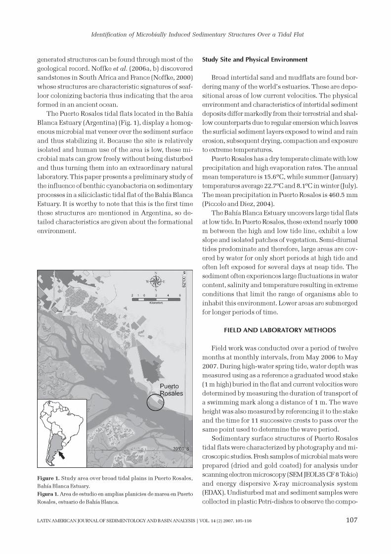
The Puerto Rosales tidal flats located in the Bahía
Blanca Estuary (Argentina) (Fig. 1), display a homog-
enous microbial mat veneer over the sediment surface
and thus stabilizing it. Because the site is relatively
isolated and human use of the area is low, these mi-
crobial mats can grow freely without being disturbed
and thus turning them into an extraordinary natural
laboratory. This paper presents a preliminary study of
the influence of benthic cyanobacteria on sedimentary
processes in a siliciclastic tidal flat of the Bahía Blanca
Estuary. It is worthy to note that this is the first time
these structures are mentioned in Argentina, so de-
tailed characteristics are given about the formational
environment.
Study Site and Physical Environment
Broad intertidal sand and mudflats are found bor-
dering many of the world’s estuaries. These are depo-
sitional areas of low current velocities. The physical
environment and characteristics of intertidal sediment
deposits differ markedly from their terrestrial and shal-
low counterparts due to regular emersion which leaves
the surficial sediment layers exposed to wind and rain
erosion, subsequent drying, compaction and exposure
to extreme temperatures.
Puerto Rosales has a dry temperate climate with low
precipitation and high evaporation rates. The annual
mean temperature is 15.6ºC, while summer (January)
temperatures average 22.7ºC and 8.1ºC in winter (July).
The mean precipitation in Puerto Rosales is 460.5 mm
(Piccolo and Diez, 2004).
The Bahía Blanca Estuary uncovers large tidal flats
at low tide. In Puerto Rosales, these extend nearly 1000
m between the high and low tide line, exhibit a low
slope and isolated patches of vegetation. Semi-diurnal
tides predominate and therefore, large areas are cov-
ered by water for only short periods at high tide and
often left exposed for several days at neap tide. The
sediment often experiences large fluctuations in water
content, salinity and temperature resulting in extreme
conditions that limit the range of organisms able to
inhabit this environment. Lower areas are submerged
for longer periods of time.
FIELD AND LABORATORY METHODS
Field work was conducted over a period of twelve
months at monthly intervals, from May 2006 to May
2007. During high-water spring tide, water depth was
measured using as a reference a graduated wood stake
(1 m high) buried in the flat and current velocities were
determined by measuring the duration of transport of
a swimming mark along a distance of 1 m. The wave
height was also measured by referencing it to the stake
and the time for 11 successive crests to pass over the
same point used to determine the wave period.
Sedimentary surface structures of Puerto Rosales
tidal flats were characterized by photography and mi-
croscopic studies. Fresh samples of microbial mats were
prepared (dried and gold coated) for analysis under
scanning electron microscopy (SEM JEOL35 CF 8 Tokio)
and energy dispersive X-ray microanalysis system
(EDAX). Undisturbed mat and sediment samples were
collected in plastic Petri-dishes to observe the compo-
Figure 1. Study area over broad tidal plains in Puerto Rosales,
Bahía Blanca Estuary.
Figura 1. Area de estudio en amplias planicies de marea en Puerto
Rosales, estuario de Bahía Blanca.
Page 4
108
Diana G. CUADRADO and Natalia V. PIZANI
LATIN AMERICAN JOURNAL OF SEDIMENTOLOGY AND BASIN ANALYSIS | VOL. 14 (2) 2007, 105-116
sition and structure under the optical microscope Nikon
SMZ 1500. Sediment samples were obtained with tube
corers (10 cm long, 3 cm in diameter) separating the
surficial mat from the rest and the latter into 1-cm thick
layers. They were stood in hydrogen peroxide for three
days and then sediment grain size was measured us-
ing a Malvern Mastersizer 2000 laser particle analyzer.
We compared the structures found in Argentina
based on a catalogue of microbial signatures (Gerdes,
et al., 2000) of sedimentary structures from two differ-
ent study sites in Europe (North Sea) and Africa (Tu-
nisian sabkha coast).
RESULTS
Typical conditions at the site are reflected by data
collected on May 11th, 2007. The intertidal study area
was flooded by seawater 12-15 cm deep during high
tide. Current speed reached up to 40 cm s-1 and waves
reached 5 cm in height with periods of 2 s. Southwest
wind speed was 5-6 m s-1 during the measurement,
although previously they reached up to 10-11 m s-1.
Another event where strong onshore winds and higher
wave heights was also examined (Cuadrado and Gómez,
2007) is discussed below.
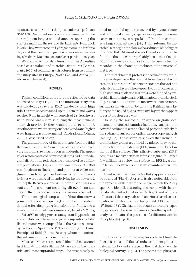
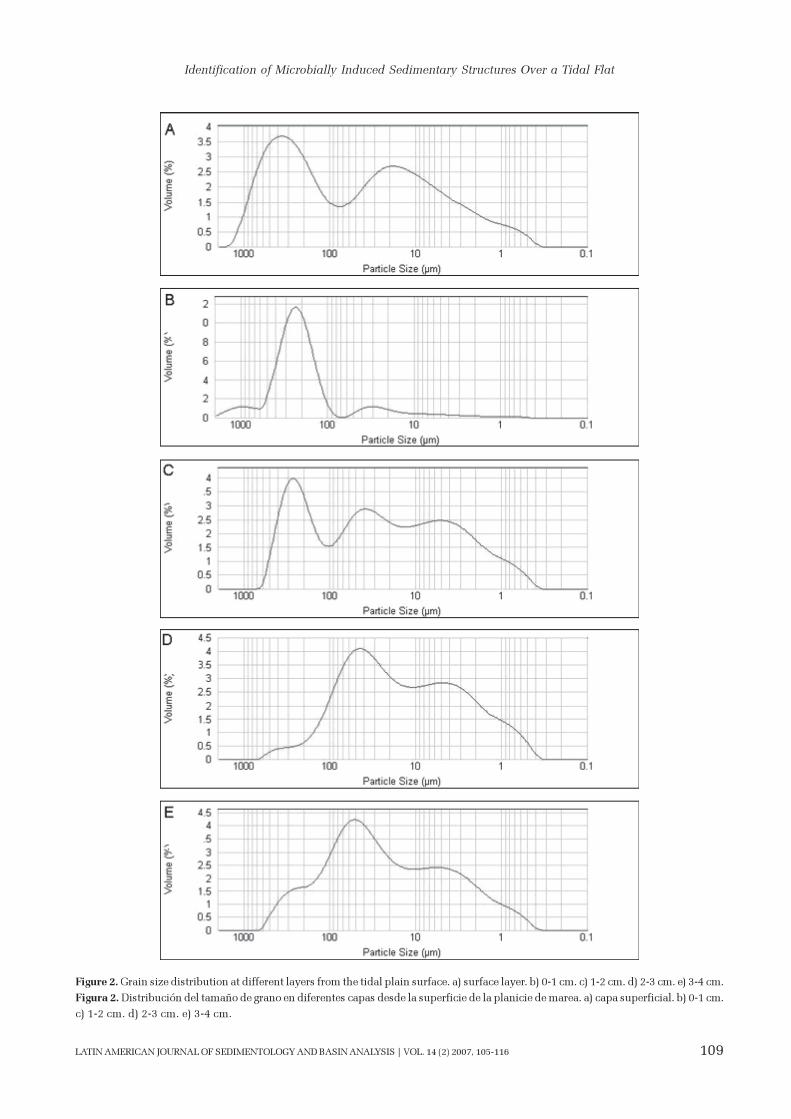
The granulometry of the sediments from the tidal
flat was measured in 1 cm thick-layers and displayed
varying grain size distribution with depth. The upper
layer which consisted of microbial mats had a bimodal
grain distribution reflecting the presence of two differ-
ent populations (Fig. 2). There was a peak at 0.350
mm (medium to fine sand) and another at 0.020 mm
(fine silt), indicating mixed sediments. Similar charac-
teristics were observed in underlying layers down to 2
cm depth. Between 2 and 4 cm depth, sand was ab-
sent and fine sediment including silt 0.040 mm and
clay 0.004 mm approximately in size was observed.
The mineralogical composition of the sediments was
primarily feldspar and quartz (Fig. 3). There were abun-
dant alterites displaying inclusions and fluids, and a
minor proportion of heavy minerals (density > 2.83 g
cm-3 at 20ºC) mostly pyroxenes (augite and hypersthene)
and amphiboles. The mineralogical composition of tidal
flat sediments was comparable to the results obtained
by Gelós and Spagnuolo (1982) studying the Canal
Principal of Bahía Blanca Estuary whom determined
the volcanic origin of its sediments.
Main occurrences of microbial films and mats found
in tidal flats of Bahía Blanca Estuary are in the inter-
tidal and lower supratidal range. The areas closely re-
lated to the tidal cycle are covered by layers of mats
and biofilms at an early stage of development. In some
cases, mats can even be peeled off from the sediment
as a large coherent piece (Fig. 4). In autumn, the mi-
crobial mat begins to colonize the sediment of the higher
intertidal flat. Different stages of development can be
found in the late winter probably because of the pat-
tern of successive colonization in the area, a feature
recorded in the changing thickness of the microbial
mat layer.
The microbial mat protects the sedimentary struc-
tures developed over the tidal flat from wave and wind
erosion. The structural characteristics are those of non
cohesive sand layers where upper bedding planes with
high contents of clastic minerals were binded by mi-
crobial films mainly made of filamentous cyanobacteria
(Fig. 5) that builds a fibrillar meshwork. Furthermore,
such mats are visible on tidal flats of Bahía Blanca Es-
tuary to the naked eye as extensive layers that appear
to resist erosion very well.
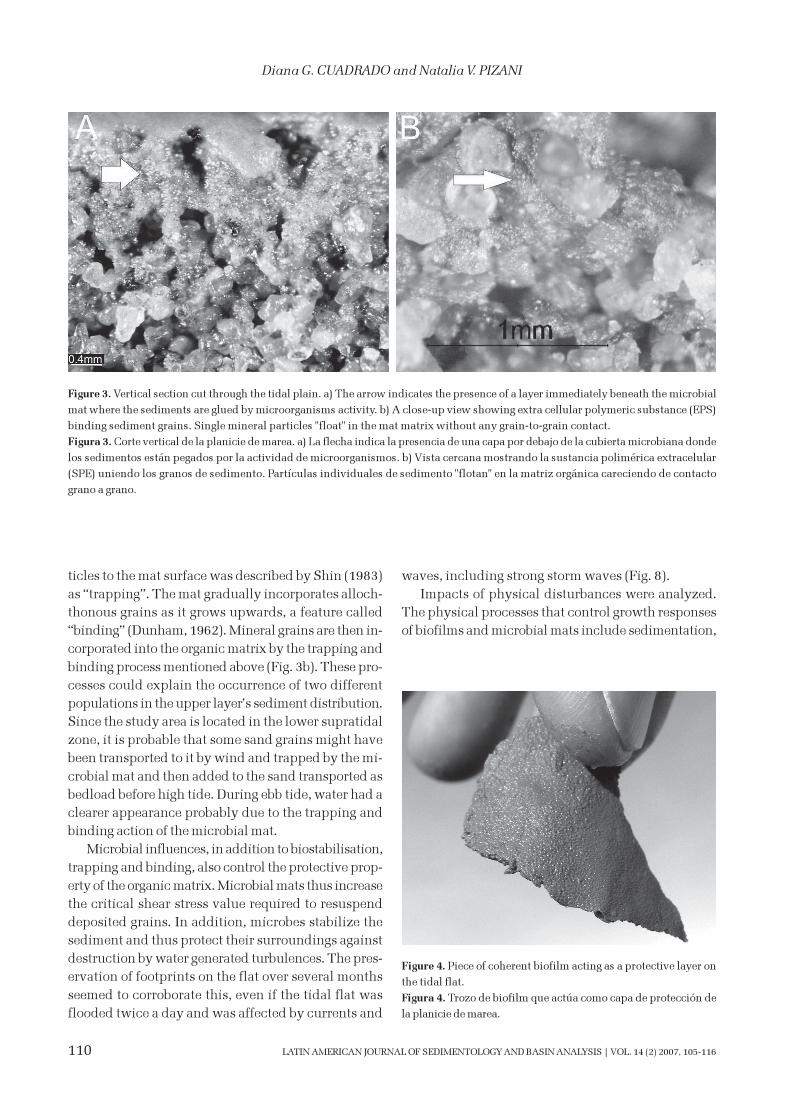
To study the microbial influence on grain sedi-
ments, undisturbed samples including surficial mat
covered sediments were collected perpendicularly to
the sediment surface for optical microscope analysis
(see Fig. 3a). These samples showed that individual
sedimentary grains are binded by microbial extra cel-
lular polymeric substances (EPS) immediately below
the tidal flat surface. This adhesive substance (EPS)
occurs as a matrix between grains in figure 3b. Only a
few millimeters below the surface the EPS layer can-
not be seen, however, some grains maintain their con-
nection.
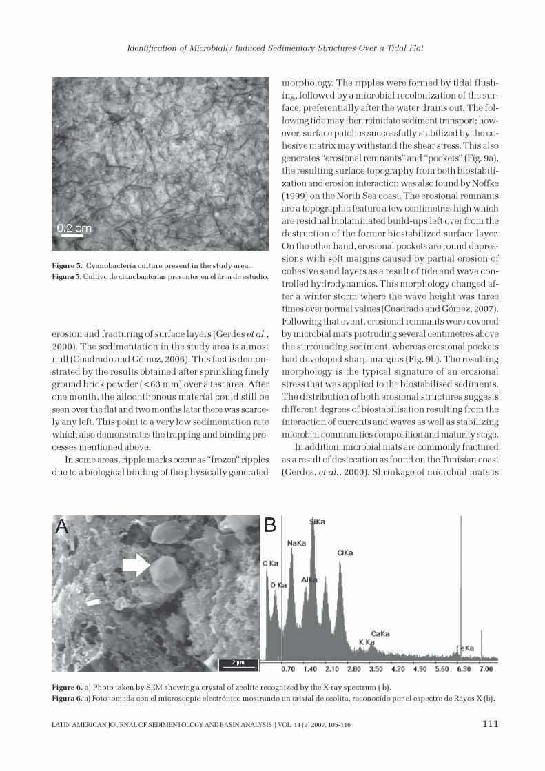
Small-sized particles with a flaky appearance can
be observed (Fig. 6). A crystal is also noticeable from
the upper middle part of the image, which the X-ray
spectrum identifies as authigenic zeolite with charac-
teristic elements of chabazite: Ca, Na, Si and Al. Iden-
tification of these crystals as chabazite is based on cor-
relation of the rhombic morphology and EDX spectrum
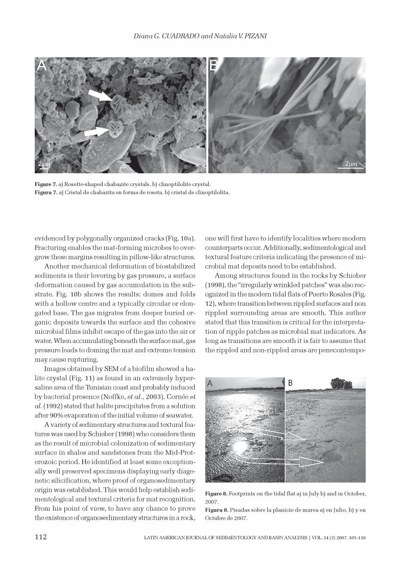
(Welton, 1984). Chabazite also occurs as rosette-shaped
crystals as can be seen in figure 7a. Another spectrum
analysis indicates the presence of a different zeolite:
clinoptilolite (Fig. 7b).
DISCUSSION
EPS was found in the samples collected from the
Puerto Rosales tidal flat as binded sediment grains lo-
cated in the top surface layer of the tidal flat due to the
biological activity (Fig. 3). The process that glues par-
Page 5
Identification of Microbially Induced Sedimentary Structures Over a Tidal Flat
109LATIN AMERICAN JOURNAL OF SEDIMENTOLOGY AND BASIN ANALYSIS | VOL. 14 (2) 2007, 105-116
Figure 2. Grain size distribution at different layers from the tidal plain surface. a) surface layer. b) 0-1 cm. c) 1-2 cm. d) 2-3 cm. e) 3-4 cm.
Figura 2. Distribución del tamaño de grano en diferentes capas desde la superficie de la planicie de marea. a) capa superficial. b) 0-1 cm.
c) 1-2 cm. d) 2-3 cm. e) 3-4 cm.
Page 6
110
Diana G. CUADRADO and Natalia V. PIZANI
LATIN AMERICAN JOURNAL OF SEDIMENTOLOGY AND BASIN ANALYSIS | VOL. 14 (2) 2007, 105-116
ticles to the mat surface was described by Shin (1983)
as “trapping”. The mat gradually incorporates alloch-
thonous grains as it grows upwards, a feature called
“binding” (Dunham, 1962). Mineral grains are then in-
corporated into the organic matrix by the trapping and
binding process mentioned above (Fig. 3b). These pro-
cesses could explain the occurrence of two different
populations in the upper layer’s sediment distribution.
Since the study area is located in the lower supratidal
zone, it is probable that some sand grains might have
been transported to it by wind and trapped by the mi-
crobial mat and then added to the sand transported as
bedload before high tide. During ebb tide, water had a
clearer appearance probably due to the trapping and
binding action of the microbial mat.
Microbial influences, in addition to biostabilisation,
trapping and binding, also control the protective prop-
erty of the organic matrix. Microbial mats thus increase
the critical shear stress value required to resuspend
deposited grains. In addition, microbes stabilize the
sediment and thus protect their surroundings against
destruction by water generated turbulences. The pres-
ervation of footprints on the flat over several months
seemed to corroborate this, even if the tidal flat was
flooded twice a day and was affected by currents and
waves, including strong storm waves (Fig. 8).
Impacts of physical disturbances were analyzed.
The physical processes that control growth responses
of biofilms and microbial mats include sedimentation,
Figure 3. Vertical section cut through the tidal plain. a) The arrow indicates the presence of a layer immediately beneath the microbial
mat where the sediments are glued by microorganisms activity. b) A close-up view showing extra cellular polymeric substance (EPS)
binding sediment grains. Single mineral particles "float" in the mat matrix without any grain-to-grain contact.
Figura 3. Corte vertical de la planicie de marea. a) La flecha indica la presencia de una capa por debajo de la cubierta microbiana donde
los sedimentos están pegados por la actividad de microorganismos. b) Vista cercana mostrando la sustancia polimérica extracelular
(SPE) uniendo los granos de sedimento. Partículas individuales de sedimento "flotan" en la matriz orgánica careciendo de contacto
grano a grano.
Figure 4. Piece of coherent biofilm acting as a protective layer on
the tidal flat.
Figura 4. Trozo de biofilm que actúa como capa de protección de
la planicie de marea.
Page 7
Identification of Microbially Induced Sedimentary Structures Over a Tidal Flat
111LATIN AMERICAN JOURNAL OF SEDIMENTOLOGY AND BASIN ANALYSIS | VOL. 14 (2) 2007, 105-116
erosion and fracturing of surface layers (Gerdes et al.,
2000). The sedimentation in the study area is almost
null (Cuadrado and Gómez, 2006). This fact is demon-
strated by the results obtained after sprinkling finely
ground brick powder (<63 mm) over a test area. After
one month, the allochthonous material could still be
seen over the flat and two months later there was scarce-
ly any left. This point to a very low sedimentation rate
which also demonstrates the trapping and binding pro-
cesses mentioned above.
In some areas, ripple marks occur as “frozen” ripples
due to a biological binding of the physically generated
morphology. The ripples were formed by tidal flush-
ing, followed by a microbial recolonization of the sur-
face, preferentially after the water drains out. The fol-
lowing tide may then reinitiate sediment transport; how-
ever, surface patches successfully stabilized by the co-
hesive matrix may withstand the shear stress. This also
generates “erosional remnants” and “pockets” (Fig. 9a),
the resulting surface topography from both biostabili-
zation and erosion interaction was also found by Noffke
(1999) on the North Sea coast. The erosional remnants
are a topographic feature a few centimetres high which
are residual biolaminated build-ups left over from the
destruction of the former biostabilized surface layer.
On the other hand, erosional pockets are round depres-
sions with soft margins caused by partial erosion of
cohesive sand layers as a result of tide and wave con-
trolled hydrodynamics. This morphology changed af-
ter a winter storm where the wave height was three
times over normal values (Cuadrado and Gómez, 2007).
Following that event, erosional remnants were covered
by microbial mats protruding several centimetres above
the surrounding sediment, whereas erosional pockets
had developed sharp margins (Fig. 9b). The resulting
morphology is the typical signature of an erosional
stress that was applied to the biostabilised sediments.
The distribution of both erosional structures suggests
different degrees of biostabilisation resulting from the
interaction of currents and waves as well as stabilizing
microbial communities composition and maturity stage.
In addition, microbial mats are commonly fractured
as a result of desiccation as found on the Tunisian coast
(Gerdes, et al., 2000). Shrinkage of microbial mats is
Figure 5. Cyanobacteria culture present in the study area.
Figura 5. Cultivo de cianobacterias presentes en el área de estudio.
Figure 6. a) Photo taken by SEM showing a crystal of zeolite recognized by the X-ray spectrum ( b).
Figura 6. a) Foto tomada con el microscopio electrónico mostrando un cristal de ceolita, reconocido por el espectro de Rayos X (b).
Page 8
112
Diana G. CUADRADO and Natalia V. PIZANI
LATIN AMERICAN JOURNAL OF SEDIMENTOLOGY AND BASIN ANALYSIS | VOL. 14 (2) 2007, 105-116
evidenced by polygonally organized cracks (Fig. 10a).
Fracturing enables the mat-forming microbes to over-
grow these margins resulting in pillow-like structures.
Another mechanical deformation of biostabilized
sediments is their levering by gas pressure, a surface
deformation caused by gas accumulation in the sub-
strate. Fig. 10b shows the results: domes and folds
with a hollow centre and a typically circular or elon-
gated base. The gas migrates from deeper buried or-
ganic deposits towards the surface and the cohesive
microbial films inhibit escape of the gas into the air or
water. When accumulating beneath the surface mat, gas
pressure leads to doming the mat and extreme tension
may cause rupturing.
Images obtained by SEM of a biofilm showed a ha-
lite crystal (Fig. 11) as found in an extremely hyper-
saline area of the Tunisian coast and probably induced
by bacterial presence (Noffke, et al., 2003). Cornée et
al. (1992) stated that halite precipitates from a solution
after 90% evaporation of the initial volume of seawater.
A variety of sedimentary structures and textural fea-
tures was used by Schieber (1998) who considers them
as the result of microbial colonization of sedimentary
surface in shales and sandstones from the Mid-Prot-
erozoic period. He identified at least some exception-
ally well preserved specimens displaying early diage-
netic silicification, where proof of organosedimentary
origin was established. This would help establish sedi-
mentological and textural criteria for mat recognition.
From his point of view, to have any chance to prove
the existence of organosedimentary structures in a rock,
one will first have to identify localities where modern
counterparts occur. Additionally, sedimentological and
textural feature criteria indicating the presence of mi-
crobial mat deposits need to be established.
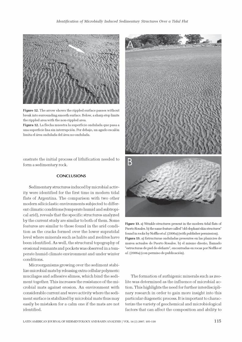
Among structures found in the rocks by Schieber
(1998), the “irregularly wrinkled patches” was also rec-
ognized in the modern tidal flats of Puerto Rosales (Fig.
12), where transition between rippled surfaces and non
rippled surrounding areas are smooth. This author
stated that this transition is critical for the interpreta-
tion of ripple patches as microbial mat indicators. As
long as transitions are smooth it is fair to assume that
the rippled and non-rippled areas are penecontempo-
Figure 7. a) Rosette-shaped chabazite crystals. b) clinoptilolite crystal.
Figura 7. a) Cristal de chabazita en forma de roseta. b) cristal de clinoptilolita.
Figure 8. Footprints on the tidal flat a) in July b) and in October,
2007.
Figura 8. Pisadas sobre la planicie de marea a) en Julio, b) y en
Octubre de 2007.
Page 9
Identification of Microbially Induced Sedimentary Structures Over a Tidal Flat
113LATIN AMERICAN JOURNAL OF SEDIMENTOLOGY AND BASIN ANALYSIS | VOL. 14 (2) 2007, 105-116
raneous, and on the contrary, if there is a sharp edge
delineating the rippled and non rippled surface, it can
be assumed that there are two different sediment lay-
ers, an upper non-rippled layer and a lower rippled
layer. The ripple patch appearance is produced when
weathering tears off portions of the upper layer, thus
forming “windows” that reveal the lower rippled layer
(Schieber, 1998). Nevertheless, this assumption does
not always prove true, since figure 12 shows that the
two above mentioned features, a smooth transition and
a sharp edge between rippled and non-rippled areas,
are simultaneously present in the same sediment layer.
The detrital grains of a featureless surface are held to-
gether by a binding property that may be weakened in
the ripple patches by wave action, e.g. storm surges. It
is possible that locally, where the microbial mat struc-
ture is weaker, erosion can tear off parts of the organic
layer and with the sandy intertidal bottom at those spots
then being eroded and creating depressions as “pock-
ets”.
Noffke et al. (2003) has detected MISS in siliciclastic
deposits from the Paleo-Archean to the Pleistocene,
where they found a record of extensive ancient micro-
bial mats covering large areas of the sea floor. This study
presents facies and sequence analyses conducted on
data sets from sandstone successions of the Paleo- and
Figure 9. a) Erosional remnants and pockets over Puerto Rosales tidal flat. b) The tidal flat after a storm.
Figura 9. a) Remanentes erosivos y depresiones en la planicie de marea de Puerto Rosales. b) La planicie de marea luego de una tormenta.
Meso-Archean of South Africa, the Neo-Proterozoic of
Namibia, and the Ordovician of the Montagne Noire,
France. The features named “erosional remnants” and
“pockets” by Noffke and Krumbein (1999) are also found
in sandstone of Paleo-Archean age (Noffke, et al., 2006a).
In her study, Noffke demonstrates the microbial origin
of the sedimentary structures in sandstones through-
out Earth history, making their detection and interpre-
tation a most valuable tool for paleobiologists. The
description of MISS in their studies resembles the
modern wrinkle structures found in Puerto Rosales tidal
flats whose bed surfaces are characterized by regular
and irregular ripple patterns (Fig. 13).
The link between sedimentary structures found in
modern environments and similar structures found in
rocks are diagenetic processes. All sediments after their
deposition are affected by diagenesis and include a
fundamental suite of physical, chemical and biological
processes that control the texture, mineralogy and fluid-
flow properties of sedimentary rocks. Usually the sedi-
ments are cemented by minerals that precipitate from a
solution. Understanding the processes and products
of diagenesis is thus a critical component in the analy-
sis of sedimentary basin evolution. Eogenesis involves
the initial interaction of the original sedimentary as-
semblage from its depositional pore water. Eogenetic
Page 10
114
Diana G. CUADRADO and Natalia V. PIZANI
LATIN AMERICAN JOURNAL OF SEDIMENTOLOGY AND BASIN ANALYSIS | VOL. 14 (2) 2007, 105-116
processes are influenced strongly by bacterial degrada-
tion of organic matter present in finer grained sedi-
ments (Kantorowicz, 1985). Under certain conditions,
microbial mats induce mineral precipitation such as
calcite (Krumbein, 1979), whereas heterotrophic bacte-
ria decompose primary producers and induce in situ
formation of minerals like pyrite (Giblin, 1988; Pye et
al., 1990). All these results were obtained from carbon-
atic environments. The present study site was on the
other hand located in a siliciclastic environment where
other minerals, different kinds of zeolites, were found.
Some of the more common environments for zeo-
lites to occur are saline, alkaline-lake deposits where
the first zeolites formed from the dissolution of natu-
ral rhyolitic glass are chabazite and/or phillipsite
(Tummer and Wirsching, 2000). Therefore, the zeolite
found in the sediment of Puerto Rosales, chabazite,
may be a product from the dissolution of glass present
in the volcanic materials of the tidal flat sediments.
These zeolites are silicates of aluminium mostly com-
posed of sodium and calcium which crystallize in a
water-rich environment under low pressure. The most
important feature is that zeolite precipitation occurs
during the fist stage of diagenesis: the eogenesis. There-
fore, the results obtained from this study clearly dem-
Figure 10. a) Polygonally fractured microbial mats. Coin is 2 cm in diameter. b) Surface deformation through bubbling caused by gas
formation in the substrate. Note that microbial mat formation is weaker only in isolated spots where the erosion may tear off pieces
of the organic layer. Consequently, the sandy surface in those exposed areas can be eroded and depressions may form.
Figura 10. a) Fracturas poligonales en coberturas microbianas. La moneda tiene 2 cm de diámetro. b) Deformación de la superficie por
burbujas debido a la formación de gas en el sustrato. Nótese que la cobertura microbiana es más débil en algunos sectores aislados
donde la erosión puede romper trozos de la capa orgánica. Consecuentemente, la superficie arenosa expuesta puede ser erosionada y
se pueden formar depresiones.
Figure 11. Image of a halite crystal.
Figura 11. Imagen de un cristal de sal.
Page 11
Identification of Microbially Induced Sedimentary Structures Over a Tidal Flat
115LATIN AMERICAN JOURNAL OF SEDIMENTOLOGY AND BASIN ANALYSIS | VOL. 14 (2) 2007, 105-116
onstrate the initial process of lithification needed to
form a sedimentary rock.
CONCLUSIONS
Sedimentary structures induced by microbial activ-
ity were identified for the first time in modern tidal
flats of Argentina. The comparison with two other
modern siliciclastic environments subjected to differ-
ent climatic conditions (temperate humid and subtropi-
cal arid), reveals that the specific structures analyzed
by the current study are similar to both of them. Some
features are similar to those found in the arid condi-
tion as the cracks formed over the lower supratidal
level where minerals such as halite and zeolites have
been identified. As well, the structural topography of
erosional remnants and pockets was observed in a tem-
perate humid climate environment and under winter
conditions.
Microorganisms growing over the sediment stabi-
lize microbial mats by releasing extra cellular polymeric
mucilages and adhesive slimes, which bind the sedi-
ment together. This increases the resistance of the mi-
crobial mats against erosion. An environment with
considerable current and wave activity where the sedi-
ment surface is stabilized by microbial mats thus may
easily be mistaken for a calm one if the mats are not
identified.
The formation of authigenic minerals such as zeo-
lite was determined as the influence of microbial ac-
tion. This highlights the need for further interdiscipli-
nary research in order to gain more insight into this
particular diagenetic process. It is important to charac-
terize the variety of geochemical and microbiological
factors that can affect the composition and ability to
Figure 12. The arrow shows the rippled surface passes without
break into surrounding smooth surface. Below, a sharp step limits
the rippled area with the non-rippled area.
Figura 12. La flecha muestra la superficie ondulada que pasa a
una superficie lisa sin interrupción. Por debajo, un agudo escalón
limita el área ondulada del área no-ondulada.
Figure 13. a) Wrinkle structures present in the modern tidal flats of
Puerto Rosales. b) the same feature called "old elephant skin structures"
found in rocks by Noffke et al. (2006a) (with publisher permission).
Figura 13. a) Estructuras onduladas presentes en las planicies de
marea actuales de Puerto Rosales. b) el mismo diseño, llamado
"estructuras de piel de elefante", encontradas en rocas por Noffke et
al. (2006a) (con permiso de publicación).
Page 12
116
Diana G. CUADRADO and Natalia V. PIZANI
LATIN AMERICAN JOURNAL OF SEDIMENTOLOGY AND BASIN ANALYSIS | VOL. 14 (2) 2007, 105-116
precipitate zeolites. Finally, the petrified structures
identified in rocks and their comparison with modern
sedimentary counterparts from tidal systems may help
with paleoenvironmental and palaeoclimatic recon-
structions.
Acknowledgements
The authors gratefully acknowledge funding for
their work from the PGI 24/ZH13 (Universidad Nacional
del Sur) and PICT´03 N°07-14652 (ANPCyT). We greatly
appreciated the help of Mr. Hugo Pellegrini in the field
and for laboratory analysis and Dr Jorge Spagnuolo for
the mineralogical analysis. The SEM images were taken
by Viviana Sorrivas (CCT BB). Useful comments by the
reviewers, Dr Thorbjørn J. Andersen and Lic. Fernando
Gómez, helped to improve the manuscript.
REFERENCES
Cornée, A., M. Dickman and G. Busson, 1992. Laminated cyanobac-
terial mats in sediments of solar salt works: some sedimen-
tological implications. Sedimentology 39:599-612.
Cuadrado, D.G. and E.A. Gómez, 2006. Acumulación en planicies
de marea causada por vegetación. Resúmenes IV Congreso
Latinoamericano de Sedimentología y XI Reunión Argentina de
Sedimentología:80, Bariloche, Argentina.
Cuadrado, D.G., E.A. Gómez, N. Pizani y E.R. Parodi, 2006.
Bioestabilización de sedimentos en planicies de marea. Resúmenes
VI Jornadas Nacionales de Ciencias del Mar y XIV Coloquio de
Oceanografía:39, Puerto Madryn, Argentina.
Cuadrado, D.G. and E.A. Gómez, 2007. Preservación de estructuras
sedimentarias en planicies de marea. Actas XII Colacmar. 3pp.
Brasil.
Decho A.W., 1990. Microbial exopolymer secretions in ocean environ-
ments: their role(s) in food webs and marine processes. Oceano-
graphic and Marine Biology: an Annual Review 28:73-153.
Dunham, R.J., 1962. Classification of carbonate rocks according to
depositional texture. Memoir American Association of Petroleum
Geologists 1:108-121.
Gelós, E. and J. Spagnuolo, 1982. Estudio composicional de los
sedimentos de fondo de la ria de Bahía Blanca entre Puerto
Cuatreros y Puerto Ingeniero White. RAGA 37(1):3-22.
Gerdes, G., Th. Klenke and N. Noffke, 2000. Microbila signatures
in peritidal siliciclastic sediments: a catalogue. Sedimentology
47:279-308.
Giblin, A., 1988. Pyrite formation in marshes during early diagenesis.
Geomicrobiology Journal 6:77-97.
Kantorowicz J.D., 1985. The petrology and diagenesis of Middle
Jurassic clastic sediments, Ravenscar Group, Yorkshire.
Sedimentology 32:833-853.
Krumbein, W.E., 1979. Photolithotropic and chemoorganotrophic
activity of bacteria and algae as related to beachrock formation
and degradation (Gulf of Aqaba, Sinai). Geomicrobiology Journal
1:139-203.
Krumbein, W.E., 1983. Stromatolites. The challenge of a term in
space and time. Precambrian Research 20:493-531.
Naylor, L., 2005. The contributions of biogeomorphology to the
emerging fiekd of geobiology. Palaeogeography, Palaeoclimatology,
Palaeoecology 219:35-51.
Noffke, N., 1999. Erosional remnants and pockets evolving from
biotic-physical interactions in a recent lower supratidal environ-
ment. Sedimentary Geology 123:175-181.
Noffke, N. and W. Krumbein, 1999. A quantitative approach to sedi-
mentary surface structures contoured by the interplay of micro-
bial colonization and physical dynamics. Sedimentology 46:417-
426.
Noffke, N., 2000. Extensive microbial mats and their influences on
the erosional and depositional dynamics of a silisiclastic cold
water environment (Lower Arenigian, Montagne Noire, France).
Sedimentary Geology 136:207-215.
Noffke, N., G. Gerdes, T. Klenke and W. E. Krumbein, 2001. Micro-
bially Induced Sedimentary Structures: A New Category within
the Classification of Primary Sedimentary Structures. Journal
of Sedimentary Research 71 (5):649-656.
Noffke, N. and A.H. Knoll, 2001. Geobiology: Its application to
sedimentary geology. Pardee Keynote Symposium, Annual
Meeting Geological Society of America, Boston. Geological
Society of America, Boulder, Colorado.
Noffke, N., G. Gerdes and T. Klenke, 2003. Benthic cyanobacteria
and their influence on the sedimentary dynamics of peritidal
depositional systems (siliciclastic, evaporitic salty, and evaporitic
carbonatic). Earth-Science Reviews 62:163-176.
Noffke N., 2005. Geobiology-a holistic scientific discipline. Palaeogeo-
graphy, Palaeoclimatology, Palaeoecology 219:1-3.
Noffke, N., N. Beukes, J. Gutzmer and R. Hazen, 2006a. Spatial and
temporal distribution of microbially induced sedimentary struc-
tures: A case study from siliciclastic storm deposits of the 2.9
Ga Witwatersrand Supergroup. South Africa. Precambrian
Research 146:35-44.
Noffke, N., K. Eriksson, R. Hazen and E. Simpson, 2006b. A new
window into Early Archena life: Microbial mats in Earth´s oldest
siliciclastic tidal deposits (3.2 Ga Moodies Group, South Africa).
Geology 34:253-256.
Paterson D.M., 1994. Microbiological mediation of sediment struc-
ture and behaviour. In Cuamette P., Stal L.J. (eds), Microbial
mats. NATO ASI Series:35-97.
Piccolo, M.C. and P.G. Diez, 2004. Meteorología del Puerto Coronel
Rosales. In M.C. Piccolo y Hoffmeyer M. (ed), Ecosistema del
Estuario de Bahía Blanca:87-90. Bahía Blanca, Argentina.
Pye, K., A.D. Dickinson, N. Schiavon, M.L. Coleman and M. Cox,
1990. Formation of siderite-Mg-calcite-iron sulfide concretions
in intertidal marsh and sandflat sediments, north Norfolk, En-
gland. Sedimentology 37:3255-343.
Schieber, J., 1998. Possible indicators of microbial mat deposits in
shales and sandstones: examples from the Mid-Proterozoic Belt
Supergroup, Montana, USA. Sedimentary Geology 120:105-124.
Shin, E.A., 1983. Tidal flat environments. American Association of
Petroleum Geologists Memoir 33:172-210.
Stal, L.J., 2000. Cyanobacterial mats and stromatolites. In Whitton,
B.A. and Potts, M. (eds), The ecology of Cyanobacteria. Netherlands:
61-120.
Trummer, B. and U. Wirsching, 2000. Formation of zeolites in saline,
alkaline-lake deposits: an experimental approach. In C.Colella
and F.A. Mumpton (eds.), Natural Zeolites for the Third
Millenium:211-225. Italy.
Walter, M.R., 1976. (ed). Stromatolites. Elsevier, Amsterdam, 790pp.
Welton, J., 1984. SEM Petrology Atlas. American Association of
Petroleum Geologists. Tulsa Oklahoma, USA, 237 pp.