Science of the Total Environment 408 (2010) 6032–6041
Contents lists available at ScienceDirect
Science of the Total Environment
j ourna l homepage: www.e lsev ie r.com/ locate /sc i totenv
Identification of nanominerals and nanoparticles in burning coal waste pilesfrom Portugal
Joana Ribeiro a, Deolinda Flores a,b, Colin R. Ward c, Luis F.O. Silva d,⁎a Centro de Geologia, Universidade do Porto, Rua do Campo Alegre, 687, 4169-007 Porto, Portugalb Departamento de Geociências, Ambiente e Ordenamento do Território, Faculdade de Ciências, Universidade do Porto, Rua do Campo Alegre, 687, 4169-007 Porto, Portugalc School of Biological, Earth and Environmental Sciences, University of New South Wales, Sydney, NSW 2052, Australiad Catarinense Institute of Environmental Research and Human Development, IPADHC, Capivari de Baixo, Santa Catarina, Brazil
Article history:Received 2 August 2010Received in revised form 16 August 2010Accepted 26 August 2010Available online 19 September 2010
Keywords:Douro CoalfieldCoal mining wastesTrace elementsCombustionCondensationHuman health
A range of carbon nanoparticles, agglomerates and mineral phases have been identified in burning coal wastepile materials from the Douro Coalfield of Portugal, as a basis for identifying their potential environmental andhuman health impacts. The fragile nature and fine particle size of these materials required novelcharacterization methods, including energy-dispersive X-ray spectrometry (EDS), field-emission scanningelectron microscope (FE-SEM), and high-resolution transmission electron microscopy (HR-TEM) techniques.The chemical composition and possible correlations with morphology of the nanominerals and associatedultra-fine particles have been evaluated in the context of human health exposure, as well as in relation tomanagement of such components in coal-fire environments.
The Douro Coalfield is the largest outcrop of terrestrial Carbonif-erous (Upper Pennsylvanian [Lower Stephanian C]) coal-bearingstrata in Portugal (Lemos de Sousa and Wagner, 1983; Wagner andLemos de Sousa, 1983; Eagar, 1983; Fernandes et al., 1987), with alength of 53 km and a width of between 30 m and 250 m (Fig. 1). Thegeographic limits of the coalfield are São Pedro de Fins (Maia) andJanarde (S. Pedro do Sul) (Pinto de Jesus, 2001).
The Douro Coalfield has been the most important coal producer inPortugal, with a large number of mines geographically dispersed. Coalmining activities, which ended in 1994, have caused several impactson the environment, one of which is the amount of discard material(mainly carbonaceous shales and lithic arenites) that was emplaced inmore than 20 waste piles throughout the coalfield area. Three of thesewaste piles, namely, S. Pedro da Cova, Midões and Lomba, arecurrently burning, following ignition caused by forest fires during thesummer of 2005.
Spontaneous combustion of coal wastes is a well-known globalphenomenon that may cause significant environmental and healthproblems (e.g. Bell et al., 2001; Finkelman, 2004; Stracher and Taylor,2004). By-products from burning of coal or coal waste may include
solid residues derived from interaction between volatilized com-pounds and the overlying material (Pone et al., 2007; Querol et al.,2008) and/or products emitted to the atmosphere in gaseous form.
Petrological and geochemical analysis of the organic and mineralmatter of burning and unburnt zones in these coal waste piles byRibeiro et al. (2010) has allowed preliminary identification of themineral and gas products that have resulted from the combustionprocess. The present work complements that previous study byidentifying and providing detailed characterization of carbon nano-particles, agglomerates and mineral phases formed in the burningwaste piles. Any health and environmental consequences associatedwith these particles would be expected to have a close relationship totheir physical and chemical characteristics, such as their size,morphology, composition, solubility, and oxidation state. The studyprovides data not previously available from bulk characterization, andthus contributes to assessing further the environmental and humanhealth impacts of burning coal waste materials.
2. Materials and methods
2.1. Sampling
Samples of neoformed minerals and the overlying material werecollected around cracks or vents along the floor and slope surfaces inactively burning areas of the S. Pedro da Cova and Lomba waste piles.Two types of neoformed minerals were observed in each waste pile:
Fig. 1. Geological and geographical setting of Douro Coalfield, Portugal (adapted from Pinto de Jesus, 2001).
6033J. Ribeiro et al. / Science of the Total Environment 408 (2010) 6032–6041
one white and the other yellow in color. The overlying materialrepresents a combination of overburden strata and discards fromwashing plants, and is made up of carbonaceous shales and lithicarenites with a variable proportion of disseminated coal.
Two samples of coal waste pile material (CWPM) from S. Pedro daCova (SP1 and SP2) and one sample from Lomba (L) were collected fordetailed analysis as part of the present study. These samples werecarefully packed into plastic boxes and labelled. Six additionalsamples from burning zones and unburned zones of each waste pile,collected for previous studies (Sant'Ovaia et al., 2010), were alsoincluded in the study to represent the material that constitutes thewaste piles. Analytical data from these samples were used to providebackground information about the mineral phases in the wastematerials generally, and to help identify the reactions that haveoccurred during the burning process.
2.2. Analytical procedures
2.2.1. X-ray diffractionThe crystalline minerals in the three CWPM samples were
identified by X-ray diffraction (XRD), using a Siemens model D5005
diffractometer with Cu K-α radiation. The samples were ground byhand in a ceramic mortar and pestle, dry mounted in aluminiumholders, and scanned from 8 to 60o 2θ (Silva et al., 2009b).
Powdered samples of the burnt and unburnt materials thatconstitute the waste piles were analysed by XRD using a PhilipsPW-1830 diffractometer with Cu K-α radiation. Scans were from 2 to60o 2θ. Quantitative analyses of the mineral phases in these sampleswere made from the diffractograms using Siroquant™, commercialinterpretation software (Taylor, 1991) based on the Rietveld XRDanalysis technique. The proportions of amorphous material in theburnt samples were determined from the XRD patterns as describedby Ward and French (2006).
2.2.2. Raman spectroscopyRaman spectroscopy was carried out using a Renishaw-model
Invia Reflex Raman system, operated in the confocal mode. The lateralresolution with this configuration was ~1 μm, and the spectralresolution was 1 cm−1 (Guedes et al., 2008). Extended scans wereperformed on each sample. The Raman spectra of carbon materialswere measured with a density filter to avoid thermal decompositionof the samples. Several Raman analyses were conducted directly on
Table 1Quantitative XRDdata (%) onwaste pilematerials (after Sant'Ovaia et al., 2010) andmineralsand nanoparticles identified inwastematerials and associated neo-formed deposits by othermethods. Analytical methods: a = FE-SEM, b = Raman, c = HR-TEM, d = XRD.
Material thatconstitutesthe waste piles
S. Pedro daCova CWPM
LombaCWPM
Burningzones
Unburnedzones
CarbonatesCalcite, CaCO3 a, c a, cSiderite, FeCO3 a, c a, c
Oxides and hydroxidesAnatase, TiO2 0.2. 0.2 a, c, d a, c, dBrucite, Mg(OH)3 c n.d.Chromite FeCr2O4 a aGoethite, Fe(OH)3 a, c a, cGibbsite, Al(OH)3 a a, cHematite, Fe2O3 0.9 a, c, d a, c, dMaghemite, Fe2O3 c cMagnetite, Fe3O4 a, c a, cQuartz, SiO2 19.8 17.4 a, b, c, d a, b, c, dRutile, TiO2 0.2 0.3
PhosphatesMonazite, (Ce, La, Th, Nd, Y)PO4 a a
SaltsAlbite, NaCl a, c, d a, dBararite, (NH4)2SiF6 c a, cSalammoniac, NH4Cl a, b, c, d a, b, c, d
SilicatesAlbite, NaAlSi3O8 a, c, d a, c, dChlorite, (Fe, Mg, Al)6(Si, Al)4O10(OH)8
0.2 1.0
Illite, K1.5Al4(Si6.5Al1.5)O20(OH)4
31.5 62.7 a, c, d a, d
Kaolinite, Al2Si2O5(OH)4 a, c, d a, c, dMetakaolin, Al2O3·2SiO2 a aMullite, Al6Si2O13 8.3 a, c, d a, c, dMuscovite, (K, NH4, Na) Al2(Si, Al)4 O10(OH)2
3.7 5.5 d d
Pyrophyllite, Al2Si4O10(OH)2 3.5 6.6 a, d a, dSulphates
Aluminite, Al2(SO4)(OH)4·7H2O
a, c a, c
Anhydrite, CaSO4 a, c, d a, cBarite, BaSO4 a, c a, cEpsomite, MgSO4.7H2O a, b, c, d a, b, c, dGypsum, CaSO4·2H2O a, b, d a, b, dHalotrichite, FeAl2(SO4)4.22H2O
a, b, c, d a, b, c, d
Hexahydrite, MgSO4·6H2O a, d aJarosite, KFe3+3(SO4)2(OH) 6 1.2 a, c, d a, c, dMelanterite FeSO4.7H2O a, c a, cMascagrite, (NH4)2SO4 a aNatrojarosite, NaFe3(SO4)2(OH)6 a aPickeringite, MgAl2(SO4)4.22H2O
a, b, c, d a, b, c, d
Schwertmannite,Fe16O16(OH)12(SO4)2
c c
SuphidesGalena, PbS a, c a, cPyrite, FeS2 a, c a, c
Sulphur a, c, d a, c, dFullerene c a, cAmorphous 28.1 a, c, d a, c, d
n.d. = not detected.
6034 J. Ribeiro et al. / Science of the Total Environment 408 (2010) 6032–6041
the CWPM particles. However, most of the analyses were performedon polished epoxy-bound pellets used for petrographic analysis.Spectra obtained from representative CWPM particles were deconvo-luted in order to calculate the Raman parameters and determine theprecise frequencies, bandwidths, and the relative intensities of thebands of the carbon materials.
2.2.3. Electron beam methodsA LEO Model 435VP SEM, fitted with a turbo pumped chamber, a
motorized stage, an Oxford energy-dispersive X-ray (EDX) spectrom-eter having resolution N133 eV, and a four-quadrant back-scatterdetector, was used to identify the minerals by conventional SEMtechniques, based on observation of whole-specimens using naturaland/or polished surfaces. The morphology, structure, and chemicalcomposition of ultrafineparticles andminerals in selected sampleswerefurther investigated using a Zeiss ULTRA plus field emission scanningelectron microscope (FE-SEM) with charge compensation for allapplications on conductive as well as non-conductive samples and a200-keV JEOL-2010F high resolution transmission electron microscope(HR-TEM) equipped with an Oxford energy dispersive X-ray detectorand a scanning (STEM) unit (Silva and DaBoit 2010; Silva et al., 2009a).The FE-SEM was equipped with an energy-dispersive X-ray spectrom-eter (SEM-EDS), and themineral identificationsweremade on the basisof morphology and grain composition using both secondary electronand back-scattered electron modes (Silva et al., 2010a).
Geometrical aberrations were measured by HR-TEM, and controlledtoprovide less than aπ/4 phase shift of the incomingelectronwave overthe probe-defining aperture of 14.5 mrad (Silva et al., 2010d). Thescanning acquisition was synchronized to the AC electrical power tominimize 60 Hz noise, and a pixel dwell time of 32 μs was chosen. EDSspectra were recorded in FE-SEM and HR-TEM images mode and thenquantified using ES Vision software that uses the thin-foil method toconvertX-ray countsof eachelement into atomicorweightpercentages.Electron diffraction patterns of the crystalline phases were recorded inSAED (selected area electron diffraction) or MBD (microbeam diffrac-tion) mode, and the d spacings were compared to the InternationalCentre for Diffraction Data (ICDD, 2010) inorganic compound powderdiffraction file to identify the crystalline phases.
Hexane, acetone, dichloromethane and methanol suspensions ofwhole-waste samples were utilized to prevent possible mineralogicalchanges in individual solvents. Each suspension was pipetted ontolacy carbon films supported by Cu grids (200 mesh) and left toevaporate before inserting the sample into the FE-SEM and HR-TEM.This method may have led to agglomeration, but is a widely usedprocedure for most minerals, including metal sulphates (Giere et al.,2006; Silva et al., 2010b). Before FE-SEM and STEM analysis, the HR-TEM specimen holder was cleaned with an Advanced Plasma System(Gatan Model 950) to minimize contamination. A drift correctionsystem was used for the STEM-EDS mapping.
2.2.4. Mineral extractionsFE-SEM and HR-TEM analyses of the CWPM samples and minerals
included within them were carried out following a sequentialextraction (SE) process. A brief explanation of the experimentalconditions and of the expected information offered by each step ofthis process is presented below.
1) For the water-soluble fraction, a 1-mg CWPM sample (fivereplicates) was mixed with Millipore-system water (1 mL) havingan electrical conductivity of 0.1–0.5 μS/cm. Samples were shaken inthe dark for 4 h, then centrifuged (3000 rpm, 10 min) and filtered(b22 μm). This extraction dissolves salts (e.g. salammoniac),jarosite, alunogen, gypsum (partially soluble in water), and otherminerals soluble in water. For the secondary sulfates and saltsformed in coal-fire environments, such an initial water-extractionstep is essential for a complete understanding of the materials
involved. The solid fraction was cold dried, suspended in acetone,pipettedon to separated lacy carbonfilms supportedbyCugrids, andleft to evaporate before inserting the sample into the FE-SEM andHR-TEM (Silva et al., 2010c). The liquid fraction was air dried/crystallized before inserting the sample into the FE-SEM;
2) For the exchangeable fraction, samples were shaken with 1 MNH4-acetate at pH 4.5 in the dark for 2 h at room temperature,then centrifuged (3000 rpm, 10 min) and filtered (b22 μm). This
6035J. Ribeiro et al. / Science of the Total Environment 408 (2010) 6032–6041
3) For poorly ordered Fe and Al oxyhydroxides/sulphates (Gaglianoet al., 2004; Regenspurg et al., 2004), a 1-mg mineral sample (fivereplicates) was mixed with 1 mL of ammonium oxalate reagent(28 g/L ammonium oxalate +15 g/L oxalic acid solution, pH~2.7).The samples were then shaken in the dark for 4 h, centrifuged(3000 rpm, 10 min) and filtered (b22 μm) (Peretyazhko et al.,2009). This extraction dissolves poorly-crystalline Fe (III) oxides(e.g. ferrihydrite, schwertmannite) in preference tomore insolublecrystalline Fe (III) oxides (e.g. goethite, hematite) (Cornell and
Fig. 2. Salammoniac XRD pattern (SP1), FE-SEM
Schwertmann, 2003; Silva and DaBoit, 2010). More than 85% of thetotal iron was released in this step;
4) Highly ordered Fe3+ hydroxides and oxides (hematite andgoethite) were partially dissolved by acid ammonium oxalate(Kumpulainen et al., 2007) using a selective extraction stepdescribed by Dold (2003). As in step 2, the extractant used was0.2 M NH4-oxalate for 2 h, but in this case the samples wereexposed to light and heated to 80 °C in a water bath;
5) For organic matter, after steps 1–4, a 0.1-mg resultant mineralsample (five replicates) was mixed with 1 mL of dimethylsulfoxide (DMSO). The samples were shaken in the dark for 12 h,
6036 J. Ribeiro et al. / Science of the Total Environment 408 (2010) 6032–6041
then centrifuged and filtered (b22 μm). This extraction dissolvesorganic anions absorbed on Al-OH/Fe-OH surfaces of minerals;
6) For residual organic and sulphides, the samples were mixed with35%H2O2 andheated in awater bath for 1 hour, then centrifuged andfiltered (b22 μm). This extraction dissolves sulphides (chalcopyrite,cinnabar, galena, orpiment, pyrite, sphalerite, stibnite, and tennan-tite-tetrahedrite).
Fig. 3. (A) Multi-walled nanotubes with various diameters (∼4–10 nm) and amplification ofdegrees of crystallinity; (C and D) HR-TEM image of very crystalline carbon contain fullere
3. Results and discussion
XRD data on the general constitution of the materials thatconstitute the waste piles (Table 1) indicate that illite and quartzare the main constituents of the samples studied from unburnedzones. Significant proportions of muscovite, kaolinite and pyrophylliteare also present, along with minor proportions of chlorite, rutile and
6037J. Ribeiro et al. / Science of the Total Environment 408 (2010) 6032–6041
anatase. Mineralogical analysis of samples from burning or alreadyburnt zones shows that the minerals found in the unburned zones arealso present, along with new mineral phases such as mullite,cristobalite, hematite and jarosite, and a significant proportion ofamorphous material. Most of these new minerals were probablyformed by thermal decomposition of clays and other minerals (e.g.pyrite) at around 900–1300 °C (Saxby, 2000), which is in accordancewith observations by Ribeiro et al. (2010).
Previous studies by Ribeiro et al. (2010) have shown that sulphurand salammoniac are the major neoformed mineral phases in the
burnt coal wastes from the Douro area. Qualitative XRD, Raman, FE-SEM, HR-TEM, and EDS investigations for the present study haverevealed a wide range of other phases (Table 1) with differentcompositions, structure, and particle size.
A combination of Raman spectroscopy, FE-SEM and XRD techniqueswere used to identify the major neoformed phases collected aroundvents near burning-zones in the coal waste piles. As indicated above,two major phases were observed: one yellow in colour identified assulphur, and one white in colour identified as salammoniac (Fig. 2). AtS. Pedro da Cova these coal-fire gas minerals occurred as well defined
6038 J. Ribeiro et al. / Science of the Total Environment 408 (2010) 6032–6041
euhedral crystals, while at Lomba they were fine grained and lessabundant. The sameminerals have been found associatedwith coalfiresbyPone et al. (2007). According toGaines et al. (1997), salammoniac is asublimation product that occurs around volcanic fumaroles and inburning coal seams and waste piles; it may, in part, be dissolved intosurface water. The ammonia usually is derived from the coal's organicmatter. Sulphur and salammoniac are formed during exhalation andcondensation of gas by interaction with surface water, atmosphericgases and surrounding rocks (Ribeiro et al., 2010).
Fig. 3 shows HR-TEM images of carbon nanoparticles obtainedfrom the CWPM samples in the present study. These images suggestthat fullerenes and multiwalled carbon nanotubes (MWNTs) wereformed by the spontaneous coal combustion and are still preserved inthe waste materials.
The presence of fullerenes in anthropogenic systems has been ofgreat interest since the discovery of these materials (Kroto et al., 1985),because their characteristic cage structure can retain some hazardouselements (Utsonomiya et al., 2002; Chen et al., 2004; Chen et al., 2005).Consequently the identification of natural occurrences may havesignificant environmental and human health implications.
Energy-dispersive X-ray spectrometry has shown that the particlesidentified in the present study may contain mercury (circled section inFig. 3A), selenium, nickel, boron, iodine, and arsenic. Because burning of
Fig. 5. (A) Jarosite identified by FE-SEM after DMSO extraction (step 3); (B) Gypsum and jaafter SE (step 3).
the coalwastewas the likely source of the fullerenes, the probable sourceof the encapsulated hazardous elements was also from the coal fires.
Selected area electron diffraction analysis combined with fastFourier transformation (FFT) provides further information on thecarbon nanostructure, especially variations in the range of structuralor nanostructural order and graphite crystallinity (Fig. 3B). Thecrystallinity of carbon depends on the ambient environment offormation, especially the temperature and cooling history. Theburning temperature of the coal waste piles is thought to be around900–1300 °C (Ribeiro et al., 2010; Sant'Ovaia et al., 2010), and suchtemperatures are high enough to produce fullerenes and multiwalledcarbon nanotube materials. The varying degrees of crystallinity of thematerials in the samples from the present study (Fig. 3B and C)suggest that the particles formed in the waste piles have experienceda variety of high-temperature thermal histories.
The carbon nanostructures present in the CWPM samples mayhave been derived from transformation of fullerenes into thenanostructures with nanotubes (Fig. 3D) ranging from 4 to 15 nm.In addition, in the organic macromolecular structure of coal, the linksbetween the adjacent aromatic units are relatively weak, and will befirst destroyed by the high energy electron; in this way, a largeamount of aromatic fragments such as biphenyl, naphthalene, pyrene,anthracene would be released (Pang and Wilson, 1993).
rosite association after water extraction (step 1); (C) Hematite nanocrystals identified
6039J. Ribeiro et al. / Science of the Total Environment 408 (2010) 6032–6041
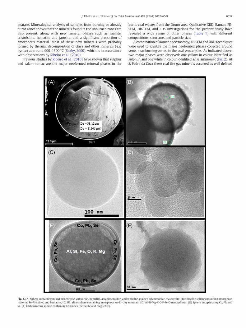
HR-TEM and FFT images also show numerous other nanocrystallineparticles containing hazardous elements in the samples studied.Ultrafine and micro Al-Si-Fe-Mg-K-Ti-S particles have been identified,showing a variety of spherulitic structures ranging in diameter from12 nm to 40 μm(Fig. 4) and creating an aggregate of mostly amorphousminerals containing potentially hazardous elements (e.g. As, Co, Cr, andPb) and very short-range structural graphitic matter. Microscopic Fe-Mg-Al-S-O spheres in sample SP1, for example (Fig. 4A), include several
crystalline components such as pickeringite, hematite, mullite, anhy-drite, arcanite, and fine-grained salammoniac-mascagnite. An ultrafinevitreous spherical particle containing Al, Si, Fe, O, K, andMg is shown inFig. 4E and C; this particle also contains some potentially hazardouselements such as Co, Cr, Se, and Pb. HR-TEM images taken from Ca-S-Si-O-Fe particles show that both amorphous and multiple nanocrystallinestructures (e.g. anhydrite as thick tubular crystals, identified using SAEDand FFT patterns) may coexist in such particles.
pickeringite; E = epsomite; H = hexahydrite; Hal = halotrichite).
6040 J. Ribeiro et al. / Science of the Total Environment 408 (2010) 6032–6041
Ultrafine carbonaceous particles occur in the waste materials (e.g.Fig. 4F and B). These are mainly soot aggregates with primary particlesizes varying from 3 to 120 nm. Depending of the temperature ofcombustion and the neighboring environment, different carbonstructures and carbonaceous spheres may be developed, and thusdetected by FE-SEM and HR-TEM techniques. The present resultsprovide a basis for a more detailed determination of hazardouselement bioavaibility, and also possibly new insights into thepotential for transportation of such particles and their associatedelements as aerosols.
The crystallinemineral phases identified by the various techniquesin the coal wastematerials include carbonates, hydroxides and oxides,phosphates, silicates, sulphates and sulphides (Table 1). The phasetransition reactions associated with formation of these materials inthe burning waste piles represent a highly dynamic process stronglyinfluenced by variations in temperature and relative humidity of thesurrounding environment (Vaniman et al., 2004; Linnow et al., 2006).Some of the metal sulphates found in the study may result fromspontaneous combustion of the coal waste piles.
Jarosite (Fig. 5A) was the principal Fe-sulphate found in the burntwaste samples. Mixed sulphates such as gypsum and jarosite (Fig. 5B)are also present, possibly formed by decomposition, fragmentation,and oxidation of sulphide minerals such as pyrite, marcasite,sphalerite, galena, pyrrhotite, and possibly carbonates, such assiderite, ankerite and calcite (see Table 1). Burning samples rich injarosite and hematite show increased concentrations of As, Cd, Cr, Pb,and Se.
Fe-oxide minerals, such as hematite (Fig. 5C), magnetite, chromite,magnesioferrite or hercynite are very common in the CWPM samplesafter spontaneous combustion. Even though Fe and O peaks are presentin the EDS spectra, the nature of the spherule indicated by the arrow inFig. 5C was further investigated by its HR-TEM image. Indexing of theFFT image for this particle (Fig. 5C) further identifies the presence ofnanohematite crystals. In general the size of the spherical iron oxides(e.g. hematite and magnetite) varied from nanometers to micrometers.In addition, several iron-bearing particles (e.g. oxides) with amorphousmicrostructures were also identified in the CWPM samples. Howeverdifferences between crystalline and amorphous minerals of similarchemical species should also be taken into account in toxicologicalstudies (Chen et al., 2004).
Several members of the MgSO4.nH2O series, epsomite, hexahy-drite, and pickeringite (Fig. 6), were identified by XRD, FE-SEM, andRaman (Table 1 and Fig. 6). Dehydration of epsomite to hexahydrite isstrongly dependent on the relative humidity. Garcia-Guinea et al.(2000) indicate that epsomite transforms (reversible reaction) tohexahydrite above 28 °C (unknown RH), while Vaniman et al. (2004)found that epsomite transforms readily to hexahydrite at ~25 °C at50–55% RH and at lower temperatures as water activity decreases. Thesometimes contradictory results of the many studies of the subjectconfirm the rather complex behavior of these sulfate salts especially incoal waste materials. In the present case, the relative abundance ofthese two phases may be expected to be quite variable, depending onthe ambient conditions in the field or in the course of the analysisprocess.
4. Conclusion
X-ray diffraction, Raman spectrometry, SEM-EDS, FE-SEM and HR-TEM investigations have provided detailed identification and character-izationof carbonnanoparticles, agglomerates andmineral phases formedin burning Portuguese coalwaste piles, and identification of the potentialeffects of such particles on the environment and human health generally.
XRD, FE-SEM, HR-TEM, and Raman spectrometry play an importantrole in nanomineral identification and geochemical interpretation. HR-TEM studies, in particular, allow mineralogical examination at a finerscale than is possible with optical or XRD techniques. Both cubic and
dendritic salammoniac formswere observed in the present study, alongwith sulfates (gypsum and jarosite) and Fe-minerals. Dehydration ofjarosite can lead to the formation of less hydrous Fe-sulfates andhematite. Hematite, with someCr in themineral structure, was noted inassociationwith jarosite. In addition, HR-TEM studies have revealed thepresence of fullerene carbons and multiwalled carbon nanotubes thatcontain some potentially hazardous elements with varying degrees ofcrystallinity. This suggests that the particles have experienced a varietyof high-temperature thermal histories.
Acknowledgements
The research for this study was carried out with support from theCatarinense Institute of Environmental Research and Human Devel-opment (IPADHC). The authors wish to acknowledge Dr. F. Macias andLuisa, for assistance with the FE-SEM and HR-TEM analyses. J. Ribeirobenefited from a PhD scholarship financed by Fundação para a Ciênciae Tecnologia (FCT), Portugal – Ref: SFRH/BD/31740/2006.
References
Bell FG, Bullock SET, Halbich TFJ, Lindsay P. Environmental impacts associated with anabandoned mine in the Witbank Coalfield, South Africa. Int J Coal Geol 2001;45:195–216.
Chen Y, Shah N, Huggins FE, Huffman GP. Investigation of the microcharacteristics ofPM2.5 in residual oil fly ash by analytical transmission electron microscopy.Environ Sci Technol 2004;38:6553–60.
Chen Y, Shah N, Huggins FE, Huffman GP, Dozier A. Characterization of ultrafine coal flyash particles by energy-filtered TEM. J Microsc 2005;217:225–34.
Cornell RM, Schwertmann U. The Iron Oxides: Structure, Properties, Reactions,Occurrence and Uses. (Second Edition). KGaA, Weinheim: Wiley-VCH VerlagGmbH & Co; 2003.
Dold B. Speciation of the most soluble phases in a sequential extraction procedureadapted for geochemical studies of copper sulfide mine waste. J Geochem Explor2003;80:55–68.
Eagar RMC. The non marine bivalve fauna of the Stephanian C of North Portugal. In:Sousa MJL, Oliveira JT, editors. The Carboniferous of Portugal: Memórias dosServiços Geológicos de Portugal, 29. ; 1983. p. 179–85.
Fernandes JP, Pinto de Jesus A, Teixeira F, Sousa MJL. Primeiros resultados palinológicosna Bacia Carbonífera do Douro (NO de Portugal). In: Grandal 'Angale A, Gutiérrez-Marco JC, Santos Fidalgo L, editors. XIII Jornadas de Paleontologia ―Fósiles deGalicia‖ y V Reunión Internacional Proyecto 351 PICG ―Paleozoico inferior delNoroeste de Gondowana‖, A Coruna, 1997, Libro de Resúmenes y Excursiones,Sociedade Espanola de Paleontologia; 1987. p. 176–9.
Finkelman RB. Potential health impacts of burning coal beds and waste banks. Int J CoalGeol 2004;51:19–24.
Gagliano WB, Brill MR, Bigham JM, Jones FS, Traina SJ. Chemistry and mineralogy ofochreous sediments in a constructed mine drainage wetland. Geochim CosmochimActa 2004;68(9):2119–28.
Gaines RV, Skinner HCW, Foord EE, Mason B, Rosenzweig A. Dana's New Mineralogy:the system of Mineralogy of James Dwight Dana and Edward Salisbury Dana. 8thed. Inc, New York: John Wiley & Sons; 1997. 1819 pp.
Garcia-Guinea J, Abella R, Sanchez-Moral S, Benito R, Martin-Ramos D. Examininghydrated minerals using optically stimulated X-ray diffraction, an inexpensivemodification of traditional diffractometers. J Sed Res 2000;70(4):964–7.
Giere R, Blackford M, Smith K. TEM study of PM2.5 emitted from coal and tirecombustion in a thermal power station. Environ Sci Technol 2006;40:6235–40.
Guedes A, Valentim B, Prieto AC, Sanz A, Flores D, Noronha F. Characterization of fly ashfrom a power plant and surroundings bymicro-Raman spectroscopy. Int J Coal Geol2008;73:359–70.
International Centre for Diffraction Data (ICDD). 2010. Powder Diffraction Database.http://www.icdd.com [accessed: 20 June 2010].
Kumpulainen S, Carlson L, Raisanen ML. Seasonal variations of ochreous precipitates inmine effluents in Finland. Appl Geochem 2007;22(4):760–77.
Lemos de Sousa MJ, Wagner RH. General description of the terrestrial Carboniferousbasins in Portugal and history of investigations. In: Lemos de Sousa MJ, Oliveira JT,editors. The Carboniferous of Portugal: Memórias dos Serviços Geológicos dePortugal, 29. ; 1983. p. 117–26.
Linnow K, Zeunert A, Steiger M. Investigation of sodium sulfate phase transitions in aporous material using humidity and temperature-controlled X-ray diffraction. AnalChem 2006;78:4683–9.
Pang LSK, Wilson MA. Nanotubes from coal. Energy Fuels 1993;7:436–7.Peretyazhko T, Zachara JM, Boily JF, Xia Y, Gassman PL, Arey BW, Burgos WD.
Pinto de Jesus, A., 2001. Génese e evolução da Bacia Carbonífera do Douro (EstefanianoC inferior, NW de Portugal): Um Modelo. PhD Thesis, University of Porto, Portugal.Vol. Text: 232 pp; Vol. Atlas: 71 pp.
6041J. Ribeiro et al. / Science of the Total Environment 408 (2010) 6032–6041
Pone JDN, Hein KAA, Stracher GB, Annegarn HJ, Finkelman RB, Blake DR, McCormack JK,Schroeder P. The spontaneous combustion of coal and its by-products in theWitbank and Sasolburg coalfields of South Africa. Int J Coal Geol 2007;72:124–40.
Querol X, Izquierdo M, Monfort E, Alvarez E, Font O, Moreno T, Alastuey A, Zhuang X, LuW, Wang Y. Environmental characterization of burnt coal gangue banks atYangquan, Shanxi Province, China. Int J Coal Geol 2008;75:93-104.
Regenspurg S, Brand A, Peiffer S. Formation and stability of schwertmannite in acidicmining lakes. Geochim Cosmochim Acta 2004;68(6):1185–97.
Ribeiro J, Ferreira da Silva E, Flores D. Burning of coal waste piles from Douro Coalfield(Portugal): petrological, geochemical and mineralogical characterization. Int J CoalGeol 2010;81:359–72.
Sant'Ovaia H, Ribeiro J, Corrêa-Ribeiro H, Gomes C, Li Z, Ward C, Flores D. An integratedstudy of mineralogy and magnetic parameters of coal waste pile materials incombustion from Douro Coalfield (Portugal): first results of a case study.Proceedings of the Second International on Coal Fire Research, Berlim, Germany;2010. p. 408–9.
Saxby JD. Minerals in coal. In: Glikson M, Mastalerz M, editors. Organic Matter andMineralisation. Kluwer Academic Publishers; 2000. p. 314–26.
Silva LFO, Moreno T, Querol X. An introductory TEM study of Fe-nanominerals withincoal fly ash. Sci Total Environ 2009a;407:4972–4.
Silva LFO, Oliveira MLS, da Boit KM, Finkelman RB. Characterization of Santa Catarina(Brazil) coal with respect to Human Health and Environmental Concerns. EnvironGeochem Health 2009b;31:475–85.
Silva LFO, DaBoit K. Nanominerals and nanoparticles in feed coal and bottom ash:implications for human health effects. Environ Monit Assess 2010, doi:10.1007/s10661-010-1449-9.
Silva LFO, Macias F, Oliveira MLS, Da Boit KM, Waanders F. Coal cleaning residues and Fe-minerals implications. EnvironMonit Assess 2010a, doi:10.1007/s10661-010-1340-8.
Silva LFO, IzquierdoM,Querol X, FinkelmanRB,OliveiraMLS,WollenschlagerM, TowlerM,Pérez-López R, Macias F. Leaching of potential hazardous elements of coal cleaningrejects. Environ Monit Assess 2010b, doi:10.1007/s10661-010-1497-1.
Silva LFO, Wollenschlager M, Oliveira MLS. A preliminary study of coal mining drainageand environmental health in the Santa Catarina region Brazil. Environ GeochemHealth 2010c, doi:10.1007/s10653-010-9322-x.
Silva LFO, Hower JC, Izquierdo M, Querol X. Complex nanominerals and ultrafineparticles assemblages in phosphogypsum of the fertilizer industry and implicationson human exposure. Sci Total Environ 2010d, doi:10.1016/j.scitotenv.2010.07.023.
Stracher GB, Taylor TP. Coal fires burning out of control around the world:thermodynamic recipe for environmental catastrophe. Int J Coal Geol 2004;59:7-17.
Taylor JC. Computer programs for standardless quantitative analysis of minerals usingthe full powder diffraction profile. Powder Diffr 1991;6:2–9.
Utsonomiya S, Jensen KA, Keeler GJ, Ewing RC. Uraninite and fullerene in atmosphericparticulates. Environ Sci Technol 2002;36:4943–7.
Vaniman DT, Bish DL, Chipera SJ, Fialips CI, Carey JW, Feldman WC. Magnesiumsulphate salts and the history of water on Mars. Nature 2004;431(7009):663–5.
Wagner RH, Lemos de Sousa MJ. The Carboniferous Megafloras of Portugal – a revisionof identifications and discussion of stratigraphic ages. In: Lemos de Sousa MJ,Oliveira JT, editors. The Carboniferous of Portugal: Memórias dos ServiçosGeológicos de Portugal, 29; 1983. p. 127–52.
Ward CR, French D. Determination of glass content and estimation of glass compositionin fly ash using quantitative X-ray diffractometry. Fuel 2006;85:2268–77.