Page 1
ISI Impact Factor:3.461. 016Bas.J.Vet.Res.Vol.15,No.4,2
144
IDENTIFICATION THE GROSS STRUCTURE OF THE ADULT OX
KIDNEY BY USING CORROSION CAST TECHNIQUE.
Nabeel Abd Murad AL-Mamoori*,Saffia Kareem Wally Alumeri*
Hazem Kareem Naser Almhanna**
*Department Of Anatomy and Histology, College Of Veterinary Medicine,University of AL-
Qadisiyah, AL- Qadisiyah, Iraq
Department Of Anatomy and Histology, College of Veterinary Medicine, University of Kufa
Kufa,Iraq
(Received 29 September 2016,,Accepted 15December 2016)
Keywords: Kidney, Corrosion cast, Ox.
ABSTRACT
In the current study, ten specimens of the adult ox kidneys have been used to identity
morphology and blood supply features by using corrosion cast technique. The mean body
weight of ox was 180±10kg. The external surface of the kidneys was elongated oval to pyramid,
lobulated flattened ventrodorsally, and light-brown in color. The right kidney is weighted 299±
24gm as well as it had two surfaces (ventral and dorsal), two borders (lateral and medial) and
two poles (cranial and caudal), while the left kidney is weighted 333± 27gm and had three
surfaces (right &left ventral and dorsal), in addition it had two borders and two poles.
The renal artery and vein have the same course and each one divided into 2-3 segmental blood
vessels. The segmental artery and vein are divided in to interlobular artery and vein which
supply medulla and cortex. The interlobular artery & vein were passed through the renal
columns and branched into arcuate and finally gave off cortical artery and vein.
The aim of this study, to exposure the normal gross internal appearance of blood vessels and
ureter of ox kidneys by using corrosion cast technique.
INTRODUCTION
The urinary system of mammalian consists of paired kidneys (right and left) that secreted the
urine after blood filtration. Each kidney drained in to ureter and convey the urine from the
pelvis of kidney to single bladder where urine is stored. Urine would be drained by single
urethra into the exterior (1). The kidneys play very an importance role in maintenance
homeostasis and excretion of metabolic waste products and other functions such as syntheses
and secretions hormones renin and erythropoietin (2 and 3).
Page 2
ISI Impact Factor:3.461. 016Bas.J.Vet.Res.Vol.15,No.4,2
145
Left and right kidneys are located in the abdominal cavity and embedded in dorsal part of body
of bovine, related nearly on each side of the caudal vena cava and abdominal aorta. However, in
the mammalian the left kidney backward more slightly than the right kidney.
Generally, in the cattle the kidney revealed externally lobulated with rough surface, while in the
other species had smooth surface (1,2 and 3).
MATERIALS AND METHODS
1- Morphological and biometrical study
Ten specimens of the kidneys of adult ox. The mean body weight of ox was 180±10kg has been
used in this study. Fresh samples were collected from AL-Diwaniyah’s abattoir immediately
after animals slaughtering. To collect the kidneys, oxen were dissected at thoracic inlet into the
pelvic cavity and removed the internal organs and take off the kidney. Kidneys had dissected
with the ureter and renal artery & vein (Major blood vessels which supply the kidney). Adipose
tissue was removed which covered the kidneys and washing the kidneys by tap water. Finally,
the length, width, weight and thickness of kidneys are measured.
2- Corrosion cast technique
This technique is used to detect the internal course of blood vessels inside kidney and ureter in
relation to kidney structure. Samples prepared as followed steps:
1-Warm normal saline solution 0.9 % inject in the renal artery, vein and ureter to clean and
remove all clots and sediment which may be found in the blood vessels and ureter.
After that inject the renal blood vessels and ureter by mixture of self-cure denture material set
(Powder and liquid 1 to 4), and it is consisted of 20% monomethyl-methacrylate powder and
80% polymethyl-methacrylate liquid and adding the suitable dyes (Red, blue and yellow ink) to
differentiate the blood vessels and the ureter (4).
2-Samples are incubated at room temperature for 24 hours for polymerization.
3-Samples put in drain opener which consist of a mixture of NaOH, Na2CO3 and NaClO) and
prepared by dissolved 1 kg in 5 liters distilled water) and left 72–96 hour to corrosion casted at
the room temperature.
4-Finally samples were washed with normal tap water, and snap image.
RESULTS
1- Morphology of kidney
A- External appearance of the kidney: Right and left kidney were embedded in sub lumbar
fossa at dorsal part of the body and surrounded capsule which are very rich in fat and adipose
tissue, moreover, kidney was light-brown in color and externally lobulated due to present of
Page 3
ISI Impact Factor:3.461. 016Bas.J.Vet.Res.Vol.15,No.4,2
146
groove which separated each lobe from other, and this grooves filled with adipose tissue. On the
other hand, right and left kidneys differ in shape, weight and dimensions.
The right kidney was pyramidal in shape and has 21±2 lobes are small and large in shape. It
consists of two surfaces (dorsal and ventral), two borders (lateral and medial) and two poles
(cranial and caudal). additionally, the dorsal surface was strongly convex and curved toward the
vertebral columns, while the ventral surface was irregular convex and contain the hilum which
against the abdominal viscera.
The medial border of right kidney was approximately straight and in related on the caudal vena
cava, while the medial border of the left kidney was directed toward the abdominal aorta and
extremities of border convex, also this border was engaged in the hilum which via it the renal
artery with nerve renal vein and ureter to enter and leave the kidney. The lateral border was
convex from side to side ( Fig.1& 2 ). At result, the mean of weight, length, widest area,
thickest area, length medial and lateral borders of the right kidney was 299± 24 gm,
15.033±0.5cm, 6.45±0.75cm, 6.75± 0.8cm, 14.133± 0.9cm and 21.333±0.88cm.
The left kidney was oval in shape and has 18.5± 1.5 lobes which differ in size. It comprised of
three surfaces (dorsal, right & left ventral), two borders (lateral and medial) and two poles
(cranial and caudal). The dorsal surface was strongly convex and the curved toward the
vertebral columns, while the ventral surface consists of two parts right and left. It is irregular
convex and contain the hilum which tend toward the abdominal viscera. The medial border was
approximately straight and tend toward the abdominal aorta, in the middle region is contained
the hilum that attached into blood and nerves supply. Furthermore, the lateral border was
convex from side to side (Fig.1). The mean of weight, length, widest area, thickest area, length
medial & lateral borders of the left kidney is 333± 27gm,16.56±0.7cm, 7.45±0.56cm, 5.2±
0.44cm, 16.5± 0.06cm & 17.6± 0.77 respectively (Table.1). The hilum of kidney revealed as
depression area which occupied the middle region of ventral surface and extend toward the
proximal third of the medial border. It refers to entrance of the ureter, artery, vein, nerves and
lymph vessels.
B- Internal Appearance of the Kidney:
After making a longitudinal incision of the kidney to describe the internal structure showed
three regions that differ in the color and texture, involves in capsule, cortex and medulla. The
capsule was a transparent strong fibrous membrane layer, cover the outer surface of the kidney
and it is easily dislocation resembling nylon bag ( Fig.3 ). The cortex is directly located under
the capsule and brown in color, rough texture and reveals radiate appearance. In addition the
cortex is extended toward the medulla between the renal pyramid to form the renal columns.
Page 4
ISI Impact Factor:3.461. 016Bas.J.Vet.Res.Vol.15,No.4,2
147
The medulla divided into many regions pyramidal in shape called renal pyramid. It is divided
into two regions according to the color. The first region was a dark- brown and form the margin
of renal pyramid, while the second region was a light-brown in color located inside of the
medulla and direct toward the renal calix. (Fig. 3).
The renal pyramid consists of two parts: base and apex, the apex was tending toward the renal
calix, while the base toward the cortex. The apex of pyramid form is ended with renal papillae
which refer to apical portion of the pyramid which was opened in the minor calyces (Fig.
3,4&5).
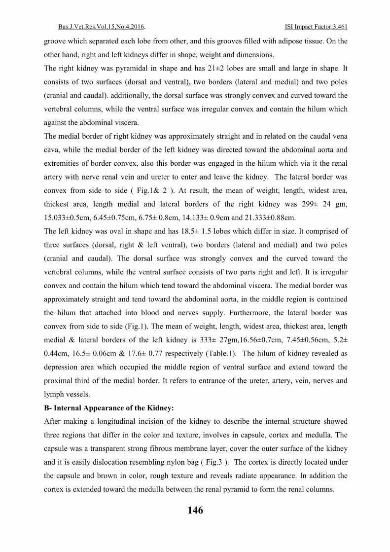
The ureter is opened at the hilum and extended more and branched into 7-8 ducts called major
calix. Each major calix divided into 19-21 collecting ducts which are funnel shape structure
called minor calix. This minor calix was surrounded by adipose tissue, and it resaved the urine
which discharge from renal papillae (Fig9&10).
2- Blood Supply Of Kidney By Using Corrosion Cast Technique
After injecting the blood vessels of the kidneys with corrosion cast technique, the renal artery
was very clear and it was originated from the ventral side of the abdominal aorta, which supply
the kidneys through the hilum (Fig.2). The renal artery at entrance of the hilum of kidney was
divided into 2-3 segmental arteries. This segmental artery after short distance reached into the
apex of the renal pyramid and divided into interlobular artery which supply medulla (Fig.4,6 &
8). Likewise, it is run course of interlobular through the renal columns between the renal
pyramid, when it reaches into the base of the renal pyramid gave branch called arcuate artery
which extend along the base of pyramid (Fig. 8).
The arcuate artery gives of several branches called cortical radiate artery which are extended
toward the cortex region (Fig. 4,5,6,7 and 8).
The renal vein was originated from the ventral side of the caudal vena cava, which enter the
kidney through the hilum of kidney ( Fig.1&2). The renal vein courses same and parallel of the
renal artery. The renal vein divided into 2-3 of segmental veins at entrance of the hilum of
kidney (Fig.4,). The segmental vein extended into short distance with segmental artery in the
same course into the apex of the renal pyramid then would be branched into interlobular vein.
The interlobular vein and with interlobular artery have the same course. It is passed through the
renal columns and form the external boundaries of the renal pyramid. it is branched into arcuate
vein when reaches at the base of the renal pyramid. The arcuate vein had same course of the
arcuate arteries and give off several branches of cortical radiate vein that extend toward the
cortex take radiate shape ( Fig.4,5,6 and7).
Page 5
ISI Impact Factor:3.461. 016Bas.J.Vet.Res.Vol.15,No.4,2
148
Table 1: Biometrical measurement of left and right kidneys in adult Ox. Mean and stander error
The parameter Right kidney Left kidney
Weight 299± 24gm 333± 27gm
Length of kidney 15.033±0.5cm 16.566± 0.7cm
Length of medial boarder 14.133±0.9 cm 16.5±0.66 cm
Length of lateral border 21.333±0.88 cm 17.6±0.77 cm
Thickness of cranial,
middle and caudal
regions respectively
3.5±0.6
cm
5.95±0.4
cm
6.75±0.8
cm
5±0.7
cm
5.2±0.44
cm
5±0.9
cm
Width of cranial, middle
and caudal regions
respectively
4.05±0.66
cm
6.45±0.75
cm
5.55±0.34
cm
5.05±0.87
cm
7.45±0.56
cm
5.65±0.34
cm
Number of lobe 21± 2 18.5± 1.5
Fig 1. Ventral 1 and dorsal 2 view of the right kidney of ox show:
A-Cranial pole. B-Caudal pole. C-Renal vein. D- Renal artery. E-Ureter. G- Adipose
tissue in hilum. H-Renal lobule. K- Lateral surface. M- Medial surface.
1 2
A A
B B
D
C
E
G
H
H
H
H
H H
M K
Page 6
ISI Impact Factor:3.461. 016Bas.J.Vet.Res.Vol.15,No.4,2
149
Fig2. Ventral view of the right and left kidneys of ox after remove capsule show:
A-Caudal vena cava. B-Abdominal aorta. C-Renal vein. D- Renal artery E-Ureter.
G- Adipose tissue in hilum. H-Renal lobule.
Left kidney Right kidney
A
B
C
D E
E
H
H
H
G G
Fig 3. Midlongitudinal section of the right kidney of ox show: A-Cranial pole. B-
Caudal pole. C- Cortex. D- Medulla. E- Renal pyramid. F- Renal papillae. G-
Adipose tissue. H-Renal lobule. K- Minor calyx. L- Renal column. M- Interlobular
artery.
A
B
C C D
D
H
F
G
G
E
K
F
L L
Page 7
ISI Impact Factor:3.461. 016Bas.J.Vet.Res.Vol.15,No.4,2
150
Fig 4. Ventral view of the arterial, venous supply and ureter of the kidney in ox by
using corrosion cast show: A- Renal artery. B- Renal vein. C- Segmental artery.
D- Segmental vein. E- Arcuate artery. G- Cortical artery and vein. F- Ureter.
H- Minor calyx. K- Adipose tissue.
A
B
C
D
E
G F
H
K
G
Fig 5. Dorsal view of the blood supply and calyx of the kidney in ox by using
corrosion cast show: A- Renal artery. B- Renal vein. C- Minor calyx. D- adipose tissue
around minor calyx. G- Cortical artery & vein.
B
A
C D
D
D
Page 8
ISI Impact Factor:3.461. 016Bas.J.Vet.Res.Vol.15,No.4,2
151
Fig 6. Ventral view of the arterial supply and ureter of the kidney in ox by
using corrosion cast show:A- Ureter. B- Major calyx. C- Minor calyx. D-
Papillary duct E- Renal artery. G- Segmental artery. H- Interlobar
A B B
C
D
E
G G
H
K K
K
Fig 7. Dorsal view of the arterial, venous supply and ureter of the kidney in
ox by using corrosion cast show: A- Renal artery. B- Major calyx. C-
A K
K
C
C
B B
D G
Fig 8. Ventral view of the arterial supply of the kidney ox by using corrosion cast
show: A- Renal artery. B- Segmental artery. C- Interlobar artery. D- Arcuate
artery. E- Cortical artery.
A
B
C C
E
E
D
Page 9
ISI Impact Factor:3.461. 016Bas.J.Vet.Res.Vol.15,No.4,2
152
DISCUSSION
Morphology of Kidney
The kidney was light-brown in color and lobulated but, the right and left kidneys differ in shape,
weight and dimensions and this result agreement with 2 and 3 in bovine that showed the kidney
is lobulated ovoid in shape, and disagreement with (2,3,4,5,6,7,8,9 and 10) in small ruminants,
horse, camel, pig, dog & cat, the kidney has smooth surface bean–shaped, this difference due to
different species.
The mean of weight, length, widest area, thickest area, length medial and lateral borders of the
right kidney was 299± 24 gm, 15.033±0.5cm, 6.45±0.75cm, 6.75± 0.8cm, 14.133± 0.9cm and
21.333±0.88cm , while the mean of weight, length, widest area, thickest area, length medial and
lateral borders of the left kidney was 333± 27gm, 16.566±0.7cm, 7.45±0.56cm, 5.2± 0.44cm ,
16.5± 0.06cm and 17.6± 0.77 respectively and this result disagreement with (11) show the
mean of weight, length, width, thickness of the right kidney was 1300±31.885gm,
21.32±0.46cm, 13.33±0.42cm and 8.475±0.085cm while the mean of weight, length, width, of
the left kidney was 952.5±52.5gm, 23.3±0.587cm, 10.45±0.42cm and 7.675±0.165cm
respectively in buffalo, this differ may be due to species of animal.
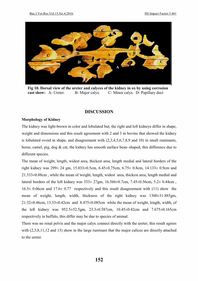
There was no renal pelvis and the major calyx connect directly with the ureter, this result agrees
with (2,3,8,11,12 and 13) show in the large ruminant that the major calices are directly attached
to the ureter.
Fig 10. Dorsal view of the ureter and calyces of the kidney in ox by using corrosion cast show: A- Ureter. B- Major calyx. C- Minor calyx. D- Papillary duct
A
A B
B
B
C
C
C
C
D
Page 10
ISI Impact Factor:3.461. 016Bas.J.Vet.Res.Vol.15,No.4,2
153
Blood supply of kidney
The renal artery originated from the ventral side of the abdominal aorta, to supply the kidneys
through the hilum and divided into segmental artery, this result agreed with (8,11,12,13,14, 15
and 16) in sheep, buffalo, dog and horse, but then again disagree with (17) which found the
accessory renal arteries originating directly from aorta.
The renal artery and vein had the same course and each one divided into 2-3 segmental arteries
and veins. The segmental artery and vein after short distance reached into the apex of the renal
pyramid and divided interlobular artery and vein which supply medulla, this result agreed with
(13,14,15,16, 18) show the segmental artery and vein divided into interlobular artery & vein.
The interlobular artery & vein pass through the renal columns and branched into arcuate, this
result agreed with (19) in pig.
In conclusion, this study asserted that during using Corrosion Cast Technique would be helped
to show internal structure of oxen kidney, in particular, veins and arteries supply better than
without this technique in gross study.
.تقنیة القالب التأكليباستخدام العجول البالغةلكلى المظهریة البنیة على التعرف
**.حازم كریم ناصر المحنة،*صفیة كریم والي العمري ،*نبیل عبد مراد المعموري
.العراق،القادسیه ، جامعة القادسیة ،كلیة الطب البیطري ،فرع التشریح واالنسجة*
.العراق،الكوفه ،جامعة الكوفة ،الطب البیطري كلیة ،فرع التشریح واالنسجة **
الخالصة
الدموي باستخدام والمددشملت الدارسة الحالیة عشرة عینات من كلیة العجول البالغة للتعرف على المواصفات المظهریة
الطولي كغم وكان المظهر الخارجي للكلیة بین البیضوي180الحیوان جسم حیث كان معدل وزن .تقنیة القالب التأكلي
و لها 24±299حیث كان وزن الكلیة الیمنى .ذات لون بني فاتح ، مسطحة بطنیا ظهریا، والهرمي الشكل مفصصه
غم ولها 27±333بینما كان وزن الكلیة الیسرى ، سطحین بطني وظهري وحافتین وحشیة وانسیة وقطبین امامي وخلفي
. ثالثة اسطح بطني ایمن وایسر وظهري وحافتین وقطبین
فروع 3-2كل واحد منهما یتفرع الى . ن نفس المساراللكلیة متمثل بالشریان والورید الكلوي حیث كان یسیر المدد الدمويان
ان الشریان والورید القطعي ینقسم الى عدة فروع من الشریان او الورید بین الفصوص لتغذیة . من الشریان والورید القطعي
Page 11
ISI Impact Factor:3.461. 016Bas.J.Vet.Res.Vol.15,No.4,2
154
ویة والتي بدورها تعطي فروع اخیرة تعرف بالفروع اوالورید بین الفصوص یتفرع الى الفروع الز اما الشریان. منطقة اللب والقشرة
.القشریة
REFERENCES
1- Shively M J (1984).Veterinary anatomy ,Basic comparative and clinical anatomy college
station Texas. University of Bristol UK. CABI publishing. Pp: 1-4.
2- Frandson R D, Wilke W L and Fails A D(2009). Anatomy and Physiology of Farm Animals.
Wiley Blackwell. (7th ed). Ch. 23. Pp: 383.
3- Dyce K M, Sack W O and Wensing C J (2010). Textbook of Veterinary Anatomy. W.B.
Saunders Company, Philadelphia. London. New York. ( 4 th Ed) Pp: 104-106, 489, 636 – 637.
4- Mishra GP, Bhatnagar S. and Singh B. (2014). Anatomical Variations In Arterial Pattern Of
Lower Segmental Artery And It Relation With Collecting System. International Journal of
Anatomy and Research, Vol 2(2)Pp:403-405.
5- Zguigal H and Ouhsine A (2004). Functional Anatomy of the Renal Pelvis in the
OneHumped Camel. J. camel science.1.Pp: 81-85.
6- Lazo P, Vlatko I, Florina P P, Nikola A, Dobrila T L(2013). Morphometrical Evaluation Of
Some Anatomical Features In Pig Kidneys: Are They Different From Human Kidneys. Mac Vet
Rev. 35 (1): 35 –42.
7- Dowelmadina I M, Elhashmi YH and Bakhiet S (2013). Morphometric Studies Of Kidneys In
One Humped Camel (Camelus Dromedarius) In Sudan. ISCCRP. Pp:109-111.
8- Sisson S S B (1911). A Text Book Of Veterinary Anatomy. W.B. Saunders Company,
Philadelphia. London. New York. Pp: 469-483.
9- Al-Asadi F S (2006). Some Morphological Studies On The Kidney Of The Sheep With
Technique To Its Arterial Segmentation. Basrah. J. Vet. Res.Vol.5,No.1.PP:44-46.
10- H, Rind M M, Ahmad R, Ahmad N, and Shah G (2003). Gross Anatomical Study on
Normal Kidneys of Adult Goat. Journal of animal and veterinary advances. 2(9): 539-541.
11- Al- Kinanny A F (2006). Anatomical, Histological and Radiological study of the kidney
and the ureter of Buffalo “Bubalus bubalis” in Iraq. A Thesis University of Baghdad.
12- Getty R (1975).The Anatomy Of Domestic Animal. 5th ed .W.B.Sanders,Co.Vol.1.
13- Sawad A A (2006). Functional Anatomy Of The Kidney In Buffaloes. Bas.J.Vet.Res. Vol.
5.No.Pp: 76-82.
Page 12
ISI Impact Factor:3.461. 016Bas.J.Vet.Res.Vol.15,No.4,2
155
14- Nagato A C, Rocha C L J, Bandeira A C B, Oliveira R M S and Bezerra F S (2013).
Morphometric and quantitative analysis of the afferent renal artery variation. J. Morphol. Sci.
Vol. 30, No. 2, Pp: 82-85.
15- Beatriz P S, Sampaio A P, Henry R W, Favorito L A and Francisco J B (2007) . Dog
Kidney: Anatomical Relationships Between Intrarenal Arteries and Kidney Collecting System.
The Anatomical Record Pp:1017–1022.
16- Ozdemir D, Zekeriya O and Ismail M (2009). Intrarenal Segmentation of the Renal Arteries
in the Kangal Dog. Kafkas ـniv Vet Fak Derg 15 (1) Pp: 41-44,
17- Chandragirish S, Nanjaiah C M, Suhas YS, Saheb S H(2014). Study On Accessory Renal
Artery. Int. J. Anat Res 2014, Vol 2(4):712-15.
18- Gahlot R, Pahuja K and Gahlot N K (2014). Study of renal arterial segmentation in
mammals by corrosion cast. Asian J. Pharm. Hea. Sci. Vol(4). 1154-1157.
19- Farag F M M (2012). The Intrarenal Venous Architecture of the Pig Kidney (Sus scrofa). J.
Vet. Anat. Vol 6 No 1, (2013) 31 – 45.