40
Identify the parts of the neuron on the slide: Cell Body Nucleus Dendrites- Carry impulses TO CELL BODY Axons- Carry impulses AWAY FROM CELL BODY
| Date post: | 15-Dec-2015 |
| Category: |
Documents |
| Upload: | esperanza-buxton |
| View: | 219 times |
| Download: | 2 times |

Identify the parts of the neuron on the slide:Cell BodyNucleusDendrites- Carry impulses TO CELL BODYAxons- Carry impulses AWAY FROM CELL BODY
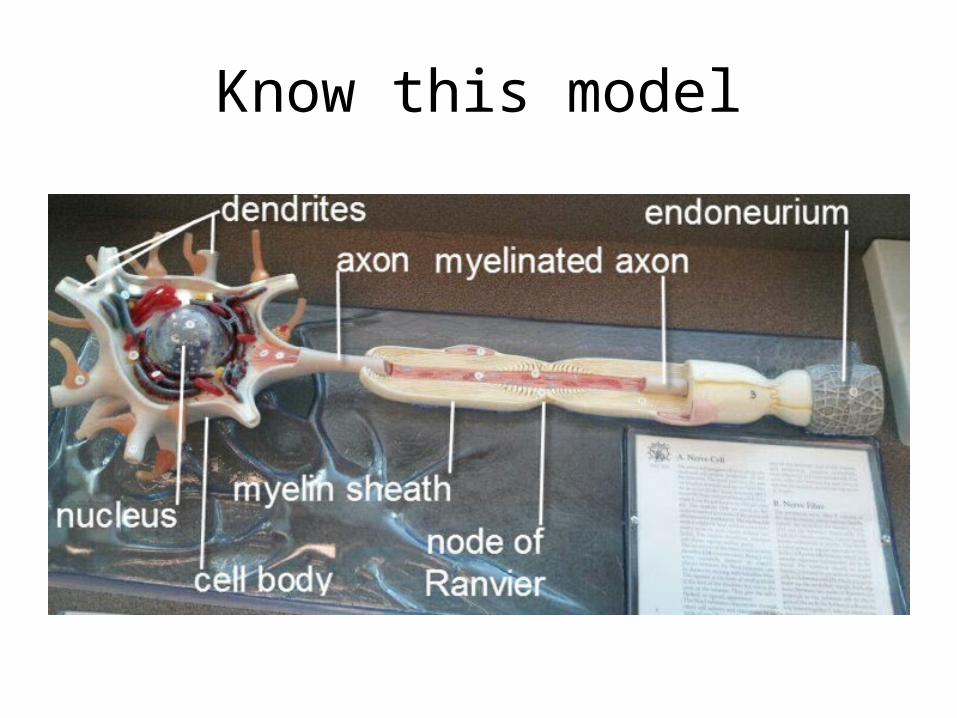
Know this model

Know the Meninges

Dura Mater- outter mostArachnoid Mater- middlePia Mater- innermost
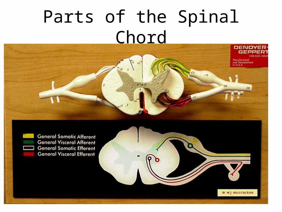
Parts of the Spinal Chord
3. Know the parts of the Spinal Cord ModelPosterior Median SulcusAnterior Median FissureCentral Canal – filled with CSF
Gray Matter-Posterior hornLateral hornAnterior hornGrey commisure- connects two halves of gray matter
White Matter-
Poterior funiculusLateral funiculusAnterior funiculus
Spinal NerveDoral root- carries sensory impulse INTO spinal cord (afferent) Dorsal root ganglion- contains SENSORY NEURON CELL BODIESVentral root- carries motor impulse AWAY from spinal cord (efferent)
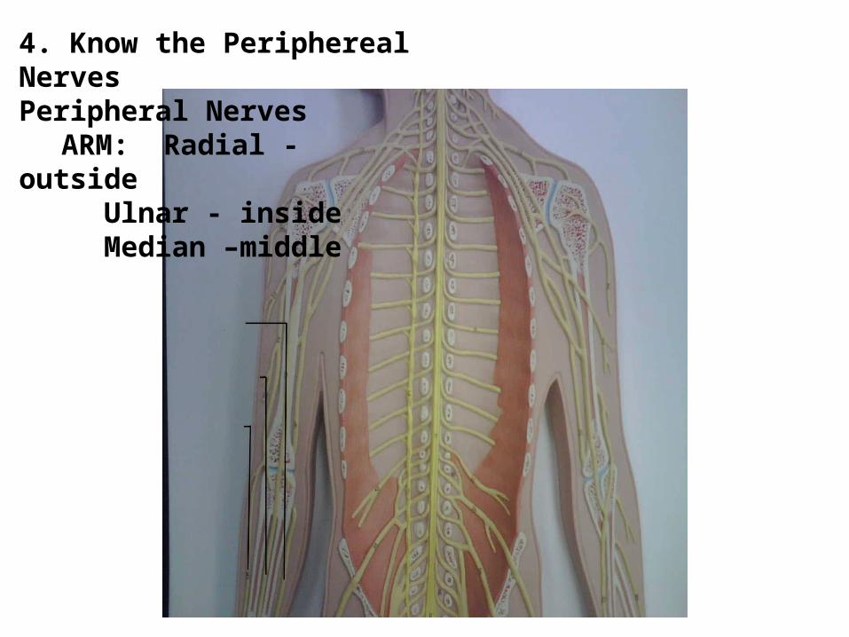
4. Know the Periphereal NervesPeripheral Nerves
ARM: Radial - outsideUlnar - insideMedian –middle

LEG: Sciatic – can only see on the left footObturator- pass through the foramenFemoral- runs down the femur
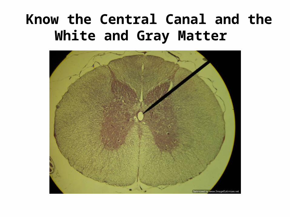
Know the Central Canal and the White and Gray Matter

Know the parts of the brain modelCerebrum
Cerebral hemispheres seperated by LONGIDUTINAL FISSURECorpus Callosum- connects the 2 cerebral hemispheres
Gyri- Precentral – motor areaPostcentral – sensory area
Sulci- Central SulcusLateral Sulcus
Central Lobes-FrontalParietalTemporalOccipital
Broca’s Area- Motor speech area

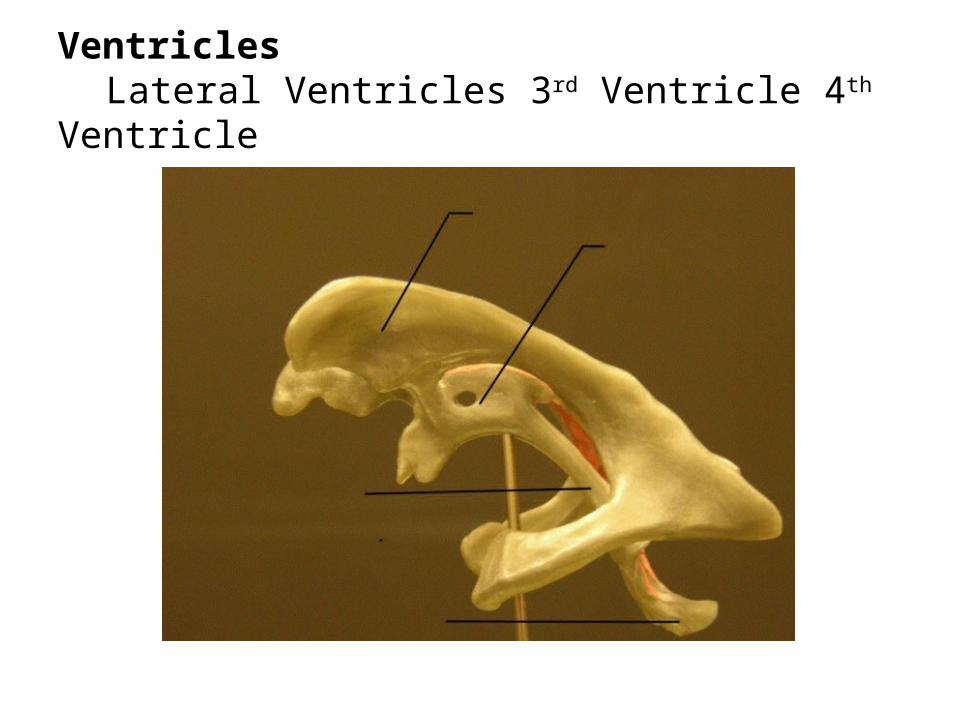
Ventricles
Lateral Ventricles 3rd Ventricle 4th Ventricle
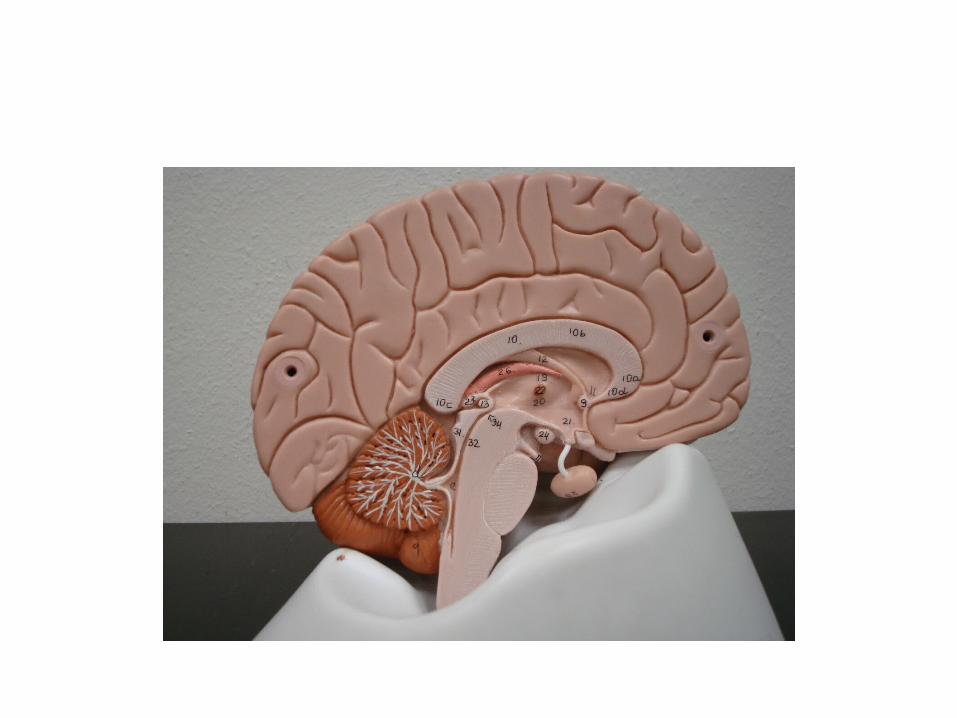
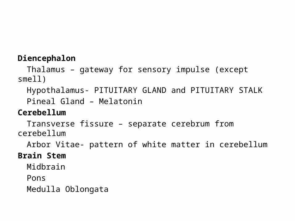
DiencephalonThalamus – gateway for sensory impulse (except smell)Hypothalamus- PITUITARY GLAND and PITUITARY STALKPineal Gland – Melatonin
CerebellumTransverse fissure – separate cerebrum from cerebellumArbor Vitae- pattern of white matter in cerebellum
Brain StemMidbrainPonsMedulla Oblongata
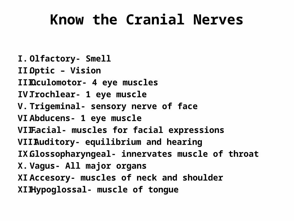
Know the Cranial Nerves
I. Olfactory- SmellII. Optic – VisionIII. Oculomotor- 4 eye musclesIV. Trochlear- 1 eye muscleV. Trigeminal- sensory nerve of faceVI. Abducens- 1 eye muscleVII. Facial- muscles for facial expressionsVIII. Auditory- equilibrium and hearingIX. Glossopharyngeal- innervates muscle of throatX. Vagus- All major organsXI. Accesory- muscles of neck and shoulderXII. Hypoglossal- muscle of tongue
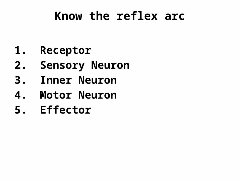
Know the reflex arc
1. Receptor2. Sensory Neuron3. Inner Neuron4. Motor Neuron5. Effector
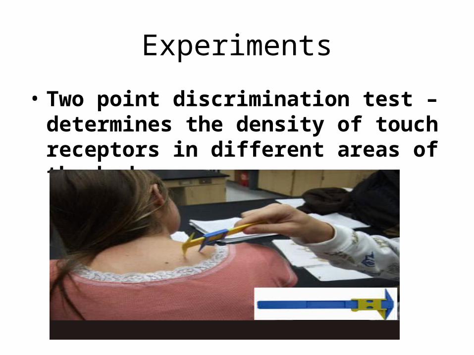
Experiments
• Two point discrimination test – determines the density of touch receptors in different areas of the body.
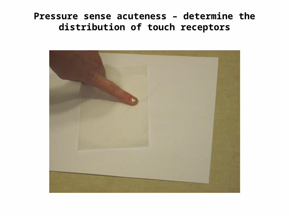
Pressure sense acuteness – determine the distribution of touch receptors
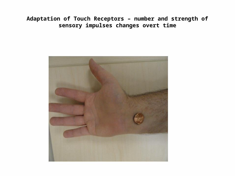
Adaptation of Touch Receptors – number and strength of sensory impulses changes
overt time
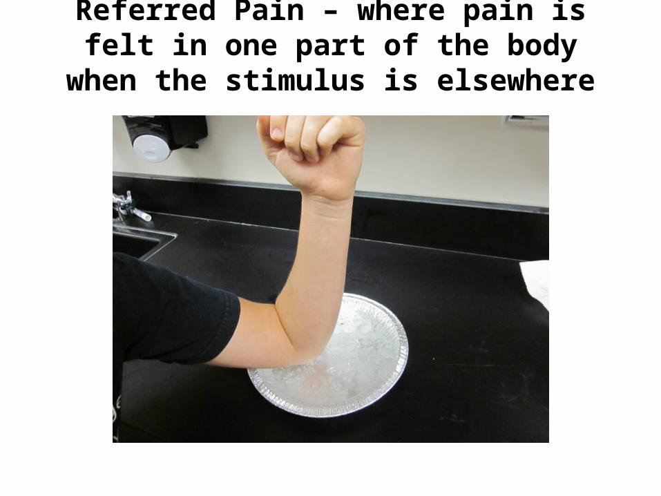
Referred Pain – where pain is felt in one part of the body when the stimulus is elsewhere

Experiments
• Localization of Taste – mapping of the taste buds
• Hearing testsRinne’s – detects conduction deafnessWeber’s – detects nerve deafness

Visual Acuity- 20/20 1st number is your eye, 2nd number is healthy eye
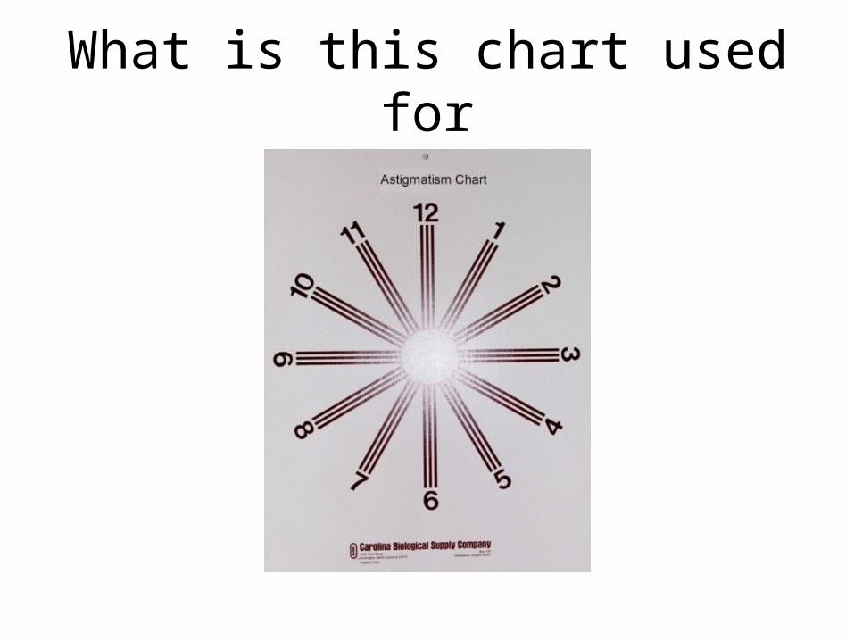
What is this chart used for
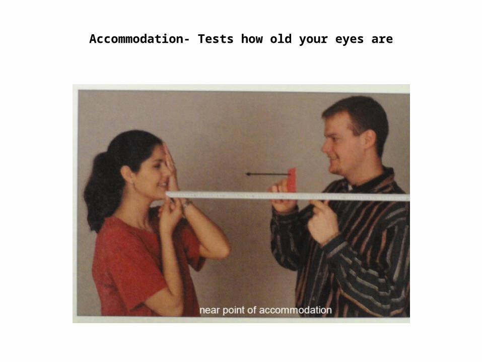
Accommodation- Tests how old your eyes are

Blind Spot Determination- The blind spot is created by the area where the optic nerve connects to the retina.

Photopupillary Reflex- Whenever the light shines in the eye the iris constricts

• Negative Color After Image – resynthesize the pigments opposites

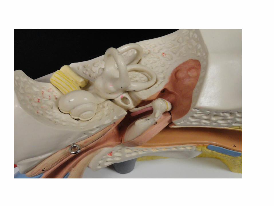
Know the anatomy of the earAuricleExternal Auditory Meatus Tympanic Cavity holds
MalleusIncusStapes
Tympanic MembraneAuditory TubeOval WindowVestibuleSemicircular canalCochleaRound WindowAuditory Nerve (Vestibulocochlear)

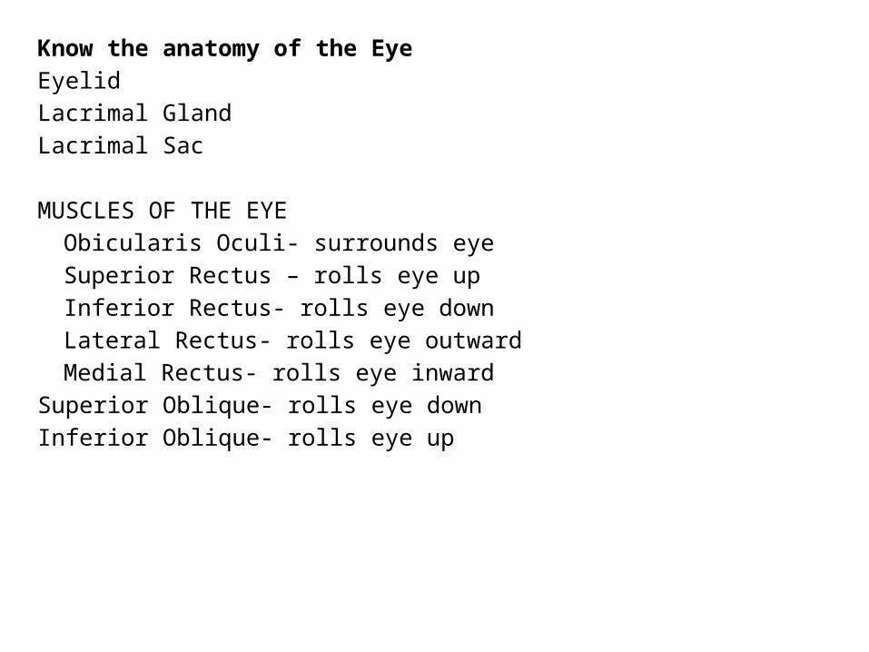
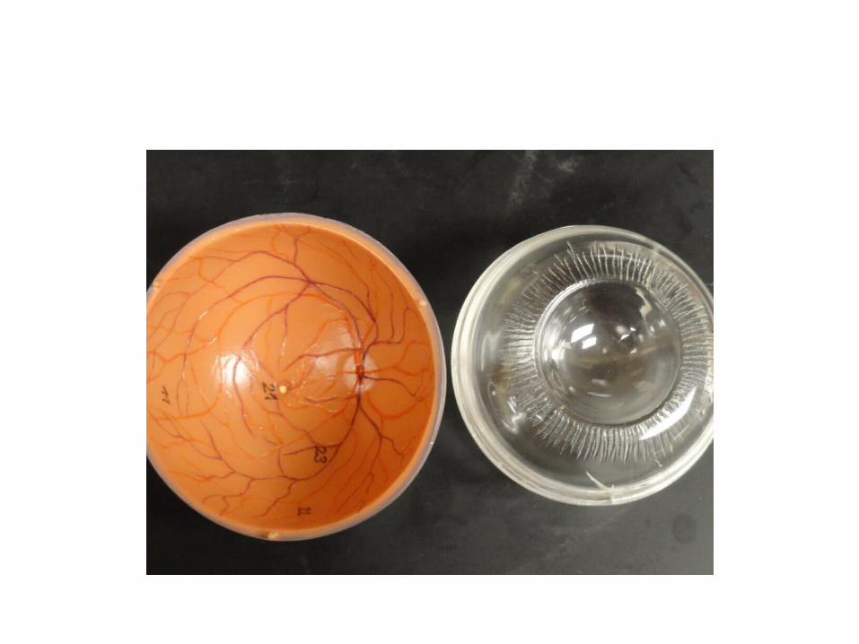
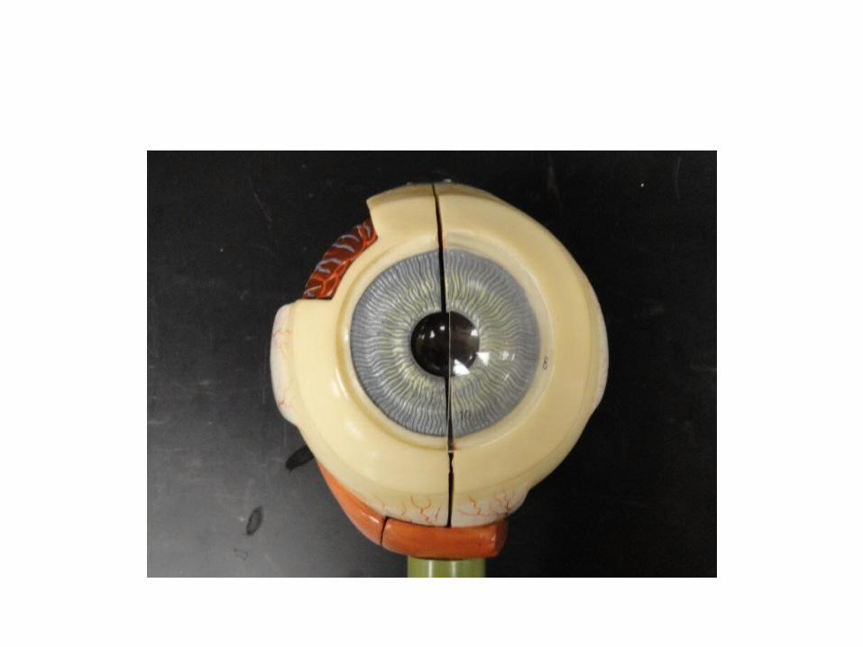
Know the anatomy of the EyeEyelidLacrimal GlandLacrimal Sac MUSCLES OF THE EYE
Obicularis Oculi- surrounds eyeSuperior Rectus – rolls eye upInferior Rectus- rolls eye downLateral Rectus- rolls eye outwardMedial Rectus- rolls eye inward
Superior Oblique- rolls eye downInferior Oblique- rolls eye up


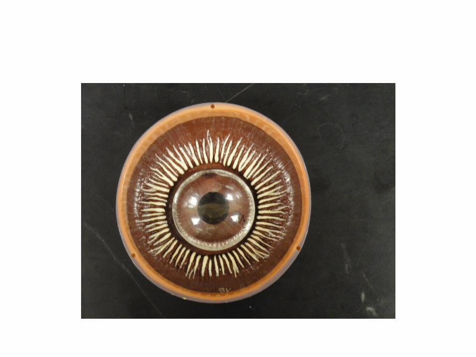
Outer tunic (layer)Sclera- whiteCornea- transparent
Middle Tunic (layer)Choroid coat- darkCiliary body with ciliary musclesSuspensory ligamentsLens- transparentIris- color varies
Pupil- hole in iris, allows light waves to reach retina, size of pupil determined by iris
Inner Tunic (layer)Retina- contains photoreceptors called RODS and CONESRODS detect light, CONES detect color
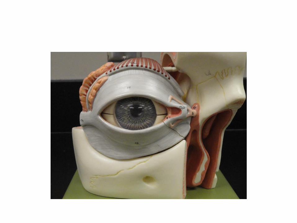

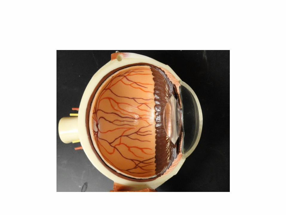

Anterior cavity- located in front of the lens and contains AQUEOUS HUMOR
Anterior chamber- between cornea and irisPosterior chamber- between iris and lens
Posterior cavity- located between lens and retina contains VITREOUS HUMOR