This article is available online at http://www.jlr.org Journal of Lipid Research Volume 44, 2003 2169 IDH1 gene transcription is sterol regulated and activated by SREBP-1a and SREBP-2 in human hepatoma HepG2 cells: evidence that IDH1 may regulate lipogenesis in hepatic cells Ishaiahu Shechter, 1 Peihua Dai, Liang Huo, and Guimin Guan Department of Surgery, F. Edward Hébert School of Medicine, Uniformed Services University of the Health Sciences, Bethesda, MD 20814-4799 Abstract The mRNA level for cytosolic NADP-dependent isocitrate dehydrogenase (IDH1) increases 2.3-fold, and en- zyme activity of NADP-isocitrate dehydrogenase (IDH) 63%, in sterol-deprived HepG2 cells. The mRNA levels of the NADP- and NAD-dependent mitochondrial enzymes show limited or lack of regulation under the same conditions. Nu- cleotide sequences that are required, and sufficient, for the sterol regulation of transcription are located within a 67 bp region of an IDH1-secreted alkaline phosphatase promoter- reporter gene. The IDH1 promoter is fully activated by the expression of SREBP-1a in the cells and, to a lesser degree, by that of SREBP-2. A 5-end truncation of 23 bp containing a CAAT and a GC-Box results in 6.5% residual activity. The promoter region involved in the activation by the sterol reg- ulatory element binding proteins (SREBPs) is located at nu- cleotides 44 to 25. Mutagenesis analysis identified within this region the IDH1-SRE sequence element GTGGGCTGAG, which binds the SREBPs. Similar to the promoter activation, electrophoretic mobility shifts of probes containing the IDH1- SRE element exhibit preferential binding to SREBP-1a, as compared with SREBP-2. These results indicate that IDH1 activity is coordinately regulated with the cholesterol and fatty acid biosynthetic pathways and suggest that it is the source for the cytosolic NADPH required by these path- ways.—Shechter, I., P. Dai, L. Huo, and G. Guan. IDH1 gene transcription is sterol regulated and activated by SREBP-1a and SREBP-2 in human hepatoma HepG2 cells: evidence that IDH1 may regulate lipogenesis in hepatic cells. J. Lipid Res. 2003. 44: 2169–2180. Supplementary key words cytosolic NADP-dependent IDH • sterol regulatory element binding protein • fatty acid synthesis • transcrip- tional regulation • cholesterogenesis Eukaryotic cells express three different forms of isoci- trate dehydrogenase (IDH). These enzymes catalyze the oxidative decarboxylation of isocitrate into -ketoglu- tarate utilizing either NAD or NADP as cosubstrates (1, 2). The role of the mitochondrial NAD-dependent IDH (IDH3) is well known, as it is the IDH that catalyzes a step in the tricarboxylic acid cycle (3). In contrast, the bio- chemical role of the two NADP-dependent IDHs (E.C. 1.1.1.42) is not entirely clear. These latter forms of the en- zyme are encoded in the nucleus and function as ho- modimers (4, 5). One NADP-IDH is localized in the mito- chondria (IDH2), but gene disruption studies in yeast have shown that it cannot replace the function of the IDH3 (6). The human cytoplasmic NADP-specific enzyme (IDH1) is encoded by the IDH1 gene, and its mRNA se- quence was reported (7). The subcellular localization of the enzyme’s protein was shown to be in both peroxi- somes and the cytoplasm (8). Interestingly, the rat enzyme was shown to localize exclusively in peroxisomes (9). The subcellular localization of IDH1 in peroxisomes is consis- tent with the presence of the peroxisomal targeting PST-1 sequence (Ala-Lys-Leu-COOH). An additional physiological role for peroxisomal IDH1 may be to provide cytosolic NADPH for several NADPH- dependent enzymes, such as 3-hydroxy-3-methylglutaryl- CoA reductase (10–13), acyl-CoA reductase (14), and 2,4- dienoyl-CoA reductase (15, 16). The activity of IDH1 is the only known source for peroxisomal NADPH. Another physiological role of peroxisomal IDH1 is to provide -ketoglutarate, which is required as a cosubstrate for the phytanoyl-CoA -hydroxylase reaction (17–20). Abbreviations: HSS, human squalene synthase; IDH, isocitrate de- hydrogenase; IDH1, cytosolic NADP-dependent IDH; IDH2, mitochon- drial NADP-dependent IDH; IDH3, mitochondrial NAD-dependent IDH; PPAR, peroxisome proliferator-activated receptor; RXR, retinoid X receptor; SCAP, SREBP cleavage-activating protein; SEAP, secreted alkaline phosphatase; SQS, squalene synthase; SRE, sterol regulatory element; SREBP, sterol regulatory element binding protein. 1 To whom correspondence should be addressed. e-mail: [email protected]Manuscript received 27 June 2003 and in revised form 8 August 2003. Published, JLR Papers in Press, August 16, 2003. DOI 10.1194/jlr.M300285-JLR200 by guest, on January 7, 2019 www.jlr.org Downloaded from
Transcript
This article is available online at http://www.jlr.org
Journal of Lipid Research
Volume 44, 2003
2169
IDH1 gene transcription is sterol regulated and activated by SREBP-1a and SREBP-2 in human hepatoma HepG2 cells: evidence that IDH1 may regulate lipogenesis in hepatic cells
Ishaiahu Shechter,
1
Peihua Dai, Liang Huo, and Guimin Guan
Department of Surgery, F. Edward Hébert School of Medicine, Uniformed Services University of the Health Sciences, Bethesda, MD 20814-4799
Abstract The mRNA level for cytosolic NADP-dependentisocitrate dehydrogenase (IDH1) increases 2.3-fold, and en-zyme activity of NADP-isocitrate dehydrogenase (IDH) 63%,in sterol-deprived HepG2 cells. The mRNA levels of theNADP- and NAD-dependent mitochondrial enzymes showlimited or lack of regulation under the same conditions. Nu-cleotide sequences that are required, and sufficient, for thesterol regulation of transcription are located within a 67 bpregion of an IDH1-secreted alkaline phosphatase promoter-reporter gene. The IDH1 promoter is fully activated by theexpression of SREBP-1a in the cells and, to a lesser degree,by that of SREBP-2. A 5
�
-end truncation of 23 bp containinga CAAT and a GC-Box results in 6.5% residual activity. Thepromoter region involved in the activation by the sterol reg-ulatory element binding proteins (SREBPs) is located at nu-cleotides
�
44 to
�
25. Mutagenesis analysis identified withinthis region the IDH1-SRE sequence element GTGGGCTGAG,which binds the SREBPs. Similar to the promoter activation,electrophoretic mobility shifts of probes containing the IDH1-SRE element exhibit preferential binding to SREBP-1a, ascompared with SREBP-2. These results indicate thatIDH1 activity is coordinately regulated with the cholesteroland fatty acid biosynthetic pathways and suggest that it isthe source for the cytosolic NADPH required by these path-
ways.
—Shechter,
I., P. Dai, L. Huo, and G. Guan.
IDH1 genetranscription is sterol regulated and activated by SREBP-1aand SREBP-2 in human hepatoma HepG2 cells: evidencethat IDH1 may regulate lipogenesis in hepatic cells.
J. LipidRes.
2003.
44:
2169–2180.
Supplementary key words
cytosolic NADP-dependent IDH
•
sterolregulatory element binding protein
•
fatty acid synthesis
•
transcrip-tional regulation
•
cholesterogenesis
Eukaryotic cells express three different forms of isoci-trate dehydrogenase (IDH). These enzymes catalyze the
oxidative decarboxylation of isocitrate into
�
-ketoglu-tarate utilizing either NAD or NADP as cosubstrates (1, 2).The role of the mitochondrial NAD-dependent IDH(IDH3) is well known, as it is the IDH that catalyzes a stepin the tricarboxylic acid cycle (3). In contrast, the bio-chemical role of the two NADP-dependent IDHs (E.C.1.1.1.42) is not entirely clear. These latter forms of the en-zyme are encoded in the nucleus and function as ho-modimers (4, 5). One NADP-IDH is localized in the mito-chondria (IDH2), but gene disruption studies in yeasthave shown that it cannot replace the function of theIDH3 (6). The human cytoplasmic NADP-specific enzyme(IDH1) is encoded by the IDH1 gene, and its mRNA se-quence was reported (7). The subcellular localization ofthe enzyme’s protein was shown to be in both peroxi-somes and the cytoplasm (8). Interestingly, the rat enzymewas shown to localize exclusively in peroxisomes (9). Thesubcellular localization of IDH1 in peroxisomes is consis-tent with the presence of the peroxisomal targeting PST-1sequence (Ala-Lys-Leu-COOH).
An additional physiological role for peroxisomal IDH1may be to provide cytosolic NADPH for several NADPH-dependent enzymes, such as 3-hydroxy-3-methylglutaryl-CoA reductase (10–13), acyl-CoA reductase (14), and 2,4-dienoyl-CoA reductase (15, 16). The activity of IDH1 isthe only known source for peroxisomal NADPH.
Another physiological role of peroxisomal IDH1 is toprovide
�
-ketoglutarate, which is required as a cosubstratefor the phytanoyl-CoA
This enzyme catalyzes a step in the degradation ofphytanoic acid, a by-product of the plant-derived phytol.Several metabolic functions for phytanic acid have beenreported: regulation of glucose metabolism in primary rathepatocytes by acting as an agonist to peroxisome prolif-erator-activated receptors (PPARs) (21, 22) and retinoidX receptor (RXR) (23); white adipocyte differentiation(24); and activation of uncoupling protein-1 gene tran-scription and brown adipocyte differentiation (25). In theautosomal disorder Refsum’s disease,
�
-oxidation of phy-tanic acid is affected in some, but not all, cases, and accu-mulation of this acid is observed in various tissues (26).
The likely function of the cytoplasmic IDH1 is to pro-duce NADPH for reductive reactions (27). Although thehexose monophosphate shunt is considered to be the ma-jor source of NADPH in the cytoplasm, genetic defects inglucose-6-phosphate dehydrogenase (G6PD), which cata-lyzes the first step in the shunt, do not affect sterol or fattyacid metabolism in humans (28). Population studies pro-vide no evidence that any disorders other than hemolyticanemia are associated with G6PD deficiency (29), and bothChinese hamster ovary cells and human fibroblasts withless than 5 percent of normal G6PD activity grow normally(30, 31). This result suggests that the contribution of IDH1to NADPH production may be significant. There are grow-ing indications that IDH1 participates in cytosolic NADPHproduction and in fatty acid biosynthesis (32, 33). In sup-port of this idea, IDH1 was also identified as a major sourceof cytosolic NADPH needed for the regeneration of re-duced glutathione, critically important in cellular defenseagainst oxidative damage (32, 34). Finally, an interestingand unexpected role for IDH1 was shown in bovine eyes,where the enzyme protein fulfills the criteria for a cornealepithelial crystallin, which may be involved in maintainingcorneal epithelial transparency (35). Because the activity ofIDH1 serves as a major source for nonmitochondrialNADPH required in multiple metabolic pathways, it is im-portant to understand how IDH1 activity is regulated. Theregulation in the peroxisomes and cytoplasm is not known.The 5
�
promoter sequence for the gene encoding IDH1 isalso not known, and information on the transcriptional reg-ulation of this gene has not been reported.
Cholesterol and fatty acid synthesis is regulated mainlyby coordinated transcription involving sterol regulatoryelements (SREs), which are the targets of three basic-helix-loop-helix leucine zipper transcription factors calledsterol regulatory element binding proteins (SREBPs)(36). These proteins are synthesized as membrane-boundendoplasmic reticulum (ER) precursors (M-SREBPs) withtwo membrane-spanning domains (37). After synthesis,the SREBPs are bound to an SREBP cleavage-activatingprotein (SCAP). The NH
2
-terminal region of SCAP con-tains eight membrane-spanning helices that serve as thesterol-sensing domain; the COOH-terminal region con-tains several copies of a WD40 sequence that binds to theSREBPs (37–41). At low membrane cholesterol levels,SCAP escorts the SREBPs to the Golgi compartment (42),where the SREBPs are processed by site-1 and site-2 pro-teases. Proteolytic cleavage releases the C-terminal SREBPs
and the soluble transcriptionally active N-terminal SREBPs(N-SREBPs) into the cytosol. The latter can then enter thenucleus and activate sterol and fatty acid synthesis. Thesterol-regulated movement of the SCAP-SREBP complexfrom the ER to the Golgi is a pivotal event that controlscholesterol homeostasis in eukaryotic cells (37, 43–45).Recently, two additional ER-bound proteins, INSIG-1 (46)and INSIG-2 (47), were shown to be involved in the regu-lated transport of the SCAP-SREBP complex to the Golgi.These two proteins have 59% identity with five putative trans-membrane domains. In the presence of high membranecholesterol content, the INSIG proteins bind to the sterol-sensing domain of SCAP, causing its retention in the ERand preventing SREBP-SCAP complex transport to theGolgi. At low cholesterol levels, the INSIGs dissociate fromthe SCAP, allowing the migration of the SREBP-SCAPcomplex to the ER budding region for transport to the Golgi(46–49). We have recently demonstrated that the ER-to-Golgi transport of the SREBP-SCAP complex is inhibitedat 20
�
C and that this inhibition in transport can be over-come by the overexpression of SCAP (50).
The N-SREBPs display differential activation of genetranscription. It was first reported that various
cis
elementsin the human hepatic squalene synthase (HSS) promoterdifferentially bind N-SREBP-1a and N-SREBP-2. Based onthat, distinct functional specificity for the two transcrip-tion factors was predicted (51, 52). A subsequent study byGoldstein and colleagues (53) established the differentialstimulation of cholesterol and unsaturated fatty acid bio-synthesis in Chinese hamster ovary cells expressing the in-dividual SREBPs. There again, the mRNA for squalenesynthase (SQS) was differentially expressed by SREBP-2and not by the two isoforms of SREBP-1. These observa-tions, together with results from studies in genetically ma-nipulated mice (54), indicate that SREBP-1 is selectivelyinvolved in the activation of genes associated with fattyacid metabolism while SREBP-2 is more specific in choles-terol homeostasis, primarily through its selective activa-tion of the SQS gene. The number of genes found to beactivated by the SREBPs is constantly increasing, and in-cludes enzymes involved in cellular cholesterol homeosta-sis and fatty acid synthesis (55–57). Our study describesthe promoter for the IDH1 gene, its transcriptional regu-lation, and the relationship between this regulation andthe regulation of fatty acid and sterol biosynthesis.
EXPERIMENTAL PROCEDURES
Cell cultures and transient transfections
Human hepatoma HepG2 cells were cultured in Eagle’s mini-mum essential medium (MEM) supplemented with 10% fetal bo-vine serum, 1 mM glutamine, 1 mM pyruvate, 100 U/ml penicil-lin, and 100
�
g/ml streptomycin, at 37
�
C under 5% CO
2
atmosphere. Transient transfections were conducted in 12-wellplates. Used for each transfection were 0.5
�
g of a given IDH1-SEAP reporter plasmid and 0.1
�
g of pCMV-
�
-galactosidase (as acontrol for transfection efficiency) plus 25 ng of one of the plas-mids pCDNA3.1 (as pCMV vector control), pCMV-CSA10 (con-
stitutively expressing the nuclear form of human SREBP-1a), orpCMV-CS2 (constitutively expressing the nuclear form of humanSREBP-2). The transfections were performed using Fugene6 Re-agent (Roche). At the time of treatment (24 h after transfection),the culture media were changed to sterol (
�
) or sterol (
�
) as de-fined below. After an additional 24 h incubation, the culture me-dium from each plate was collected and assayed for secreted al-kaline phosphatase (SEAP) activity by luminometry using theGreat EscAPe SEAP Reporter System kit from Clontech. Thecells were then lysed, and the whole-cell extracts were assayed for
�
-galactosidase activity as previously described (52). RelativeSEAP activity is expressed as the ratio of SEAP activity (light unit)to
�
-galactosidase activity. Sterol (
�
) and sterol (
�
) conditionswere achieved by addition of either 1
�
g/ml 25-OH cholesterolplus 10
�
g/ml cholesterol or 5
�
g/ml lovastatin, respectively, toMEM supplemented with 10% lipid-depleted serum (LDS).
Preparation of IDH1 promoter-reporter constructs
The 5
�
flanking region of the
IDH1
gene was amplified by PCRfrom HepG2 genomic DNA using a pair of primers set for the de-sired region, based on sequence information of the
IDH1
gene inhuman chromosome 2 (GenBank accession number AC016697).The sequences of the two primers used are: 5
�
-GTGGTACCTC-CACCGTTTTCTAAGGCTTCACATC-3
�
(forward) and 5
�
-CTCA-AGCTTGATGATATGCTGGCGAAGAGTTGGGG-3
�
(reverse). Theamplified DNA fragment is in the region
�
962 to
�
170 relativeto the
IDH1
gene transcription initiation site (see below). TheDNA was then ligated into the
Kpn
I (5
�
) and
Hind
III (3
�
) sites ofthe vector pTAL-SEAP (Clontech) utilizing the two linker se-quences built into the primers. This resulting construct is desig-nated pIDH1-962-SEAP and was used as the longest SEAP pro-moter-reporter for the human
IDH1
gene. For 5
�
truncation ofthe promoter, a PCR-based, site-directed mutagenesis procedurewas employed. Briefly, the plasmid pIDH1-962-SEAP was used asthe template. A series of oligonucleotides that correspond to thedesired 5
�
end of the
IDH1
promoter were used as forward prim-ers, all containing a
Kpn
I site. The reverse primer was an oligo-nucleotide located 3
�
to the cloning
Kpn
I site. The PCR reactionswere performed using the Expand Long Template PCR System(Roche). The resulting PCR products were digested with
Kpn
I andself-ligated to produce the SEAP-reporter plasmids of
IDH1
withvarious 5
�
promoter ends. The sequences of the forward primersused to generate the different promoter-reporters and the desig-nations of the plasmids are: pIDH1-481-SEAP, 5
�
-GCTAGGTAC-CGCATTAGGCAGCGCGGAACCCCCTAG-3
�
; pIDH1-219-SEAP,5
�
-GCTAGGTACCTTCGCTGTCGGGATTCGGGACTGAATC-3
�
;pIDH1-91-SEAP, 5
�
-GCTAGGTACCATCCCACGGGAATTGGCG-TGTGGCG-3
�
; pIDH1-67-SEAP, 5
�
-GCTAGGTACCGGCGATTG-GAGGCGTGTCGGGGGCG-3
�
; pIDH1-44-SEAP, 5
�
-GCTAGGTA-CCGGGGCTGGGGGAGGTGGGCTGAGGA-3
�
; pIDH1-25-SEAP,5
�
-GCTAGGTACCTGAGGAGGCGGGGCCTGGGAGGGG-3
�
; andpIDH1-6-SEAP, 5
�
-GCTAGGTACCGAGGGGACAAAGCCGGGA-AGAGGAAA-3
�
.
Site-directed mutagenesis of IDH1 promoter
Replacement mutation of the IDH1-SRE-1 element in the hu-man IDH1 promoter was obtained using the QuikChange™ Site-Directed Mutagenesis Kit (Stratagene). The primers used in thisprocedure were the pair of primers used in the electrophoreticmobility shift assay to produce the IDH1
�
40/
�
5 m5 probe (seeFig. 7B), in which the sequence TGAT of the proposed IDH1-SREelement was replaced by the sequence GTTT. This mutation pro-cedure produced the IDH1-67m5-SEAP mutant promoter-reporterconstruct.
Determination of transcription initiation sites
To determine the transcription initiation site(s) of the
IDH1
gene,a 5
�
-rapid amplification of cDNA ends (RACE) was performed(58) using the 5
�
-Race System kit from Invitrogen. The gene-specificprimers GSP1 (5
�
-TCCTCAACCCTCTTCTCATC-3
�
) and GSP2(5
�
-TTCGTCTCATTTCATCTCCT-3
�
) used are located at
�
42and
�
234 3
�
to the AUG translation initiator site. The variousRACE clones isolated were sequenced, and the most 5
�
end se-quences were determined.
Preparation of poly(A
�
) RNA and Northern blot analysis
HepG2 cells (1
10
6
) were plated onto 150 mm plates andgrown for 2 days at 37
�
C then treated as described above. Twosets of plates were treated. One set was kept at 37
�
C, and theother was transferred and kept at 20
�
C. At the end of the 24 htreatment period, the cells were harvested and total RNA wasprepared using Tryzol Reagent (Invitrogen). Poly(A
�
) RNA wasextracted from total RNA using the FastTrack
®
2.0 Kit (Invitro-gen). For Northern blots, 4
�
g poly(A
�
) RNA from each samplewas separated on 1.2% agarose gel, transferred onto nylonmembranes, and probed with different
32
P-radiolabeled cDNAprobes. IDH1 and IDH2 cDNA probes were generated by RT-PCR based on known mRNA sequences, and the probes forIDH3-
�
and IDH3-
were excised from clones containing corre-sponding cDNA sequences obtained from American Type Cul-ture Collection. Quantitation of specific signal intensities wasperformed by densitometry using ImageQuant software (Molec-ular Dynamics).
Assay for NADP-dependent IDH activity
HepG2 cells were cultured and treated with sterol (
�
) and ste-rol (
�
) medium as described above. Whole-cell lysates were pre-pared by sonication of cell suspensions in a buffer containing 50mM Tris-HCl (pH 7.9), and 1 mM EDTA, followed by centrifuga-tion to obtain clear extracts. NADP-dependent IDH enzymaticactivity assays were performed at 25
�
C using the Farrell proce-dure (59). Enzyme activity is expressed in
�
mol increase ofNADPH per minute.
Electrophoretic mobility shift assay
Electrophoretic mobility shift assay (EMSA) was carried out asdescribed previously (60). Briefly, annealed oligo probes wereend-labeled with
-
32
P-ATP using T4 polynucleotide kinase. Forthe binding reaction, 8
10
4
dpm of each probe was incubatedwith
�
50 ng of purified SREBP protein (60) in a buffer contain-ing 25 mM Tris-HCl (pH 8.0), 50 mM KCl, 6.25 mM MgCl
2
, 0.5mM EDTA, 0.5 mM DTT, 50
�
g/ml of polydeoxyinosinic-deoxy-cytidylic acid, and 10% glycerol for 30 min on ice. The mixtureswere then separated by electrophoresis on a nondenaturing 30%polyacrylamide gel. Detection of radiolabeled signals was doneby autoradiography. The probes used in the EMSA were all syn-thetic oligonucleotides; sequences are described in each experi-ment (below). For positive control, an oligonucleotide probe con-taining the HSS-SRE-1 sequence in the human SQS promoterwas used [5
�
-TAGAGTGTTATCACGCCAGTCTCCTT-3
�
(60)].
RESULTS
Sterol-mediated regulation of IDH1 mRNA level
The possible regulatory role of sterols in IDH1 expres-sion was investigated by the addition of sterols to HepG2cells, and analysis of IDH1 mRNA level. At 37
�
C, a 2.3-foldinduction of the IDH1 mRNA level was observed after 24 hin sterol-depleted cells, as compared with sterol-loaded
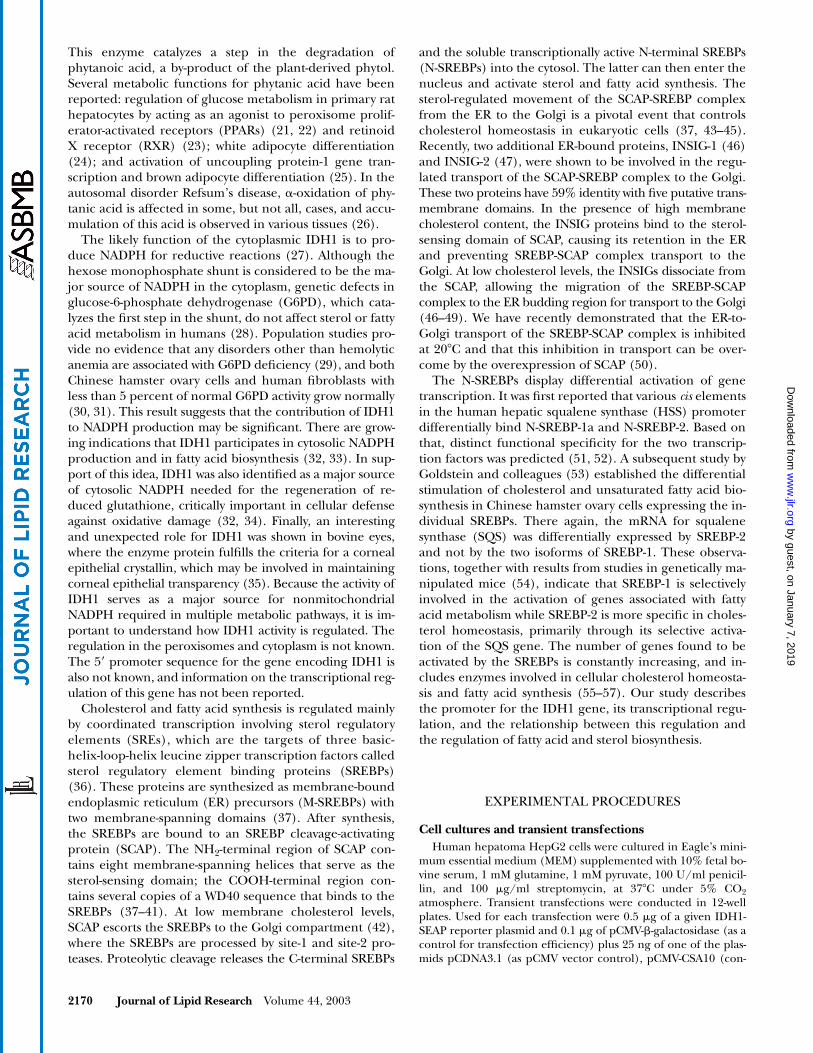
cells (Fig. 1). Under the same conditions, the sterol regu-lation of the NADP-dependent mitochondrial IDH2 wasonly 1.3-fold, and no sterol regulation was observed forthe mRNA levels of either the �- or the -subunits of theNAD-dependent mitochondrial IDH3. We have recentlydemonstrated that sterol regulation of enzymes in thecholesterol and fatty acid biosynthetic pathways is dimin-ished or completely absent in cells maintained at 20�C,due to lack of processing of the SREBPs at this tempera-ture (50). It appears that a similar low-temperature effectis observed for the regulation of IDH1. The sterol-regu-lated increase in IDH1 mRNA level is inhibited in sterol-deprived cells maintained at 20�C, and the mRNA levelsare lower than those observed in cells at 37�C, regardlessof the presence of sterols. The limited sterol regulation ofIDH2 is also absent at 20�C, but the level of mRNA for thisenzyme is still rather substantial. No temperature effect orsterol regulation was observed for the mRNA of either the�- or the -subunits of IDH3 (Fig. 1). These observations,together with our earlier studies (50), indicate a coordi-nated regulation between cholesterol and fatty acid syn-thesis and the expression of the IDH1 gene. They alsopoint to SREBPs as the likely common regulatory tran-scription factors for all of these genes. Lack of, or limited,sterol regulation of the mRNA for the other IDH genesmay suggest that IDH1 is the major source for NADPH forsterol and fatty acid biosynthesis.
Sterol regulation of NADP-dependent IDH enzyme activity
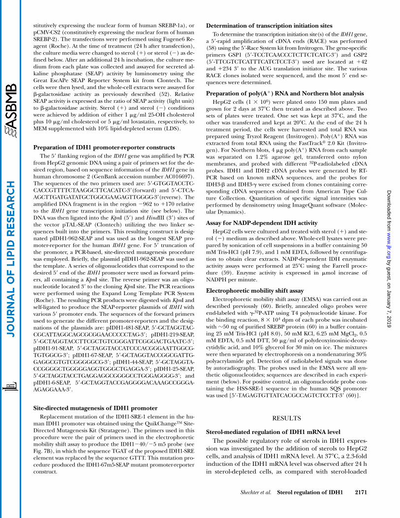
The 24 h sterol effect on the NADP-dependent IDH ac-tivity in HepG2 extracts is shown in Fig. 2. Sterol depriva-
tion of cells for this period of time results in a 63% in-crease in NADP-dependent IDH activity compared withthe activity in cells grown in the presence of sterols. Be-cause this enzyme assay cannot distinguish between theactivities of IDH1 and IDH2, it is not possible to assess thecontribution of each to the observed activity and the par-ticipation of either in the sterol regulation. Nevertheless,a significant sterol effect on the NADP-dependent activityin total cell extract is observed.
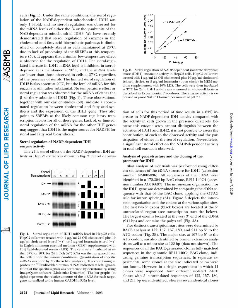
Analysis of gene structure and the cloning of the promoter for IDH1
Blast analysis of GenBank was performed using differ-ent sequences of the cDNA structure for IDH1 (accessionnumber NM005896). All sequences of the cDNA wereidentified in a 172,304 bp BAC clone, RP11-140C4 (acces-sion number AC016697). The intron-exon organization forthe IDH1 gene was determined by comparing the cDNA se-quence with that of the BAC clone, applying the GT-AGrule for intron splicing (61). Figure 3 depicts the intron-exon organization and the codons at the various splice sites.The first two 5� exons (black boxes) are located at the 5�untranslated region (see transcription start site below).The largest exon is located at the very 3� end of the cDNA(941 bp) and contains the polyA tail (Fig. 3A).
Five distinct transcription start sites were determined byRACE analysis at 122, 157, 167, 180, and 211 bp 5� to theATG codon (Fig. 3B). The major site, at 167 bp 5� to theATG codon, was also identified by primer extension analy-sis, as well as a minor site at 122 bp (data not shown). Thesequences of all the RACE-generated clones fully matchedsequences in the genomic RP11-140C4 BAC clone, indi-cating genuine transcription sequences. In separate ex-periments, some clones at the size indicated below werenot found. However, in a single experiment in which 11clones were sequenced, four different isolated RACEclones with 5� untranslated sequences of 122, 157, 180,and 211 bp were identified, whereas seven identical clones
Fig. 1. Sterol regulation of IDH1 mRNA level in HepG2 cells.HepG2 cells were treated with 1 �g/ml 25-OH cholesterol plus 10�g/ml cholesterol [sterol(�)], or 5 �g/ml lovastatin (sterol(�)]in Eagle’s minimum essential medium (MEM) supplemented with10% lipid-depleted serum (LDS). The cells were incubated at ei-ther 37�C or 20�C for 24 h. Poly(A�) RNA was then prepared fromthe cells under the various conditions. Quantitation of specificmRNAs was done by Northern blot analyses (left section) using asprobes the 32P-radiolabled human cDNAs indicated at left. Quanti-tation of the specific signals was performed by densitometry, usingImageQuant software (Molecular Dynamics). The bar graphs (atright) represent the relative amounts of the mRNA for each targetgene normalized to the human GAPDH mRNA level.
Fig. 2. Sterol regulation of NADP-dependent isocitrate dehydrog-enase (IDH1) enzymatic activity in HepG2 cells. HepG2 cells weretreated with 1 �g/ml 25-OH cholesterol plus 10 �g/ml cholesterol(closed circle), or 5 �g/ml lovastatin (open circle) in MEM me-dium supplemented with 10% LDS. The cells were then incubatedat 37�C for 24 h. IDH1 activity was measured in whole-cell lysate asdescribed in Experimental Procedures. The enzyme activity is ex-pressed as �mol NADPH formed per minute at pH 7.4.
with a 5� untranslated sequence of 167 bp were found.Thus, it appears that clones with a 5� untranslated se-quence of 167 bp are at approximately 7 frequency. Themost 5� adenosine of the longest clone (211 bp 5� to theATG) was designated 1. Based on the above, the sequenceof �962 to �170 was amplified for the construction of thepIDH1-962-SEAP promoter-reporter.
Sterol regulation of pIDH1-962-SEAP promoter-reporter activity
The IDH1 promoter activity is enhanced 2-fold in cellsgrown in media containing 10% LDS, as compared withthe activity in cells grown in the presence of 10% FBS(Fig. 4). The promoter activity in cells grown in the pres-ence of FBS is suppressed by the addition of sterols to themedia. Addition of sterols completely diminishes the acti-vation by LDS, and the IDH1 promoter activity is sup-pressed regardless of the type of serum in the media. As
expected for a sterol-mediated regulation, addition of lov-astatin to the growth media greatly enhanced the IDH1promoter activity. A 5.4-fold enhancement in activity is ob-served in lipid-deprived cells in which the promoter isfully activated (LDS � lovastatin), compared with sup-pressed conditions (LDS � sterols). Thus, the regulationof the IDH1 promoter activity is sterol mediated, similarto the observed variations in IDH1 mRNA levels seen inFig. 1.
Regulation of pIDH1-962-SEAP promoter-reporter activity by SREBP
Because the SREBP transcription factors are involved insterol-mediated regulation of genes involved in choles-terol and fatty acid biosynthesis, it is anticipated that acti-vation of the sterol-responsive IDH1 promoter is similarlyregulated by these transcription factors. Indeed, the activ-ity of the pIDH1-962-SEAP promoter increases in re-
Fig. 3. The intron-exon organization for the IDH1 gene and transcription initiation sites. The intron-exonorganization for the IDH1 gene was determined by comparing the IDH1 cDNA sequence with that of theIDH1 BAC clone RP11-140C4 (accession number AC016697) applying the GT-AG rule for intron splicing.The intron-exon organization and the codons at the various splice sites are shown (A). The first two 5� exons(black boxes) are located at the 5� untranslated region (see 5�-UTR below). The largest exon is located at thevery 3� end of the cDNA (941 bp) and contains the polyA tail. Nucleotide sequences of five 5�-UTRs of thehuman IDH1 mRNA are shown (B). The five distinct transcription start sites were determined by rapid am-plification of cDNA ends (RACE) analysis at 122, 157, 167, 180, and 211 bp 5� to the ATG codon and are in-dicated by arrows. The first few nucleotides for each initiation site are also shown. In a single representativeexperiment, in which 11 clones were sequenced, four different isolated RACE clones with 5� untranslated se-quences of 122, 157, 180, and 211 bp were identified, whereas seven identical clones with a 5� untranslatedsequence of 167 bp (bold arrow) were found. Distances between the initiation sites as well as the size of theshortest 5�-UTR are labeled as base pair at the bottom of the scheme.
sponse to increasing amounts of pCMV-CSA10 (SREBP-1a) and pCMV-CS2 (SREBP-2) DNA in transiently trans-fected HepG2 cells (Fig. 5). The dose-response activationcurves for SREBP-1a and SREBP-2 are similar, but activa-tion by SREBP-2 is consistently lower. Almost identical ac-tivation of the IDH1 promoter was observed by transfec-tion of pCMV-CSA10 and pCMV-CS1c, which encodesexpression of SREBP-1c, indicating a similar response ofthe reporter gene to the two SREBP-1 expression vectors(data not shown).
Sterol regulation and activation by SREBPs of fusion genes containing successively 5�-truncated IDH1 promoter
A series of successively 5�-truncated IDH1 promoter-SEAP reporter genes was prepared by PCR methodology(Fig. 6A). Significant sterol-dependent regulation of pro-moter activity is observed in fusion genes containing pro-moter sequences of 67 bp or longer (Fig. 6B). In fact,IDH1-67-SEAP shows the most response (7.9-fold) to ste-rol regulation compared with longer promoters. The pro-moter-reporter genes longer than 67 bp also show a signif-icant response to an expressed SREBP-1a or SREBP-2. Theactivation of each of the promoters by SREBP-1a is consis-tently higher than the activation by SREBP-2. For IDH1-67-SEAP, the activation by SREBP-1a is �2-fold higherthan the activation by SREBP-2. Additional 23 bp 5� trun-cation (IDH1-44-SEAP) resulted in a dramatic loss of pro-moter activity and response to sterols. However, residualresponse to the expressed SREBPs is still observed. Withinthis truncated 23 nucleotide (nt) sequence reside an in-verted CCAAT box [�62]ATTGG-[�58] and a GC-Boxsequence [�55]CGGGGGCGGGGCT-[�43] (Fig. 6A). Thus,the loss of promoter activity in this reporter gene may be
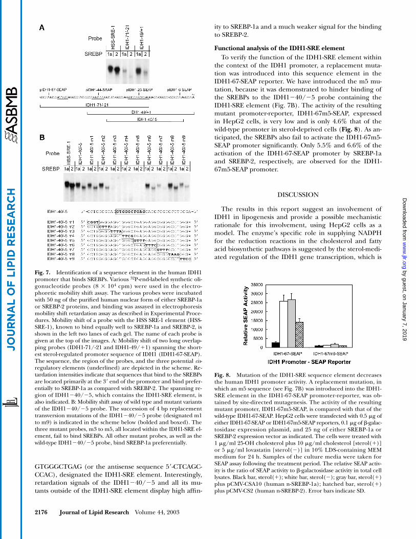
due to a lack of sequences for the binding of NF1/NFY, orto an SP1 transcription factor, or both. A truncation of ad-ditional 19 nts resulted in an almost complete loss of pro-moter activity. This latter truncation (IDH1-25-SEAP) wasso designed in order to cleave at an inverted SRE-1-likeelement, designated IDH1-SRE [�30]GTGGGCTGA-[�22](Fig. 6A) suspected for the binding of and the activation bythe SREBP transcription factors (see below for gel retarda-tion studies).
Mutation analyses of the interaction of the SREBPs with promoter sequences of IDH1
For the analyses of the interaction of the SREBPs withthe IDH1 promoter, different synthetic radiolabeledprobes were prepared based on the promoter sequences.The binding of the SREBPs to these probes was assayed inelectrophoretic mobility shift assays, and the results werecompared with the binding of the SREBPs to a probe con-taining the SRE-1 element in the HSS promoter (HSS-SRE-1) (52). SREBP-1a and SREBP-2 similarly retard theHSS-SRE-1 probe, resulting in radioactive signals of ap-proximately the same intensity (Fig. 7A). A significantlyweaker binding of SREBP-1a to the probe IDH1�71/�21is observed. This probe contains at its very 3� end theIDH1-SRE-1 sequence. Relatively weak signals in the mo-bility shift assay indicate that the IDH1�71/�21 probedoes not bind to the SREBP-2 protein in a significant way.The IDH1�49/�1 probe displays a much stronger bind-ing to the SREBP-1a protein. This probe contains theIDH1-SRE-1 sequence element at its core, and its bindingto SREBP-1a is comparable in signal intensity to that of
Fig. 4. Sterol regulation of human IDH1 gene promoter activity.HepG2 cells were transiently transfected with pIDH1-962-SEAP anda �-galactosidase expression vector. Two main experimental groupswere formed, one of cells maintained in 10% FBS and the other ofcells maintained in 10% LDS. The media in one-third of the cul-tures of each experimental group were supplemented with either 1�g/ml 25-OH cholesterol plus 10 �g/ml cholesterol (black bars), 5�g/ml lovastatin (white bars), or no additional treatment (graybars), and the cells were further incubated for 24 h. The culturemedia were assayed for secreted alkaline phosphatase (SEAP) activ-ity, and the whole-cell extracts were assayed for �-galactosidase ac-tivity. The relative SEAP activity is expressed as the ratio of SEAP to�-galactosidase activities (n � 6). Error bars indicate SD.
Fig. 5. Activation of the IDH1 promoter by SREBP-1a and SREBP-2.HepG2 cells (105) were transiently cotransfected with 0.5 �g ofpIDH1-962-SEAP, 0.1 �g of �-galactosidase expression vector, andeither of two plasmids, pCMV-CSA10 and pCMV-CS2, which expressthe nuclear form of human SREBP-1a (closed circles) or thehuman SREBP-2 (open circles), respectively. The amount of trans-fected DNA encoding the sterol regulatory element binding pro-teins (SREBPs) varied between 2 to 31 ng per transfection, asindicated. After 24 h, the growth media were replaced with mediasupplemented with 1 �g/ml 25-OH cholesterol plus 10 �g/ml cho-lesterol and the cells were incubated for an additional 24 h. Themedia were then collected for SEAP assay and the whole-cell lysateswere assayed for �-galactosidase activity. Relative SEAP activity is ex-pressed as the ratio of SEAP to �-galactosidase activities (n � 3). Er-ror bars indicate SD.
the HSS-SRE-1 probe. Only a weak retardation signal isobserved for the IDH1�49/�1 probe with the SREBP-2protein, indicating a weaker interaction with this proteincompared with that with SREBP-1a (Fig. 7A).
On the basis of these results, the IDH1�40/�5 syn-thetic probe was designed for mutation analysis of the in-teraction of the SREBPs with sequence elements in theIDH1 promoter. This shorter probe also contains theIDH1-SRE sequence element at its core and displays retar-
dation by the SREBPs similar to the retardation observedfor the longer IDH1�49/�1 probe (Fig. 7B). A successive4 nt scanning mutation analysis indicates that the eight 5�bp (mutations m1 and m2) and the sixteen 3� bp (muta-tions m6 to m9) of the probe are not essential for the re-tardation and are therefore not involved in the binding tothe SREBPs. However, mutations m3, m4, and m5 of theIDH1�40/�5 probe completely abolish the mobilityshift. These three mutations are within the sequence
Fig. 6. Sterol regulation and activation by SREBPs of successively 5�-translated IDH1-SEAP promoter-reporter genes. Successively 5�-truncated promoter fragments of the human IDH1 gene fused to a SEAP re-porter were generated by site-directed mutagenesis. A schematic representation of the various truncations isshown in A. The white bars represent different lengths of human IDH1 promoter, and the black bars repre-sent the 5�-UTR of the human IDH1 mRNA. Promoter lengths, relative to transcription initiation site, are in-dicated at the left of each construct. The DNA sequence between nucleotides �64 and �20 is shown above,with three potential regulatory elements underlined and in bold. The names and consensus sequences ofthese three elements are given below each sequence, with arrows indicating their normal orientation. Pro-moter activities of the various constructs in transiently transfected HepG2 cells grown under different condi-tions are shown in B. The cells were transfected with 0.5 �g of the IDH1-SEAP reporters, 0.1 �g of �-galac-tosidase control plasmid for transfection efficiency, and 25 ng of either SREBP-1a or SREBP-2 expressionvectors (pCMV-CSA10 or pCMV-CS2, respectively). The cells were treated with either 1 �g/ml 25-OH choles-terol plus 10 �g/ml cholesterol [sterol(�)] or with 5 �g/ml lovastatin [sterol(�)] in 10% LDS-containingMEM medium for 24 h, and the culture media were assayed for SEAP activity. The relative SEAP activity is ex-pressed as the ratio of SEAP to �-galactosidase activity in the cell lysates (n � 3). Black bar, sterol(�); whitebar, sterol(�); gray bar, sterol(�) plus pCMV-CSA10 (human n-SREBP-1a); hatched bar, sterol(�) pluspCMV-CS2 (human n-SREBP-2). Error bars indicate SD.
GTGGGCTGAG (or the antisense sequence 5�-CTCAGC-CCAC), designated the IDH1-SRE element. Interestingly,retardation signals of the IDH1�40/�5 and all its mu-tants outside of the IDH1-SRE element display high affin-
ity to SREBP-1a and a much weaker signal for the bindingto SREBP-2.
Functional analysis of the IDH1-SRE elementTo verify the function of the IDH1-SRE element within
the context of the IDH1 promoter, a replacement muta-tion was introduced into this sequence element in theIDH1-67-SEAP reporter. We have introduced the m5 mu-tation, because it was demonstrated to hinder binding ofthe SREBPs to the IDH1�40/�5 probe containing theIDH1-SRE element (Fig. 7B). The activity of the resultingmutant promoter-reporter, IDH1-67m5-SEAP, expressedin HepG2 cells, is very low and is only 4.6% that of thewild-type promoter in sterol-deprived cells (Fig. 8). As an-ticipated, the SREBPs also fail to activate the IDH1-67m5-SEAP promoter significantly. Only 5.5% and 6.6% of theactivation of the IDH1-67-SEAP promoter by SREBP-1aand SREBP-2, respectively, are observed for the IDH1-67m5-SEAP promoter.
DISCUSSION
The results in this report suggest an involvement ofIDH1 in lipogenesis and provide a possible mechanisticrationale for this involvement, using HepG2 cells as amodel. The enzyme’s specific role in supplying NADPHfor the reduction reactions in the cholesterol and fattyacid biosynthetic pathways is suggested by the sterol-medi-ated regulation of the IDH1 gene transcription, which is
Fig. 7. Identification of a sequence element in the human IDH1promoter that binds SREBPs. Various 32P-end-labeled synthetic oli-gonucleotide probes (8 104 cpm) were used in the electro-phoretic mobility shift assay. The various probes were incubatedwith 50 ng of the purified human nuclear form of either SREBP-1aor SREBP-2 proteins, and binding was assayed in electrophoresismobility shift retardation assay as described in Experimental Proce-dures. Mobility shift of a probe with the HSS SRE-1 element (HSS-SRE-1), known to bind equally well to SREBP-1a and SREBP-2, isshown in the left two lanes of each gel. The name of each probe isgiven at the top of the images. A: Mobility shift of two long overlap-ping probes (IDH1-71/-21 and IDH1-49/�1) spanning the short-est sterol-regulated promoter sequence of IDH1 (IDH1-67-SEAP).The sequence, the region of the probes, and the three potential cis-regulatory elements (underlined) are depicted in the scheme. Re-tardation intensities indicate that sequences that bind to the SREBPsare located primarily at the 3� end of the promoter and bind prefer-entially to SREBP-1a as compared with SREBP-2. The spanning re-gion of IDH1�40/�5, which contains the IDH1-SRE element, isalso indicated. B: Mobility shift assay of wild type and mutant variantsof the IDH1�40/�5 probe. The succession of 4 bp replacementtransversion mutations of the IDH1�40/�5 probe (designated m1to m9) is indicated in the scheme below (bolded and boxed). Thethree mutant probes, m3 to m5, all located within the IDH1-SRE el-ement, fail to bind SREBPs. All other mutant probes, as well as thewild-type IDH1�40/�5 probe, bind SREBP-1a preferentially.
Fig. 8. Mutation of the IDH1-SRE sequence element decreasesthe human IDH1 promoter activity. A replacement mutation, inwhich an m5 sequence (see Fig. 7B) was introduced into the IDH1-SRE element in the IDH1-67-SEAP promoter-reporter, was ob-tained by site-directed mutagenesis. The activity of the resultingmutant promoter, IDH1-67m5-SEAP, is compared with that of thewild-type IDH1-67-SEAP. HepG2 cells were transfected with 0.5 �g ofeither IDH1-67-SEAP or IDH1-67m5-SEAP reporters, 0.1 �g of �-galac-tosidase expression plasmid, and 25 ng of either SREBP-1a orSREBP-2 expression vector as indicated. The cells were treated with1 �g/ml 25-OH cholesterol plus 10 �g/ml cholesterol [sterol(�)]or 5 �g/ml lovastatin [sterol(�)] in 10% LDS-containing MEMmedium for 24 h. Samples of the culture media were taken forSEAP assay following the treatment period. The relative SEAP activ-ity is the ratio of SEAP activity to �-galactosidase activity in total celllysates. Black bar, sterol(�); white bar, sterol(�); gray bar, sterol(�)plus pCMV-CSA10 (human n-SREBP-1a); hatched bar, sterol(�)plus pCMV-CS2 (human n-SREBP-2). Error bars indicate SD.
coordinated with the transcription of all other regulatedgenes in these pathways. This coordinated regulation isachieved through the SREBP transcription factors, whichare long recognized to have a pivotal role in the lipid-medi-ated regulation of the lipogenic pathways (37, 43–45).
The biochemical source of the cytosolic NADPH re-quired for the cholesterol and fatty acid biosynthetic reac-tions has not been firmly established. Recently, it was sug-gested that the reactions catalyzed by the three cytosolicenzymes malic enzyme, G6PD, and 6-phosphogluconatedehydrogenase supply the necessary NADPH (43). How-ever, there is no evidence to suggest that the regulation ofany of these enzymes is linked to the production of sterolsand fatty acids. In fact, there are several reports suggestingthat G6PD, or the reactions catalyzed by the hexosemonophosphate shunt, may not be the source of theNADPH needed to drive lipogenesis in humans or in cul-tured cells (28–31). Because the exact cellular function ofIDH1 has not been established, its potential in generatingsignificant amounts of NADPH in the cytosol may be un-derestimated. The significant contribution of IDH1 inproducing cytosolic NADPH was actually demonstrated in
one early comparative study in which IDH1 in rat liver wasshown to be 16- and 18-fold more active in producingNADPH than were G6PD and malic enzyme, respectively(62).
The regulation of IDH1 gene transcription by sterols isnow demonstrated in HepG2 cells (Fig. 1). The magni-tude of the regulation of the IDH1 mRNA level by sterolsexceeds by far that of the mitochondrial dehydrogenaseIDH2 mRNA, which displays limited response, or that ofthe �- and -subunits of IDH3 mRNA, which do not dis-play any response to sterols. The regulatory response tosterols is also demonstrated in the modulation of enzymeactivity (Fig. 2). Because this enzyme assay, performedwith total-cell extracts, cannot distinguish between the ac-tivities of IDH1 and IDH2, the level of the sterol regula-tion of IDH1 cannot be determined precisely, because of abackground contribution of activity by IDH2. However,based on the data shown in Fig. 1, the activity of IDH2 isnot likely to be highly regulated and, thus, the sterol re-sponse could be attributed mostly to IDH1. Future pro-duction of IDH1-specific antibodies should resolve this is-sue. The 20�C suppression of IDH1 mRNA level and its
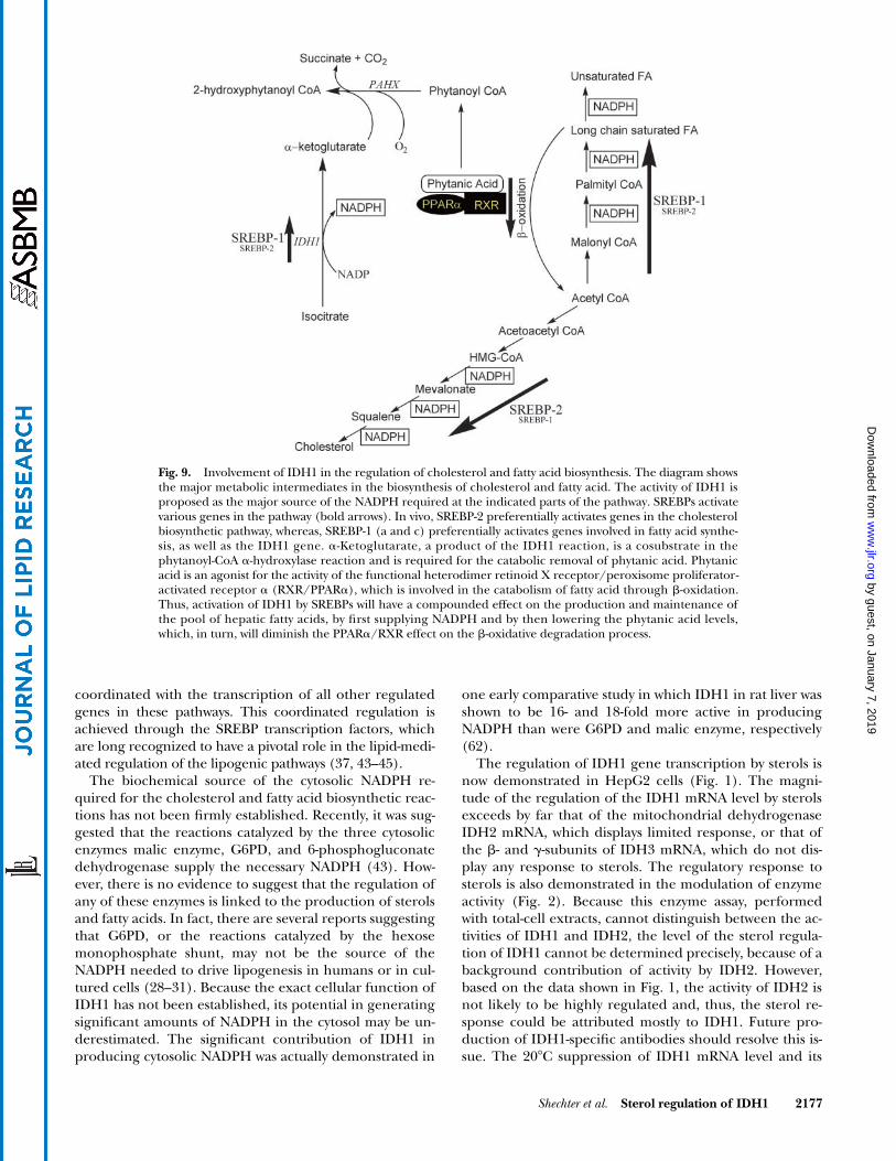
Fig. 9. Involvement of IDH1 in the regulation of cholesterol and fatty acid biosynthesis. The diagram showsthe major metabolic intermediates in the biosynthesis of cholesterol and fatty acid. The activity of IDH1 isproposed as the major source of the NADPH required at the indicated parts of the pathway. SREBPs activatevarious genes in the pathway (bold arrows). In vivo, SREBP-2 preferentially activates genes in the cholesterolbiosynthetic pathway, whereas, SREBP-1 (a and c) preferentially activates genes involved in fatty acid synthe-sis, as well as the IDH1 gene. �-Ketoglutarate, a product of the IDH1 reaction, is a cosubstrate in thephytanoyl-CoA �-hydroxylase reaction and is required for the catabolic removal of phytanic acid. Phytanicacid is an agonist for the activity of the functional heterodimer retinoid X receptor/peroxisome proliferator-activated receptor � (RXR/PPAR�), which is involved in the catabolism of fatty acid through �-oxidation.Thus, activation of IDH1 by SREBPs will have a compounded effect on the production and maintenance ofthe pool of hepatic fatty acids, by first supplying NADPH and by then lowering the phytanic acid levels,which, in turn, will diminish the PPAR�/RXR effect on the �-oxidative degradation process.
sterol regulation (Fig. 1) provided the first evidence thatthis regulation, similar to the sterol-mediated regulationof all other enzymes in the cholesterol biosynthetic path-way, is effected through the SREBPs (50). The function ofSREBPs in the sterol regulation of the IDH1 gene tran-scription is demonstrated by the activation of the IDH1promoter-reporter genes by the SREBPs (Figs. 4–6). In allof these experiments, consistently, the activation bySREBP-1a is significantly higher than that obtained by theexpression of SREBP-2. For IDH1-67-SEAP, the shorteststerol-responsive reporter gene, the activation by SREBP-1ais more than 2-fold higher than that obtained with SREBP-2(Fig. 6B).
On the basis of mutation analyses of electrophoreticmobility shift (Fig. 7B) and promoter activity (Fig. 8), thecis-regulatory element in the IDH1 promoter targetedby the SREBPs is most likely the IDH1-SRE sequence[�30]5�-GTGGGCTGAG[�21] (or the antisense sequence5�-CTCAGCCCAC), which resembles (but in a reverse ori-entation to) the SRE-1 element in the LDL receptor gene(63). Unlike the HSS-SRE-1 sequence element in the SQSgene, this sequence element preferentially binds toSREBP-1a, as compared with a much weaker binding toSRRBP-2 (Fig. 7). This difference in binding may explainthe differential activation of the IDH1 promoter by thetwo SREBPs (Figs. 4–6). The most pronounced differencein sequence between the consensus SRE-1 and the IDH1-SRE elements is an AT to CT change in the most 5� end ofthe latter (Fig. 6A). Yet, this seemingly minute change insequence uniquely results in a preferential binding of theIDH1-SRE sequence to SREBP-1a. In an earlier report, wehave demonstrated that the sequence 5�-TCCACCCCAC,present in the HSS promoter [originally designated HSS-SRE(8/10)], exclusively binds to SREBP-2 and not toSREBP-1a (52). Here again, the most pronounced differ-ence in the sequence is at the 5� end, where a TC replacesthe AT in the consensus SRE-1 element. Thus, it appearsthat the specificity in binding to (and presumably activa-tion by) the various SREBPs resides, in part, in the two 5�nucleotides of the SRE motif. This observation, which ap-pears to be correct for the HSS, IDH1, and LDL receptorpromoters, will have to be further examined by mobilityshift assays of variant SRE motifs. If correct, specific re-sponse in transcription to the various SREBPs may be pre-dicted by promoter sequence analysis. Whatever the ex-planation, the activation of the IDH1 promoter is effectedin lipid-depleted cells mostly by SREBP-1a and SREBP-1c(data not shown for the latter).
The shortest IDH1 promoter-reporter gene that dis-plays sterol regulation is IDH1-67-SEAP (Fig. 6). IDH1-44-SEAP, which is a 23 bp shorter promoter-reporter gene,still contains the intact IDH1-SRE element but lacks theCAAT and the GC-Box elements at its 5� end. These ele-ments are known to bind the NFY/NF1and SP1 transcriptionfactors, respectively. The IDH1-44-SEAP promoter-reporteractivity is very low, is no longer responsive to sterols, and inthe presence of SREBPs, is only a fraction of the activity ofthe longer promoters. Thus, the earlier observation that ac-tivation of gene transcription by the SREBPs requires the
coactivators NFY/NF1 or SP1 (51, 64–67) is also correctfor the IDH1 gene, which further supports the idea that,functionally, this gene is a member of the SREBP-activatedgene family.
The preferential activation of the IDH1 promoter bySREBP-1a compared with SREBP-2 may indicate that theregulation of IDH1 activity may be coordinated more withfatty acid biosynthesis than with sterol production (43).Thus, it is expected that activation of IDH1 is a responseto a shortage in hepatic fatty acid supply. The subcellularlocalization of IDH1 in both cytosol and peroxisomes ide-ally situates it to respond to the lipogenic requirement ofthe cell. The coproduct of the reaction, �-ketoglutarate,which is required as a cosubstrate for the phytanoyl-CoA�-hydroxylase reaction (17–20), is required for the cata-bolic removal of phytanic acid, a known agonist to PPAR�(21, 22). PPAR� is highly expressed in heart, muscle, kid-ney, and endothelial cells, but mostly in liver (27, 34),where its function is in the catabolism of fatty acidsthrough the regulation of genes encoding mitochondrialand peroxisomal enzymes important for �-oxidation (68).In addition, phytanic acid was also found to bind and acti-vate the RXR (23), which forms a functional heterodimerwith PPAR�. Therefore, an increase in IDH1 activity willhave a compounded effect on the production and mainte-nance of the hepatic fatty acid pool, by first supplyingNADPH, which will enhance fatty acid biosynthesis, andby then lowering the phytanic acid levels, which, in turn,will diminish the PPAR�/RXR effect on the �-oxidativedegradation process (Fig. 9). Indeed, the expression ofIDH1 in rats is primarily in liver, where the above pro-cesses are likely to occur (27, 34).
The available data, and the results presented herein,strongly suggest that the activity of IDH1 is required as amajor source for cytosolic NADPH, which is specificallytargeted as a substrate in the biosynthesis of cellular fattyacids and cholesterol. This activity also produces �-ketoglu-tarate, which indirectly regulates the �-oxidation of fattyacids through the degradation of the PPAR�/RXR agonistphytanic acid. Thus, IDH1 activity is involved in maintain-ing cellular cholesterol and fatty acid homeostasis throughsynthesis and degradation. Additional data, from studiesusing gene overexpression and knockout methodologiesin cultured cells and animals, are needed to assess the spe-cific function of IDH1 under various lipogenic conditions.If the activity of IDH1 is proven to be the major biochemi-cal NADPH-producing reaction for these processes, it maybe worthwhile to consider IDH1 as a target enzyme forlipid-lowering pharmacological strategies.
This work was supported by Grant HL-048540 from the Na-tional Institutes of Health.
REFERENCES
1. Illingworth, J. A., and K. F. Tipton. 1970. Purification and proper-ties of the nicotinamide-adenine dinucleotide phosphate-depen-
dent isocitrate dehydrogenase from pig liver cytoplasm. Biochem. J.118: 253–258.
2. Plaut, G. W., M. Cook, and T. Aogaichi. 1983. The subcellular loca-tion of isozymes of NADP-isocitrate dehydrogenase in tissues frompig, ox and rat. Biochim. Biophys. Acta. 760: 300–308.
3. Plaut, G. W., C. M. Smith, and W. L. Alworth. 1974. Biosynthesis ofwater-soluble vitamins. Annu. Rev. Biochem. 43: 899–922.
4. Ramachandran, N., and R. F. Colman. 1980. Chemical character-ization of distinct subunits of pig heart DPN-specific isocitrate de-hydrogenase. J. Biol. Chem. 255: 8859–8864.
5. Keys, D. A., and L. McAlister-Henn. 1990. Subunit structure, ex-pression, and function of NAD(H)-specific isocitrate dehydroge-nase in Saccharomyces cerevisiae. J. Bacteriol. 172: 4280–4287.
6. Haselbeck, R. J., and L. McAlister-Henn. 1993. Function and ex-pression of yeast mitochondrial NAD- and NADP-specific isocitratedehydrogenases. J. Biol. Chem. 268: 12116–12122.
7. Nekrutenko, A., D. M. Hillis, J. C. Patton, R. D. Bradley, and R. J.Baker. 1998. Cytosolic isocitrate dehydrogenase in humans, mice,and voles and phylogenetic analysis of the enzyme family. Mol. Biol.Evol. 15: 1674–1684.
8. Geisbrecht, B. V., and S. J. Gould. 1999. The human PICD geneencodes a cytoplasmic and peroxisomal NADP(�)-dependentisocitrate dehydrogenase. J. Biol. Chem. 274: 30527–30533.
9. Yoshihara, T., T. Hamamoto, R. Munakata, R. Tajiri, M. Ohsumi,and S. Yokota. 2001. Localization of cytosolic NADP-dependentisocitrate dehydrogenase in the peroxisomes of rat liver cells: bio-chemical and immunocytochemical studies. J. Histochem. Cytochem.49: 1123–1132.
10. Keller, G. A., M. C. Barton, D. J. Shapiro, and S. J. Singer. 1985.3-Hydroxy-3-methylglutaryl-coenzyme A reductase is present inperoxisomes in normal rat liver cells. Proc. Natl. Acad. Sci. USA. 82:770–774.
11. Keller, G. A., M. Pazirandeh, and S. Krisans. 1986. 3-Hydroxy-3-methylglutaryl coenzyme A reductase localization in rat liver per-oxisomes and microsomes of control and cholestyramine-treatedanimals: quantitative biochemical and immunoelectron micro-scopical analyses. J. Cell Biol. 103: 875–886.
12. Krisans, S. K. 1996. Cell compartmentalization of cholesterol bio-synthesis. Ann. N. Y. Acad. Sci. 804: 142–164.
13. Biardi, L., and S. K. Krisans. 1996. Compartmentalization of cho-lesterol biosynthesis. Conversion of mevalonate to farnesyl diphos-phate occurs in the peroxisomes. J. Biol. Chem. 271: 1784–1788.
14. Wanders, R. J., and J. M. Tager. 1998. Lipid metabolism in peroxi-somes in relation to human disease. Mol. Aspects Med. 19: 69–154.
15. Fransen, M., P. P. Van Veldhoven, and S. Subramani. 1999. Identifi-cation of peroxisomal proteins by using M13 phage protein VI ph-age display: molecular evidence that mammalian peroxisomescontain a 2,4-dienoyl-CoA reductase. Biochem. J. 340: 561–568.
16. Geisbrecht, B. V., X. Liang, J. C. Morrell, H. Schulz, and S. J.Gould. 1999. The mouse gene PDCR encodes a peroxisomaldelta(2), delta(4)-dienoyl-CoA reductase. J. Biol. Chem. 274:25814–25820.
17. Jansen, G. A., S. J. Mihalik, P. A. Watkins, H. W. Moser, C. Jakobs, S.Denis, and R. J. Wanders. 1996. Phytanoyl-CoA hydroxylase ispresent in human liver, located in peroxisomes, and deficient inZellweger syndrome: direct, unequivocal evidence for the new, re-vised pathway of phytanic acid alpha-oxidation in humans. Bio-chem. Biophys. Res. Commun. 229: 205–210.
18. Mihalik, S. J., A. M. Rainville, and P. A. Watkins. 1995. Phytanicacid alpha-oxidation in rat liver peroxisomes. Production of alpha-hydroxyphytanoyl-CoA and formate is enhanced by dioxygenasecofactors. Eur. J. Biochem. 232: 545–551.
19. Croes, K., M. Casteels, E. De Hoffmann, G. P. Mannaerts, and P. P.Van Veldhoven. 1996. Alpha-oxidation of 3-methyl-substituted fattyacids in rat liver. Production of formic acid instead of CO2, cofactorrequirements, subcellular localization and formation of a 2-hydroxy-3-methylacyl-CoA intermediate. Eur. J. Biochem. 240: 674–683.
20. Verhoeven, N. M., and C. Jakobs. 2001. Human metabolism of phy-tanic acid and pristanic acid. Prog. Lipid Res. 40: 453–466.
21. Heim, M., J. Johnson, F. Boess, I. Bendik, P. Weber, W. Hunziker,and B. Fluhmann. 2002. Phytanic acid, a natural peroxisome pro-liferator-activated receptor (PPAR) agonist, regulates glucose me-tabolism in rat primary hepatocytes. FASEB J. 16: 718–720.
22. Ellinghaus, P., C. Wolfrum, G. Assmann, F. Spener, and U. Seedorf.1999. Phytanic acid activates the peroxisome proliferator-activatedreceptor alpha (PPARalpha) in sterol carrier protein 2-/sterol car-rier protein x-deficient mice. J. Biol. Chem. 274: 2766–2772.
23. Kitareewan, S., L. T. Burka, K. B. Tomer, C. E. Parker, L. J. Deterd-ing, R. D. Stevens, B. M. Forman, D. E. Mais, R. A. Heyman, T. Mc-Morris, and C. Weinberger. 1996. Phytol metabolites are circulat-ing dietary factors that activate the nuclear receptor RXR. Mol.Biol. Cell. 7: 1153–1166.
24. Schluter, A., P. Yubero, R. Iglesias, M. Giralt, and F. Villarroya.2002. The chlorophyll-derived metabolite phytanic acid induceswhite adipocyte differentiation. Int. J. Obes. Relat. Metab. Disord. 26:1277–1280.
25. Schluter, A., M. J. Barbera, R. Iglesias, M. Giralt, and F. Villarroya.2002. Phytanic acid, a novel activator of uncoupling protein-1gene transcription and brown adipocyte differentiation. Biochem. J.362: 61–69.
26. Wierzbicki, A. S., M. D. Lloyd, C. J. Schofield, M. D. Feher, and F. B.Gibberd. 2002. Refsum’s disease: a peroxisomal disorder affectingphytanic acid alpha-oxidation. J. Neurochem. 80: 727–735.
27. Jennings, G., S. Sechi, P. Stevenson, R. Tuckey, D. Parmelee, and L.McAlister-Henn. 1994. Cytosolic NADP(�)-dependent isocitratedehydrogenase. Isolation of rat cDNA and study of tissue-specificand developmental expression of mRNA. J. Biol. Chem. 269: 23128–23134.
28. Luzzatto, L., A. Mehta, and T. Vulliamy. 2001. Glucose 6-phosphatedehydrogenase deficiency. In The Metabolic and Molecular Bases ofInherited Diseases. 8th edition. Vol. 3. C. R. Scriver, A. L. Beaudet,W. S. Sly, and D. Valle, editors. McGraw-Hill, New York. 4517–4553.
29. Heller, P., W. R. Best, R. B. Nelson, and J. Becktel. 1979. Clinicalimplications of sickle-cell trait and glucose-6-phosphate dehydrog-enase deficiency in hospitalized black male patients. N. Engl. J.Med. 300: 1001–1005.
30. Rosenstraus, M., and L. A. Chasin. 1975. Isolation of mammaliancell mutants deficient in glucose-6-phosphate dehydrogenase ac-tivity: linkage to hypoxanthine phosphoribosyl transferase. Proc.Natl. Acad. Sci. USA. 72: 493–497.
31. Town, M., M. Athanasiou-Metaxa, and L. Luzzatto. 1990. In-tragenic interspecific complementation of glucose 6-phosphatedehydrogenase in human-hamster cell hybrids. Somat. Cell Mol.Genet. 16: 97–108.
32. Winkler, B. S., N. DeSantis, and F. Solomon. 1986. MultipleNADPH-producing pathways control glutathione (GSH) contentin retina. Exp. Eye Res. 43: 829–847.
33. Belfiore, F., and S. Iannello. 1995. Fatty acid synthesis fromglutamate in the adipose tissue of normal subjects and obese pa-tients: an enzyme study. Biochem. Mol. Med. 54: 19–25.
34. Lee, S. M., H. J. Koh, D. C. Park, B. J. Song, T. L. Huh, and J. W.Park. 2002. Cytosolic NADP(�)-dependent isocitrate dehydroge-nase status modulates oxidative damage to cells. Free Radic. Biol.Med. 32: 1185–1196.
35. Sun, L., T. T. Sun, and R. M. Lavker. 1999. Identification of a cyto-solic NADP�-dependent isocitrate dehydrogenase that is prefer-entially expressed in bovine corneal epithelium. A corneal epithe-lial crystallin. J. Biol. Chem. 274: 17334–17341.
36. Yokoyama, C., X. Wang, M. R. Briggs, A. Admon, J. Wu, X. Hua, J. L.Goldstein, and M. S. Brown. 1993. SREBP-1, a basic-helix-loop-helix-leucine zipper protein that controls transcription of the lowdensity lipoprotein receptor gene. Cell. 75: 187–197.
37. Brown, M. S., and J. L. Goldstein. 1999. A proteolytic pathway thatcontrols the cholesterol content of membranes, cells, and blood.Proc. Natl. Acad. Sci. USA. 96: 11041–11048.
38. Nohturfft, A., M. S. Brown, and J. L. Goldstein. 1998. Topology ofSREBP cleavage-activating protein, a polytopic membrane proteinwith a sterol-sensing domain. J. Biol. Chem. 273: 17243–17250.
39. Sakai, J., R. B. Rawson, P. J. Espenshade, D. Cheng, A. C. Seeg-miller, J. L. Goldstein, and M. S. Brown. 1998. Molecular identifi-cation of the sterol-regulated luminal protease that cleavesSREBPs and controls lipid composition of animal cells. Mol. Cell. 2:505–514.
40. Sakai, J., A. Nohturfft, J. L. Goldstein, and M. S. Brown. 1998.Cleavage of sterol regulatory element-binding proteins (SREBPs)at site-1 requires interaction with SREBP cleavage-activating pro-tein. Evidence from in vivo competition studies. J. Biol. Chem. 273:5785–5793.
41. Hua, X., A. Nohturfft, J. L. Goldstein, and M. S. Brown. 1996. Ste-rol resistance in CHO cells traced to point mutation in SREBPcleavage-activating protein. Cell. 87: 415–426.
42. Nohturfft, A., D. Yabe, J. L. Goldstein, M. S. Brown, and P. J. Espen-shade. 2000. Regulated step in cholesterol feedback localized tobudding of SCAP from ER membranes. Cell. 102: 315–323.
43. Horton, J. D., J. L. Goldstein, and M. S. Brown. 2002. SREBPs: acti-vators of the complete program of cholesterol and fatty acid syn-thesis in the liver. J. Clin. Invest. 109: 1125–1131.
44. Edwards, P. A., D. Tabor, H. R. Kast, and A. Venkateswaran. 2000.Regulation of gene expression by SREBP and SCAP. Biochim. Bio-phys. Acta. 1529: 103–113.
45. Goldstein, J. L., R. B. Rawson, and M. S. Brown. 2002. Mutantmammalian cells as tools to delineate the sterol regulatory ele-ment-binding protein pathway for feedback regulation of lipid syn-thesis. Arch. Biochem. Biophys. 397: 139–148.
46. Yang, T., P. J. Espenshade, M. E. Wright, D. Yabe, Y. Gong, R. Ae-bersold, J. L. Goldstein, and M. S. Brown. 2002. Crucial step incholesterol homeostasis: sterols promote binding of SCAP toINSIG-1, a membrane protein that facilitates retention of SREBPsin ER. Cell. 110: 489–500.
47. Yabe, D., M. S. Brown, and J. L. Goldstein. 2002. Insig-2, a secondendoplasmic reticulum protein that binds SCAP and blocks exportof sterol regulatory element-binding proteins. Proc. Natl. Acad. Sci.USA. 99: 12753–12758.
48. Espenshade, P. J., W. P. Li, and D. Yabe. 2002. Sterols block bind-ing of COPII proteins to SCAP, thereby controlling SCAP sortingin ER. Proc. Natl. Acad. Sci. USA. 99: 11694–11699.
49. Loewen, C. J. R., and T. P. Levine. 2002. Cholesterol homeostasis:not until the SCAP lady INSIGs. Curr. Biol. 12: R779–R781.
50. Shechter, I., P. Dai, M. A. Roseman, S. D. Gupta, B. B. Boyer, andG. Guan. 2003. Low-temperature effect on the sterol-dependentprocessing of SREBPs and transcription of related genes in HepG2cells. J. Lipid Res. 44: 1581–1590.
51. Guan, G., P. H. Dai, and I. Shechter. 1998. Differential transcrip-tional regulation of the human squalene synthase gene by sterolregulatory element-binding proteins (SREBP) 1a and 2 and in-volvement of 5� DNA sequence elements in the regulation. J. Biol.Chem. 273: 12526–12535.
52. Guan, G., P. H. Dai, T. F. Osborne, J. B. Kim, and I. Shechter. 1997.Multiple sequence elements are involved in the transcriptionalregulation of the human squalene synthase gene. J. Biol. Chem.272: 10295–10302.
53. Pai, J. T., O. Guryev, M. S. Brown, and J. L. Goldstein. 1998. Differ-ential stimulation of cholesterol and unsaturated fatty acid biosyn-thesis in cells expressing individual nuclear sterol regulatory ele-ment-binding proteins. J. Biol. Chem. 273: 26138–26148.
54. Horton, J. D., I. Shimomura, M. S. Brown, R. E. Hammer, J. L.Goldstein, and H. Shimano. 1998. Activation of cholesterol synthe-sis in preference to fatty acid synthesis in liver and adipose tissueof transgenic mice overproducing sterol regulatory element-bind-ing protein-2. J. Clin. Invest. 101: 2331–2339.
55. Shimano, H. 2001. Sterol regulatory element-binding proteins
56. Tabor, D. E., J. B. Kim, B. M. Spiegelman, and P. A. Edwards. 1998.Transcriptional activation of the stearoyl-CoA desaturase 2 gene bysterol regulatory element-binding protein/adipocyte determina-tion and differentiation factor 1. J. Biol. Chem. 273: 22052–22058.
57. Fielding, C., and P. Fielding. 2000. Cholesterol and caveolae: struc-tural and functional relationships. Biochim. Biophys. Acta. 1529:210–222.
58. Frohman, M. A., M. K. Dush, and G. R. Martin. 1988. Rapid pro-duction of full-length cDNAs from rare transcripts: amplificationusing a single gene-specific oligonucleotide primer. Proc. Natl.Acad. Sci. USA. 85: 8998–9002.
59. Farrell, H. M., Jr. 1980. Purification and properties of NADP�:isocitrate dehydrogenase from lactating bovine mammary gland.Arch. Biochem. Biophys. 204: 551–559.
60. Guan, G., G. Jiang, R. L. Koch, and I. Shechter. 1995. Molecularcloning and functional analysis of the promoter of the humansqualene synthase gene. J. Biol. Chem. 270: 21958–21965.
61. Ohshima, Y., and Y. Gotoh. 1987. Signals for the selection of asplice site in pre-mRNA. Computer analysis of splice junction se-quences and like sequences. J. Mol. Biol. 195: 247–259.
62. Veech, R. L., L. V. Eggleston, and H. A. Krebs. 1969. The redoxstate of free nicotinamide-adenine dinucleotide phosphate in thecytoplasm of rat liver. Biochem. J. 115: 609–619.
63. Smith, J. R., T. F. Osborne, J. L. Goldstein, and M. S. Brown. 1990.Identification of nucleotides responsible for enhancer activity ofsterol regulatory element in low density lipoprotein receptor gene.J. Biol. Chem. 265: 2306–2310.
64. Magana, M. M., S. H. Koo, H. C. Towle, and T. F. Osborne. 2000.Different sterol regulatory element-binding protein-1 isoforms uti-lize distinct co-regulatory factors to activate the promoter for fattyacid synthase. J. Biol. Chem. 275: 4726–4733.
65. Jackson, S. M., J. Ericsson, R. Mantovani, and P. A. Edwards. 1998.Synergistic activation of transcription by nuclear factor Y and ste-rol regulatory element binding protein. J. Lipid Res. 39: 767–776.
66. Ericsson, J., S. M. Jackson, and P. A. Edwards. 1996. Synergisticbinding of sterol regulatory element-binding protein and NF-Y tothe farnesyl diphosphate synthase promoter is critical for sterol-regulated expression of the gene. J. Biol. Chem. 271: 24359–24364.
67. Jackson, S. M., J. Ericsson, T. F. Osborne, and P. A. Edwards. 1995.NF-Y has a novel role in sterol-dependent transcription of two cho-lesterogenic genes. J. Biol. Chem. 270: 21445–21448.
68. Desvergne, B., A. IJpenberg, P. R. Devchand, and W. Wahli. 1998.The peroxisome proliferator-activated receptors at the cross-roadof diet and hormonal signalling. J. Steroid Biochem. Mol. Biol. 65:65–74.