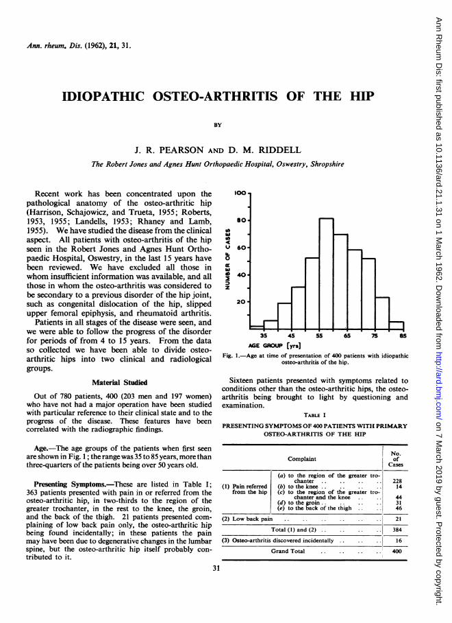
Ann. rheum. Dis. (1962), 21, 31. IDIOPATHIC OSTEO-ARTHRITIS OF THE HIP BY J. R. PEARSON AND D. M. RIDDELL The Robert Jones and Agnes Hunt Orthopaedic Hospital, Oswestry, Shropshire Recent work has been concentrated upon the pathological anatomy of the osteo-arthritic hip (Harrison, Schajowicz, and Trueta, 1955; Roberts, 1953, 1955; Landells, 1953; Rhaney and Lamb, 1955). We have studied the disease from the clinical aspect. All patients with osteo-arthritis of the hip seen in the Robert Jones and Agnes Hunt Ortho- paedic Hospital, Oswestry, in the last 15 years have been reviewed. We have excluded all those in whom insufficient information was available, and all those in whom the osteo-arthritis was considered to be secondary to a previous disorder of the hip joint, such as congenital dislocation of the hip, slipped upper femoral epiphysis, and rheumatoid arthritis. Patients in all stages of the disease were seen, and we were able to follow the progress of the disorder for periods of from 4 to 15 years. From the data so collected we have been able to divide osteo- arthritic hips into two clinical and radiological groups. Material Studied Out of 780 patients, 400 (203 men and 197 women) who have not had a major operation have been studied with particular reference to their clinical state and to the progress of the disease. These features have been correlated with the radiographic findings. Age.-The age groups of the patients when first seen are shown in Fig. 1; the range was 35 to 85 years, more than three-quarters of the patients being over 50 years old. Presenting Symptoms.-These are listed in Table I; 363 patients presented with pain in or referred from the osteo-arthritic hip, in two-thirds to the region of the greater trochanter, in the rest to the knee, the groin, and the back of the thigh. 21 patients presented com- plaining of low back pain only, the osteo-arthritic hip being found incidentally; in these patients the pain may have been due to degenerative changes in the lumbar spine, but the osteo-arthritic hip itself probably con- tributed to it. U 6 ki *0 40 z 20- 35 45 55 65 75 85 AGE GROUP (yrs Fig. 1.-Age at time of presentation of 400 patients with idiopathic osteo-arthritis of the hip. Sixteen patients presented with symptoms related to conditions other than the osteo-arthritic hips, the osteo- arthritis being brought to light by questioning and examination. TABLE I PRESENTING SYMPTOMS OF 400 PATIENTS WITH PRIMARY OSTEO-ARTHRITIS OF THE HIP No. Complaint of Cases (a) to the region of the greater tro- chanter .228 (1) Pain referred (b) to the knee .14 from the hip (c) to the region of the greater tro- chanter and the knee .. 44 (d) to the groin 31 (e) to the back of the thigh .. 46 (2) Low back pain .21 Total (I) and (2) .384 (3) Osteo-arthritis discovered incidentally.16 Grand Total 400 31 on 7 March 2019 by guest. Protected by copyright. http://ard.bmj.com/ Ann Rheum Dis: first published as 10.1136/ard.21.1.31 on 1 March 1962. Downloaded from
Transcript
Ann. rheum. Dis. (1962), 21, 31.
IDIOPATHIC OSTEO-ARTHRITIS OF THE HIP
BY
J. R. PEARSON AND D. M. RIDDELLThe Robert Jones and Agnes Hunt Orthopaedic Hospital, Oswestry, Shropshire
Recent work has been concentrated upon thepathological anatomy of the osteo-arthritic hip(Harrison, Schajowicz, and Trueta, 1955; Roberts,1953, 1955; Landells, 1953; Rhaney and Lamb,1955). We have studied the disease from the clinicalaspect. All patients with osteo-arthritis of the hipseen in the Robert Jones and Agnes Hunt Ortho-paedic Hospital, Oswestry, in the last 15 years havebeen reviewed. We have excluded all those inwhom insufficient information was available, and allthose in whom the osteo-arthritis was considered tobe secondary to a previous disorder of the hip joint,such as congenital dislocation of the hip, slippedupper femoral epiphysis, and rheumatoid arthritis.
Patients in all stages of the disease were seen, andwe were able to follow the progress of the disorderfor periods of from 4 to 15 years. From the dataso collected we have been able to divide osteo-arthritic hips into two clinical and radiologicalgroups.
Material Studied
Out of 780 patients, 400 (203 men and 197 women)who have not had a major operation have been studiedwith particular reference to their clinical state and to theprogress of the disease. These features have beencorrelated with the radiographic findings.
Age.-The age groups of the patients when first seenare shown in Fig. 1; the range was 35 to 85 years, more thanthree-quarters of the patients being over 50 years old.
Presenting Symptoms.-These are listed in Table I;363 patients presented with pain in or referred from theosteo-arthritic hip, in two-thirds to the region of thegreater trochanter, in the rest to the knee, the groin,and the back of the thigh. 21 patients presented com-plaining of low back pain only, the osteo-arthritic hipbeing found incidentally; in these patients the painmay have been due to degenerative changes in the lumbarspine, but the osteo-arthritic hip itself probably con-tributed to it.
U 6ki*0
40
z
20-
35 45 55 65 75 85
AGE GROUP (yrsFig. 1.-Age at time of presentation of 400 patients with idiopathic
osteo-arthritis of the hip.
Sixteen patients presented with symptoms related toconditions other than the osteo-arthritic hips, the osteo-arthritis being brought to light by questioning andexamination.
TABLE I
PRESENTING SYMPTOMS OF 400 PATIENTS WITH PRIMARYOSTEO-ARTHRITIS OF THE HIP
No.Complaint of
Cases
(a) to the region of the greater tro-chanter.228
(1) Pain referred (b) to the knee.14from the hip (c) to the region of the greater tro-
chanter and the knee .. 44(d) to the groin 31(e) to the back of the thigh .. 46
(2) Low back pain.21
Total (I) and (2).384
(3) Osteo-arthritis discovered incidentally.16
Grand Total 400
31
on 7 March 2019 by guest. P
rotected by copyright.http://ard.bm
j.com/
Ann R
heum D
is: first published as 10.1136/ard.21.1.31 on 1 March 1962. D
The severity of pain was estimated by the dosage ofanalgesics necessary to control it, the presence of nightpain, the distance the patient could walk, and the needto use one or two sticks. It was found that there was norelation between the degree of limitation of movementand the radiological appearances and intensity of pain.No patient complained primarily of stiffness. The
length of history varied from one week to many years,and had no correlation with the stage of the osteo-arthritis. In 49 patients the symptoms began afteran injury to the hip.Radiographs.-In each case a series of radiographs
was available, the series extending over several years.These were examined with regard to the following points:
(1) The degree and site of diminution in "jointspace",
(2) The amount and site of sclerosis:(3) The presence and sites of cyst formation:(4) The sites of osteophytes.
ResultsClinical and radiological examination showed
that the disease falls into two patterns, each of whichpossesses distinct characteristics, all the casesreviewed conformed to one of these patterns.The early stages of both patterns are similar.
The initial loss of movement is always one of internalrotation and of extension. In all cases, further
limitation of extension leads to a flexion deformity,and, at the same time, limitation of flexion occurs.The subsequent progress of the disease differs in thetwo types.
Adduction-External Rotation Type.-Abductionand external rotation decreases, and finally all side-to-side movement is lost. The final position isone of flexion, adduction, and external rotation,either quite stiff, or with a few degrees of free flexionand no other movement.
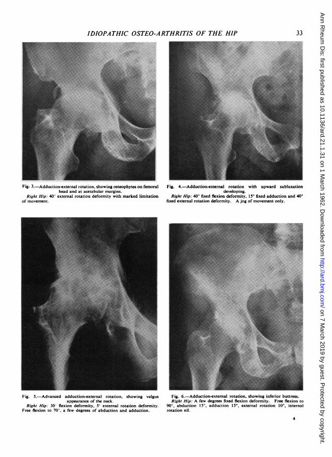
Radiologically, the maximal loss of 'joint space"and the greatest degree of sclerosis and cyst forma-tion appear in the superior part of the joint(Fig. 2). Osteophytes are present all round theacetabular margin and around the rim of the headof the femur (Fig. 3, opposite). In these hips,upward subluxation of the head of the femur oftenoccurs (Fig. 4, opposite). The neck may present avalgus appearance (Fig. 5, opposite). A buttress isseen on the inferior border of the neck, and is oftenpresent before the development of an adductiondeformity (Fig. 6, opposite).
Fig. 7 (overleaf) shows that this buttress issituated in the postero-inferior part of the neck:the significance of this buttress has been discussedby Wiberg (1939) and Roberts (1953).
Fig. 2.-Early case of adduction-external rotation, showing maximal loss of "joint space". and greatest sclerosis and most cysts in thesuperior part of the joint, left hip.
Right Hip: 30' fixed flexion deformity, 5 fixed abduction and external rotation deformity. Free flexion 65 virtually no other movement.Left Hip: 10' fixed deformity. Free flexion 90 , a few degrees of abduction and adduction, no rotation.
32
on 7 March 2019 by guest. P
rotected by copyright.http://ard.bm
j.com/
Ann R
heum D
is: first published as 10.1136/ard.21.1.31 on 1 March 1962. D
Fig. 3.-Adduction-external rotation, showing osteophytes on femoral Fig. 4.-Adduction-external rotation with upward subluxationhead and at acetabular margins. developing.
Right Hip: 40) external rotation deformity with marked limitation Right Hip: 40' fixed flexion deformity, 15' fixed adduction and 40'of movement. fixed external rotation deformity. A jog of movement only.
Fig. 5.-Advanced adduction-external rotation, showing valgus Fig. 6.-Adduction-external rotation, showing inferior buttress.appearance of the neck. Right Hip: A few degrees fixed flexion deformity. Free flexion to
Right Hip: 30- flexion deformity, 5' external rotation deformity. 90°, abduction 15', adduction 15', external rotation 10', internalFree flexion to 70-, a few degrees of abduction and adduction. rotation nil.
4
on 7 March 2019 by guest. P
rotected by copyright.http://ard.bm
j.com/
Ann R
heum D
is: first published as 10.1136/ard.21.1.31 on 1 March 1962. D
Fig. 7.-Adduction-external rotation.Left Hip: Laternal view of neck, showing posterior and inferior
position of buttress.
Non-Adducted Type.-Adduction and externalrotation decrease, until all side-to-side movementis lost, so that the final position is one of flexionand external rotation, either in neutral abductionadduction or with an abduction deformity-anadduction deformity is unknown in this type. Asin the first type, the hip may be completely stiffor may retain a few degrees of free flexton.
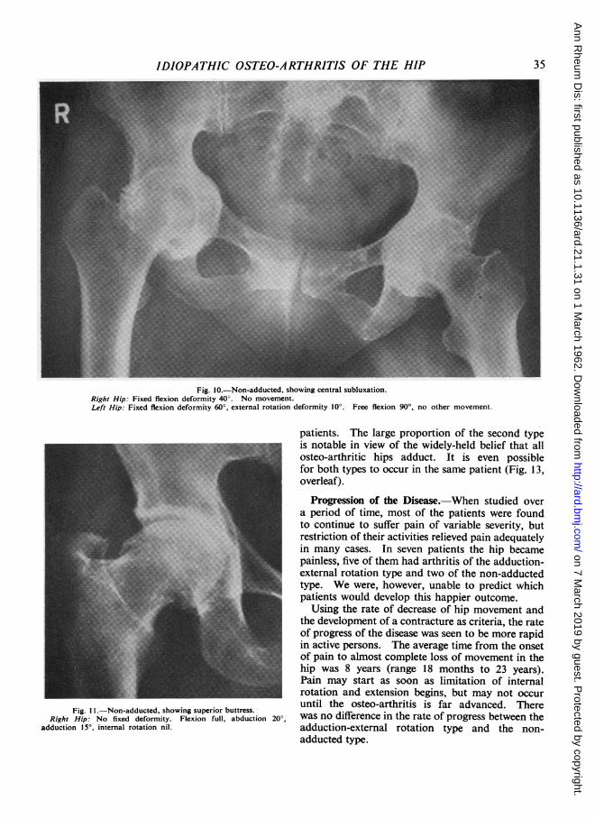
Radiologically, the greatest loss of "joint space",and the greatest sclerosis and cyst formation appearmedially in the deepest part of the joint (Fig. 8).Osteophytes are present around the acetabularmargin and around the rim of the head of the femur(Fig. 9).These hips progress to a central subluxation
(Fig. 10, opposite). The appearance of a buttressis seen to occur superiorly in the neck (Fig. 11,opposite).The majority have the appearance of a varus
neck (Fig. 12, overleaf), although this is not invari-able, some of these cases being associated with a Fig. 9.-Non-adducted, showing femoral head osteophytes andvalgus neck. acetabular marginal osteophytes.
Right Hip: 5' fixed flexion deformity. Free flexion 90', abductionThe adduction-external rotation type was found and adduction limited by one-third, internal rotation nil.
in 311 patients and the non-adducted type in 89
34
on 7 March 2019 by guest. P
rotected by copyright.http://ard.bm
j.com/
Ann R
heum D
is: first published as 10.1136/ard.21.1.31 on 1 March 1962. D
IDIOPATHIC OSTEO-ARTHRITIS OF THE HIP_X j~~~~~~~~~~~~~~~~~~~~~~~~
Fig. 10.-Non-adducted, showing central subluxation.Right Hip: Fixed flexion deformity 40°. No movement.Left Hip: Fixed flexion deformity 60°, external rotation deformity 10°. Free flexion 90°, no other movement.
Fig. I .-Non-adducted, showing superior buttress.Right Hip: No fixed deformity. Flexion full, abduction 20°,
adduction 15°, internal rotation nil.
patients. The large proportion of the second typeis notable in view of the widely-held belief that allosteo-arthritic hips adduct. It is even possiblefor both types to occur in the same patient (Fig. 13,overleaf).
Progression of the Disease.-When studied overa period of time, most of the patients were foundto continue to suffer pain of variable severity, butrestriction of their activities relieved pain adequatelyin many cases. In seven patients the hip becamepainless, five of them had arthritis of the adduction-external rotation type and two of the non-adductedtype. We were, however, unable to predict whichpatients would develop this happier outcome.Using the rate of decrease of hip movement and
the development of a contracture as criteria, the rateof progress of the disease was seen to be more rapidin active persons. The average time from the onsetof pain to almost complete loss of movement in thehip was 8 years (range 18 months to 23 years).Pain may start as soon as limitation of internalrotation and extension begins, but may not occuruntil the osteo-arthritis is far advanced. Therewas no difference in the rate of progress between theadduction-external rotation type and the non-adducted type.
35
on 7 March 2019 by guest. P
rotected by copyright.http://ard.bm
j.com/
Ann R
heum D
is: first published as 10.1136/ard.21.1.31 on 1 March 1962. D
Fig. 13.-Adduction-external rotation on the left side and non- adduction on the right side.Right Hip: 30' fixed flexion deformity. Free flexion to 60°, jog of abduction, no rotation.Left Hip: 70° fixed flexion deformity, 5° adduction deformity. No free movement.
on 7 March 2019 by guest. P
rotected by copyright.http://ard.bm
j.com/
Ann R
heum D
is: first published as 10.1136/ard.21.1.31 on 1 March 1962. D
Radiological progression of the osteo-arthritisoccurred in the following order:
(1) Loss of "joint space";(2) Sclerosis and osteophytic changes appearing
together;(3) Cyst formation;(4) Subluxation associated with flattening of
the head.
Limitation of internal rotation and extension,and also a flexion contracture, are frequently foundbefore obvious radiological evidence of the diseaseis present. On the other hand, marked radiologicalchanges can be present with a surprisingly goodrange of movement.
Discussion
Limitation of movement of the hip joint can bedue to any of the following factors: capsular con-tracture and muscle spasm, contracture of musclesand their overlying fascia (particularly the fasciaover the ilio psoas), mechanical block due to osteo-phytes, and incongruity of joint surfaces. Capsularcontracture and muscle spasm are probably th-most important causes of true limitation of move-ment and of deformity of the hip. Bony block dueto osteophytes is rare, and can occur only in thelater stages of the disease when osteophyte formationis excessive. Similarly, limitation of movement due
to incongruity of the joint surfaces can occur onlywhen there is gross disorganization of the joint.Contracture of muscle and fascia are found in thepresence of any long-standing deformity in the hip.Walmsley (1928) reported that in the position of
full extension of the hip the capsule is tight in all itscomponents; it follows from this that contractureoccurring in any part of the capsule must produceloss of extension, and must ultimately lead to aflexion contracture of the hip. Roberts (1953) hasshown that, if the postero-inferior part of the capsuleis contracted, a limitation of abduction and internalrotation will occur, leading to an adduction-externalrotation deformity-this would correspond to theadduction-external rotation type.We have correlated the clinical feature of both
types with the appearances at operation and ofpost mortem specimens. In the adduction-externalrotation type, the capsule is thickened postero-inferiorly and division allows abduction and someinternal rotation to occur. In the non-adductedtype, the capsule is thickened postero-superiorly,and division above and behind relieves the abductioncontracture, if present, and allows adduction andinternal rotation to occur.We examined microscopically pieces of capsule
excised from the postero-superior and postero-inferior portion of the capsule in both types, andcompared them with the thickness of the rest of thecapsule. Fig. 14 shows the increase in the thicknessof the capsule.
A~~~~~~~~~~~~~~~~~~~
Fig. 14.-Microphotograph of capsule taken from an osteo-arthritic hip of the non-adductedtype, showing increased thickness in the postero-superior portion.
Left: Thickened capsule. Right: Normal capsule.
37
on 7 March 2019 by guest. P
rotected by copyright.http://ard.bm
j.com/
Ann R
heum D
is: first published as 10.1136/ard.21.1.31 on 1 March 1962. D
ANNALS OF THE RHEUMATIC DISEASESThe distance between the capsular attachments of
the hip joint was measured on a dry specimen invarying positions of the joint, using an imaginaryclock face on the acetabular rim and the base of theneck of the femur (Fig. 15).
Fig. 15.-Posterior aspect of hip joint, showing line of attachment ofcapsule and points from which measurements were taken.
Table II shows that in adduction and externalrotation deformity most shrinkage occurs postero-inferiorly, and that in abduction and externalrotation deformity the greatest shrinkage occurspostero-superiorly.
In all these cases, the radiological findings can becorrelated with this differential shortening in thecapsule. Where this occurs postero-inferiorly,producing an adduction and external rotationdeformity, the maximal strain falls on the superiorpart of the joint, leading to degenerative changesin this part of the joint and giving the radiologicalpicture shown in the adduction-external rotationtype.When the capsular contracture is postero-
superior, leading to loss of adduction and ultimatelyto abduction deformity, the maximal strain fallson the medial part of the joint, producing degenera-tive changes here, and central subluxation, as seenin the non-adducted type. We do not feel that thiscapsule contracture is the initial cause of the defor-mity; it is probable that muscle spasm produces theinitial deformity and that adaptive changes in thecapsule occur later.
In this series of patients, various forms of con-servative treatment were tried, including most formsof physiotherapy. Active stretching exercises, in-tended to prevent contracture occurring or progress-ing, relieved pain for a considerable period if thepatients themselves continued to do these exercisesdiligently.
TABLE II
MEASUREMENT OF VARYING HIP POSITIONS ON DRY SPECIMENS OF ILIUM AND FEMUR OFCAPSULAR ATTACHMENT
(Average of ten measurements in millimetres + or -neutral position)
When real or apparent shortening had occurredin the later stage of osteo-arthritis, a "raise" tothe shoe on the affected side was the most effectiveway of relieving pain, particularly if it was combinedwith the use of a stick and restriction of activities.
Summary
(1) 400 patients with idiopathic osteo-arthritis ofthe hip treated conservatively have been reviewed.
(2) Two clinical and radiological patterns havebeen described, and their causation discussed andrelated to the area of capsular contracture.
(3) The final position of the hip may be one offlexion, adduction, and external rotation deformity,or one of flexion, external rotation, and neutralor slight abduction deformity. This depends mainlyupon the site of capsular contracture.
We wish to thank D. Lloyd Griffiths, Director of theDepartment of Orthopaedics, Manchester Royal Infir-mary, and Robert Roaf, Director of Clinical Studiesand Research, the Robert Jones and Agnes HuntOrthopaedic Hospital, Oswestry, for their great help andencouragement in writing this paper, the ConsultantStaff of the Robert Jones and Agnes Hunt OrthopaedicHospital, Oswestry, for allowing us to review theirpatients. Our thanks are also due to Dr. R. Ollerenshaw,Department of Medical Illustration, Manchester RoyalInfirmary, to Beverley Southern, clinical photographer,and to our secretary, Mrs. Meriel Jackson.
REFERENCESHarrison, M. H. M., Schajowicz, F., and Trueta, J.
(1953). J. Bone Jt Surg., 35B, 598.Landells, J. W. (1953). Ibid., 35B, 643.Roberts, G. C. Lloyd (1953). Ibid., 35B, 627.
(1955). Ibid., 37B, 8.Rhaney, K., and Lamb, D. W. (1955). Ibid., 37B, 663.Walmsley, T. (1928). Ibid., 10, 40.Wiberg, G. (1939). Acta chir. scand., 83, Suppl. 58.
Coxarthrose idiopathiqueRESUME
(1) On passe en revue 400 cas de coxarthrose idio-pathique, traits par des procedes non sanglants.
(2) On decrit deux types cliniques et radiologiques,on discute leurs causes et on indique le rapport entrecelles-ci et des regions de contracture capsulaire.
(3) En position finale la hanche peut presenter unedeformation en flexion, adduction et rotation externeou bien en flexion, rotation externe et abduction legereou neutre. Cela depend surtout du siege de la con-tracture capsulaire.
Osteoartritis idiopatica de la caderaSUMARIO
(1) Se revistan 400 casos de osteoartritis idiopaticade la cadera tratados sin intervenci6n quirurgica.
(2) Se describen dos tipos clinicos y radiol6gicos y sediscuten sus causas, relacionandolas con regiones decontractura capsular.
(3) En posici6n final la cadera puede presenter unadeformaci6n en fiexi6n, aducci6n y rotaci6n externa oen fiexi6n, rotaci6n externa y abducci6n ligera o neutra.Esto estriba sobre todo en el sitio de la contracturacapsular.
39
on 7 March 2019 by guest. P
rotected by copyright.http://ard.bm
j.com/
Ann R
heum D
is: first published as 10.1136/ard.21.1.31 on 1 March 1962. D