12
IDIOPATHIC SYRINGOMYELIA- ALDER HEY EXPERIENCE Saxena A, Buxton N Department of Neurosurgery Alder Hey Children’s NHS Foundation Trust, Liverpool, U.K.
IDIOPATHIC SYRINGOMYELIA-ALDER HEY EXPERIENCE
Saxena A, Buxton N Department of Neurosurgery
Alder Hey Children’s NHS Foundation Trust, Liverpool, U.K.
CONFLICT OF INTEREST DISCLOSURE
• Grants/research support- none• Consultant- none• Stock/shareholder- none• Royalties- none• Other financial support- none• Employee- none
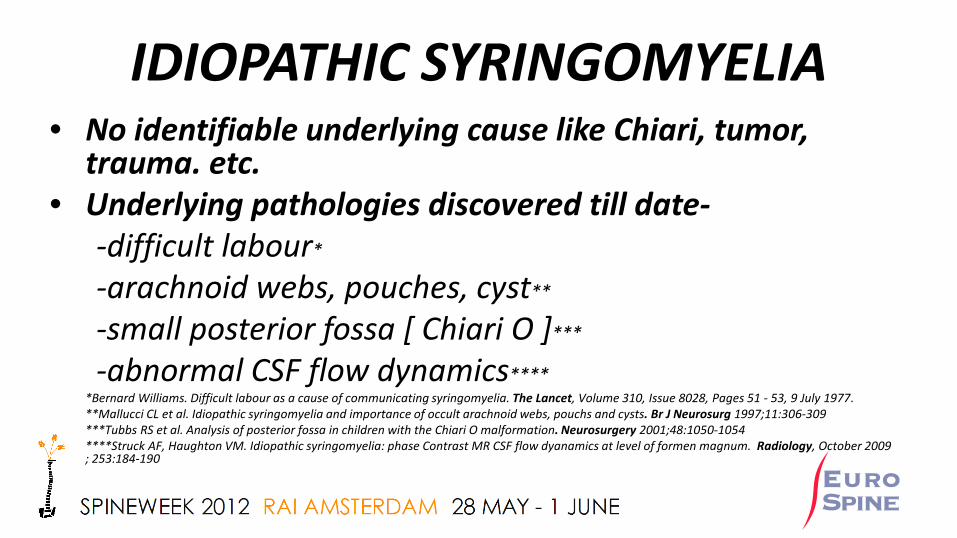
IDIOPATHIC SYRINGOMYELIA• No identifiable underlying cause like Chiari, tumor,
trauma. etc.• Underlying pathologies discovered till date-
-difficult labour*
-arachnoid webs, pouches, cyst**
-small posterior fossa [ Chiari O ]***
-abnormal CSF flow dynamics*****Bernard Williams. Difficult labour as a cause of communicating syringomyelia. The Lancet, Volume 310, Issue 8028, Pages 51 - 53, 9 July 1977. **Mallucci CL et al. Idiopathic syringomyelia and importance of occult arachnoid webs, pouchs and cysts. Br J Neurosurg 1997;11:306-309***Tubbs RS et al. Analysis of posterior fossa in children with the Chiari O malformation. Neurosurgery 2001;48:1050-1054****Struck AF, Haughton VM. Idiopathic syringomyelia: phase Contrast MR CSF flow dyanamics at level of formen magnum. Radiology, October 2009 ; 253:184-190
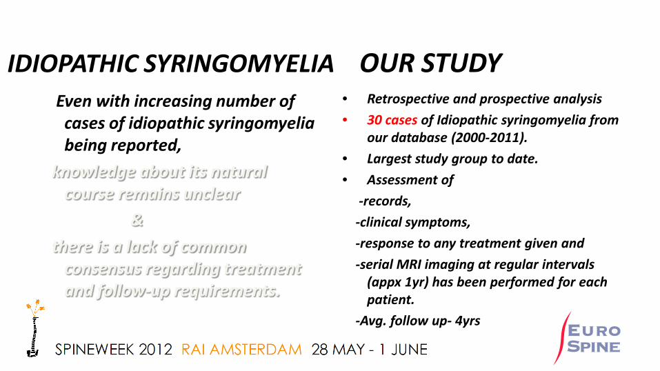
IDIOPATHIC SYRINGOMYELIA OUR STUDYEven with increasing number of
cases of idiopathic syringomyeliabeing reported,
knowledge about its natural course remains unclear
&there is a lack of common
consensus regarding treatment and follow-up requirements.
• Retrospective and prospective analysis• 30 cases of Idiopathic syringomyelia from
our database (2000-2011).• Largest study group to date.• Assessment of
-records, -clinical symptoms, -response to any treatment given and -serial MRI imaging at regular intervals
(appx 1yr) has been performed for each patient.
-Avg. follow up- 4yrs
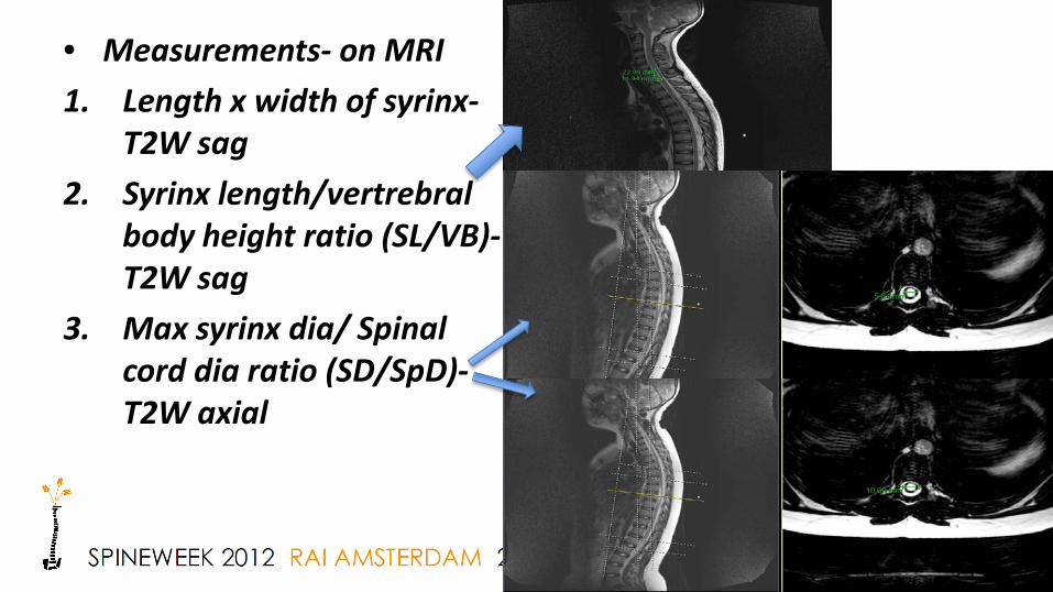
• Measurements- on MRI1. Length x width of syrinx-
T2W sag 2. Syrinx length/vertrebral
body height ratio (SL/VB)-T2W sag
3. Max syrinx dia/ Spinal cord dia ratio (SD/SpD)-T2W axial
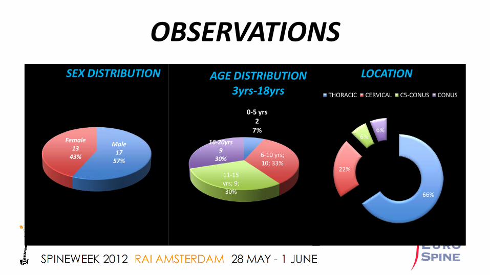
OBSERVATIONS
Male17
57%
Female13
43%
SEX DISTRIBUTION
0-5 yrs2
7%
6-10 yrs; 10; 33%
11-15 yrs; 9; 30%
16-20yrs9
30%
AGE DISTRIBUTION3yrs-18yrs
66%
22%
6%6%
LOCATION
THORACIC CERVICAL C5-CONUS CONUS
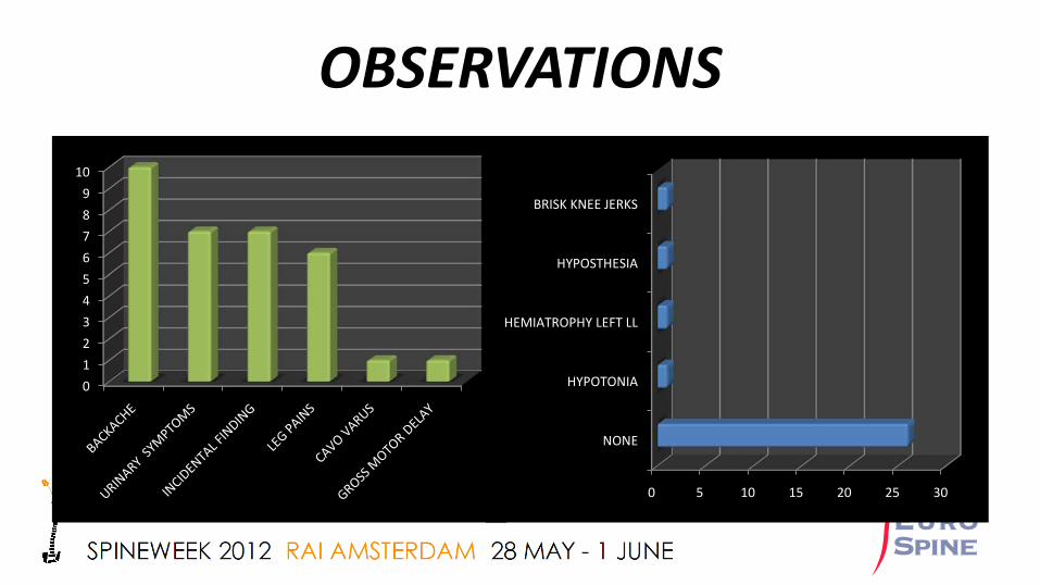
OBSERVATIONS
0123456789
10
0 5 10 15 20 25 30
NONE
HYPOTONIA
HEMIATROPHY LEFT LL
HYPOSTHESIA
BRISK KNEE JERKS

TREATMENT SURGICAL GROUP
ANALGESICS36%
NONE29%
UROLOGICAL16% PHYSIOTHERA
PY6%
DIVISON OF FILUM
10%
FMD3%
Location P.complaints
Clinical signs
Size MRI Surgery Outcome Post Op size
Sy dia/Sp dia ratio
C6-C7 Backache, leg pain, frequency
None 32 x 2 Normal postionConus M
FT Division
Clinical improvement
resolved
0.2 0
Conus Urgency, ferquency, backache
None 27 x 1.5
Normal postionConus M
FT division
Clinical improvement
27 x 1.5
0.2 0.15
T10-L1 Incidental MRI finding
None 26.5 x 3-42 x 5
Normal postionConus M
FT Division
Under follow up
0.4-0.6
Post-op MRI awaited
C4-T8 Backache, Urgency
Hypo sthesiaL/L
164 x 12
Crowdingof PF
FMD Clinical improvement
154 x 5
0.7 0.58
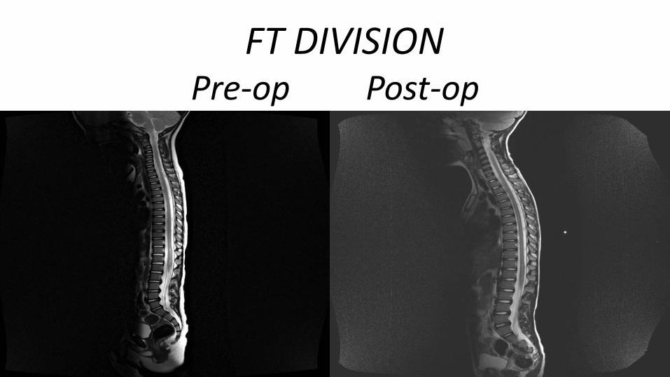
FT DIVISIONPre-op Post-op

FMDPre op Post op
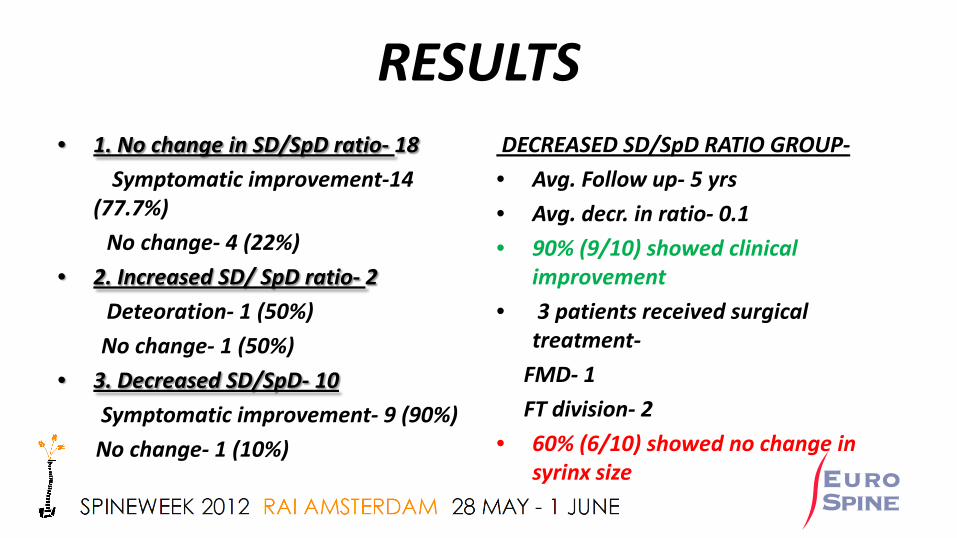
RESULTS• 1. No change in SD/SpD ratio- 18
Symptomatic improvement-14 (77.7%)
No change- 4 (22%)• 2. Increased SD/ SpD ratio- 2
Deteoration- 1 (50%)No change- 1 (50%)
• 3. Decreased SD/SpD- 10Symptomatic improvement- 9 (90%)
No change- 1 (10%)
DECREASED SD/SpD RATIO GROUP-• Avg. Follow up- 5 yrs• Avg. decr. in ratio- 0.1• 90% (9/10) showed clinical
improvement• 3 patients received surgical
treatment-FMD- 1FT division- 2
• 60% (6/10) showed no change in syrinx size

CONCLUSION• We believe that group of truly idiopathic syringomyelia is shrinking.• “Treat the patient, not the scan” - Consider division of F T if symptoms fit even if
scans may not.• Early, pre-pubertal FT division/FMD should be considered esp. in patients with
scoliosis• Size of syrinx may not be the only radiological parameter to guide treatment and
follow-up. • Follow up may only be required until SD / SpD ratio decreases or symptoms
improve.• There is a need for further Large multi-institutional prospective studies.
THANKYOU


