of August 20, 2014. This information is current as Human Cancer IL-2: The First Effective Immunotherapy for Steven A. Rosenberg http://www.jimmunol.org/content/192/12/5451 doi: 10.4049/jimmunol.1490019 2014; 192:5451-5458; ; J Immunol References http://www.jimmunol.org/content/192/12/5451.full#ref-list-1 , 50 of which you can access for free at: cites 83 articles This article Subscriptions http://jimmunol.org/subscriptions is online at: The Journal of Immunology Information about subscribing to Permissions http://www.aai.org/ji/copyright.html Submit copyright permission requests at: Email Alerts http://jimmunol.org/cgi/alerts/etoc Receive free email-alerts when new articles cite this article. Sign up at: Print ISSN: 0022-1767 Online ISSN: 1550-6606. All rights reserved. 9650 Rockville Pike, Bethesda, MD 20814-3994. The American Association of Immunologists, Inc., is published twice each month by The Journal of Immunology at Mirand Library, Roswell Park Cancer Inst on August 20, 2014 http://www.jimmunol.org/ Downloaded from at Mirand Library, Roswell Park Cancer Inst on August 20, 2014 http://www.jimmunol.org/ Downloaded from
Transcript
of August 20, 2014.This information is current as
Human CancerIL-2: The First Effective Immunotherapy for
Email Alertshttp://jimmunol.org/cgi/alerts/etocReceive free email-alerts when new articles cite this article. Sign up at:
Print ISSN: 0022-1767 Online ISSN: 1550-6606. All rights reserved.9650 Rockville Pike, Bethesda, MD 20814-3994.The American Association of Immunologists, Inc.,
is published twice each month byThe Journal of Immunology
IL-2: The First Effective Immunotherapy for HumanCancerSteven A. Rosenberg
The ability of IL-2 to expand T cells with maintenanceof functional activity has been translated into the firstreproducible effective human cancer immunotherapies.The administration of IL-2 can lead to durable, com-plete, and apparently curative regressions in patients withmetastatic melanoma and renal cancer. The growth oflarge numbers of tumor-infiltrating lymphocytes within vitro anti-cancer activity in IL-2 has led to the devel-opment of cell transfer therapies that are highly effectivein patients with melanoma. The genetic modification ofT cells with genes encoding ab TCRs or chimeric Agreceptors and the administration of these cells after ex-pansion in IL-2 have extended effective cell transfer ther-apy to other cancer types. The Journal of Immunology,2014, 192: 5451–5458.
In November 1984, a 33-year-old woman with metastaticmelanoma who had progressed through multiple priortreatments received the aggressive infusion of rIL-2.Within
one month after treatment, biopsy of one of her tumors showedextensive necrosis, after two months, all tumor deposits wereshrinking, and a few months later, all evidence of cancer wasgone. This patient has remained disease-free for the past 29years. She was the first cancer patient to respond to the admin-istration of IL-2 and, thus, the first to demonstrate that a purelyimmunologic maneuver that stimulated T lymphocytes couldmediate complete destruction of large, invasive, vascularizedcancers in humans. This patient and thousands that subse-quently received IL-2 played a major role in the introduction ofimmunotherapy into the mainstream of cancer treatment.Several seminal events in the development of modern cel-
lular immunology set the stage for the translation of immu-nologic concepts into effective immunotherapies for patientswith cancer. The origins of cellular immunology are recent.The study of Abs dominated studies of immunology until themiddle of the 20th century when it became apparent that thecellular arm of the immune system played a major role inimmunologic reactions. The word “lymphocyte” was not listedin the index of the 1958 issue of The Journal of Immunology,and the ability of lymphocytes to directly identify and interactwith Ags and components on the surface of other cells was notappreciated. The study of the direct role of lymphocytes indelayed hypersensitivity reactions and the importance of cellular
immune reactions as mediators of tissue rejection broughtthe lymphocyte into the forefront of studies of immunology(1). These studies, however, were severely hampered by theinability to sustain the survival and growth of lymphocytesex vivo until the identification in 1976 of a T cell growth factor(now known as IL-2), produced by lymphocytes, that was ca-pable of growing T lymphocytes in vitro (2). The availabilityof this growth factor propelled studies of cellular immunologyforward at a rapid pace.IL-2 is a 15.5-kDa cytokine secreted predominately by Ag-
simulated CD41 T cells, but it can also be produced byCD81 cells, NK cells, and activated dendritic cells. IL-2 canstimulate cells that express either a trimeric high-affinity IL-2receptor containing the a-, b-, and g-chains or a low-affinitydimeric receptor consisting of only the b- and g-chains. In CD8cells, IL-2 can simulate cell growth, as well as differentiation intomemory and more terminally differentiated lymphocytes. IL-2is the predominant factor responsible for the maintenance ofCD41 regulatory T cells and plays a role in the differentiationof CD4 T cells into a variety of subsets with different T cellfunctions. Translation of information derived from in vitro andmurine tumor models led to the administration of this non-specific T cell growth factor to patients with cancer and ulti-mately to the growth and adoptive cell transfer (ACT) of naturalor genetically modified autologous human antitumor T cellsexpanded in vitro in IL-2 to treat a variety of cancer types (3).These findings have had a profound impact on the ability tomanipulate the cellular immune system to successfully treatpatients with cancer, and they represented the first reproducibledemonstrations that manipulations of the immune system couldmediate the regression of large human cancers.Examples of durable cancer regressions in patients with meta-
static cancer resulting from IL-2–based therapies are shown inFig. 1, including a patient with kidney cancer treated with IL-2,a patient with melanoma treated with autologous tumor-infiltrating lymphocytes (TILs), and patients with lymphomaor synovial cell sarcoma treated with autologous lymphocytesgenetically engineered with chimeric Ag receptors (CARs) orTCRs that recognize the patient’s cancer.
IL-2 administration as a cancer treatment
The inability to sustain the survival and growth of T lympho-cytes in vitro was a perplexing problem in the 1970s. Althoughresponder T cells from mixed lymphocyte cultures could surviveand grow for many months when cells were stimulated by
Surgery Branch, National Cancer Institute, Bethesda, MD 20892
Address correspondence and reprint requests to Dr. Steven A. Rosenberg, NationalInstitutes of Health, 9000 Rockville Pike, CRC Building, Room 3W-3940, Bethesda,MD 20892. E-mail address: [email protected]
Abbreviations used in this article: ACT, adoptive cell transfer; CAR, chimeric Ag recep-tor; LAK, lymphokine-activated killer; TBI, total body irradiation; TIL, tumor-infiltrating lymphocyte.
allogeneic lymphocytes every 1–2 wk, the factors responsiblefor this sustained growth were unknown. An important steptoward understanding this phenomenon was a report in 1976that the culture of pools of lymphocytes from multiple donorsin medium containing PHA gave rise to a supernatant factorthat when continuously supplied could sustain the growth ofT lymphocytes from normal human bone marrow for manymonths (2). The growing cells were identified as T cells basedon their ability to form rosettes of sheep erythrocytes, themajor marker of T cells at that time. These growing cells weredistinguished from growing B cells by the absence of EBV,
which could promote the long-term growth of B cells. Thesefindings were quickly followed by studies, both in mice andhumans, demonstrating that supernatants derived from lectin-stimulated normal lymphocytes could sustain the growth ofboth mouse and human lymphocytes in vitro with mainte-nance of Ag recognition (4–8). Purification of the super-natants showed that the growth-promoting properties wereindependent of the continued presence of the lectin in thesupernatant (9–11). The ability of this T cell growth factor(shortly thereafter named IL-2) to mediate T cell survival andsustain function in vitro suggested that its administration could
FIGURE 1. Upper panel, Fifty-six–year-old male with metastatic renal
cell cancer to the liver and subcarinal
lymph nodes was treated with high-dose
bolus IL-2 in January 1994. Patient
underwent a complete regression of all
disease and remains disease-free 20 y
later. Upper middle panel, Fifty-four–
year-old male with metastatic melanoma
to the lungs and liver was treated with
autologous TILs plus IL-2 following
a lymphodepleting regimen in Decem-
ber 2003. The patient underwent a
complete regression of all disease and
remains disease-free .10 y later. Lowermiddle panel, Fifty-year-old male with
follicular non-Hodgkin’s lymphoma
at multiple sites in the abdomen, me-
diastinum, and axillary lymph nodes
treated with genetically engineered au-
tologous peripheral lymphocytes express-
ing a gene encoding an anti-CD19
chimeric Ag receptor in May 2009.
The patient underwent a dramatic
regression of all disease following two
cycles of treatment and is progression-
free .4 y later. Lower panel, Sixty-seven–year-old female with metastatic
synovial sarcoma to the lung and right
pelvis treated with genetically engi-
neered autologous peripheral lympho-
cytes expressing a gene encoding a
TCR reactive with the NY-ESO-1
cancer testes Ag in August 2010. The
patient was treated in August 2010
and has undergone a dramatic partial
regression now ongoing .3 y later.
5452 TRANSLATING IMMUNOLOGY: IMMUNOTHERAPY WITH IL-2
potentially stimulate functional T cells in vivo; however, thesestudies were severely hampered by the inability to obtain largeamounts of purified IL-2.A human Jurkat T cell tumor line was identified that secreted
high levels of IL-2 following PHA stimulation, and three lots ofpurified preparations of IL-2 were produced for clinical testingby the E.I. DuPont Company (12) between July 1983 andFebruary 1984. Sixteen patients with advanced cancer weretreated with this purified natural IL-2 (13). IL-2 administra-tion resulted in dose-related fever, chills, malaise, and mildreversible hepatic dysfunction, but no antitumor activity wasnoted in these patients.Progress in the administration of IL-2, however, was stim-
ulated by the cloning of the gene encoding IL-2 in 1983 (14)and the subsequent production and characterization in 1984of the biological activity of rIL-2 produced in Escherichia coli(15, 16). PHA-stimulated human lymphocytes provided ayield of 0.7 mg/l supernatant, which was improved to 3 mg/lwhen PHA was used to stimulate the Jurkat T cell tumor line.In contrast, recombinant E. coli could produce 100 mg IL-2/l.The production of rIL-2 finally made it possible to evaluatethe impact of the in vivo administration of large amounts ofIL-2 in cancer-bearing mice and humans.Early experiments demonstrated that the administration of
rIL-2 to tumor-bearing mice could mediate the regression ofsmall established pulmonary metastases as well as s.c. tumors inanimal models (17), although it was necessary to reach sig-nificant IL-2–related toxicity before antitumor effects were seen.The modest results seen using IL-2 in animal models, however,provided the impetus for the first administration of rIL-2 inhumans. Twenty patients were reported in 1985 (23 treated)who received a wide variety of different regimens and doses ofrIL-2 (18). The half-life of IL-2 in humans was ∼7 min witha later delayed clearance consistent with a two-compartmentmodel as IL-2 was released from extravascular space into theplasma compartment. Marked depletion of all lymphoid cellswas seen almost immediately after IL-2 administration, whichrebounded after IL-2 was discontinued. Significant toxicitiesbecame apparent in these early studies, including fever, chills,malaise, arthralgias, and unexpected capillary leak, which ledto weight gain from marked fluid retention. Levels of IFN andother cytokines were found in the serum, and although theseinteresting immunologic changes were seen, there was no evi-dence of tumor regression in any of these cancer patients treatedwith IL-2 alone.It was early noted that the exposure of normal mouse
splenocytes or human PBMCs to supernatants containing IL-2could generate cells, later called lymphokine-activated killer(LAK) cells, that without further stimulation could recognizeand kill cultured tumor cell lines and fresh human cancer cellsin vitro (19–22). LAK cell precursors were not T cells andappeared related to the NK lineage. Multiple studies of theadoptive transfer of these LAK cells grown in vitro and ad-ministered to tumor-bearing mice showed in vivo antitumoractivity, but only in models in which tumors were treated be-fore they became vascularized (23–26). These studies stimu-lated a clinical trial in 30 patients with advanced cancer, firstusing i.v. administration of LAK cells generated with naturalIL-2 and later LAK cells generated with rIL-2 (27). No anti-tumor responses were seen in any of these patients. Murinemodels indicated that the administration of IL-2 could increase
the in vivo activity of LAK cells, and this work stimulated moreaggressive attempts to administer IL-2 at the maximum toler-ated doses in conjunction with LAK cells in humans.These higher IL-2 doses finally led to the first demonstration
that IL-2 administration was capable of mediating tumor re-gression in humans, and the results of this study were publishedin December 1985 (28). Twenty-five patients with metastaticcancer, in whom standard therapy had failed, were treated inthe Surgery Branch at the National Cancer Institute with in-creasing doses of IL-2 until toxicity precluded further doseescalation. Although most patients treated in the early phaseof this study received 60,000 IU/kg every 8 h, patients sub-sequently treated received 180,000 or 600,000 IU/kg. An every8 h schedule of the bolus infusion of IL-2 was establishedbased on the in vivo half-life of IL-2 and assured that serumlevels were continuously maintained at concentrations neces-sary to stimulate high-affinity IL-2 receptors during the courseof IL-2 administration. In this first series of 25 patients, in-cluding the first patient mentioned at the beginning of thisreview, 4 of 7 patients with metastatic melanoma and 3 of 3patients with metastatic renal cancer exhibited regression ofmetastatic cancer, and thus these cancer types were the targetsin many subsequent studies evaluating the clinical effective-ness of IL-2. A generalized capillary leak syndrome was inducedby IL-2 in vivo that resulted in interstitial pulmonary infiltratesand substantial weight gain in patients. Similarly, serum cre-atinine and bilirubin levels were elevated in about half of thepatients. The side effects were transient and returned to base-line following treatment.Importantly, however, this was the first demonstration that a
purely immunologic maneuver could lead to tumor regressionin humans and stimulated substantial activity exploring theadministration of IL-2 to patients (29). Tumors do not expressIL-2 receptors and thus the antitumor activity was the resultof IL-2 stimulation of immune cells. In 1987, the SurgeryBranch at the National Cancer Institute reported the resultsof the first 157 consecutive patients with advanced cancer treatedwith high-dose IL-2, either alone or in conjunction with LAKcells (30). A maximum dose of 600,000–720,000 IU/kg every8 h was established as the maximum tolerated dose. Thirty ofthe 157 patients showed objective cancer regressions, including13 of 57 patients with renal cell cancer (23%) and 12 of 42patients with melanoma (29%). Cancer regressions were du-rable and seven of the nine complete responses remained inremission at the time of publication and continued for yearsthereafter. A randomized trial of 181 patients with metastaticmelanoma or renal cancer comparing treatment with IL-2alone or in conjunction with LAK cells showed that the an-titumor effects were due to IL-2 alone, and thus LAK celladministration was omitted in future studies (31).These results then led to an explosion of studies utilizing IL-2
in patients with metastatic cancer using either a high-dose bolusregimen or a continuous infusion of IL-2 (32–34). Patients withmetastatic melanoma or metastatic renal cell cancer wereuniquely responsive to high-dose IL-2 administration, and ex-cept for patients with advanced non-Hodgkin’s lymphomas(35) only rare responses were seen in patients with other tumortypes. Complete durable regressions of metastatic disease werea hallmark of IL-2 therapy in melanoma and renal cancer. In1994 the Surgery Branch at the National Cancer Institutereported on 283 consecutive patients treated with high-
dose bolus IL-2 with metastatic melanoma or renal cancer(32), which was updated to 409 consecutive patients 4 ylater. This later study revealed a 15% incidence of objectiveregressions in 182 patients with metastatic melanoma (7%were complete) and a 19% overall response rate in 227 patientswith metastatic renal cancer (9% were complete) (33). Twenty-seven of the 33 completely responding patients (82%) re-mained in ongoing continuous complete response from 39to .148 mo from the onset of treatment and appeared tobe cured. Tumor regressions were seen at all organ sites. Multi-institutional studies confirmed these single institution results.The continuous infusion of high-dose IL-2 was evaluated by theNational Biotherapy Study Group in multiple trials usingthe continuous infusion of IL-2, often in combination withthe administration of cells or other cytokines, and similar anti-tumor responses were seen (36). Two hundred fifty-five con-secutive patients with metastatic kidney cancer from multipleinstitutions were entered into seven phase 2 clinical trials ofhigh-dose bolus IL-2 sponsored by the Chiron Corporationresulting in an overall objective response rate of 14% with5% achieving complete responses (37). The median durationof partial responses was 19 mo and the median response du-ration of complete responses was not reached because 8 of the12 complete responders were ongoing, including many longerthan 2 y. A recent analysis of 259 consecutive patients withmetastatic kidney cancer treated in the Surgery Branch at theNational Cancer Institute between 1986 and 2006 with high-dose IL-2 revealed that 23 (9%) patients experienced a com-plete response and only 4 of 23 developed disease recurrence(38). Thirty patients (11%) achieved a partial response for anoverall objective response rate of 20%. Based on the results ofthe durability of responses in both single institution as well asmulti-institutional studies, the U.S. Food and Drug Admin-istration approved high-dose bolus IL-2 for the treatment ofpatients with metastatic renal cancer in 1992, thus becomingthe first immunotherapy approved for the treatment of patientswith cancer.As these studies were being conducted, similar results were
being achieved in multi-institutional studies of IL-2 for thetreatment of patients with metastatic melanoma. Two hundredseventy patients were entered into clinical trials conducted at22 different institutions using the high-dose bolus IL-2 regimen(39). The overall objective response rate was 16% with 17complete responders (6%) and 26 partial responders (10%).The median duration in patients who achieved a completeresponse had not been reached with 10 of the 17 completeresponders ongoing at 24–106 mo. Based on the durabilityof these responses, IL-2 was approved by the U.S. Food andDrug Administration for the treatment of patients with meta-static melanoma in 1998. Higher response rates were seen inpatients with s.c. or cutaneous metastases, although the greatmajority of patients treated had visceral disease.The early toxicities seen accompanying the administration of
high-dose IL-2 were unexpected, but were a harbinger of tox-icities seen with many immunologic maneuvers that followed.The underlying toxicity of IL-2 results from a capillary leak thatleads to fluid extravasation into visceral organs that can com-promise their function. Although serious biochemical abnor-malities can be seen in the liver and kidney, these all returnedto normal following the completion of treatment. An unusualaspect of the administration of IL-2 was the continuation of
dosing until grade 3 or 4 toxicity was reached. There was asignificant learning curve in the administration of IL-2 beforeit was realized that many of these toxicities were completelyreversible (40). In the first 157 patients reported from theSurgery Branch at the National Cancer Institute there were4 treatment-related deaths (28). The neutrophil chemotacticdefects induced by IL-2 were probably associated with the highincidence of central line sepsis that accompanied IL-2 admin-istration (41), although a prospective randomized study dem-onstrated that this complication could be virtually eliminatedby the use of prophylactic oxacillin (42). As information waslearned about the tolerance to IL-2, the administered doses inthe first cycle of therapy decreased from an initial median of13 doses to 7 doses without any decrease in the ongoing ob-jective or complete response rates (43). A detailed study of1241consecutive metastatic cancer patients treated with high-dose i.v. bolus IL-2 in the Surgery Branch at the NationalCancer Institute showed a substantial decrease in toxicities inpatients when IL-2 administration experience increased (43).Although treatment-related deaths were initially in the 2 to4% range, mortalities dropped consistently, and in the last809 consecutive patients reported and treated in the SurgeryBranch at the National Cancer Institute in 1998 there were nodeaths related to treatment with IL-2. Thus, with appropriatemanagement and experience high-dose IL-2 can be safely ad-ministered to patients with metastatic cancer with treatment-related mortalities ,1%.
The role of IL-2 in ACT of cancer
The use of IL-2 to grow T cells in vitro that can be used foradoptive cell therapy, as well as the administration of IL-2 tosupport the growth and survival of antitumor cells infused intopatients, has been of major importance in translating basicstudies of IL-2 to the clinic.Prior to the advent of IL-2, attempts to adoptively transfer
T cells with antitumor activity as a therapeutic modality inanimal tumor models were dependent on the ability to generatecells with antitumor activity by in vivo immunization (reviewedin Ref. 44). Although adoptive transfer of these T cells couldmediate modest antitumor activity, the inability to grow thesecells ex vivo severely limited progress, and the lack of humancells with specific antitumor reactivity was a major obstacle tothe application of this approach in humans.An important aspect of early studies of IL-2 in vitro was the
demonstration that lymphocytes expanded long-term in IL-2could retain specific Ag reactivity (4–8). Clonal T cell linesfrom mouse and human lymphocytes could be generated bylimiting dilution and growth in IL-2 (45–47). Early mousemodels of adoptive cell therapy using T cells resulting fromimmunization of mice with the Friend virus–induced leuke-mia, FBL-3, showed that the adoptive transfer of T cells i.p.to mice bearing i.p. and nodal FBL-3 exhibited in vivo an-titumor effects (48). It was not at all clear, however, that largeblastic-activated T cells grown in vitro in IL-2 could mediatesystemic antitumor effects because early studies showed a dra-matic accumulation of these large cells in the lungs for 1–2 dfollowing i.v. administration (49, 50). An early test of theimpact of the i.v. administration of cells grown in IL-2 dem-onstrated that the rejection of skin grafts in mice could beaccelerated by the i.v. adoptive transfer of syngeneic cells withalloreactivity expanded in IL-2 (51). Similarly, in an extension
5454 TRANSLATING IMMUNOLOGY: IMMUNOTHERAPY WITH IL-2
of prior work performed using the FBL-3 leukemia (8), a pal-pable local tumor in the footpad as well as disseminated me-tastases could be cured by the i.v. administration of specificimmune lymphocytes expanded in IL-2 (52). This provideda clear demonstration that i.v. injection of cells grown in IL-2could circulate and manifest immune effector functions againstpalpable vascularized tumors in vivo.Although T cells could be generated against the viral-induced
FBL-3 lymphomamodel, a major obstacle to the application ofthis cell transfer approach was the inability to generate T cellreactivity against transplantable solid tumors in mice oragainst naturally growing human cancers. Lymphocytes in-filtrating into the stroma of solid tumors unexpectedly pro-vided a source of such antitumor T cells. The growth of single-cell suspensions of a variety of murine and human solidtumors in IL-2 resulted in pure cultures of TILs free ofcontaminating tumor cells (19). The early growth of LAK cellsin these cultures was limited, and as cells continued to growthe resulting lymphocytes often exhibited specific tumor re-activity against the tumor of origin and not other tumors.The first report of the adoptive immunotherapy of murine
tumors using syngeneic TILs plus IL-2 showed that largeestablished, vascularized pulmonary, as well as hepatic metas-tases could be eliminated by this approach (53). Lymphode-pletion prior to administration of specific TILs could curemost mice bearing visceral metastastic deposits from theMC38 colon adenocarcinoma, as well as mice bearing largemethylcholanthrene-induced sarcomas. An important stepin the development of cell transfer therapy for the treatmentof human cancers was the demonstration that TILs obtainedfrom resected metastatic melanoma deposits and grown in IL-2developed specific cytolytic immune responses against the au-tologous human tumor (54). In vitro studies demonstrating thetumor specificity of human TILs stimulated the developmentof methods to grow human TILs in large numbers (55, 56) andthen led to the first report of the ability of adoptive cell transferto mediate the regression of large established cancers in patientswith metastatic melanoma (57). In a paper reported in 1988from the Surgery Branch at the National Cancer Institute, 20patients with metastatic melanoma were treated with in vitro–expanded autologous TILs in conjunction with IL-2. Objectivecancer regression was seen in 9 of 15 patients not previouslytreated with IL-2 and in 2 of 5 patients who had progressedthrough prior treatment with IL-2. Regression of cancers wasseen in the lungs, liver, bones, skin, and s.c. sites. Studies ofthe adoptive transfer of [111In]-labeled TILs into patients withmetastatic melanoma showed accumulation in the lungs for∼24 h and subsequent accumulation of administered TILs intumor deposits (50, 58). From May 1987 through December1992, 86 patients with metastatic melanoma were treated with145 courses of autologous TILs plus high-dose bolus i.v. IL-2at 720,000 IU/kg every 8 h (59). Because of evidence fromanimal models that prior lymphodepletion could improve theantitumor impact of cell administration, 57 of the 86 patientsreceived a single i.v. dose of 25 mg/kg cyclophosphamide ∼36 hprior to the TIL infusion. The overall objective response ratein these patients was 34% and was similar whether patientsreceived prior cyclophosphamide or had previously been treatedwith high-dose IL-2. The rate of response was greater in patientstreated with TILs from younger cultures, TILs with shorterdoubling times, and TILs with higher lytic capacity against
autologous tumor targets (60). Many of these responses, how-ever, were of short duration with minimal apparent persistenceof the transferred cells. In studies using retroviral insertionof a neomycin phophotransferase gene to mark TILs, barely0.01% of the administered cells could be detected in thecirculation days after cell transfer (61). Attempts to improvethe antitumor effects of TIL transfer in humans using highlyselected tumor-reactive CD81 clones reactive with melano-cyte differentiation Ags did not result in objective tumor re-gression in patients with melanoma, suggesting that thepolyclonal nature of tumor reactivity in TILs and possiblythe presence of CD41 cells were necessary to induce tumorrejection (62, 63).Based on multiple animal models demonstrating that lym-
phodepletion was necessary for the effectiveness of the trans-ferred cells, a trial was conducted in the Surgery Branch at theNational Cancer Institute that dramatically changed the fieldof ACT in humans (64). A vigorous nonmyeloablative con-ditioning regimen consisting of high-dose cyclophosphamideand fludarabine was given immediately before cell transfer,which resulted in complete elimination of normal lymphocytesfrom peripheral blood for ∼8 d before reconstitution ofendogenous lymphocytes occurred. Six of 13 patients in thistrial exhibited an objective cancer response, and for the firsttime there was substantial persistence and sometimes a clonalrepopulation of the transferred cells that could represent 75%of all circulating CD81 cells at 6–12 mo after infusion (64).In a subsequent study of 43 patients, a 49% objective responserate was achieved, and substantial persistence of the transferredcells was seen in most responding patients (65, 66). Two ad-ditional pilot trials of ACT in 25 patients each were conductedin the Surgery Branch at the National Cancer Institute toevaluate the impact of increased lymphodepletion by addingeither 2 or 12 Gy total body irradiation (TBI) to the pre-parative chemotherapy regimen prior to the adoptive transferof cells (65, 66). Objective response rates by ResponseEvaluation Criteria in Solid Tumors criteria in these two trialswere 52 and 72%, respectively. Five of 25 (20%) patientsreceiving 2 Gy TBI and 10 of 25 (40%) of patients receiving12 Gy TBI achieved a complete response. Nineteen of the 20complete responders in this total of 93 patients remained incomplete regression at 70–114 mo following the adoptivetransfer of cells. Seventeen of the 20 completely respondingpatients had visceral metastases and all but 2 of the completeresponders had progressive disease following other prior sys-temic therapy, including IL-2, chemotherapy, and anti-CTLA4.These rates of overall response and durable complete responsesin patients with metastatic melanoma exceed those of othertreatments for patients with this disease. Similar results usingthe adoptive transfer of TILs following a lymphodepletingpreparative regimen have been reported by groups at the ShebaMedical Center in Israel (67), as well as the M.D. AndersonHospital (68) and the Moffit Cancer Center in the UnitedStates (69).Thus, a continuous improvement in the treatment of pa-
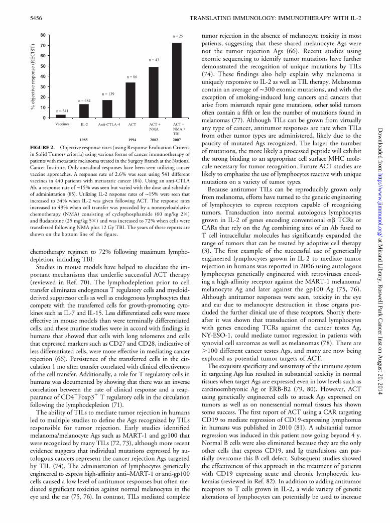
tients with metastatic melanoma has been achieved utilizingIL-2–based immunotherapies (Fig. 2). Response rates in theSurgery Branch at the National Cancer Institute increasedfrom 15% seen with IL-2 alone to 34% using ACT/TILs to49% with ACT/TILs preceded by a preparative lymphodepleting
chemotherapy regimen to 72% following maximum lympho-depletion, including TBI.Studies in mouse models have helped to elucidate the im-
portant mechanisms that underlie successful ACT therapy(reviewed in Ref. 70). The lymphodepletion prior to celltransfer eliminates endogenous T regulatory cells and myeloid-derived suppressor cells as well as endogenous lymphocytes thatcompete with the transferred cells for growth-promoting cyto-kines such as IL-7 and IL-15. Less differentiated cells were moreeffective in mouse models than were terminally differentiatedcells, and these murine studies were in accord with findings inhumans that showed that cells with long telomeres and cellsthat expressed markers such as CD27 and CD28, indicative ofless differentiated cells, were more effective in mediating cancerrejection (66). Persistence of the transferred cells in the cir-culation 1 mo after transfer correlated with clinical effectivenessof the cell transfer. Additionally, a role for T regulatory cells inhumans was documented by showing that there was an inversecorrelation between the rate of clinical response and a reap-pearance of CD41Foxp31 T regulatory cells in the circulationfollowing the lymphodepletion (71).The ability of TILs to mediate tumor rejection in humans
led to multiple studies to define the Ags recognized by TILsresponsible for tumor rejection. Early studies identifiedmelanoma/melanocyte Ags such as MART-1 and gp100 thatwere recognized by many TILs (72, 73), although more recentevidence suggests that individual mutations expressed by au-tologous cancers represent the cancer rejection Ags targetedby TIL (74). The administration of lymphocytes geneticallyengineered to express high-affinity anti–MART-1 or anti-gp100cells caused a low level of antitumor responses but often me-diated significant toxicities against normal melanocytes in theeye and the ear (75, 76). In contrast, TILs mediated complete
tumor rejection in the absence of melanocyte toxicity in mostpatients, suggesting that these shared melanocyte Ags werenot the tumor rejection Ags (66). Recent studies usingexomic sequencing to identify tumor mutations have furtherdemonstrated the recognition of unique mutations by TILs(74). These findings also help explain why melanoma isuniquely responsive to IL-2 as well as TIL therapy. Melanomascontain an average of ∼300 exomic mutations, and with theexception of smoking-induced lung cancers and cancers thatarise from mismatch repair gene mutations, other solid tumorsoften contain a fifth or less the number of mutations found inmelanomas (77). Although TILs can be grown from virtuallyany type of cancer, antitumor responses are rare when TILsfrom other tumor types are administered, likely due to thepaucity of mutated Ags recognized. The larger the numberof mutations, the more likely a processed peptide will exhibitthe strong binding to an appropriate cell surface MHC mole-cule necessary for tumor recognition. Future ACT studies arelikely to emphasize the use of lymphocytes reactive with uniquemutations on a variety of tumor types.Because antitumor TILs can be reproducibly grown only
from melanoma, efforts have turned to the genetic engineeringof lymphocytes to express receptors capable of recognizingtumors. Transduction into normal autologous lymphocytesgrown in IL-2 of genes encoding conventional ab TCRs orCARs that rely on the Ag combining sites of an Ab fused toT cell intracellular molecules has significantly expanded therange of tumors that can be treated by adoptive cell therapy(3). The first example of the successful use of geneticallyengineered lymphocytes grown in IL-2 to mediate tumorrejection in humans was reported in 2006 using autologouslymphocytes genetically engineered with retroviruses encod-ing a high-affinity receptor against the MART-1 melanoma/melanocyte Ag and later against the gp100 Ag (75, 76).Although antitumor responses were seen, toxicity in the eyeand ear due to melanocyte destruction in those organs pre-cluded the further clinical use of these receptors. Shortly there-after it was shown that transduction of normal lymphocyteswith genes encoding TCRs against the cancer testes Ag,NY-ESO-1, could mediate tumor regression in patients withsynovial cell sarcomas as well as melanomas (78). There are.100 different cancer testes Ags, and many are now beingexplored as potential tumor targets of ACT.The exquisite specificity and sensitivity of the immune system
in targeting Ags has resulted in substantial toxicity in normaltissues when target Ags are expressed even in low levels such ascarcinoembryonic Ag or ERB-B2 (79, 80). However, ACTusing genetically engineered cells to attack Ags expressed ontumors as well as on nonessential normal tissues has shownsome success. The first report of ACT using a CAR targetingCD19 to mediate regression of CD19-expressing lymphomasin humans was published in 2010 (81). A substantial tumorregression was induced in this patient now going beyond 4 y.Normal B cells were also eliminated because they are the onlyother cells that express CD19, and Ig transfusions can par-tially overcome this B cell defect. Subsequent studies showedthe effectiveness of this approach in the treatment of patientswith CD19 expressing acute and chronic lymphocytic leu-kemias (reviewed in Ref. 82). In addition to adding antitumorreceptors to T cells grown in IL-2, a wide variety of geneticalterations of lymphocytes can potentially be used to increase
the antitumor activity of the transferred cells in ACT. Theintroduction of a gene encoding a single-chain IL-12 has beenshown to increase the antitumor activity of transferred cells inmouse models, and clinical trials using TILs transduced toexpress single-chain IL-12 have begun (83).
ConclusionsThe translation of basic findings concerning IL-2 had a pro-found impact on the development of cancer immunotherapy.The administration of IL-2 as well as the adoptive transfer ofantitumor T cells grown in IL-2 represented the first effectiveimmunotherapies for cancer in humans and have provided oneof the first curative systemic therapies for any solid tumor.These studies played a major role in enabling immunotherapyto join the mainstream of cancer treatment.
DisclosuresThe author has no financial conflicts of interest.
References1. Mitchison, N. A. 1955. Studies on the immunological response to foreign tumor
transplants in the mouse. I. The role of lymph node cells in conferring immunity byadoptive transfer. J. Exp. Med. 102: 157–177.
2. Morgan, D. A., F. W. Ruscetti, and R. G. Gallo. 1976. Selective in vitro growth ofT lymphocytes from normal human bone marrows. Science 193: 1007–1008.
3. Rosenberg, S. A. 2011. Cell transfer immunotherapy for metastatic solid cancer:what clinicians need to know. Nat. Rev. Clin. Oncol. 8: 577–585.
4. Gillis, S., and K. A. Smith. 1977. Long term culture of tumour-specific cytotoxicT cells. Nature 268: 154–156.
5. Rosenberg, S. A., S. Schwarz, and P. J. Spiess. 1978. In vitro growth of murineT cells. II. Growth of in vitro sensitized cells cytotoxic for alloantigens. J. Immunol.121: 1951–1955.
6. Strausser, J. L., and S. A. Rosenberg. 1978. In vitro growth of cytotoxic humanlymphocytes. I. Growth of cells sensitized in vitro to alloantigens. J. Immunol. 121:1491–1495.
7. Gillis, S., P. E. Baker, F. W. Ruscetti, and K. A. Smith. 1978. Long-term culture ofhuman antigen-specific cytotoxic T-cell lines. J. Exp. Med. 148: 1093–1098.
8. Cheever, M. A., P. D. Greenberg, and A. Fefer. 1981. Specific adoptive therapy ofestablished leukemia with syngeneic lymphocytes sequentially immunized in vivoand in vitro and nonspecifically expanded by culture with interleukin 2. J. Immunol.126: 1318–1322.
9. Kurnick, J. T., K.-O. Gronvik, A. K. Kimura, J. B. Lindblom, V. T. Skoog,O. Sjoberg, and H. Wigzell. 1979. Long term growth in vitro of human T cell blastswith maintenance of specificity and function. J. Immunol. 122: 1255–1260.
10. Rosenberg, S. A., S. Schwarz, P. J. Spiess, and J. M. Brown. 1980. In vitro growth ofmurine T cells. III. Method for separation of T cell growth factor (TCGF) fromconcanavalin A and biological activity of the resulting TCGF. J. Immunol. Methods33: 337–350.
11. Lotze, M. T., and S. A. Rosenberg. 1981. In vitro growth of cytotoxic humanlymphocytes. III. The preparation of lectin-free T cell growth factor (TCGF) andan analysis of its activity. J. Immunol. 126: 2215–2220.
12. Robb, R. J., R. M. Kutny, and V. Chowdhry. 1983. Purification and partial se-quence analysis of human T-cell growth factor. Proc. Natl. Acad. Sci. USA 80: 5990–5994.
13. Lotze, M. T., L. W. Frana, S. O. Sharrow, R. J. Robb, and S. A. Rosenberg. 1985.In vivo administration of purified human interleukin 2. I. Half-life and immuno-logic effects of the Jurkat cell line-derived interleukin 2. J. Immunol. 134: 157–166.
14. Taniguchi, T., H. Matsui, T. Fujita, C. Takaoka, N. Kashima, R. Yoshimoto, andJ. Hamuro. 1983. Structure and expression of a cloned cDNA for human inter-leukin-2. Nature 302: 305–310.
15. Devos, R., G. Plaetinck, H. Cheroutre, G. Simons, W. Degrave, J. Tavernier,E. Remaut, and W. Fiers. 1983. Molecular cloning of human interleukin 2 cDNAand its expression in E. coli. Nucleic Acids Res. 11: 4307–4323.
16. Rosenberg, S. A., E. A. Grimm, M. McGrogan, M. Doyle, E. Kawasaki, K. Koths,and D. F. Mark. 1984. Biological activity of recombinant human interleukin-2produced in Escherichia coli. Science 223: 1412–1414.
17. Rosenberg, S. A., J. J. Mule, P. J. Spiess, C. M. Reichert, and S. L. Schwarz. 1985.Regression of established pulmonary metastases and subcutaneous tumor mediatedby the systemic administration of high-dose recombinant interleukin 2. J. Exp. Med.161: 1169–1188.
18. Lotze, M. T., Y. L. Matory, S. E. Ettinghausen, A. A. Rayner, S. O. Sharrow,C. A. Seipp, M. C. Custer, and S. A. Rosenberg. 1985. In vivo administration ofpurified human interleukin 2. II. Half life, immunologic effects, and expansion ofperipheral lymphoid cells in vivo with recombinant IL 2. J. Immunol. 135: 2865–2875.
19. Yron, I., T. A. Wood, Jr., P. J. Spiess, and S. A. Rosenberg. 1980. In vitro growth ofmurine T cells. V. The isolation and growth of lymphoid cells infiltrating syngeneicsolid tumors. J. Immunol. 125: 238–245.
20. Grimm, E. A., A. Mazumder, H. Z. Zhang, and S. A. Rosenberg. 1982. Lym-phokine-activated killer cell phenomenon. Lysis of natural killer-resistant fresh solidtumor cells by interleukin 2-activated autologous human peripheral blood lymphocytes.J. Exp. Med. 155: 1823–1841.
21. Grimm, E. A., K. M. Ramsey, A. Mazumder, D. J. Wilson, J. Y. Djeu, andS. A. Rosenberg. 1983. Lymphokine-activated killer cell phenomenon. II. Precursorphenotype is serologically distinct from peripheral T lymphocytes, memory cyto-toxic thymus-derived lymphocytes, and natural killer cells. J. Exp. Med. 157: 884–897.
22. Yang, J. C., J. J. Mule, and S. A. Rosenberg. 1986. Murine lymphokine-activatedkiller (LAK) cells: phenotypic characterization of the precursor and effector cells. J.Immunol. 137: 715–722.
23. Mule, J. J., and S. A. Rosenberg. 1985. Successful adoptive immunotherapy ofestablished metastases with lymphokine activated killer cells and recombinant in-terleukin-2. In Immune Responses to Metastases. R. B. Herberman, R. H. Wiltrout,and E. Gorelik, eds. CRC, Boca Raton, FL, p. 69–94.
24. Rosenberg, S. A. 1985. Lymphokine-activated killer cells: a new approach to im-munotherapy of cancer. J. Natl. Cancer Inst. 75: 595–603.
25. Ettinghausen, S. E., and S. A. Rosenberg. 1986. Immunotherapy of murine sar-comas using lymphokine activated killer cells: optimization of the schedule androute of administration of recombinant interleukin-2. Cancer Res. 46: 2784–2792.
26. Lafreniere, R., and S. A. Rosenberg. 1985. Successful therapy of hepatic metastasesfrom several murine tumors using lymphokine activated killer cells and recombinantinterleukin-2. Surg. Forum 36: 392–394.
27. Lotze, M. T., B. R. Line, D. J. Mathisen, and S. A. Rosenberg. 1980. The in vivodistribution of autologous human and murine lymphoid cells grown in T cellgrowth factor (TCGF): implications for the adoptive immunotherapy of tumors.J. Immunol. 125: 1487–1493.
28. Rosenberg, S. A., M. T. Lotze, L. M. Muul, S. Leitman, A. E. Chang,S. E. Ettinghausen, Y. L. Matory, J. M. Skibber, E. Shiloni, J. T. Vetto, et al. 1985.Observations on the systemic administration of autologous lymphokine-activatedkiller cells and recombinant interleukin-2 to patients with metastatic cancer. N.Engl. J. Med. 313: 1485–1492.
29. Lotze, M. T., A. E. Chang, C. A. Seipp, C. Simpson, J. T. Vetto, and S. A. Rosenberg.1986. High-dose recombinant interleukin 2 in the treatment of patients with dis-seminated cancer. Responses, treatment-related morbidity, and histologic findings.JAMA 256: 3117–3124.
30. Rosenberg, S. A., M. T. Lotze, L. M. Muul, A. E. Chang, F. P. Avis, S. Leitman,W. M. Linehan, C. N. Robertson, R. E. Lee, J. T. Rubin, et al. 1987. A progressreport on the treatment of 157 patients with advanced cancer using lymphokine-activated killer cells and interleukin-2 or high-dose interleukin-2 alone. N. Engl. J.Med. 316: 889–897.
31. Rosenberg, S. A., M. T. Lotze, J. C. Yang, S. L. Topalian, A. E. Chang,D. J. Schwartzentruber, P. Aebersold, S. Leitman, W. M. Linehan, C. A. Seipp,et al. 1993. Prospective randomized trial of high-dose interleukin-2 alone or inconjunction with lymphokine-activated killer cells for the treatment of patients withadvanced cancer. J. Natl. Cancer Inst. 85: 622–632.
32. Rosenberg, S. A., J. C. Yang, S. L. Topalian, D. J. Schwartzentruber, J. S. Weber,D. R. Parkinson, C. A. Seipp, J. H. Einhorn, and D. E. White. 1994. Treatment of283 consecutive patients with metastatic melanoma or renal cell cancer using high-dose bolus interleukin 2. JAMA 271: 907–913.
33. Rosenberg, S. A., J. C. Yang, D. E. White, and S. M. Steinberg. 1998. Durability ofcomplete responses in patients with metastatic cancer treated with high-doseinterleukin-2: identification of the antigens mediating response. Ann. Surg. 228:307–319.
34. Yang, J. C., R. M. Sherry, S. M. Steinberg, S. L. Topalian, D. J. Schwartzentruber,P. Hwu, C. A. Seipp, L. Rogers-Freezer, K. E. Morton, D. E. White, et al. 2003.Randomized study of high-dose and low-dose interleukin-2 in patients with met-astatic renal cancer. J. Clin. Oncol. 21: 3127–3132.
35. Weber, J. S., J. C. Yang, S. L. Topalian, D. J. Schwartzentruber, D. E. White, andS. A. Rosenberg. 1992. The use of interleukin-2 and lymphokine-activated killercells for the treatment of patients with non-Hodgkin’s lymphoma. J. Clin. Oncol.10: 33–40.
36. Dillman, R. O., C. Church, R. K. Oldham, W. H. West, L. Schwartzberg, andR. Birch. 1993. Inpatient continuous-infusion interleukin-2 in 788 patients withcancer. The National Biotherapy Study Group experience. Cancer 71: 2358–2370.
37. Fyfe, G. A., R. I. Fisher, S. A. Rosenberg, M. Sznol, and D. R. Parkinson. 1996.Long-term response data for 255 patients with metastatic renal cell carcinomatreated with high-dose recombinant interleukin-2 therapy. J. Clin. Oncol. 14: 2410–2411.
38. Klapper, J. A., S. G. Downey, F. O. Smith, J. C. Yang, M. S. Hughes,U. S. Kammula, R. M. Sherry, R. E. Royal, S. M. Steinberg, and S. A. Rosenberg.2008. High-dose interleukin-2 for the treatment of metastatic renal cell carcinoma:a retrospective analysis of response and survival in patients treated in the surgerybranch at the National Cancer Institute between 1986 and 2006. Cancer 113: 293–301.
39. Atkins, M. B., M. T. Lotze, J. P. Dutcher, R. I. Fisher, G. Weiss, K. Margolin,J. Abrams, M. Sznol, D. Parkinson, M. Hawkins, et al. 1999. High-dose re-combinant interleukin 2 therapy for patients with metastatic melanoma: analysis of270 patients treated between 1985 and 1993. J. Clin. Oncol. 17: 2105–2116.
40. Schwartzentruber, D. J. 1995. Biologic therapy with interleukin-2: clinical appli-cations. Principles of administration and management of side effects. In BiologicTherapy of Cancer, 2nd ed. V. T. DeVita, Jr., S. Hellman, and S. A. Rosenberg, eds.Lippincott Williams & Wilkins, Philadelphia, p. 235–249.
41. Klempner, M. S., R. Noring, J. W. Mier, and M. B. Atkins. 1990. An acquiredchemotactic defect in neutrophils from patients receiving interleukin-2 immuno-therapy. N. Engl. J. Med. 322: 959–965.
42. Bock, S. N., R. E. Lee, B. Fisher, J. T. Rubin, D. J. Schwartzentruber, J. P. Wei,D. P. E. Callender, J. C. Yang, M. T. Lotze, P. A. Pizzo, and S. A. Rosenberg. 1990.A prospective randomized trial evaluating prophylactic antibiotics to prevent triple-lumen catheter-related sepsis in patients treated with immunotherapy. J. Clin.Oncol. 8: 161–169.
43. Kammula, U. S., D. E. White, and S. A. Rosenberg. 1998. Trends in the safety ofhigh dose bolus interleukin-2 administration in patients with metastatic cancer.Cancer 83: 797–805.
44. Rosenberg, S. A., and W. D. Terry. 1977. Passive immunotherapy of cancer inanimals and man. Adv. Cancer Res. 25: 323–388.
45. Baker, P. E., S. Gillis, and K. A. Smith. 1979. Monoclonal cytolytic T-cell lines. J.Exp. Med. 149: 273–278.
46. Rosenberg, S. A., P. J. Spiess, and S. Schwarz. 1980. In vitro growth of murineT cells. IV. Use of T-cell growth factor to clone lymphoid cells. Cell. Immunol. 54:293–306.
47. Lotze, M. T., J. L. Strausser, and S. A. Rosenberg. 1980. In vitro growth of cyto-toxic human lymphocytes. II. Use of T cell growth factor (TCGF) to clone humanT cells. J. Immunol. 124: 2972–2978.
48. Greenberg, P. D., M. A. Cheever, and A. Fefer. 1981. Eradication of disseminatedmurine leukemia by chemoimmunotherapy with cyclophosphamide and adoptivelytransferred immune syngeneic Lyt-1122 lymphocytes. J. Exp. Med. 154: 952–963.
49. Mathisen, D. J., and S. A. Rosenberg. 1980. Comparison of in vivo cell distributionfollowing either intraperitoneal or intravenous injection of lymphoid cells. Trans-plantation 29: 347–349.
50. Fisher, B., B. S. Packard, E. J. Read, J. A. Carrasquillo, C. S. Carter, S. L. Topalian,J. C. Yang, P. Yolles, S. M. Larson, and S. A. Rosenberg. 1989. Tumor localizationof adoptively transferred indium-111 labeled tumor infiltrating lymphocytes inpatients with metastatic melanoma. J. Clin. Oncol. 7: 250–261.
51. Rosenstein, M., T. Eberlein, M. M. Kemeny, P. H. Sugarbaker, and S. A. Rosenberg.1981. In vitro growth of murine T cells. VI. Accelerated skin graft rejection caused byadoptively transferred cells expanded in T cell growth factor. J. Immunol. 127: 566–571.
52. Eberlein, T. J., M. Rosenstein, and S. A. Rosenberg. 1982. Regression of a dis-seminated syngeneic solid tumor by systemic transfer of lymphoid cells expanded ininterleukin 2. J. Exp. Med. 156: 385–397.
53. Rosenberg, S. A., P. Spiess, and R. Lafreniere. 1986. A new approach to theadoptive immunotherapy of cancer with tumor-infiltrating lymphocytes. Science233: 1318–1321.
54. Muul, L. M., P. J. Spiess, E. P. Director, and S. A. Rosenberg. 1987. Identificationof specific cytolytic immune responses against autologous tumor in humans bearingmalignant melanoma. J. Immunol. 138: 989–995.
55. Topalian, S. L., L. M. Muul, D. Solomon, and S. A. Rosenberg. 1987. Expansion ofhuman tumor infiltrating lymphocytes for use in immunotherapy trials. J. Immunol.Methods 102: 127–141.
56. Topalian, S. L., D. Solomon, F. P. Avis, A. E. Chang, D. L. Freerksen,W. M. Linehan, M. T. Lotze, C. N. Robertson, C. A. Seipp, P. Simon, et al. 1988.Immunotherapy of patients with advanced cancer using tumor-infiltrating lymphocytesand recombinant interleukin-2: a pilot study. J. Clin. Oncol. 6: 839–853.
57. Rosenberg, S. A., B. S. Packard, P. M. Aebersold, D. Solomon, S. L. Topalian,S. T. Toy, P. Simon, M. T. Lotze, J. C. Yang, C. A. Seipp, et al. 1988. Use oftumor-infiltrating lymphocytes and interleukin-2 in the immunotherapy ofpatients with metastatic melanoma. A preliminary report. N. Engl. J. Med. 319:1676–1680.
58. Griffith, K. D., E. J. Read, J. A. Carrasquillo, C. S. Carter, J. C. Yang, B. Fisher,P. Aebersold, B. S. Packard, M. Y. Yu, and S. A. Rosenberg. 1989. In vivo distri-bution of adoptively transferred indium-111-labeled tumor infiltrating lymphocytesand peripheral blood lymphocytes in patients with metastatic melanoma. J. Natl.Cancer Inst. 81: 1709–1717.
59. Rosenberg, S. A., J. R. Yannelli, J. C. Yang, S. L. Topalian, D. J. Schwartzentruber,J. S. Weber, D. R. Parkinson, C. A. Seipp, J. H. Einhorn, and D. E. White. 1994.Treatment of patients with metastatic melanoma with autologous tumor-infiltrating lymphocytes and interleukin 2. J. Natl. Cancer Inst. 86: 1159–1166.
60. Schwartzentruber, D. J., S. S. Hom, R. Dadmarz, D. E. White, J. R. Yannelli,S. M. Steinberg, S. A. Rosenberg, and S. L. Topalian. 1994. In vitro predictors oftherapeutic response in melanoma patients receiving tumor-infiltrating lymphocytesand interleukin-2. J. Clin. Oncol. 12: 1475–1483.
61. Rosenberg, S. A., P. M. Aebersold, K. Cornetta, A. Kasid, R. A. Morgan, R. Moen,E. M. Karson, M. T. Lotze, J. C. Yang, S. L. Topalian, et al. 1990. Gene transferinto humans: immunotherapy of patients with advanced melanoma, using tumor-infiltrating lymphocytes modified by retroviral gene transduction. N. Engl. J. Med.323: 570–578.
62. Dudley, M. E., J. Wunderlich, M. I. Nishimura, D. Yu, J. C. Yang, S. L. Topalian,D. J. Schwartzentruber, P. Hwu, F. M. Marincola, R. Sherry, et al. 2001. Adoptivetransfer of cloned melanoma-reactive T lymphocytes for the treatment of patientswith metastatic melanoma. J. Immunother. 24: 363–373.
63. Dudley, M. E., J. R. Wunderlich, J. C. Yang, P. Hwu, D. J. Schwartzentruber,S. L. Topalian, R. M. Sherry, F. M. Marincola, S. F. Leitman, C. A. Seipp, et al.2002. A phase I study of nonmyeloablative chemotherapy and adoptive transfer ofautologous tumor antigen-specific T lymphocytes in patients with metastatic mel-anoma. J. Immunother. 25: 243–251.
64. Dudley, M. E., J. R. Wunderlich, P. F. Robbins, J. C. Yang, P. Hwu,D. J. Schwartzentruber, S. L. Topalian, R. Sherry, N. P. Restifo, A. M. Hubicki,
et al. 2002. Cancer regression and autoimmunity in patients after clonal repopu-lation with antitumor lymphocytes. Science 298: 850–854.
65. Dudley, M. E., J. C. Yang, R. Sherry, M. S. Hughes, R. Royal, U. Kammula,P. F. Robbins, J. Huang, D. E. Citrin, S. F. Leitman, et al. 2008. Adoptive celltherapy for patients with metastatic melanoma: evaluation of intensive myeloablativechemoradiation preparative regimens. J. Clin. Oncol. 26: 5233–5239.
66. Rosenberg, S. A., J. C. Yang, R. M. Sherry, U. S. Kammula, M. S. Hughes,G. Q. Phan, D. E. Citrin, N. P. Restifo, P. F. Robbins, J. R. Wunderlich, et al.2011. Durable complete responses in heavily pretreated patients with metastaticmelanoma using T-cell transfer immunotherapy. Clin. Cancer Res. 17: 4550–4557.
67. Besser, M. J., R. Shapira-Frommer, O. Itzhaki, A. J. Treves, D. B. Zippel, D. Levy,A. Kubi, N. Shoshani, D. Zikich, Y. Ohayon, et al. 2013. Adoptive transfer oftumor-infiltrating lymphocytes in patients with metastatic melanoma: intent-to-treatanalysis and efficacy after failure to prior immunotherapies. Clin. Cancer Res. 19:4792–4800.
68. Radvanyi, L. G., C. Bernatchez, M. Zhang, P. S. Fox, P. Miller, J. Chacon, R. Wu,G. Lizee, S. Mahoney, G. Alvarado, et al. 2012. Specific lymphocyte subsets predictresponse to adoptive cell therapy using expanded autologous tumor-infiltratinglymphocytes in metastatic melanoma patients. Clin. Cancer Res. 18: 6758–6770.
69. Pilon-Thomas, S., L. Kuhn, S. Ellwanger, W. Janssen, E. Royster, S. Marzban,R. Kudchadkar, J. Zager, G. Gibney, V. K. Sondak, et al. 2012. Efficacy of adoptivecell transfer of tumor-infiltrating lymphocytes after lymphopenia induction formetastatic melanoma. J. Immunother. 35: 615–620.
70. Gattinoni, L., D. J. Powell, Jr., S. A. Rosenberg, and N. P. Restifo. 2006. Adoptiveimmunotherapy for cancer: building on success. Nat. Rev. Immunol. 6: 383–393.
71. Yao, X., M. Ahmadzadeh, Y. C. Lu, D. J. Liewehr, M. E. Dudley, F. Liu,D. S. Schrump, S. M. Steinberg, S. A. Rosenberg, and P. F. Robbins. 2012. Levelsof peripheral CD41FoxP31 regulatory T cells are negatively associated with clinicalresponse to adoptive immunotherapy of human cancer. Blood 119: 5688–5696.
72. Kawakami, Y., S. Eliyahu, C. H. Delgado, P. F. Robbins, K. Sakaguchi, E. Appella,J. R. Yannelli, G. J. Adema, T. Miki, and S. A. Rosenberg. 1994. Identification ofa human melanoma antigen recognized by tumor-infiltrating lymphocytes associatedwith in vivo tumor rejection. Proc. Natl. Acad. Sci. USA 91: 6458–6462.
73. Kawakami, Y., S. Eliyahu, C. H. Delgado, P. F. Robbins, L. Rivoltini, S. L. Topalian,T. Miki, and S. A. Rosenberg. 1994. Cloning of the gene coding for a shared humanmelanoma antigen recognized by autologous T cells infiltrating into tumor. Proc. Natl.Acad. Sci. USA 91: 3515–3519.
74. Robbins, P. F., Y. C. Lu, M. El-Gamil, Y. F. Li, C. Gross, J. Gartner, J. C. Lin,J. K. Teer, P. Cliften, E. Tycksen, et al. 2013. Mining exomic sequencing data toidentify mutated antigens recognized by adoptively transferred tumor-reactiveT cells. Nat. Med. 19: 747–752.
75. Morgan, R. A., M. E. Dudley, J. R. Wunderlich, M. S. Hughes, J. C. Yang,R. M. Sherry, R. E. Royal, S. L. Topalian, U. S. Kammula, N. P. Restifo, et al.2006. Cancer regression in patients after transfer of genetically engineered lymphocytes.Science 314: 126–129.
76. Johnson, L. A., R. A. Morgan, M. E. Dudley, L. Cassard, J. C. Yang, M. S. Hughes,U. S. Kammula, R. E. Royal, R. M. Sherry, J. R. Wunderlich, et al. 2009. Genetherapy with human and mouse T-cell receptors mediates cancer regression andtargets normal tissues expressing cognate antigen. Blood 114: 535–546.
77. Vogelstein, B., N. Papadopoulos, V. E. Velculescu, S. Zhou, L. A. Diaz, Jr., andK. W. Kinzler. 2013. Cancer genome landscapes. Science 339: 1546–1558.
78. Robbins, P. F., R. A. Morgan, S. A. Feldman, J. C. Yang, R. M. Sherry,M. E. Dudley, J. R. Wunderlich, A. V. Nahvi, L. J. Helman, C. L. Mackall, et al.2011. Tumor regression in patients with metastatic synovial cell sarcoma andmelanoma using genetically engineered lymphocytes reactive with NY-ESO-1. J.Clin. Oncol. 29: 917–924.
79. Parkhurst, M. R., J. C. Yang, R. C. Langan, M. E. Dudley, D. A. Nathan,S. A. Feldman, J. L. Davis, R. A. Morgan, M. J. Merino, R. M. Sherry, et al. 2011.T cells targeting carcinoembryonic antigen can mediate regression of metastaticcolorectal cancer but induce severe transient colitis. Mol. Ther. 19: 620–626.
80. Morgan, R. A., J. C. Yang, M. Kitano, M. E. Dudley, C. M. Laurencot, andS. A. Rosenberg. 2010. Case report of a serious adverse event following the ad-ministration of T cells transduced with a chimeric antigen receptor recognizingERBB2. Mol. Ther. 18: 843–851.
81. Kochenderfer, J. N., W. H. Wilson, J. E. Janik, M. E. Dudley, M. Stetler-Stevenson, S. A. Feldman, I. Maric, M. Raffeld, D. A. Nathan, B. J. Lanier, et al.2010. Eradication of B-lineage cells and regression of lymphoma in a patient treatedwith autologous T cells genetically engineered to recognize CD19. Blood 116: 4099–4102.
82. Kochenderfer, J. N., and S. A. Rosenberg. 2013. Treating B-cell cancer with T cellsexpressing anti-CD19 chimeric antigen receptors. Nat. Rev. Clin. Oncol. 10: 267–276.
83. Kerkar, S. P., P. Muranski, A. Kaiser, A. Boni, L. Sanchez-Perez, Z. Yu,D. C. Palmer, R. N. Reger, Z. A. Borman, L. Zhang, et al. 2010. Tumor-specificCD81 T cells expressing interleukin-12 eradicate established cancers in lympho-depleted hosts. Cancer Res. 70: 6725–6734.
84. Rosenberg, S. A., J. C. Yang, and N. P. Restifo. 2004. Cancer immunotherapy:moving beyond current vaccines. Nat. Med. 10: 909–915.
85. Prieto, P. A., J. C. Yang, R. M. Sherry, M. S. Hughes, U. S. Kammula, D. E. White,C. L. Levy, S. A. Rosenberg, and G. Q. Phan. 2012. CTLA-4 blockade with ipi-limumab: long-term follow-up of 177 patients with metastatic melanoma. Clin.Cancer Res. 18: 2039–2047.
5458 TRANSLATING IMMUNOLOGY: IMMUNOTHERAPY WITH IL-2