1 ILAE Classification & Definition of Epilepsy Syndromes in the Neonate and Infant: Position Statement by the ILAE Task Force on Nosology and Definitions Authors: Sameer M Zuberi 1 , Elaine Wirrell 2 , Elissa Yozawitz 3 , Jo M Wilmshurst 4 , Nicola Specchio 5 , Kate Riney 6 , Ronit Pressler 7 , Stephane Auvin 8 , Pauline Samia 9 , Edouard Hirsch 10 , O Carter Snead 11 , Samuel Wiebe 12 , J Helen Cross 13 , Paolo Tinuper 14,15 , Ingrid E Scheffer 16 , Rima Nabbout 17 1. Paediatric Neurosciences Research Group, Royal Hospital for Children & Institute of Health & Wellbeing, University of Glasgow, Member of European Reference Network EpiCARE, Glasgow, UK. 2. Divisions of Child and Adolescent Neurology and Epilepsy, Department of Neurology, Mayo Clinic, Rochester MN, USA. 3. Isabelle Rapin Division of Child Neurology of the Saul R Korey Department of Neurology, Montefiore Medical Center, Bronx, NY USA. 4. Department of Paediatric Neurology, Red Cross War Memorial Children’s Hospital, Neuroscience Institute, University of Cape Town, South Africa. 5. Rare and Complex Epilepsy Unit, Department of Neuroscience, Bambino Gesu’ Children’s Hospital, IRCCS, Member of European Reference Network EpiCARE, Rome, Italy 6. Neurosciences Unit, Queensland Children's Hospital, South Brisbane, Queensland, Australia. Faculty of Medicine, University of Queensland, Queensland, Australia. 7. Clinical Neuroscience, UCL- Great Ormond Street Institute of Child Health, London, UK. Department of Clinical Neurophysiology, Great Ormond Street Hospital for Children NHS Foundation Trust, Member of European Reference Network EpiCARE London, UK 8. Université de Paris, AP-HP, Hôpital Robert-Debré, INSERM NeuroDiderot, DMU Innov-RDB, Neurologie Pédiatrique, Member of European Reference Network EpiCARE, Paris, France. 9. Department of Paediatrics and Child Health, Aga Khan University, East Africa.
Transcript
1
ILAE Classification & Definition of Epilepsy Syndromes in the Neonate and Infant: Position
Statement by the ILAE Task Force on Nosology and Definitions
Authors:
Sameer M Zuberi1, Elaine Wirrell2, Elissa Yozawitz3, Jo M Wilmshurst4, Nicola Specchio5, Kate
Riney6, Ronit Pressler7, Stephane Auvin8, Pauline Samia9, Edouard Hirsch10, O Carter Snead11,
Samuel Wiebe12, J Helen Cross13, Paolo Tinuper14,15, Ingrid E Scheffer16, Rima Nabbout17
1. Paediatric Neurosciences Research Group, Royal Hospital for Children & Institute of
Health & Wellbeing, University of Glasgow, Member of European Reference Network
EpiCARE, Glasgow, UK.
2. Divisions of Child and Adolescent Neurology and Epilepsy, Department of Neurology,
Mayo Clinic, Rochester MN, USA.
3. Isabelle Rapin Division of Child Neurology of the Saul R Korey Department of
Neurology, Montefiore Medical Center, Bronx, NY USA.
4. Department of Paediatric Neurology, Red Cross War Memorial Children’s Hospital,
Neuroscience Institute, University of Cape Town, South Africa.
5. Rare and Complex Epilepsy Unit, Department of Neuroscience, Bambino Gesu’
Children’s Hospital, IRCCS, Member of European Reference Network EpiCARE,
Rome, Italy
6. Neurosciences Unit, Queensland Children's Hospital, South Brisbane, Queensland,
Australia. Faculty of Medicine, University of Queensland, Queensland, Australia.
7. Clinical Neuroscience, UCL- Great Ormond Street Institute of Child Health, London,
UK. Department of Clinical Neurophysiology, Great Ormond Street Hospital for
Children NHS Foundation Trust, Member of European Reference Network EpiCARE
London, UK
8. Université de Paris, AP-HP, Hôpital Robert-Debré, INSERM NeuroDiderot, DMU
Innov-RDB, Neurologie Pédiatrique, Member of European Reference Network
EpiCARE, Paris, France.
9. Department of Paediatrics and Child Health, Aga Khan University, East Africa.
2
10. Neurology Epilepsy Unit “Francis Rohmer”, INSERM 1258, FMTS, Strasbourg
University, France.
11. Pediatric Neurology, Hospital for Sick Children, Faculty of Medicine, University of
Toronto, Toronto, ON, Canada.
12. Department of Clinical Neurosciences, University of Calgary, Calgary, AB, Canada.
13. Programme of Developmental Neurosciences, UCL NIHR BRC Great Ormond Street
Institute of Child Health, Great Ormond Street Hospital for Children, Member of
European Reference Network EpiCARE, London and Young Epilepsy Lingfield, UK.
14. Department of Biomedical and Neuromotor Sciences. University of Bologna.
15. IRCCS Istituto delle Scienze Neurologiche. Bologna, Italy.
16. University of Melbourne, Austin Health and Royal Children’s Hospital, Florey
Institute, Murdoch Children’s Research Institute, Melbourne, Australia.
17. Reference Centre for Rare Epilepsies, Department of Pediatric Neurology, Necker–
Enfants Malades Hospital, APHP, Member of European Reference Network EpiCARE,
Institut Imagine, INSERM, UMR 1163, Université de Paris, Paris, France.
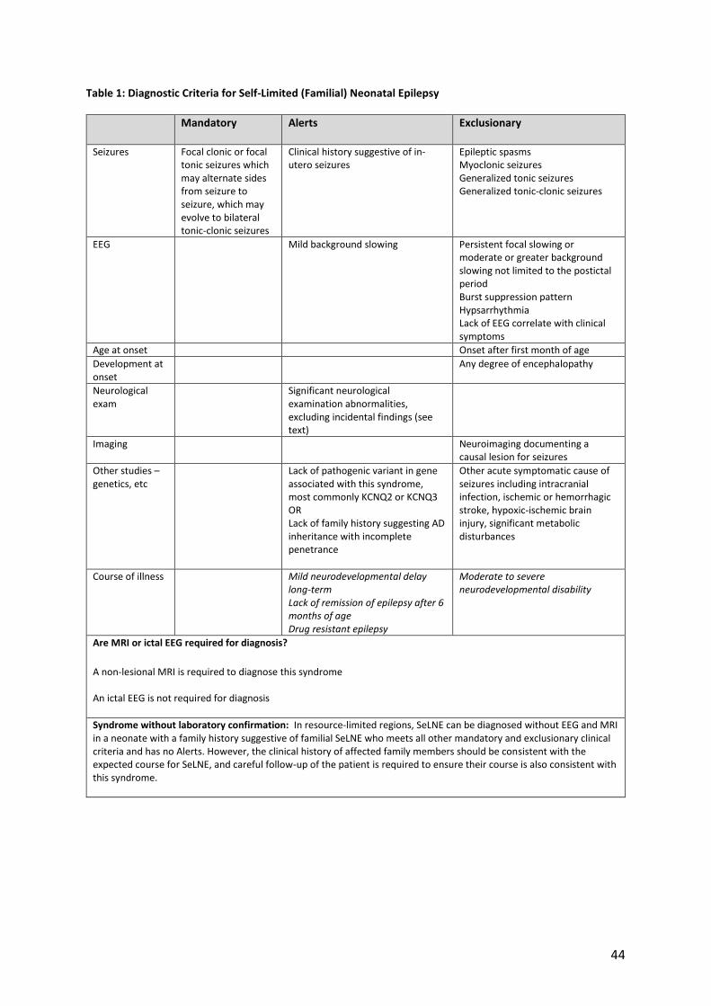
EEG Mild background slowing Persistent focal slowing or moderate or greater background slowing not limited to the postictal period Burst suppression pattern Hypsarrhythmia Lack of EEG correlate with clinical symptoms
Age at onset Onset after first month of age
Development at onset
Any degree of encephalopathy
Neurological exam
Significant neurological examination abnormalities, excluding incidental findings (see text)
Imaging Neuroimaging documenting a causal lesion for seizures
Other studies – genetics, etc
Lack of pathogenic variant in gene associated with this syndrome, most commonly KCNQ2 or KCNQ3 OR Lack of family history suggesting AD inheritance with incomplete penetrance
Other acute symptomatic cause of seizures including intracranial infection, ischemic or hemorrhagic stroke, hypoxic-ischemic brain injury, significant metabolic disturbances
Course of illness Mild neurodevelopmental delay long-term Lack of remission of epilepsy after 6 months of age Drug resistant epilepsy
Moderate to severe neurodevelopmental disability
Are MRI or ictal EEG required for diagnosis?
A non-lesional MRI is required to diagnose this syndrome An ictal EEG is not required for diagnosis
Syndrome without laboratory confirmation: In resource-limited regions, SeLNE can be diagnosed without EEG and MRI in a neonate with a family history suggestive of familial SeLNE who meets all other mandatory and exclusionary clinical criteria and has no Alerts. However, the clinical history of affected family members should be consistent with the expected course for SeLNE, and careful follow-up of the patient is required to ensure their course is also consistent with this syndrome.
45
Table 2: Diagnostic Criteria for Self-Limited (Familial) Neonatal-Infantile Epilepsy
Mandatory Alerts Exclusionary
Seizures Focal clonic or focal tonic seizures which may alternate sides from seizure to seizure, and may evolve to bilateral tonic-clonic seizures
EEG Mild background slowing Persistent focal slowing or moderate or greater background slowing not limited to the postictal period Burst suppression pattern Hypsarrhythmia Lack of EEG correlate with clinical symptoms
Age at onset 1 day to 23 months
Development at onset
Encephalopathy
Neurological exam
Significant neurological examination abnormalities, excluding incidental findings (see text)
Imaging Neuroimaging documenting a causal lesion for seizures
Other studies – genetics, etc
A history of prior acute symptomatic seizures including intracranial infection, ischemic or hemorrhagic stroke, hypoxic-ischemic brain injury, significant metabolic disturbances Lack of pathogenic variant in genes associated with this syndrome (usually SCN2A)
Course of illness Mild neurodevelopmental delay long-term Lack of remission of epilepsy by age 2 years Drug resistant epilepsy
Moderate to severe neurodevelopmental disability
Are MRI or ictal EEG required for diagnosis? A non-lesional MRI is required to diagnose this syndrome. An ictal EEG is not required for diagnosis.
Syndrome without laboratory confirmation: In resource-limited regions, Self-limited neonatal-infantile epilepsy can be diagnosed without EEG and MRI in a neonate with a family history suggestive of familial self-limited neonatal-infantile epilepsy who meets all other mandatory and exclusionary clinical criteria and has no Alerts. However, the clinical history of affected family members should be consistent with the expected course for SeLNIE, and careful follow-up of the patient is required to ensure their course is also consistent with this syndrome.
46
Table 3: Diagnostic Criteria for Self-Limited (Familial) Infantile Epilepsy
Mandatory Alerts Exclusionary
Seizures Focal seizures occur with behavioural arrest, impaired awareness, automatisms, head/eye version, and clonic movements (often alternating from one side to the other and progressing to a hemiclonic or focal to bilateral tonic-clonic seizure). Seizures are usually brief (<3 minutes).
Prolonged or focal hemiclonic seizures (>10 minutes)
Lack of pathogenic variants found in PRRT2, SCN2A, KCNQ2 or KCNQ3 OR Lack of family history suggesting AD inheritance with incomplete penetrance
Course of illness Lack of remission by late childhood
Neurocognitive regression with myoclonic seizures, ataxia, spasticity
Are MRI or ictal EEG required for diagnosis? A non-lesional MRI is required to diagnose this syndrome. An ictal EEG is not required for diagnosis.
Syndrome without laboratory confirmation: In resource-limited regions, SeLIE can be diagnosed without EEG and MRI in an infant with a family history suggestive of familial SeLIE who meets all other mandatory and exclusionary clinical criteria and has no Alerts. However, the clinical history of affected family members should be consistent with the expected course for SeLIE, and careful follow-up of the patient is required to ensure their course is also consistent with this syndrome.
47
Table 4: Diagnostic Criteria for Febrile Seizures Plus
Mandatory Alerts Exclusionary
Seizures Febrile seizures persisting after 6 years of age and/or afebrile seizures
Absence of familial history of GEFS+ (although in some cases, obtaining an accurate family history may be challenging)
Seizures due to other acute causes such as infection, metabolic disturbances
Course of illness Drug resistant seizures Lack of remission by puberty
Cognitive Regression
Are MRI or ictal EEG required for diagnosis? An MRI is not required for diagnosis. An ictal EEG is not required for diagnosis.
Syndrome without laboratory confirmation: In resource-limited regions, Febrile seizures Plus can be diagnosed without EEG and MRI provided the patient meets all other mandatory and exclusionary clinical criteria and has no Alerts.
48
Table 5: Diagnostic Criteria for Myoclonic Epilepsy in Infancy
Mandatory Alerts Exclusionary
Seizures Myoclonic seizures (see text)
Afebrile generalized tonic-clonic seizure or generalized clonic at time of epilepsy onset
Any of the following seizure types:
• Absence seizures
• Atonic seizures
• Epileptic spasms
• Focal impaired awareness seizures
• Hemiconvulsive seizures
• Myoclonic-absence seizures
• Tonic Seizures
EEG Normal background
PPR at low frequency photic stimulation (suggest CLN2 disease) Lack of generalized spike-wave discharge on sleep recording
Recorded myoclonic event without EEG correlate Hypsarrhythmia Generalized slow spike-wave (<2.5 Hz)
Age at onset Age at onset of myoclonic seizures <4 months or >3 years
Development at onset
Speech delay at time of diagnosis Moderate to profound ID
Neurological exam
Significant neurological examination abnormalities, excluding incidental findings (see text)
Dysmorphism or other congenital anomalies (suggests chromosomal disorder)
Imaging Significant neuroimaging abnormalities
Other studies – genetics, etc
Low CSF glucose or pathogenic SLC2A1 variants
Course of illness Neurocognitive regression
Are MRI or ictal EEG required for diagnosis? A non-lesional MRI is required for diagnosis. An ictal EEG is not required for diagnosis but should be strongly considered if the interictal sleep recording does not show generalized spike-wave to confirm that myoclonus is epileptic.
Syndrome without laboratory confirmation: In resource-limited regions, at a minimum, a sleep EEG showing generalized spike-wave is required to make this diagnosis.
49
Table 6: Diagnostic Criteria for Early Infantile Developmental and Epileptic Encephalopathy
Mandatory Alerts Exclusionary
Seizures Tonic and/or myoclonic seizures
EEG Either burst suppression or multifocal discharges
Diffuse slowing
Age at onset Birth to 3 months (adjusted for prematurity)
Development at onset
Normal development at onset, although it is acknowledged that this can be challenging to accurately assess historically
Neurological exam at onset
Normal neurological examination, although it is acknowledged that this can be challenging to assess historically or in an infant who has had very frequent seizures and/or received ASMs that may alter their exam.
Early Comorbidities
Developmental impairment is present prior to or shortly after seizure onset
Course of illness Abnormal neurodevelopment including intellectual disability (see text)
Are MRI or ictal EEG required for diagnosis? An MRI is not required for diagnosis but is strongly recommended to exclude structural causes. An ictal EEG is not required in an infant with characteristic clinical features where the interictal EEG shows burst-suppression, multi-focal discharges with diffuse slowing.
Syndrome without laboratory confirmation: In resource-limited regions, this syndrome cannot be diagnosed without an interictal EEG.
50
Table 7: Diagnostic Criteria for Epilepsy of Infancy with Migrating Focal Seizures
Mandatory Alerts Exclusionary
Seizures Focal/multifocal tonic or clonic seizures, with or without subtle behavioral arrest and prominent autonomic features Seizures migrate from one hemisphere or lobe to another clinically. Seizure frequency rapidly increases in the first weeks and months, often progressing to status epilepticus
Myoclonic seizures
EEG Ictal recording shows a migrating pattern (this might be missed if a prolonged video EEG is not performed)
Multifocal discharges
Suppression burst pattern prior to medication
Single persistent epileptic focus on EEG
Hypsarrhythmia
Age at onset <12 months Onset 6-12 months
Development at onset
Severe delay prior to seizure onset
Neurological exam
Significant abnormalities on neurological examination prior to seizure onset
Comorbidities Developmental plateauing or regression with frequent seizures
Imaging Abnormal neuroimaging with structural causal lesion
Course of illness
Neurodevelopmental delay
Seizure freedom
Lack of brain atrophy on MRI
Are MRI or ictal EEG required for diagnosis? An MRI is required for diagnosis to exclude a causal structural etiology. An ictal EEG may not be required if clinical migration is observed. However, an ictal EEG is strongly recommended to document a migrating pattern.
Syndrome without laboratory confirmation: In resource-limited regions, EIMFS can be diagnosed on clinical observation of seizure migration without EEG or MRI, provided all other clinical mandatory and exclusionary criteria are met.
51
Table 8: Diagnostic Criteria for Epilepsy of Infantile Spasms Syndrome
Mandatory Alerts Exclusionary
Seizures Clusters of flexor, extensor or mixed epileptic spasms
EEG Either hypsarrhythmia, multifocal or focal epileptiform discharges
Normal interictal EEG Suppression-burst pattern on EEG
Normal EEG during recorded clinical events of suspected spasms
Age at onset 1-24 months (while epileptic spasms may begin later, this would not be ISS)
Age at onset 1-2 months
Comorbidities Developmental slowing after spasm onset
Are MRI or ictal EEG required for diagnosis? An MRI is not required for diagnosis but is highly recommended to evaluate for underlying cause. An ictal EEG is not required for diagnosis provided the interictal study shows hypsarrhythmia. In the absence of hysparrhythmia, an ictal recording is required.
Possible evolving syndrome: Infants with preceding brain injury, developmental brain malformations or specific genetic conditions, including early-infantile DEE, who show significant interictal EEG abnormalities (high amplitude, background slowing and/or multifocal discharges) should be carefully watched for the development of clinical epileptic spasms. However, the syndrome of ISS cannot be diagnosed prior to onset of the mandatory seizure type.
Syndrome without laboratory confirmation: In resource-limited regions, an interictal EEG is highly recommended. However, if EEG is unavailable, if clear clusters of typical epileptic spasms are witnessed by an experienced clinician (in person or on video recording), with the other clinical mandatory and exclusionary criteria, ISS can be diagnosed.
52
Table 9: Diagnostic Criteria for Dravet Syndrome
Mandatory Alerts Exclusionary
Seizures Recurrent hemiclonic seizures (which often alternate sides from seizure to seizure), focal to bilateral tonic-clonic and/or generalized tonic clonic seizures
No history of prolonged seizures (>10 minutes)
Lack of fever sensitivity as a seizure trigger
Epileptic spasms Early infantile SCN1A DEE
EEG Normal EEG background without interictal discharges after age 2 years
Age at onset 1-20 months 1-2 months or 16-20 months
Development at onset
Developmental delay at seizure onset
Neurological exam Focal neurological findings (other than Todds paresis)
Imaging MRI showing a causal focal lesion
Other testing: ie genetics etc
Lack of pathogenic SCN1A or other causal variant
Course of illness Drug resistant epilepsy Intellectual disability
Good efficacy with prophylactic sodium-channel agents including carbamazepine, oxcarbazepine and phenytoin
Are MRI or ictal EEG required for diagnosis?
An MRI is not required for diagnosis but is highly recommended to exclude other causes. An ictal EEG is not required for diagnosis.
Possible evolving syndrome: In a child <12 months who presents with a prolonged hemiclonic or bilateral tonic-clonic seizure with fever, and no other underlying cause, the possibility of Dravet syndrome should be considered. Further convulsive seizures (often with fever, and if prolonged or hemiclonic) would allow more definitive diagnosis of Dravet syndrome. A diagnosis would be further supported by the finding of a pathogenic SCN1A variant.
Syndrome without laboratory confirmation: In resource-limited regions, Dravet syndrome can be diagnosed in children without Alerts who meet all other clinical mandatory and exclusionary criteria, without EEG, MRI and genetic testing.
53
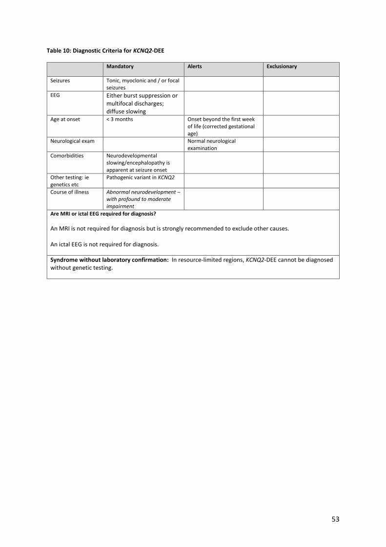
Table 10: Diagnostic Criteria for KCNQ2-DEE
Mandatory Alerts Exclusionary
Seizures Tonic, myoclonic and / or focal seizures
EEG Either burst suppression or multifocal discharges; diffuse slowing
Age at onset < 3 months Onset beyond the first week of life (corrected gestational age)
Neurological exam Normal neurological examination
Comorbidities Neurodevelopmental slowing/encephalopathy is apparent at seizure onset
Other testing: ie genetics etc
Pathogenic variant in KCNQ2
Course of illness Abnormal neurodevelopment – with profound to moderate impairment
Are MRI or ictal EEG required for diagnosis?
An MRI is not required for diagnosis but is strongly recommended to exclude other causes. An ictal EEG is not required for diagnosis.
Syndrome without laboratory confirmation: In resource-limited regions, KCNQ2-DEE cannot be diagnosed without genetic testing.
54
Table 11: Diagnostic Criteria for Early-onset Vitamin-dependent (Pyridoxine/ Pyridox(am)ine 5’-Phosphate) DEE
Mandatory Alerts Exclusionary
Seizures Variable seizure types which may include:
• Focal/multifocal seizures
• Epileptic spasms
• Generalized tonic seizures
• Generalized clonic seizures Seizures are drug-resistant and frequent (often evolving to status epilepticus) but rapidly respond to pyridoxine (pyridoxine-dependent-DEE) or pyridoxal-5-phosphate
(Pyridox(am)ine 5’-Phosphate-DEE)
supplementation
EEG Abnormal with slowing and focal/multifocal discharges or burst suppression pattern
Age at onset Age >3 years at onset (there are rare, later-onset forms of pyridoxine-dependent epilepsy)
Neurological exam Lack of encephalopathy and irritability
Other testing: ie genetics etc
Laboratory testing providing confirmatory evidence, which may include either: 1. Metabolic features: Increased α-aminoadipic semialdehyde and/or pipecolic acid in urine, plasma and/or CSF (pyridoxine-dependent-DEE) or low pyridoxal-5-phosphate in CSF (Pyridox(am)ine 5’-Phosphate-DEE) OR 2. Genetic features: pathogenic variants in ALDH7A1 or PLBP (pyridoxine dependent-DEE) or PNPO gene (Pyridox(am)ine 5’-Phosphate-DEE)
Course of illness Seizures that show sustained marked reduction or cessation with lifelong pyridoxine or pyridoxal-5-phosphate.
Normal neurodevelopmental outcome
Are MRI or ictal EEG required for diagnosis?
An MRI is not required for diagnosis but is strongly recommended to exclude other causes. An ictal EEG is not required for diagnosis.
Syndrome without laboratory confirmation: In resource-limited regions, Pyridoxine or Pyridox(am)ine 5’-Phosphate-DEE can be diagnosed in children without Alerts who meet all other mandatory and exclusionary clinical criteria and whose seizures cease with pyridoxine or P5P supplementation, recur when supplementation is stopped, and cease again with re-introduction of supplementation.
55
Table 12: Diagnostic Criteria for CDKL5-DEE
Mandatory Alerts Exclusionary
Seizures Seizures which may include tonic seizures, epileptic spasms, generalized tonic-clonic seizures and/or focal seizures. Hypermotor-tonic-spasms sequence seizures are characteristic but not seen in all cases
Absence of epileptic spasms in the first year of life
EEG Normal EEG background without interictal discharges after 4 months of age
Age at onset Onset of epilepsy >3 months
Development at onset
Normal development prior to seizure onset
Neurological exam Normal tone Lack of encephalopathy
Other testing: ie genetics etc
Pathogenic variant in the CDKL5 gene (X-linked but females outnumber males by 4:1)
Course of illness Profound to severe intellectual disability Drug resistant epilepsy
Are MRI or ictal EEG required for diagnosis?
An MRI is not required for diagnosis but is strongly recommended to exclude other causes. An ictal EEG is not required for diagnosis.
Syndrome without laboratory confirmation: In resource-limited regions, CDKL5-DEE cannot be diagnosed without confirmatory genetic testing.
56
Table 13: Diagnostic Criteria for PCDH19 Clustering Epilepsy
Mandatory Alerts Exclusionary
Seizures Focal seizures (fearful screaming typical) and tonic-clonic seizures, typically in clusters; may be triggered by fever
Prolonged hemiclonic seizures in infancy (consider Dravet) No clustering
EEG Absence of epileptiform discharge (which is usually focal, but rarely may be generalized)
Age at seizure onset 1.5-60 months in females; 5-96 months in males
Other testing: ie genetics etc
PCDH19 pathogenic variant: see text regarding unusual inheritance pattern
Are MRI or ictal EEG required for diagnosis? An MRI is not required for diagnosis but is strongly recommended to exclude other causes. An ictal EEG is not required for diagnosis.
Possible evolving syndrome: This syndrome should be considered in an infant girl who presents with a first cluster of febrile seizures.
Syndrome without laboratory confirmation: In resource-limited regions, PCDH19 clustering epilepsy could be a provisionally diagnosed without confirmatory genetic testing, specifically in the setting of a family history suggestive of X-linked dominant inheritance with male sparing.
57
Table 14: Diagnostic Criteria for GLUT1DS-DEE
Mandatory Alerts Exclusionary
Seizures Seizures which may be focal or generalized, including absence seizures (often beginning before 3 years of age)
Neurological exam Focal neurological findings (other than Todds paresis)
Other testing: ie genetics etc
Pathogenic SLC2A1 variant OR Low fasting CSF glucose and CSF/plasma glucose ratio*
Other documented etiology for hypoglycorrhachia
Course of illness Intellectual disability
Seizures that are controlled with medication Lack of improvement in seizures with ketogenic diet Lack of movement disorders such as ataxia, paroxysmal exercise-induced dyskinesia, dystonia
Are MRI or ictal EEG required for diagnosis? An MRI is not required for diagnosis but is strongly recommended to exclude other causes. An ictal EEG is not required for diagnosis.
Syndrome without laboratory confirmation: In resource-limited regions, GLUT1DS-DEE can be diagnosed without EEG, MRI or genetic studies in children without Alerts who meet all other mandatory and exclusionary clinical criteria. CSF studies are required for diagnosis.
*CSF glucose may not be as low in later-onset epilepsies associated with GLUT1 deficiency
58
Table 15: Diagnostic Criteria for Sturge Weber Syndrome
Mandatory Alerts Exclusionary
Seizures Focal motor or autonomic seizures with or without impaired awareness, which may evolve to bilateral tonic-clonic seizures
EEG Lack of asymmetrical background with reduction in voltage and slowing over the affected hemisphere
Neurological exam Lack of facial capillary hemangioma affecting the V1 dermatome
Imaging MRI showing leptomeningeal enhancement suggestive of leptomeningeal angioma, with cortical calcification and focal cerebral atrophy developing with time
Course of illness Lack of abnormal neurological examination – may be limited to visual field deficit Lack of intellectual disability ranging from mild to profound
Are MRI or ictal EEG required for diagnosis? An MRI is required for diagnosis. Changes may be very subtle or absent on MRI done prior to 2 months of age. An ictal EEG is not required for diagnosis.
Syndrome without laboratory confirmation: In resource-limited regions, Sturge Weber syndrome can be presumptively diagnosed without EEG or MRI in persons without Alerts who meet all other mandatory clinical criteria.
59
Table 16: Diagnostic Criteria for Gelastic Seizures with Hypothalamic Hamartoma
Mandatory Alerts Exclusionary
Seizures Gelastic seizures with mechanical, mirthless laughter, inappropriate to context
Seizure frequency less than daily
EEG Generalized or focal background slowing (excluding immediate postictal period) Gelastic seizures may lack ictal EEG correlate
Age at onset Onset >5 years of age
Development at onset Clear developmental delay at seizure onset
Neurological exam Focal neurological findings (other than Todds paresis) or generalized hypotonia
Imaging Hypothalamic hamartoma (may require thin slices through the hypothalamic region to confirm)
Course of illness Drug resistant epilepsy
Lack of behavioral problems including aggression, impulsivity and hyperactivity
Are MRI or ictal EEG required for diagnosis? An MRI is required for diagnosis. An ictal EEG is not required for diagnosis. Furthermore, gelastic seizures may lack ictal correlate on EEG.
Syndrome without laboratory confirmation: In resource-limited regions, HH-GS cannot be diagnosed in the absence of an MRI, as gelastic seizures may arise from other brain regions.
60
Figure Legends:
Figure 1: Organization of Epilepsy Syndromes that Begin in the Neonates and Infants
Syndromes are broadly divided into Self-Limited Epilepsies (where there is likely to be spontaneous remission)
and Developmental and Epileptic Encephalopathies (disorders where there is developmental impairment related
to both the underlying aetiology independent of epileptiform activity and the epileptic encephalopathy).
Etiology-specific epilepsy syndromes are due to specific genetic, structural, metabolic, immune or infectious
etiologies, and have consistent electroclinical features, management and prognostic implications. Most etiology-
specific syndromes that begin in the neonatal or infantile period are DEEs.
61
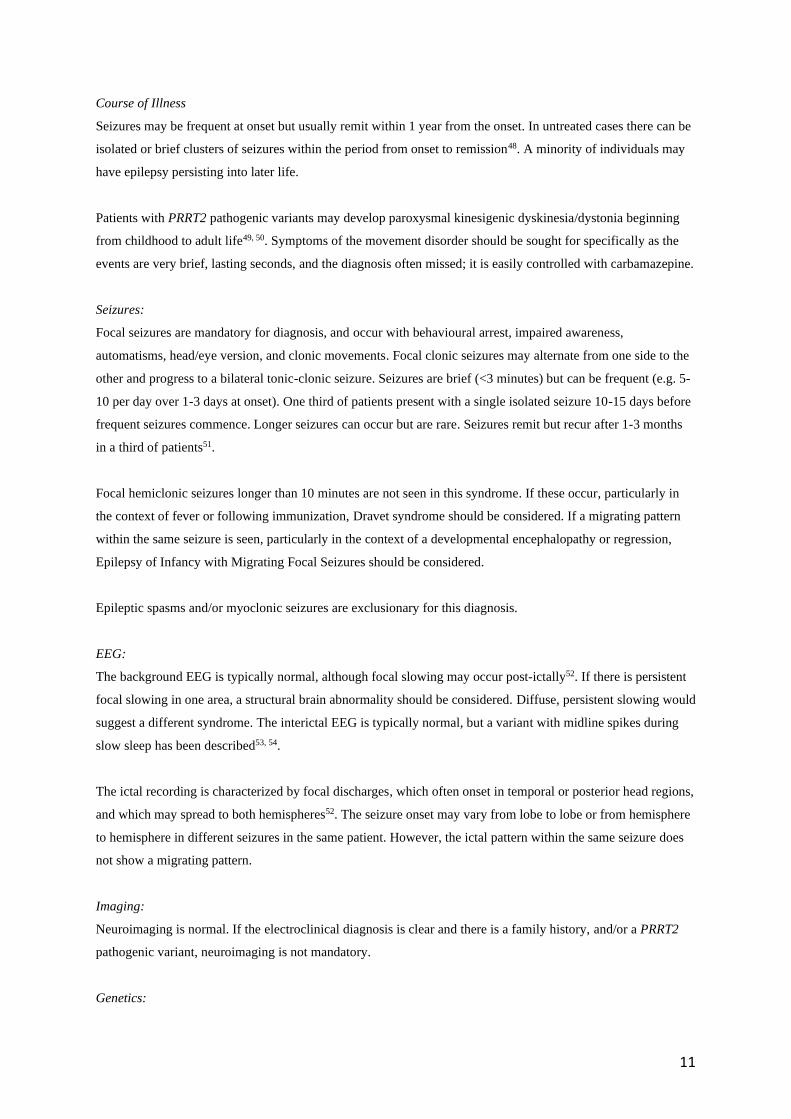
Figure 2: The ” theta pointu alternant ” interictal pattern on EEG. This pattern is characterized by runs of non-
reactive theta activity, that may be intermixed with sharp waves, and frequently shows inter-hemispheric
asynchrony.
62
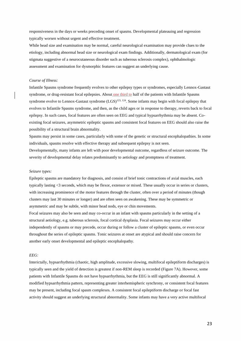
Figure 3: Seizure recorded in a three-day-old neonate with Self-limited Familial Neonatal Epilepsy, born at full-
term. Seizures begin with a tonic and/or apneic phase concomitant with diffuse bilateral, but asymmetrical
flattening of the background activity and polygraphic EMG recording from both deltoids showing tonic
contraction of both upper limbs (tonic and/or apneic phase). This is followed by left frontal rhythmic, high
amplitude slow waves, which evolve to sharp waves and spread to left temporal and central regions, and then to
the right hemisphere, with eventual bilateral clonic movements of the limbs.
63
Figure 4: 14 month old boy, who presented with myoclonic seizures. His development was normal for age and
he was diagnosed with Myoclonic Epilepsy in Infancy. The EEG shows generalized spike wave discharge, with
a clinical myoclonic jerk seen in the EMG lead.
64
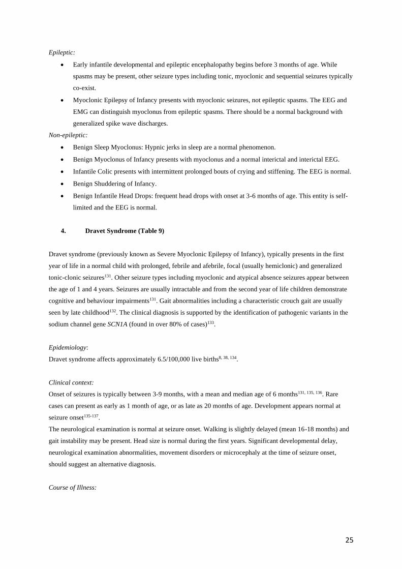
Figure 5: 4 week old boy with EIDEE. He presented on day 2 of life with sequential seizures with a prominent
tonic component and severe encephalopathy. The EEG (20 microvolt/mm, 30 mm/sec) shows a burst-
suppression pattern. Genetic testing showed a KCNQ2 pathogenic variant. The patient showed a marked
reduction in seizures with carbamazepine but remained profoundly delayed.
65
Figure 6: EEG recording of a 5-month girl with Epilepsy in Infancy with Migrating Focal Seizures related
to SCN8A pathogenic variant. The EEG shows a prolonged seizure of 7 minutes starting in the left temporal
area (green frame) and progressively becoming bilateral (pink frame) then migrates to the right hemisphere
involving mainly the right temporal area (blue frame) and ending on the right hemisphere (orange frame).
66
Figure 7A and B: A 7 month old boy presented with a 1 month history of flexor spasms. He had shown normal
developmental progress until onset of spasms but then had decreased visual interest. His interictal EEG (Figure
7A) shows a hypsarrhythmia pattern. The ictal recording (Figure 7B) shows a high amplitude sharp wave
followed by a relative decrement.
Figure 7A
Figure 7B
67
Figure 8: Differentiation of Spasm from Myoclonic and Tonic Seizure (from Fusco L and Vigevano F, Epilepsia
1993)125 . Both EMG and EEG channels are shown. A: Myoclonic jerk, B: Tonic Seizure, C: Spasms. Note the
EMG correlate of a spasm appears as a rhombus, and the EEG correlate as a slow wave, with an inverse phase
reversal of the vertex region.
68
Figure 9: GLUT1 DEE
9a EEG in a 9 year old child with Glut1DS prior to ketogenic diet showing moderate to high amplitude slow
background with mixed theta and delta frequencies.
9b EEG in same child as in 9a after 3 months on the ketogenic diet showing much faster background rhythms. A
similar change can be seen with food taken immediately prior to or during the EEG.
69
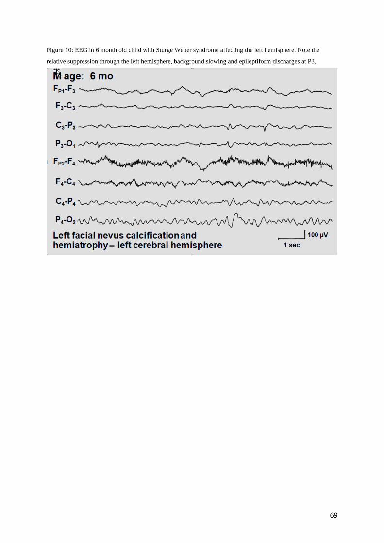
Figure 10: EEG in 6 month old child with Sturge Weber syndrome affecting the left hemisphere. Note the
relative suppression through the left hemisphere, background slowing and epileptiform discharges at P3.
70
1. Scheffer IE, Berkovic S, Capovilla G, Connolly MB, French J, Guilhoto L, et al. ILAE classification of the epilepsies: Position paper of the ILAE Commission for Classification and Terminology. Epilepsia 2017;58:512-521. 2. Proposal for classification of epilepsies and epileptic syndromes. Commission on Classification and Terminology of the International League Against Epilepsy. Epilepsia 1985;26:268-278. 3. Wirrell E, Nabbout R, Scheffer IE, Alsaadi T, Bogacz A, French JA, et al. Methodology for Classification and Definition of Epilepsy Syndromes: Report of the ILAE Task Force on Nosology and Definitions. Epilepsia, in press. 2021. 4. Hauser WA, Annegers JF, Kurland LT. Incidence of epilepsy and unprovoked seizures in Rochester, Minnesota: 1935-1984. Epilepsia 1993;34:453-468. 5. Wirrell EC, Grossardt BR, Wong-Kisiel LC, Nickels KC. Incidence and classification of new-onset epilepsy and epilepsy syndromes in children in Olmsted County, Minnesota from 1980 to 2004: a population-based study. Epilepsy Res 2011;95:110-118. 6. Eltze CM, Chong WK, Cox T, Whitney A, Cortina-Borja M, Chin RF, et al. A population-based study of newly diagnosed epilepsy in infants. Epilepsia 2013;54:437-445. 7. Camfield CS, Camfield PR, Gordon K, Wirrell E, Dooley JM. Incidence of epilepsy in childhood and adolescence: a population-based study in Nova Scotia from 1977 to 1985. Epilepsia 1996;37:19-23. 8. Symonds JD, Elliot K, Shetty J. Amrstrong M., Brunklaus A. et al. The early childhood epilepsies: epidemiology, classification, aetiology, genomics, socio-economic determinants and outcomes. Brain, in press. 2021. 9. Senanayake N, Roman GC. Epidemiology of epilepsy in developing countries. Bull World Health Organ 1993;71:247-258. 10. Singh A, Trevick S. The Epidemiology of Global Epilepsy. Neurol Clin 2016;34:837-847. 11. Newton CR, Garcia HH. Epilepsy in poor regions of the world. Lancet 2012;380:1193-1201. 12. Berg AT, Langfitt JT, Testa FM, Levy SR, DiMario F, Westerveld M, et al. Global cognitive function in children with epilepsy: a community-based study. Epilepsia 2008;49:608-614. 13. Wirrell E, Wong-Kisiel L, Mandrekar J, Nickels K. Predictors and course of medically intractable epilepsy in young children presenting before 36 months of age: a retrospective, population-based study. Epilepsia 2012;53:1563-1569. 14. Moseley BD, Wirrell EC, Wong-Kisiel LC, Nickels K. Early onset epilepsy is associated with increased mortality: a population-based study. Epilepsy Res 2013;105:410-414. 15. Berg AT, Zelko FA, Levy SR, Testa FM. Age at onset of epilepsy, pharmacoresistance, and cognitive outcomes: a prospective cohort study. Neurology 2012;79:1384-1391. 16. Wilson SJ, Micallef S, Henderson A, Rayner G, Wrennall JA, Spooner C, et al. Developmental outcomes of childhood-onset temporal lobe epilepsy: a community-based study. Epilepsia 2012;53:1587-1596. 17. Bahi-Buisson N, Nectoux J, Rosas-Vargas H, Milh M, Boddaert N, Girard B, et al. Key clinical features to identify girls with CDKL5 mutations. Brain 2008;131:2647-2661. 18. Tarquinio DC, Hou W, Berg A, Kaufmann WE, Lane JB, Skinner SA, et al. Longitudinal course of epilepsy in Rett syndrome and related disorders. Brain 2017;140:306-318. 19. Rett A, Teubel R. Neugeborenenkraempfe im Rahmen einer epileptisch belasteten Familie. Wien Klin Wschr 1964;76:609-613. 20. Trivisano M, Pietrafusa N, Terracciano A, Marini C, Mei D, Darra F, et al. Defining the electroclinical phenotype and outcome of PCDH19-related epilepsy: A multicenter study. Epilepsia 2018;59:2260-2271.
71
21. Kolc KL, Sadleir LG, Scheffer IE, Ivancevic A, Roberts R, Pham DH, et al. A systematic review and meta-analysis of 271 PCDH19-variant individuals identifies psychiatric comorbidities, and association of seizure onset and disease severity. Mol Psychiatry 2019;24:241-251. 22. Scheffer IE, Turner SJ, Dibbens LM, Bayly MA, Friend K, Hodgson B, et al. Epilepsy and mental retardation limited to females: an under-recognized disorder. Brain 2008;131:918-927. 23. Stamberger H, Nikanorova M, Willemsen MH, Accorsi P, Angriman M, Baier H, et al. STXBP1 encephalopathy: A neurodevelopmental disorder including epilepsy. Neurology 2016;86:954-962. 24. Matricardi S, Darra F, Spalice A, Basti C, Fontana E, Dalla Bernardina B, et al. Electroclinical findings and long-term outcomes in epileptic patients with inv dup (15). Acta Neurol Scand 2018;137:575-581. 25. Howell KB, Freeman JL, Mackay MT, Fahey MC, Archer J, Berkovic SF, et al. The severe epilepsy syndromes of infancy: A population-based study. Epilepsia 2021;62:358-370. 26. Berg AT, Berkovic SF, Brodie MJ, Buchhalter J, Cross JH, van Emde Boas W, et al. Revised terminology and concepts for organization of seizures and epilepsies: report of the ILAE Commission on Classification and Terminology, 2005-2009. Epilepsia 2010;51:676-685. 27. Ronen GM, Rosales TO, Connolly M, Anderson VE, Leppert M. Seizure characteristics in chromosome 20 benign familial neonatal convulsions. Neurology 1993;43:1355-1360. 28. Grinton BE, Heron SE, Pelekanos JT, Zuberi SM, Kivity S, Afawi Z, et al. Familial neonatal seizures in 36 families: Clinical and genetic features correlate with outcome. Epilepsia 2015;56:1071-1080. 29. Shevell MI, Sinclair DB, Metrakos K. Benign familial neonatal seizures: clinical and electroencephalographic characteristics. Pediatr Neurol 1986;2:272-275. 30. Maihara T, Tsuji M, Higuchi Y, Hattori H. Benign familial neonatal convulsions followed by benign epilepsy with centrotemporal spikes in two siblings. Epilepsia 1999;40:110-113. 31. Dedek K, Kunath B, Kananura C, Reuner U, Jentsch TJ, Steinlein OK. Myokymia and neonatal epilepsy caused by a mutation in the voltage sensor of the KCNQ2 K+ channel. Proc Natl Acad Sci USA 2001;98:12272-12277. 32. Sands TT, Balestri M, Bellini G, Mulkey SB, Danhaive O, Bakken EH, et al. Rapid and safe response to low-dose carbamazepine in neonatal epilepsy. Epilepsia 2016;57:2019-2030. 33. Dehan M, Quilleron D, Navelet Y, D'Allest AM, Vial M, Retbi JM, et al. Les convulsions du cinquieme jour de vie: un nouveau syndrome? Arch Fr Ped 1977;34:730-742. 34. Hirsch E, Velez A, Sellal F, Maton B, Grinspan A, Malafosse A, et al. Electroclinical signs of benign neonatal familial convulsions. Ann Neurol 1993;34:835-841. 35. Biervert C, Schroeder BC, Kubisch C, Berkovic SF, Propping P, Jentsch TJ, et al. A potassium channel mutation in neonatal human epilepsy. Science 1998;279:403-406. 36. Charlier C, Singh NA, Ryan SG, Lewis TB, Reus BE, Leach RJ, et al. A pore mutation in a novel KQT-like potassium channel gene in an idiopathic epilepsy family. Nat Genet 1998;18:53-55. 37. Singh NA, Charlier C, Stauffer D, DuPont BR, Leach RJ, Melis R, et al. A novel potassium channel gene, KCNQ2, is mutated in an inherited epilepsy of newborns. Nat Genet 1998;18:25-29. 38. Symonds JD, Zuberi SM, Stewart K, McLellan A, O'Regan M, MacLeod S, et al. Incidence and phenotypes of childhood-onset genetic epilepsies: a prospective population-based national cohort. Brain 2019;142:2303-2318. 39. Heron SE, Cox K, Grinton BE, Zuberi SM, Kivity S, Afawi Z, et al. Deletions or duplications in KCNQ2 can cause benign familial neonatal seizures. J Med Genet 2007;44:791-796. 40. Kaplan RE, Lacey DJ. Benign familial neonatal-infantile seizures. Am J Med Genet 1983;16:595-599. 41. Heron SE, Crossland KM, Andermann E, Phillips HA, Hall AJ, Bleasel A, et al. Sodium-channel defects in benign familial neonatal-infantile seizures. Lancet 2002;360:851-852. 42. Zara F, Specchio N, Striano P, Robbiano A, Gennaro E, Paravidino R, et al. Genetic testing in benign familial epilepsies of the first year of life: clinical and diagnostic significance. Epilepsia 2013;54:425-436.
72
43. Berkovic SF, Heron SE, Giordano L, Marini C, Guerrini R, Kaplan RE, et al. Benign familial neonatal-infantile seizures: characterization of a new sodium channelopathy. Ann Neurol 2004;55:550-557. 44. Herlenius E, Heron SE, Grinton BE, Keay D, Scheffer IE, Mulley JC, et al. SCN2A mutations and benign familial neonatal-infantile seizures: the phenotypic spectrum. Epilepsia 2007;48:1138-1142. 45. Vigevano F, Fusco L, Di Capua M, Ricci S, Sebastianelli R, Lucchini P. Benign infantile familial convulsions. Eur J Pediatr 1992;151:608-612. 46. Szepetowski P, Rochette J, Berquin P, Piussan C, Lathrop GM, Monaco AP. Familial infantile convulsions and paroxysmal choreoathetosis: a new neurological syndrome linked to the pericentromeric region of human chromosome 16. Am J Hum Genet 1997;61:889-898. 47. Ramos-Lizana J, Martinez-Espinosa G, Rodriguez-Lucenilla MI, Aguirre-Rodriguez J, Aguilera-Lopez P. [Frequency, semiology and prognosis of benign infantile epilepsy]. Rev Neurol 2018 16;66:254-260. 48. Okumura A, Hayakawa F, Kuno K, Watanabe K. Benign partial epilepsy in infancy. Arch Dis Child 1996;74:19-21. 49. Cloarec R, Bruneau N, Rudolf G, Massacrier A, Salmi M, Bataillard M, et al. PRRT2 links infantile convulsions and paroxysmal dyskinesia with migraine. Neurology 2012;79:2097-2103. 50. Heron SE, Dibbens LM. Role of PRRT2 in common paroxysmal neurological disorders: a gene with remarkable pleiotropy. J Med Genet 2013;50:133-139. 51. Caraballo RH, Cersosimo RO, Espeche A, Fejerman N. Benign familial and non-familial infantile seizures: a study of 64 patients. Epileptic Disord 2003;5:45-49. 52. Vigevano F. Benign familial infantile seizures. Brain Dev 2005;27:172-177. 53. Bureau M, Cokar O, Maton B, Genton P, Dravet C. Sleep-related, low voltage Rolandic and vertex spikes: an EEG marker of benignity in infancy-onset focal epilepsies. Epileptic Disord 2002;4:15-22. 54. Flesler S, Sakr D, Cersosimo R, Caraballo R. Benign infantile focal epilepsy with midline spikes and waves during sleep: a new epileptic syndrome or a variant of benign focal epilepsy? Epileptic Disord 2010;12:205-211. 55. Gardella E, Becker F, Moller RS, Schubert J, Lemke JR, Larsen LH, et al. Benign infantile seizures and paroxysmal dyskinesia caused by an SCN8A mutation. Ann Neurol 2016;79:428-436. 56. Singh R, Scheffer IE, Crossland K, Berkovic SF. Generalized epilepsy with febrile seizures plus: a common childhood-onset genetic epilepsy syndrome. Ann Neurol 1999;45:75-81. 57. Singh R, Andermann E, Whitehouse WP, Harvey AS, Keene DL, Seni MH, et al. Severe myoclonic epilepsy of infancy: extended spectrum of GEFS+? Epilepsia 2001;42:837-844. 58. Zhang YH, Burgess R, Malone JP, Glubb GC, Helbig KL, Vadlamudi L, et al. Genetic epilepsy with febrile seizures plus: Refining the spectrum. Neurology 2017;89:1210-1219. 59. Scheffer IE, Harkin LA, Grinton BE, Dibbens LM, Turner SJ, Zielinski MA, et al. Temporal lobe epilepsy and GEFS+ phenotypes associated with SCN1B mutations. Brain 2007;130:100-109. 60. Myers KA, Burgess R, Afawi Z, Damiano JA, Berkovic SF, Hildebrand MS, et al. De novo SCN1A pathogenic variants in the GEFS+ spectrum: Not always a familial syndrome. Epilepsia 2017;58:e26-e30. 61. Scheffer IE, Berkovic SF. Generalized epilepsy with febrile seizures plus. A genetic disorder with heterogeneous clinical phenotypes. Brain 1997;120 ( Pt 3):479-490. 62. Bonanni P, Malcarne M, Moro F, Veggiotti P, Buti D, Ferrari AR, et al. Generalized epilepsy with febrile seizures plus (GEFS+): clinical spectrum in seven Italian families unrelated to SCN1A, SCN1B, and GABRG2 gene mutations. Epilepsia 2004;45:149-158. 63. Wallace RH, Wang DW, Singh R, Scheffer IE, George AL, Jr., Phillips HA, et al. Febrile seizures and generalized epilepsy associated with a mutation in the Na+-channel beta1 subunit gene SCN1B. Nat Genet 1998;19:366-370.
73
64. Escayg A, MacDonald BT, Meisler MH, Baulac S, Huberfeld G, An-Gourfinkel I, et al. Mutations of SCN1A, encoding a neuronal sodium channel, in two families with GEFS+2. Nat Genet 2000;24:343-345. 65. Myers KA, Scheffer IE, Berkovic SF, Commission IG. Genetic literacy series: genetic epilepsy with febrile seizures plus. Epileptic Disord 2018;20:232-238. 66. Wolking S, May P, Mei D, Moller RS, Balestrini S, Helbig KL, et al. Clinical spectrum of STX1B-related epileptic disorders. Neurology 2019;92:e1238-e1249. 67. Verrotti A, Matricardi S, Pavone P, Marino R, Curatolo P. Reflex myoclonic epilepsy in infancy: a critical review. Epileptic Disord 2013;15:114-122. 68. Auvin S, Pandit F, De Bellecize J, Badinand N, Isnard H, Motte J, et al. Benign myoclonic epilepsy in infants: electroclinical features and long-term follow-up of 34 patients. Epilepsia 2006;47:387-393. 69. Dravet C, Bureau M, Genton P. Benign myoclonic epilepsy of infancy: electroclinical symptomatology and differential diagnosis from the other types of generalized epilepsy of infancy. Epilepsy Res Suppl 1992;6:131-135. 70. Zuberi SM, O'Regan ME. Developmental outcome in benign myoclonic epilepsy in infancy and reflex myoclonic epilepsy in infancy: a literature review and six new cases. Epilepsy Res 2006;70 Suppl 1:S110-115. 71. Mangano S, Fontana A, Cusumano L. Benign myoclonic epilepsy in infancy: neuropsychological and behavioural outcome. Brain Dev 2005;27:218-223. 72. Rossi PG, Parmeggiani A, Posar A, Santi A, Santucci M. Benign myoclonic epilepsy: long-term follow-up of 11 new cases. Brain Dev 1997;19:473-479. 73. Dravet C. [The behavioral disorders in epilepsy]. Rev Neurol (Paris) 2002;158:4S33-38. 74. Todt H, Muller D. The therapy of benign myoclonic epilepsy in infants. Epilepsy Res Suppl 1992;6:137-139. 75. Pressler RM, Cilio MR, Mizrahi EM, Moshe SL, Nunes ML, Plouin P, et al. The ILAE classification of seizures and the epilepsies: Modification for seizures in the neonate. Position paper by the ILAE Task Force on Neonatal Seizures. Epilepsia 2021;62:615-628. 76. Aicardi J, Ohtahara S. Severe neonatal epilepsies with suppression burst. In: Roger J, Bureau M, Dravet C, Dreifuss FE, Perret A, Wolf P, editors. Epileptic syndromes in infancy, childhood and adolescence. 2nd. ed: John Libbey; 2005. p. 13-22. 77. Yamamoto H, Okumura A, Fukuda M. Epilepsies and epileptic syndromes starting in the neonatal period. Brain Dev 2011;33:213-220. 78. Ohtahara S, Ishida T, Oka E. On the specific age-dependent epileptic syndromes: the early infantile epileptic encephalopathy with suppression-burst. No To Hattatsu 1976;8:270-280. 79. Lombroso CT. Early myoclonic encephalopathy, early infantile epileptic encephalopathy, and benign and severe infantile myoclonic epilepsies: a critical review and personal contributions. J Clin Neurophysiol 1990;7:380-408. 80. Murakami N, Ohtsuka Y, Ohtahara S. Early infantile epileptic syndromes with suppression-bursts: early myoclonic encephalopathy vs. Ohtahara syndrome. Jpn J Psychiatry Neurol 1993;47:197-200. 81. Olson HE, Kelly M, LaCoursiere CM, Pinsky R, Tambunan D, Shain C, et al. Genetics and genotype-phenotype correlations in early onset epileptic encephalopathy with burst suppression. Ann Neurol 2017;81:419-429. 82. Pearl PL. Amenable Treatable Severe Pediatric Epilepsies. Semin Pediatr Neurol 2016;23:158-166. 83. Fusco L, Pachatz C, Di Capua M, Vigevano F. Video/EEG aspects of early-infantile epileptic encephalopathy with suppression-bursts (Ohtahara syndrome). Brain Dev 2001;23:708-714. 84. Pisano T, Numis AL, Heavin SB, Weckhuysen S, Angriman M, Suls A, et al. Early and effective treatment of KCNQ2 encephalopathy. Epilepsia 2015;56:685-691.
74
85. Howell KB, McMahon JM, Carvill GL, Tambunan D, Mackay MT, Rodriguez-Casero V, et al. SCN2A encephalopathy: A major cause of epilepsy of infancy with migrating focal seizures. Neurology 2015;85:958-966. 86. Wolff M, Johannesen KM, Hedrich UBS, Masnada S, Rubboli G, Gardella E, et al. Genetic and phenotypic heterogeneity suggest therapeutic implications in SCN2A-related disorders. Brain 2017;140:1316-1336. 87. Mills PB, Surtees RA, Champion MP, Beesley CE, Dalton N, Scambler PJ, et al. Neonatal epileptic encephalopathy caused by mutations in the PNPO gene encoding pyridox(am)ine 5'-phosphate oxidase. Hum Mol Genet 2005;14:1077-1086. 88. Radaelli G, de Souza Santos F, Borelli WV, Pisani L, Nunes ML, Scorza FA, et al. Causes of mortality in early infantile epileptic encephalopathy: A systematic review. Epilepsy Behav 2018;85:32-36. 89. Allen NM, Mannion M, Conroy J, Lynch SA, Shahwan A, Lynch B, et al. The variable phenotypes of KCNQ-related epilepsy. Epilepsia 2014;55:e99-105. 90. Weckhuysen S, Ivanovic V, Hendrickx R, Van Coster R, Hjalgrim H, Moller RS, et al. Extending the KCNQ2 encephalopathy spectrum: clinical and neuroimaging findings in 17 patients. Neurology 2013;81:1697-1703. 91. Serino D, Specchio N, Pontrelli G, Vigevano F, Fusco L. Video/EEG findings in a KCNQ2 epileptic encephalopathy: a case report and revision of literature data. Epileptic Disord 2013;15:158-165. 92. Kato M, Yamagata T, Kubota M, Arai H, Yamashita S, Nakagawa T, et al. Clinical spectrum of early onset epileptic encephalopathies caused by KCNQ2 mutation. Epilepsia 2013;54:1282-1287. 93. Weckhuysen S, Mandelstam S, Suls A, Audenaert D, Deconinck T, Claes LR, et al. KCNQ2 encephalopathy: emerging phenotype of a neonatal epileptic encephalopathy. Ann Neurol 2012;71:15-25. 94. Numis AL, Angriman M, Sullivan JE, Lewis AJ, Striano P, Nabbout R, et al. KCNQ2 encephalopathy: delineation of the electroclinical phenotype and treatment response. Neurology 2014;82:368-370. 95. Zerem A, Lev D, Blumkin L, Goldberg-Stern H, Michaeli-Yossef Y, Halevy A, et al. Paternal germline mosaicism of a SCN2A mutation results in Ohtahara syndrome in half siblings. Eur J Paediatr Neurol 2014;18:567-571. 96. Gardella E, Marini C, Trivisano M, Fitzgerald MP, Alber M, Howell KB, et al. The phenotype of SCN8A developmental and epileptic encephalopathy. Neurology 2018;91:e1112-e1124. 97. Vatta M, Tennison MB, Aylsworth AS, Turcott CM, Guerra MP, Eng CM, et al. A novel STXBP1 mutation causes focal seizures with neonatal onset. J Child Neurol 2012;27:811-814. 98. Milh M, Villeneuve N, Chouchane M, Kaminska A, Laroche C, Barthez MA, et al. Epileptic and nonepileptic features in patients with early onset epileptic encephalopathy and STXBP1 mutations. Epilepsia 2011;52:1828-1834. 99. El Kosseifi C, Cornet MC, Cilio MR. Neonatal Developmental and Epileptic Encephalopathies. Semin Pediatr Neurol 2019;32:100770. 100. Chitre M, Nahorski MS, Stouffer K, Dunning-Davies B, Houston H, Wakeling EL, et al. PEHO syndrome: the endpoint of different genetic epilepsies. J Med Genet 2018;55:803-813. 101. Coppola G, Plouin P, Chiron C, Robain O, Dulac O. Migrating partial seizures in infancy: a malignant disorder with developmental arrest. Epilepsia 1995;36:1017-1024. 102. Coppola G. Malignant migrating partial seizures in infancy: an epilepsy syndrome of unknown etiology. Epilepsia 2009;50 Suppl 5:49-51. 103. Barcia G, Fleming MR, Deligniere A, Gazula VR, Brown MR, Langouet M, et al. De novo gain-of-function KCNT1 channel mutations cause malignant migrating partial seizures of infancy. Nat Genet 2012;44:1255-1259. 104. Burgess R, Wang S, McTague A, Boysen KE, Yang X, Zeng Q, et al. The Genetic Landscape of Epilepsy of Infancy with Migrating Focal Seizures. Ann Neurol 2019;86:821-831.
75
105. McTague A, Appleton R, Avula S, Cross JH, King MD, Jacques TS, et al. Migrating partial seizures of infancy: expansion of the electroclinical, radiological and pathological disease spectrum. Brain 2013;136:1578-1591. 106. Kuchenbuch M, Barcia G, Chemaly N, Carme E, Roubertie A, Gibaud M, et al. KCNT1 epilepsy with migrating focal seizures shows a temporal sequence with poor outcome, high mortality and SUDEP. Brain 2019;142:2996-3008. 107. Marsh E, Melamed SE, Barron T, Clancy RR. Migrating partial seizures in infancy: expanding the phenotype of a rare seizure syndrome. Epilepsia 2005;46:568-572. 108. Caraballo RH, Fontana E, Darra F, Cassar L, Negrini F, Fiorini E, et al. Migrating focal seizures in infancy: analysis of the electroclinical patterns in 17 patients. J Child Neurol 2008;23:497-506. 109. Kuchenbuch M, Benquet P, Kaminska A, Roubertie A, Carme E, de Saint Martin A, et al. Quantitative analysis and EEG markers of KCNT1 epilepsy of infancy with migrating focal seizures. Epilepsia 2019;60:20-32. 110. Jocic-Jakubi B, Lagae L. Malignant migrating partial seizures in Aicardi syndrome. Dev Med Child Neurol 2008;50:790-792. 111. Lee EH, Yum MS, Jeong MH, Lee KY, Ko TS. A case of malignant migrating partial seizures in infancy as a continuum of infantile epileptic encephalopathy. Brain Dev 2012;34:768-772. 112. Selioutski O, Seltzer LE, Burchfiel J, Paciorkowski AR, Erba G. Characteristic Features of the Interictal EEG Background in 2 Patients With Malignant Migrating Partial Epilepsy in Infancy. J Clin Neurophysiol 2015;32:e23-29. 113. Moller RS, Heron SE, Larsen LH, Lim CX, Ricos MG, Bayly MA, et al. Mutations in KCNT1 cause a spectrum of focal epilepsies. Epilepsia 2015;56:e114-120. 114. Barcia G, Chemaly N, Kuchenbuch M, Eisermann M, Gobin-Limballe S, Ciorna V, et al. Epilepsy with migrating focal seizures: KCNT1 mutation hotspots and phenotype variability. Neurol Genet 2019;5:e363. 115. Ohba C, Kato M, Takahashi N, Osaka H, Shiihara T, Tohyama J, et al. De novo KCNT1 mutations in early-onset epileptic encephalopathy. Epilepsia 2015;56:e121-128. 116. Barba C, Darra F, Cusmai R, Procopio E, Dionisi Vici C, Keldermans L, et al. Congenital disorders of glycosylation presenting as epileptic encephalopathy with migrating partial seizures in infancy. Dev Med Child Neurol 2016;58:1085-1091. 117. Fukuyama Y. History of clinical identification of West syndrome--in quest after the classic. Brain Dev 2001;23:779-787. 118. O'Callaghan FJ, Lux AL, Darke K, Edwards SW, Hancock E, Johnson AL, et al. The effect of lead time to treatment and of age of onset on developmental outcome at 4 years in infantile spasms: evidence from the United Kingdom Infantile Spasms Study. Epilepsia 2011;52:1359-1364. 119. Ohtahara S, Yamatogi Y. Epileptic encephalopathies in early infancy with suppression-burst. J Clin Neurophysiol 2003;20:398-407. 120. Riikonen R. Epidemiological data of West syndrome in Finland. Brain Dev 2001;23:539-541. 121. Pavone P, Striano P, Falsaperla R, Pavone L, Ruggieri M. Infantile spasms syndrome, West syndrome and related phenotypes: what we know in 2013. Brain Dev 2014;36:739-751. 122. Cowan LD, Hudson LS. The epidemiology and natural history of infantile spasms. J Child Neurol 1991;6:355-364. 123. Gastaut H, Roger J, Soulayrol R, Saint-Jean M, Tassinari CA, Regis H, et al. [Epileptic encephalopathy of children with diffuse slow spikes and waves (alias "petit mal variant") or Lennox syndrome]. Ann Pediatr (Paris) 1966;13:489-499. 124. Trevathan E, Murphy CC, Yeargin-Allsopp M. Prevalence and descriptive epidemiology of Lennox-Gastaut syndrome among Atlanta children. Epilepsia 1997;38:1283-1288. 125. Fusco L, Vigevano F. Ictal clinical electroencephalographic findings of spasms in West syndrome. Epilepsia 1993;34:671-678.
76
126. Osborne JP, Lux AL, Edwards SW, Hancock E, Johnson AL, Kennedy CR, et al. The underlying etiology of infantile spasms (West syndrome): information from the United Kingdom Infantile Spasms Study (UKISS) on contemporary causes and their classification. Epilepsia 2010;51:2168-2174. 127. Wirrell EC, Laux L, Donner E, Jette N, Knupp K, Meskis MA, et al. Optimizing the Diagnosis and Management of Dravet Syndrome: Recommendations From a North American Consensus Panel. Pediatr Neurol 2017;68:18-34 e13. 128. Saltik S, Kocer N, Dervent A. Magnetic resonance imaging findings in infantile spasms: etiologic and pathophysiologic aspects. J Child Neurol 2003;18:241-246. 129. Poulat AL, Lesca G, Sanlaville D, Blanchard G, Lion-Francois L, Rougeot C, et al. A proposed diagnostic approach for infantile spasms based on a spectrum of variable aetiology. Eur J Paediatr Neurol 2014;18:176-182. 130. Aydinli N, Caliskan M, Ozmen M, Tonguc E. Neuroradiologic aspects of West syndrome. Pediatr Neurol 1998;19:211-216. 131. Dravet C. Severe myoclonic epilepsy in infants and its related syndromes. Epilepsia 2000;41 Suppl 9:7. 132. Rodda JM, Scheffer IE, McMahon JM, Berkovic SF, Graham HK. Progressive gait deterioration in adolescents with Dravet syndrome. Arch Neurol 2012;69:873-878. 133. Cetica V, Chiari S, Mei D, Parrini E, Grisotto L, Marini C, et al. Clinical and genetic factors predicting Dravet syndrome in infants with SCN1A mutations. Neurology 2017;88:1037-1044. 134. Wu YW, Sullivan J, McDaniel SS, Meisler MH, Walsh EM, Li SX, et al. Incidence of Dravet Syndrome in a US Population. Pediatrics 2015;136:e1310-1315. 135. Dravet C, Bureau M, Oguni H. Dravet Syndrome (Severe Myoclonic Epilepsy in Infancy). Epileptic Syndromes in Infancy, Childhood and Adolescence. France: John Libbey Eurotext; 2012. p. 125-156. 136. Brunklaus A, Ellis R, Reavey E, Forbes GH, Zuberi SM. Prognostic, clinical and demographic features in SCN1A mutation-positive Dravet syndrome. Brain 2012;135:2329-2336. 137. Wolff M, Casse-Perrot C, Dravet C. Severe myoclonic epilepsy of infants (Dravet syndrome): natural history and neuropsychological findings. Epilepsia 2006;47 Suppl 2:45-48. 138. Genton P, Velizarova R, Dravet C. Dravet syndrome: the long-term outcome. Epilepsia 2011;52 Suppl 2:44-49. 139. Losito E, Kuchenbuch M, Chemaly N, Laschet J, Chiron C, Kaminska A, et al. Age-related "Sleep/nocturnal" tonic and tonic clonic seizure clusters are underdiagnosed in patients with Dravet Syndrome. Epilepsy Behav 2017;74:33-40. 140. Nabbout R, Chemaly N, Chipaux M, Barcia G, Bouis C, Dubouch C, et al. Encephalopathy in children with Dravet syndrome is not a pure consequence of epilepsy. Orphanet J Rare Dis. 2013;8:176. 141. Ragona F, Brazzo D, De Giorgi I, Morbi M, Freri E, Teutonico F, et al. Dravet syndrome: early clinical manifestations and cognitive outcome in 37 Italian patients. Brain Dev 2010;32:71-77. 142. Villeneuve N, Laguitton V, Viellard M, Lepine A, Chabrol B, Dravet C, et al. Cognitive and adaptive evaluation of 21 consecutive patients with Dravet syndrome. Epilepsy Behav 2014;31:143-148. 143. Scheffer IE, Nabbout R. SCN1A-related phenotypes: Epilepsy and beyond. Epilepsia 2019;60 Suppl 3:S17-S24. 144. Nabbout R, Desguerre I, Sabbagh S, Depienne C, Plouin P, Dulac O, et al. An unexpected EEG course in Dravet syndrome. Epilepsy Res 2008;81:90-95. 145. Guerrini R, Dravet C, Genton P, Belmonte A, Kaminska A, Dulac O. Lamotrigine and seizure aggravation in severe myoclonic epilepsy. Epilepsia 1998;39:508-512. 146. Dalic L, Mullen SA, Roulet Perez E, Scheffer I. Lamotrigine can be beneficial in patients with Dravet syndrome. Dev Med Child Neurol 2015;57:200-202.
77
147. Specchio N, Balestri M, Trivisano M, Japaridze N, Striano P, Carotenuto A, et al. Electroencephalographic features in Dravet syndrome: five-year follow-up study in 22 patients. J Child Neurol 2012;27:439-444. 148. Guerrini R, Striano P, Catarino C, Sisodiya SM. Neuroimaging and neuropathology of Dravet syndrome. Epilepsia 2011;52 Suppl 2:30-34. 149. Gaily E, Anttonen AK, Valanne L, Liukkonen E, Traskelin AL, Polvi A, et al. Dravet syndrome: new potential genetic modifiers, imaging abnormalities, and ictal findings. Epilepsia 2013;54:1577-1585. 150. Myers CT, Hollingsworth G, Muir AM, Schneider AL, Thuesmunn Z, Knupp A, et al. Parental Mosaicism in "De Novo" Epileptic Encephalopathies. N Engl J Med 2018;378:1646-1648. 151. Sadleir LG, Mountier EI, Gill D, Davis S, Joshi C, DeVile C, et al. Not all SCN1A epileptic encephalopathies are Dravet syndrome: Early profound Thr226Met phenotype. Neurology 2017;89:1035-1042. 152. Berecki G, Bryson A, Terhag J, Maljevic S, Gazina EV, Hill SL, et al. SCN1A gain of function in early infantile encephalopathy. Ann Neurol 2019;85:514-525. 153. Steel D, Symonds JD, Zuberi SM, Brunklaus A. Dravet syndrome and its mimics: Beyond SCN1A. Epilepsia 2017;58:1807-1816. 154. Shellhaas RA, Wusthoff CJ, Tsuchida TN, Glass HC, Chu CJ, Massey SL, et al. Profile of neonatal epilepsies: Characteristics of a prospective US cohort. Neurology 2017;89:893-899. 155. Dedek K, Fusco L, Teloy N, Steinlein OK. Neonatal convulsions and epileptic encephalopathy in an Italian family with a missense mutation in the fifth transmembrane region of KCNQ2 Epilepsy. Res 2003;54:21-27. 156. Millichap JJ, Park KL, Tsuchida T, Ben-Zeev B, Carmant L, Flamini R, et al. KCNQ2 encephalopathy: Features, mutational hot spots, and ezogabine treatment of 11 patients. Neurol Genet 2016;2:e96. 157. Milh M, Boutry-Kryza N, Sutera-Sardo J, Mignot C, Auvin S, Lacoste C, et al. Similar early characteristics but variable neurological outcome of patients with a de novo mutation of KCNQ2. Orphanet J Rare Dis 2013;8:80. 158. Allen NM, Weckhuysen S, Gorman K, King MD, Lerche H. Genetic potassium channel-associated epilepsies: Clinical review of the Kv family. Eur J Paediatr Neurol 2020;24:105-116. 159. Goto A, Ishii A, Shibata M, Ihara Y, Cooper EC, Hirose S. Characteristics of KCNQ2 variants causing either benign neonatal epilepsy or developmental and epileptic encephalopathy. Epilepsia 2019;60:1870-1880. 160. Plecko B. Pyridoxine and pyridoxalphosphate-dependent epilepsies. Handb Clin Neurol 2013;113:1811-1817. 161. Coughlin CR, 2nd, Swanson MA, Spector E, Meeks NJL, Kronquist KE, Aslamy M, et al. The genotypic spectrum of ALDH7A1 mutations resulting in pyridoxine dependent epilepsy: A common epileptic encephalopathy. J Inherit Metab Dis 2019;42:353-361. 162. Ebinger M, Schultze C, Konig S. Demographics and diagnosis of pyridoxine-dependent seizures. J Pediatr 1999;134:795-796. 163. Baxter P. Epidemiology of pyridoxine dependent and pyridoxine responsive seizures in the UK. Arch Dis Child 1999;81:431-433. 164. Jiao X, Xue J, Gong P, Wu Y, Zhang Y, Jiang Y, et al. Clinical and genetic features in pyridoxine-dependent epilepsy: a Chinese cohort study. Dev Med Child Neurol 2020;62:315-321. 165. Srinivasaraghavan R, Parameswaran N, Mathis D, Burer C, Plecko B. Antiquitin Deficiency with Adolescent Onset Epilepsy: Molecular Diagnosis in a Mother of Affected Offsprings. Neuropediatrics 2018;49:154-157. 166. Mills PB, Camuzeaux SS, Footitt EJ, Mills KA, Gissen P, Fisher L, et al. Epilepsy due to PNPO mutations: genotype, environment and treatment affect presentation and outcome. Brain 2014;137:1350-1360.
78
167. Mills PB, Footitt EJ, Mills KA, Tuschl K, Aylett S, Varadkar S, et al. Genotypic and phenotypic spectrum of pyridoxine-dependent epilepsy (ALDH7A1 deficiency). Brain 2010;133:2148-2159. 168. Coughlin CR, 2nd, Tseng LA, Abdenur JE, Ashmore C, Boemer F, Bok LA, et al. Consensus guidelines for the diagnosis and management of pyridoxine-dependent epilepsy due to alpha-aminoadipic semialdehyde dehydrogenase deficiency. J Inherit Metab Dis 2021;44:178-192. 169. Bok LA, Halbertsma FJ, Houterman S, Wevers RA, Vreeswijk C, Jakobs C, et al. Long-term outcome in pyridoxine-dependent epilepsy. Dev Med Child Neurol 2012;54:849-854. 170. de Rooy RLP, Halbertsma FJ, Struijs EA, van Spronsen FJ, Lunsing RJ, Schippers HM, et al. Pyridoxine dependent epilepsy: Is late onset a predictor for favorable outcome? Eur J Paediatr Neurol 2018;22:662-666. 171. Hatch J, Coman D, Clayton P, Mills P, Calvert S, Webster RI, et al. Normal Neurodevelopmental Outcomes in PNPO Deficiency: A Case Series and Literature Review. JIMD Rep 2016;26:91-97. 172. Stockler S, Plecko B, Gospe SM, Jr., Coulter-Mackie M, Connolly M, van Karnebeek C, et al. Pyridoxine dependent epilepsy and antiquitin deficiency: clinical and molecular characteristics and recommendations for diagnosis, treatment and follow-up. Mol Genet Metab 2011;104:48-60. 173. Coman D, Lewindon P, Clayton P, Riney K. PNPO Deficiency and Cirrhosis: Expanding the Clinical Phenotype? JIMD Rep 2016;25:71-75. 174. Bennett CL, Chen Y, Hahn S, Glass IA, Gospe SM, Jr. Prevalence of ALDH7A1 mutations in 18 North American pyridoxine-dependent seizure (PDS) patients. Epilepsia 2009;50:1167-1175. 175. Darin N, Reid E, Prunetti L, Samuelsson L, Husain RA, Wilson M, et al. Mutations in PROSC Disrupt Cellular Pyridoxal Phosphate Homeostasis and Cause Vitamin-B6-Dependent Epilepsy. Am J Hum Genet 2016;99:1325-1337. 176. Lindy AS, Stosser MB, Butler E, Downtain-Pickersgill C, Shanmugham A, Retterer K, et al. Diagnostic outcomes for genetic testing of 70 genes in 8565 patients with epilepsy and neurodevelopmental disorders. Epilepsia 2018;59:1062-1071. 177. Kothur K, Holman K, Farnsworth E, Ho G, Lorentzos M, Troedson C, et al. Diagnostic yield of targeted massively parallel sequencing in children with epileptic encephalopathy. Seizure 2018;59:132-140. 178. Olson HE, Demarest ST, Pestana-Knight EM, Swanson LC, Iqbal S, Lal D, et al. Cyclin-Dependent Kinase-Like 5 Deficiency Disorder: Clinical Review. Pediatr Neurol 2019;97:18-25. 179. Demarest ST, Olson HE, Moss A, Pestana-Knight E, Zhang X, Parikh S, et al. CDKL5 deficiency disorder: Relationship between genotype, epilepsy, cortical visual impairment, and development. Epilepsia 2019;60:1733-1742. 180. Fehr S, Wilson M, Downs J, Williams S, Murgia A, Sartori S, et al. The CDKL5 disorder is an independent clinical entity associated with early-onset encephalopathy. Eur J Hum Genet 2013;21:266-273. 181. Fehr S, Wong K, Chin R, Williams S, de Klerk N, Forbes D, et al. Seizure variables and their relationship to genotype and functional abilities in the CDKL5 disorder. Neurology 2016;87:2206-2213. 182. Klein KM, Yendle SC, Harvey AS, Antony JH, Wallace G, Bienvenu T, et al. A distinctive seizure type in patients with CDKL5 mutations: Hypermotor-tonic-spasms sequence. Neurology 2011;76:1436-1438. 183. Bahi-Buisson N, Kaminska A, Boddaert N, Rio M, Afenjar A, Gerard M, et al. The three stages of epilepsy in patients with CDKL5 mutations. Epilepsia 2008;49:1027-1037. 184. Melani F, Mei D, Pisano T, Savasta S, Franzoni E, Ferrari AR, et al. CDKL5 gene-related epileptic encephalopathy: electroclinical findings in the first year of life. Dev Med Child Neurol 2011;53:354-360. 185. Depienne C, Bouteiller D, Keren B, Cheuret E, Poirier K, Trouillard O, et al. Sporadic infantile epileptic encephalopathy caused by mutations in PCDH19 resembles Dravet syndrome but mainly affects females. PLoS Genet 2009;5:e1000381.
79
186. Chemaly N, Losito E, Pinard JM, Gautier A, Villeneuve N, Arbues AS, et al. Early and long-term electroclinical features of patients with epilepsy and PCDH19 mutation. Epileptic Disord 2018;20:457-467. 187. Lotte J, Bast T, Borusiak P, Coppola A, Cross JH, Dimova P, et al. Effectiveness of antiepileptic therapy in patients with PCDH19 mutations. Seizure 2016;35:106-110. 188. Kolc KL, Sadleir LG, Depienne C, Marini C, Scheffer IE, Moller RS, et al. A standardized patient-centered characterization of the phenotypic spectrum of PCDH19 girls clustering epilepsy. Transl Psychiatry 2020;10:127. 189. Breuillard D, Leunen D, Chemaly N, Auclair L, Pinard JM, Kaminska A, et al. Autism spectrum disorder phenotype and intellectual disability in females with epilepsy and PCDH-19 mutations. Epilepsy Behav 2016;60:75-80. 190. Trivisano M, Specchio N. The role of PCDH19 in refractory status epilepticus. Epilepsy Behav 2019;101:106539. 191. de Lange IM, Rump P, Neuteboom RF, Augustijn PB, Hodges K, Kistemaker AI, et al. Male patients affected by mosaic PCDH19 mutations: five new cases. Neurogenetics 2017;18:147-153. 192. Symonds JD, Joss S, Metcalfe KA, Somarathi S, Cruden J, Devlin AM, et al. Heterozygous truncation mutations of the SMC1A gene cause a severe early onset epilepsy with cluster seizures in females: Detailed phenotyping of 10 new cases. Epilepsia 2017;58:565-575. 193. Wang D, Pascual JM, De Vivo D. Glucose Transporter Type 1 Deficiency Syndrome. In: Adam MP, Ardinger HH, Pagon RA, Wallace SE, Bean LJH, Stephens K, et al., editors. GeneReviews((R)). Seattle (WA)1993. 194. De Vivo DC, Trifiletti RR, Jacobson RI, Ronen GM, Behmand RA, Harik SI. Defective glucose transport across the blood-brain barrier as a cause of persistent hypoglycorrhachia, seizures, and developmental delay. N Engl J Med 1991;325:703-709. 195. Pong AW, Geary BR, Engelstad KM, Natarajan A, Yang H, De Vivo DC. Glucose transporter type I deficiency syndrome: epilepsy phenotypes and outcomes. Epilepsia 2012;53:1503-1510. 196. Kossoff EH, Zupec-Kania BA, Auvin S, Ballaban-Gil KR, Christina Bergqvist AG, Blackford R, et al. Optimal clinical management of children receiving dietary therapies for epilepsy: Updated recommendations of the International Ketogenic Diet Study Group. Epilepsia Open 2018;3:175-192. 197. Klepper J, Akman CI, Armeno E, Auvin S, Cervanka M, et al. Glut1 Deficiency Syndrome: State of the art 2020 and recommendations of the international Glut1DS study group. Epilepsia Open 2020;5:354-65. 198. Wolking S, Becker F, Bast T, Wiemer-Kruel A, Mayer T, Lerche H, et al. Focal epilepsy in glucose transporter type 1 (Glut1) defects: case reports and a review of literature. J Neurol 2014;261:1881-1886. 199. Leen WG, Klepper J, Verbeek MM, Leferink M, Hofste T, van Engelen BG, et al. Glucose transporter-1 deficiency syndrome: the expanding clinical and genetic spectrum of a treatable disorder. Brain 2010;133:655-670. 200. Pearson TS, Pons R, Engelstad K, Kane SA, Goldberg ME, De Vivo DC. Paroxysmal eye-head movements in Glut1 deficiency syndrome. Neurology.2017;88:1666-1673. 201. Leen WG, de Wit CJ, Wevers RA, van Engelen BG, Kamsteeg EJ, Klepper J, et al. Child neurology: differential diagnosis of a low CSF glucose in children and young adults. Neurology 2013;81:e178-181. 202. Mullen SA, Suls A, De Jonghe P, Berkovic SF, Scheffer IE. Absence epilepsies with widely variable onset are a key feature of familial GLUT1 deficiency. Neurology 2010;75:432-440. 203. Leen WG, Taher M, Verbeek MM, Kamsteeg EJ, van de Warrenburg BP, Willemsen MA. GLUT1 deficiency syndrome into adulthood: a follow-up study. J Neurol 2014;261:589-599. 204. Arsov T, Mullen SA, Damiano JA, Lawrence KM, Huh LL, Nolan M, et al. Early onset absence epilepsy: 1 in 10 cases is caused by GLUT1 deficiency. Epilepsia 2012;53:e204-207. 205. Leary LD, Wang D, Nordli DR, Jr., Engelstad K, De Vivo DC. Seizure characterization and electroencephalographic features in Glut-1 deficiency syndrome. Epilepsia 2003;44:701-707.
80
206. Vaudano AE, Olivotto S, Ruggieri A, Gessaroli G, De Giorgis V, Parmeggiani A, et al. Brain correlates of spike and wave discharges in GLUT1 deficiency syndrome. Neuroimage Clin 2017;13:446-454. 207. Ismayilova N, Hacohen Y, MacKinnon AD, Elmslie F, Clarke A. GLUT-1 deficiency presenting with seizures and reversible leukoencephalopathy on MRI imaging. Eur J Paediatr Neurol 2018;22:1161-1164. 208. Klepper J, Engelbrecht V, Scheffer H, van der Knaap MS, Fiedler A. GLUT1 deficiency with delayed myelination responding to ketogenic diet. Pediatr Neurol 2007;37:130-133. 209. Akman CI, Provenzano F, Wang D, Engelstad K, Hinton V, Yu J, et al. Topography of brain glucose hypometabolism and epileptic network in glucose transporter 1 deficiency. Epilepsy Res 2015;110:206-215. 210. Gras D, Cousin C, Kappeler C, Fung CW, Auvin S, Essid N, et al. A simple blood test expedites the diagnosis of glucose transporter type 1 deficiency syndrome. Ann Neurol 2017;82:133-138. 211. Shirley MD, Tang H, Gallione CJ, Baugher JD, Frelin LP, Cohen B, et al. Sturge-Weber syndrome and port-wine stains caused by somatic mutation in GNAQ. N Engl J Med 2013;368:1971-1979. 212. Sujansky E, Conradi S. Outcome of Sturge-Weber syndrome in 52 adults. Am J Med Genet 1995;57:35-45. 213. Dutkiewicz AS, Ezzedine K, Mazereeuw-Hautier J, Lacour JP, Barbarot S, Vabres P, et al. A prospective study of risk for Sturge-Weber syndrome in children with upper facial port-wine stain. J Am Acad Dermatol 2015;72:473-480. 214. Waelchli R, Aylett SE, Robinson K, Chong WK, Martinez AE, Kinsler VA. New vascular classification of port-wine stains: improving prediction of Sturge-Weber risk. Br J Dermatol 2014;171:861-867. 215. Bar C, Pedespan JM, Boccara O, Garcelon N, Levy R, Grevent D, et al. Early magnetic resonance imaging to detect presymptomatic leptomeningeal angioma in children with suspected Sturge-Weber syndrome. Dev Med Child Neurol 2020;62:227-233. 216. Aydin A, Cakmakci H, Kovanlikaya A, Dirik E. Sturge-Weber syndrome without facial nevus. Pediatr Neurol 2000;22:400-402. 217. Sujansky E, Conradi S. Sturge-Weber syndrome: age of onset of seizures and glaucoma and the prognosis for affected children. J Child Neurol 1995;10:49-58. 218. Bosnyak E, Behen ME, Guy WC, Asano E, Chugani HT, Juhasz C. Predictors of Cognitive Functions in Children With Sturge-Weber Syndrome: A Longitudinal Study. Pediatr Neurol 2016;61:38-45. 219. Alkonyi B, Chugani HT, Karia S, Behen ME, Juhasz C. Clinical outcomes in bilateral Sturge-Weber syndrome. Pediatr Neurol 2011;44:443-449. 220. Comi A. Current Therapeutic Options in Sturge-Weber Syndrome. Semin Pediatr Neurol 2015;22:295-301. 221. Pascual-Castroviejo I, Pascual-Pascual SI, Velazquez-Fragua R, Viano J. Sturge-Weber syndrome: study of 55 patients. Can J Neurol Sci 2008;35:301-307. 222. Pinto AL, Chen L, Friedman R, Grant PE, Poduri A, Takeoka M, et al. Sturge-Weber Syndrome: Brain Magnetic Resonance Imaging and Neuropathology Findings. Pediatr Neurol 2016;58:25-30. 223. Pinto A, Sahin M, Pearl PL. Epileptogenesis in neurocutaneous disorders with focus in Sturge Weber syndrome. F1000Res 2016;5. 224. Ville D, Enjolras O, Chiron C, Dulac O. Prophylactic antiepileptic treatment in Sturge-Weber disease. Seizure 2002;11:145-150. 225. Kossoff EH, Bachur CD, Quain AM, Ewen JB, Comi AM. EEG evolution in Sturge-Weber syndrome. Epilepsy Res 2014;108:816-819. 226. Bar C, Kaminska A, Nabbout R. Spikes might precede seizures and predict epilepsy in children with Sturge-Weber syndrome: A pilot study. Epilepsy Res 2018;143:75-78.
81
227. Brandberg G, Raininko R, Eeg-Olofsson O. Hypothalamic hamartoma with gelastic seizures in Swedish children and adolescents. Eur J Paediatr Neurol 2004;8:35-44. 228. Kerrigan JF. Hypothalamic hamartoma and gelastic epilepsy. In: Shorvon S, Andermann F, Guerrini R, editors. The Causes of Epilepsy: Common and Uncommon Causes in Adults and Children: Cambridge University Press; 2011. p. 449-453. 229. Shahar E, Kramer U, Mahajnah M, Lerman-Sagie T, Goez R, Gross V, et al. Pediatric-onset gelastic seizures: clinical data and outcome. Pediatr Neurol 2007;37:29-34. 230. Frattali CM, Liow K, Craig GH, Korenman LM, Makhlouf F, Sato S, et al. Cognitive deficits in children with gelastic seizures and hypothalamic hamartoma. Neurology 2001;57:43-46. 231. Quiske A, Frings L, Wagner K, Unterrainer J, Schulze-Bonhage A. Cognitive functions in juvenile and adult patients with gelastic epilepsy due to hypothalamic hamartoma. Epilepsia 2006;47:153-158. 232. Kerrigan JF, Ng YT, Prenger E, Krishnamoorthy KS, Wang NC, Rekate HL. Hypothalamic hamartoma and infantile spasms. Epilepsia 2007;48:89-95. 233. Shim KW, Chang JH, Park YG, Kim HD, Choi JU, Kim DS. Treatment modality for intractable epilepsy in hypothalamic hamartomatous lesions. Neurosurgery 2008;62:847-856. 234. Delalonde O, Fohlen M. Disconnecting surgical treatment of hypothalamic hamartoma in children and adults with refractory epilepsy and proposal of a new classification. Neurol Med Chir (Tokyo) 2003;43:61-68. 235. Hildebrand MS, Griffin NG, Damiano JA, Cops EJ, Burgess R, Ozturk E, et al. Mutations of the Sonic Hedgehog Pathway Underlie Hypothalamic Hamartoma with Gelastic Epilepsy. Am J Hum Genet 2016;99:423-429. 236. West WJ. On a peculiar form of infantile convulsions. Lancet 1841;1:724-725.


![Personalized translational epilepsy research - novel ... · focal (mostly lesional) epilepsy syndromes who are candidates for epilepsy surgery [6]. The ... characterized by hypo-,](https://static.documents.pub/doc/80x56/5f2c017e847cd27046085bd0/personalized-translational-epilepsy-research-novel-focal-mostly-lesional.jpg)


