Image processing for cardiac and vascular applications
Isabelle [email protected]
http://perso.telecom-paristech.fr/bloch
LTCI, Telecom ParisTech
Cardio-vascular imaging – p.1/26
Image processing for cardiac imaging
1. For diagnosis in cardiology: segmentation, derived measures, perfusion,
movement.
2. For oncology applications (heart = organ at risk).
Requirements and validation depend on the application.
Cardio-vascular imaging – p.2/26
Segmentation for diagnosis• Examples from R. El Berbari’s PhD (collaboration with LIF and HEGP).
• Contraction and late enhancement images.
• Evaluation of left ventricle cinetics.
• Quantification of transmurality of myocardium infarctus.
One slice during the cardiac cycle
Late enhancement
Cardio-vascular imaging – p.3/26
Segmentation method
Cardio-vascular imaging – p.4/26
Segmentation method
Optimal value of λ
Cardio-vascular imaging – p.4/26
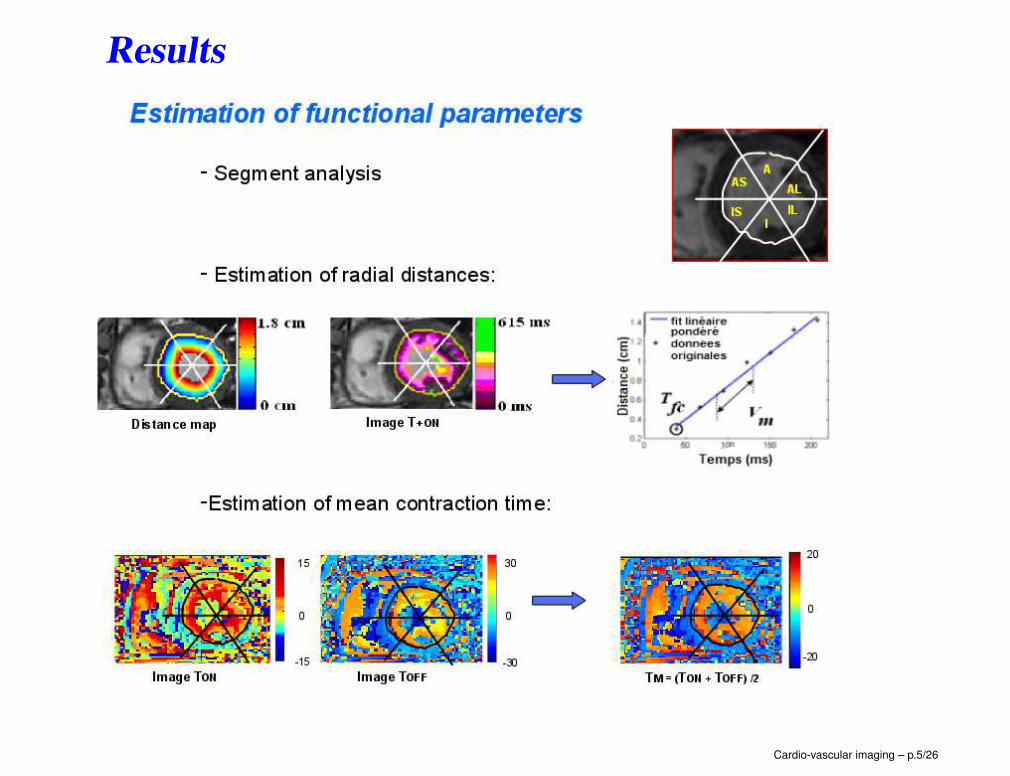
Results
Cardio-vascular imaging – p.5/26
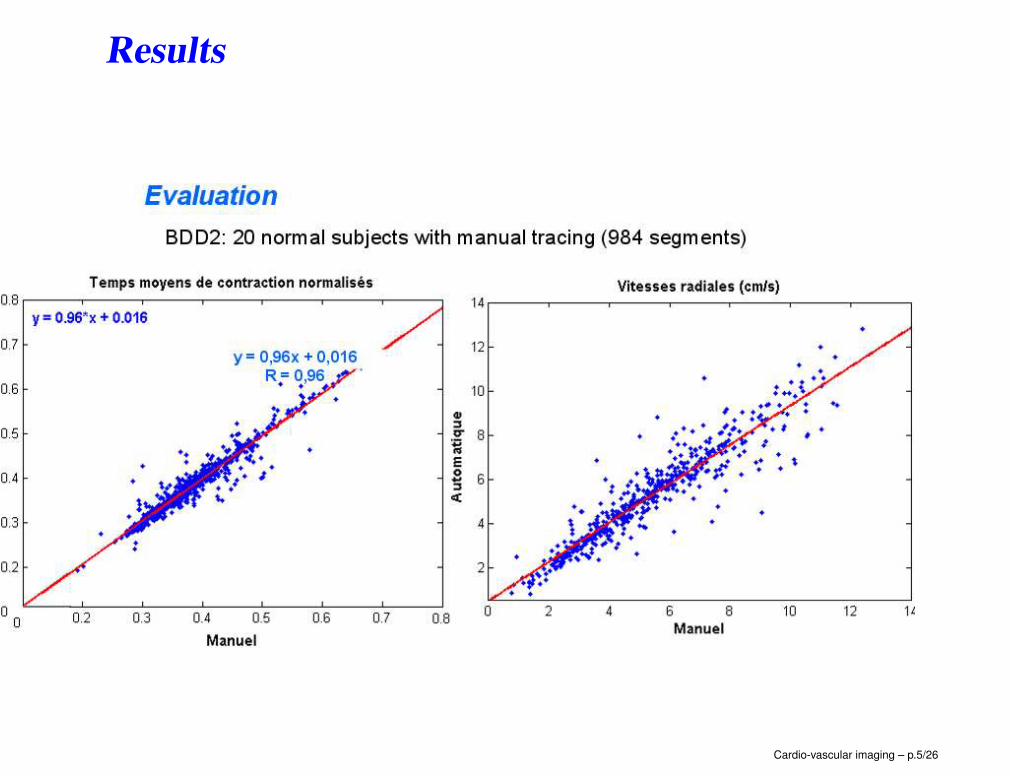
Results
Cardio-vascular imaging – p.5/26
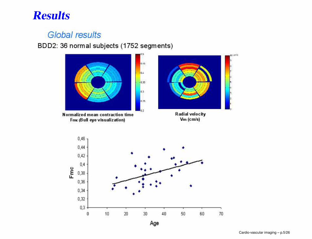
Results
Cardio-vascular imaging – p.5/26
Results
Cardio-vascular imaging – p.5/26
Results
Cardio-vascular imaging – p.5/26
Results
Cardio-vascular imaging – p.5/26
Results
Cardio-vascular imaging – p.5/26
Late enhancement images
Cardio-vascular imaging – p.6/26
Late enhancement images
Cardio-vascular imaging – p.6/26
Late enhancement images
Cardio-vascular imaging – p.6/26
Late enhancement images
Cardio-vascular imaging – p.6/26
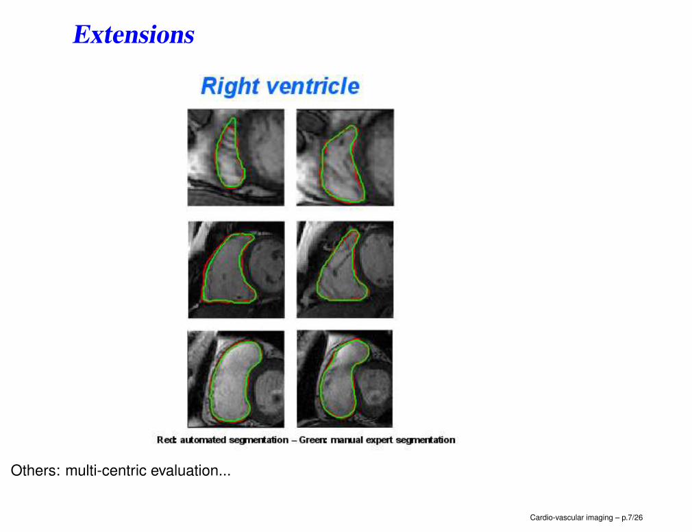
Extensions
Others: multi-centric evaluation...
Cardio-vascular imaging – p.7/26
Heart segmentation for oncology applications
(A. Moreno, J. Wojak)
Using structural constraints
Cardio-vascular imaging – p.8/26
Heart segmentation for oncology applications
(A. Moreno, J. Wojak)
Using structural constraints and a breathing model
Cardio-vascular imaging – p.8/26
Heart segmentation for oncology applications
(A. Moreno, J. Wojak)
Cardio-vascular imaging – p.8/26
Heart segmentation for oncology applications
(A. Moreno, J. Wojak)
Using shape constraints
Magenta = structural constraints, red = shape constraints, green = manual
Cardio-vascular imaging – p.8/26
Heart segmentation for oncology applications
(A. Moreno, J. Wojak)
Follow-up
Cardio-vascular imaging – p.8/26
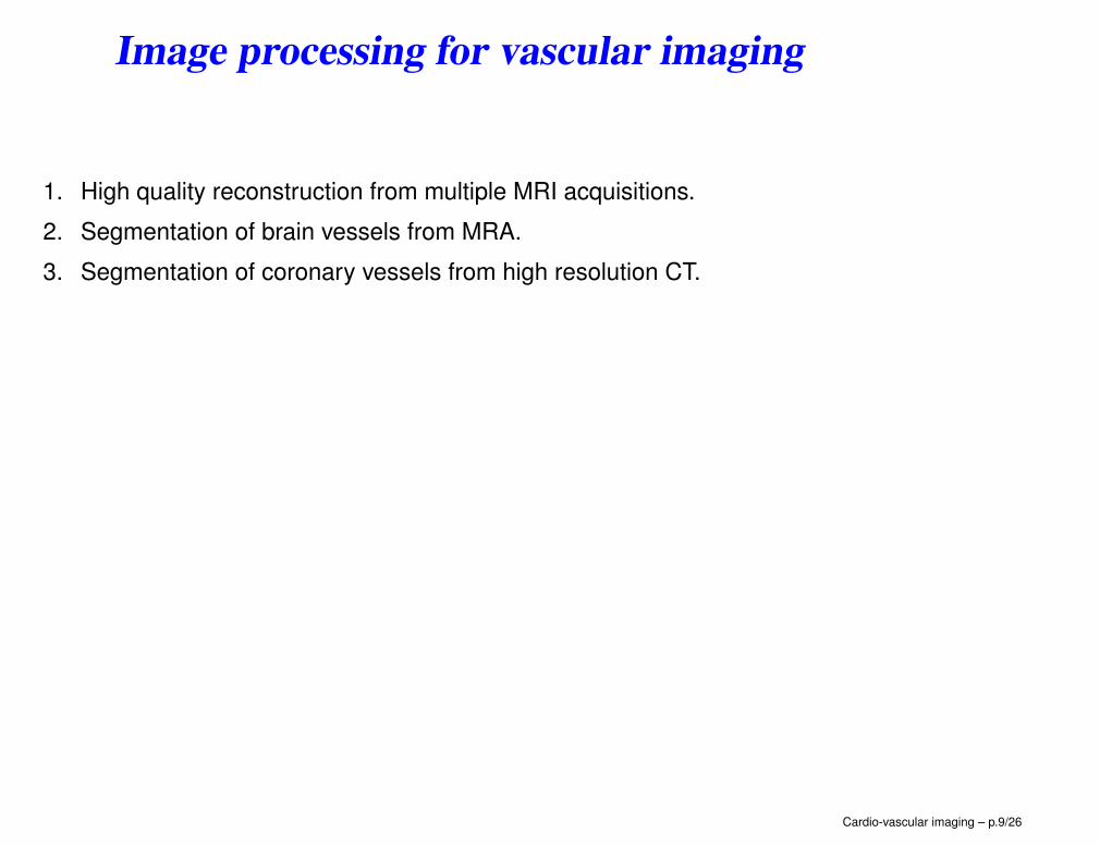
Image processing for vascular imaging
1. High quality reconstruction from multiple MRI acquisitions.
2. Segmentation of brain vessels from MRA.
3. Segmentation of coronary vessels from high resolution CT.
Cardio-vascular imaging – p.9/26
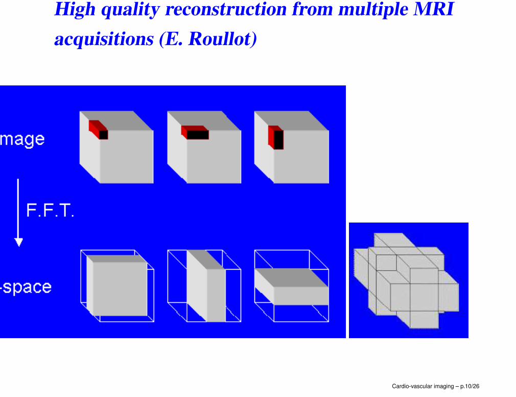
High quality reconstruction from multiple MRI
acquisitions (E. Roullot)
Cardio-vascular imaging – p.10/26
High quality reconstruction from multiple MRI
acquisitions (E. Roullot)
Cardio-vascular imaging – p.10/26
High quality reconstruction from multiple MRI
acquisitions (E. Roullot)
result_anime
Cardio-vascular imaging – p.10/26
Vessel segmentation for...
• better visualization,
• diagnosis assistance (detection, quantification),
• virtual endoscopy...
Some issues:
• classical ones: resolution, noise, partial volume effect...
• vessel specific: thin structures, bifurcations, anomalies...
Three important components
• models (hypotheses),
• features (image information),
• extraction techniques.
Cardio-vascular imaging – p.11/26
Segmentation of brain vessels from MRA (B.
Verdonck)
Cardio-vascular imaging – p.12/26
Segmentation of brain vessels from MRA (B.
Verdonck)
Cardio-vascular imaging – p.12/26
Segmentation of brain vessels from MRA (B.
Verdonck)
Cardio-vascular imaging – p.12/26
Segmentation of brain vessels from MRA (B.
Verdonck)
Cardio-vascular imaging – p.12/26
Segmentation of brain vessels from MRA (B.
Verdonck)
Cardio-vascular imaging – p.12/26
Segmentation of coronary vessels from high res-
olution CT (D. Lesage)
• Collaboration with Siemens Corporate Research.
• High resolution CT: ∼ 0.33 mm.
• Vessel model.
• Local features and measurements (flux).
• Segmentation expressed as a tracking process in a Bayesian framework, solved
by:
• minimal path,
• particle filter.
Cardio-vascular imaging – p.13/26
Segmentation of coronary vessels from high res-
olution CT (D. Lesage)
Cardio-vascular imaging – p.13/26
Tracking based approach
Cardio-vascular imaging – p.14/26
Overview
Cardio-vascular imaging – p.15/26
Flux based measure
Cardio-vascular imaging – p.16/26
Comparison with other measures
Cardio-vascular imaging – p.17/26
Minimal path approach
Cardio-vascular imaging – p.18/26
Metric choice
Cardio-vascular imaging – p.19/26
Result example
Cardio-vascular imaging – p.20/26
Particle filter
Cardio-vascular imaging – p.21/26
Evolution
Cardio-vascular imaging – p.22/26
Result examples and evaluation
Cardio-vascular imaging – p.23/26
Result examples and evaluation
Cardio-vascular imaging – p.23/26
Comparison of the two approaches
Evaluation on the Rotterdam database (http://coronary.bigr.nl).
Measure Minimal path Particle filter
(H = 4) (N = 1000)
Overlap 85 % 86.2 %
Distance to the central line (mm) 0.31 0.25
Error on radius (mm) 0.2 0.2
Computation time 1 min 4 min
• FP: less false positives (more robust stopping criterion).
• FP: more precise (no discretization of space).
• MP: less false negative (missing branches).
Cardio-vascular imaging – p.24/26
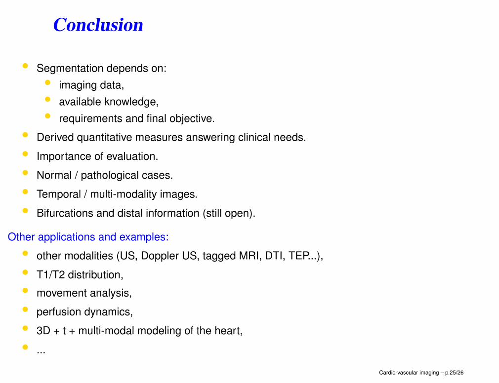
Conclusion
• Segmentation depends on:
• imaging data,
• available knowledge,
• requirements and final objective.
• Derived quantitative measures answering clinical needs.
• Importance of evaluation.
• Normal / pathological cases.
• Temporal / multi-modality images.
• Bifurcations and distal information (still open).
Other applications and examples:
• other modalities (US, Doppler US, tagged MRI, DTI, TEP...),
• T1/T2 distribution,
• movement analysis,
• perfusion dynamics,
• 3D + t + multi-modal modeling of the heart,
• ...
Cardio-vascular imaging – p.25/26
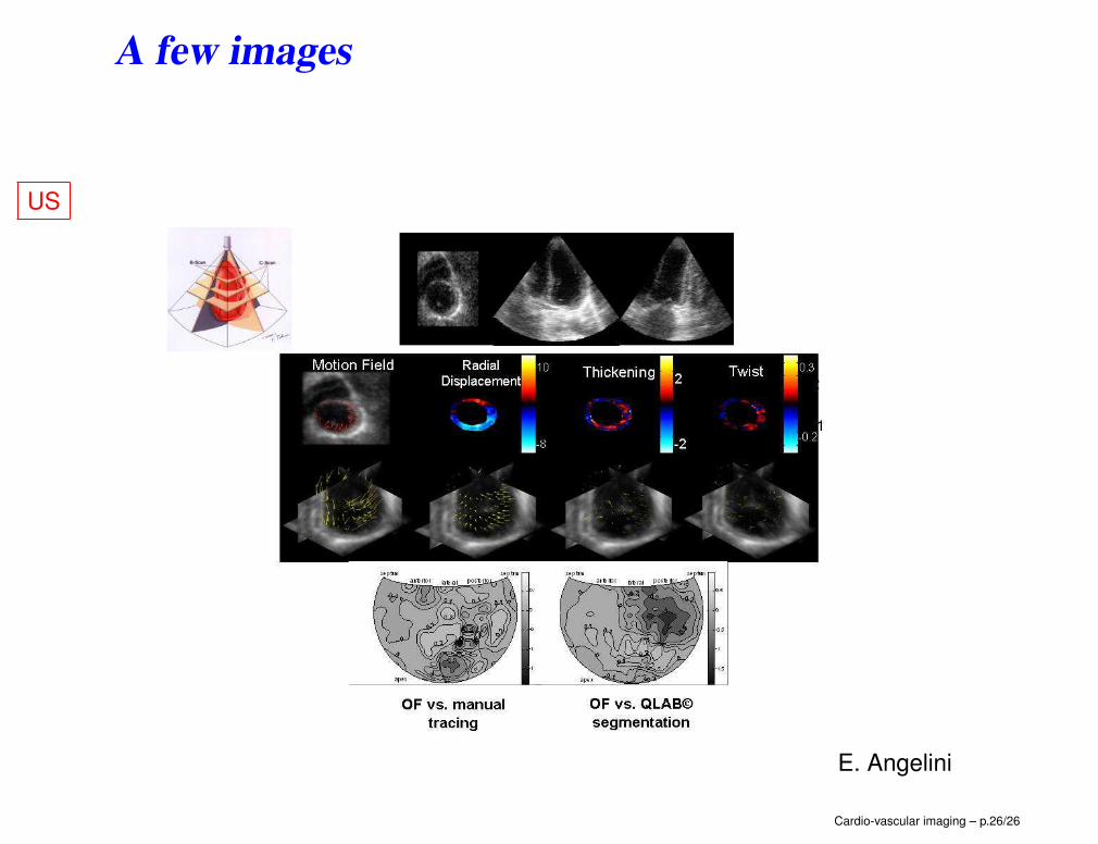
A few images
US
E. Angelini
Cardio-vascular imaging – p.26/26
A few images
TEP
Cardio-vascular imaging – p.26/26
A few images
Tagged MRI
Cardio-vascular imaging – p.26/26
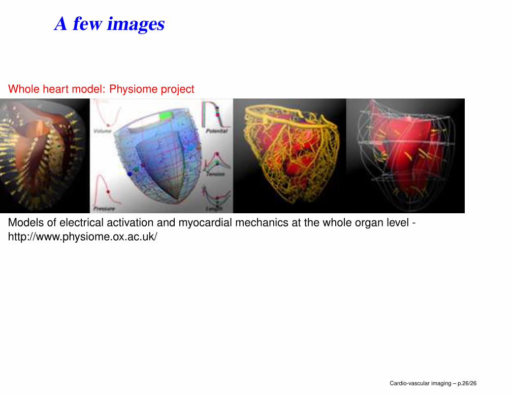
A few images
Whole heart model: Physiome project
Models of electrical activation and myocardial mechanics at the whole organ level -
http://www.physiome.ox.ac.uk/
Cardio-vascular imaging – p.26/26