Imaging Basics: with photons/electrons “Small is beautiful....” Electron Microscopy ETH Zurich (EMEZ), [email protected]! Question to audience: ! Did you use any imaging techniques? ! LM..., EM..., MRI..., PET..., X-ray ....? ! Who uses Image processing? ! Who is “comfortable” with contrasting/ staining technique? ! Who is familiar with labeling techniques? ! Who can explain the duality of the probes? ! Who is familiar with the resolution range of any mode? ! Do you know the difference between frequency and wavelength...? Content: - why imaging - why 3D... - Imaging modes... - Imaging “space”... - Imaging principle - Waves or particle - Duality - Photon - no mass high speed - Electron - mass and fast - Imaging modalities e.g in LM and EM....& Applications - Resolution.... - other Imaging modes..... X-ray Tomo, Imaging, PET, MRI... Aim of Imaging is primarily: Get information on: - Structure/ Morphology - Chemical composition/molecular composition - Regional function As fast as possible, non-invasive and as natural as possible along all length scale to understand structure- function and time domain of life.
! Question to audience:! Did you use any imaging techniques?! LM..., EM..., MRI..., PET..., X-ray....?! Who uses Image processing?! Who is “comfortable” with contrasting/
staining technique?! Who is familiar with labeling techniques?! Who can explain the duality of the probes?! Who is familiar with the resolution range of
any mode?! Do you know the difference between
frequency and wavelength...?
Content:
- why imaging - why 3D...
- Imaging modes...
- Imaging “space”...
- Imaging principle
- Waves or particle - Duality
- Photon - no mass high speed
- Electron - mass and fast
- Imaging modalities e.g in LM and EM....& Applications
- Resolution....
- other Imaging modes..... X-ray Tomo, Imaging, PET, MRI...
Aim of Imaging is primarily:
Get information on:
- Structure/ Morphology- Chemical composition/molecular composition- Regional function
As fast as possible, non-invasive and as natural as possible along all length scale to understand structure-function and time domain of life.
� � � � � � � � �Description of the “whole”.....
- A question of perspective and methodic approach...
Perspectives and the truth:
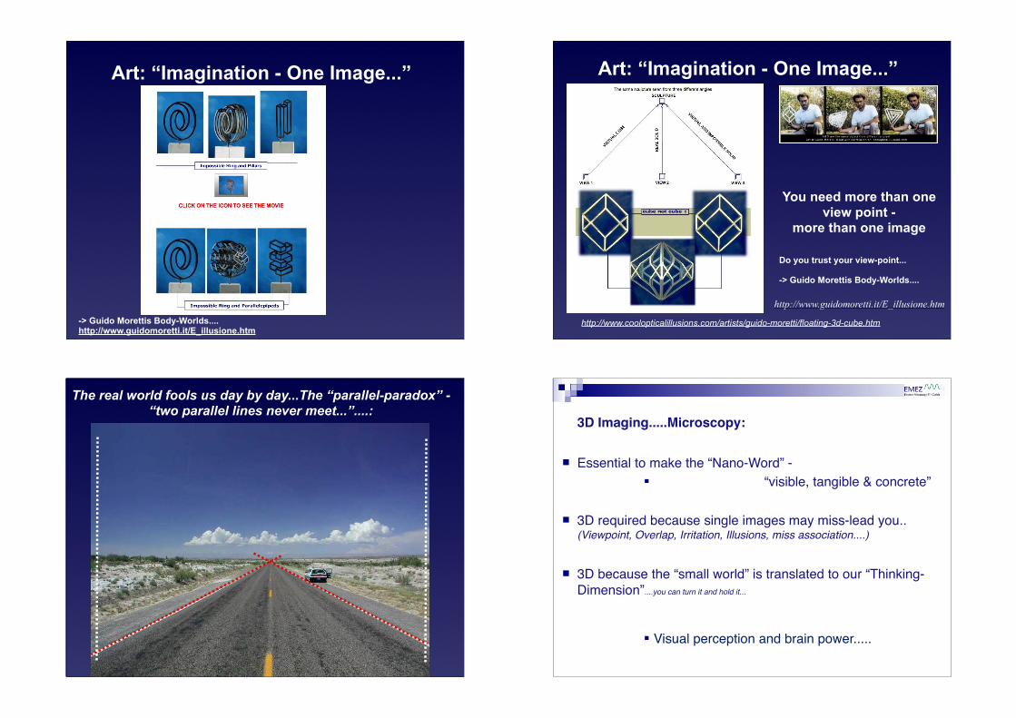
Is there one only Image of a object?
A View - A series of views...:
Vor etwa 100 Jahren versuchte Claude Monet die Gesamtheit (hier z.B. eines Heuschobers) durch mehrere Bilder aus unterschiedlichsten Perspektiven, Lichtverhältnissen und Jahreszeiten zu erfassen und zu beschreiben und so den Betrachter zum „Gesamten“ zu führen....
Your feet in Rom - your head in Hamburg if an Atom is just 0,1mm large
How does an image emerge or how is it formed ?
Self emitter
Remission:Scattering/ Absorption/Conversion
Eye/Detector...
Eye/Detector...
Rays (Waves):
Light (Photons - lampe, laser...)
Particle (Electrons, Ions,...)
Sound (Ultrasound, electromagnetic...)
Remission:Scattering/ Absorption/Conversion
Eye/Detector...
Remission:Scattering/ Absorption/Conversion
Eye/Detector...
for human eye visible part of spectrum
Evolution - human eye & visible spectra... Imaging Modalities
in Life-Science IR,PET
LM, CLSM
EM,IMSPM
X-ray,NMR
MRI, MEG, EEG, fMRI
www.neuroscience.cam.ac.uk
„CERN“
The “ Imaging Space“
im!ag!ing Date 1967: the action or process of producing an image especially by means other than visible light
Aim: enlarge samples to make them visible for the human eye or processable in a PC (Pixel/Voxel)
NMR
X-ray
NMR
X-ray
1-100nm
The “ Imaging Space“
im!ag!ing Date 1967: the action or process of producing an image especially by means other than visible light
Aim: enlarge samples to make them visible for the human eye or processable in a PC (Pixel/Voxel)
NMR
X-ray
NMR
X-ray
1-100nm
The “ Imaging Space“
im!ag!ing Date 1967: the action or process of producing an image especially by means other than visible light
Aim: enlarge samples to make them visible for the human eye or processable in a PC (Pixel/Voxel)
NMR
NMR
X-ray
X-ray
1-100nm1-100nm
Imaging: Principle “Information transfer chain”
What is Electromagnetic Radiation?Electromagnetic radiation is energy – we describe it as a wave – visible light is only a small portion
The characteristics which distinguish different types of light are the electromagnetic radiation's
- wavelength - frequency - energy
Wavelength: – the distance between two peaks (or two troughs) of the wave.Frequency: – the number of wavelengths passing a given point in one second.
->The longer the wavelength, the more time it takes for a full wave to pass a given point (or the fewer waves pass the point in a given time).
Since frequency is wavelengths per second, as the wavelength becomes longer the frequency decreases, and vice versa.
Energy: - is directly proportional to the frequency-- if the frequency increases, so does the energy of the radiation and vice versa.
! frequencyf
Wikipedia....
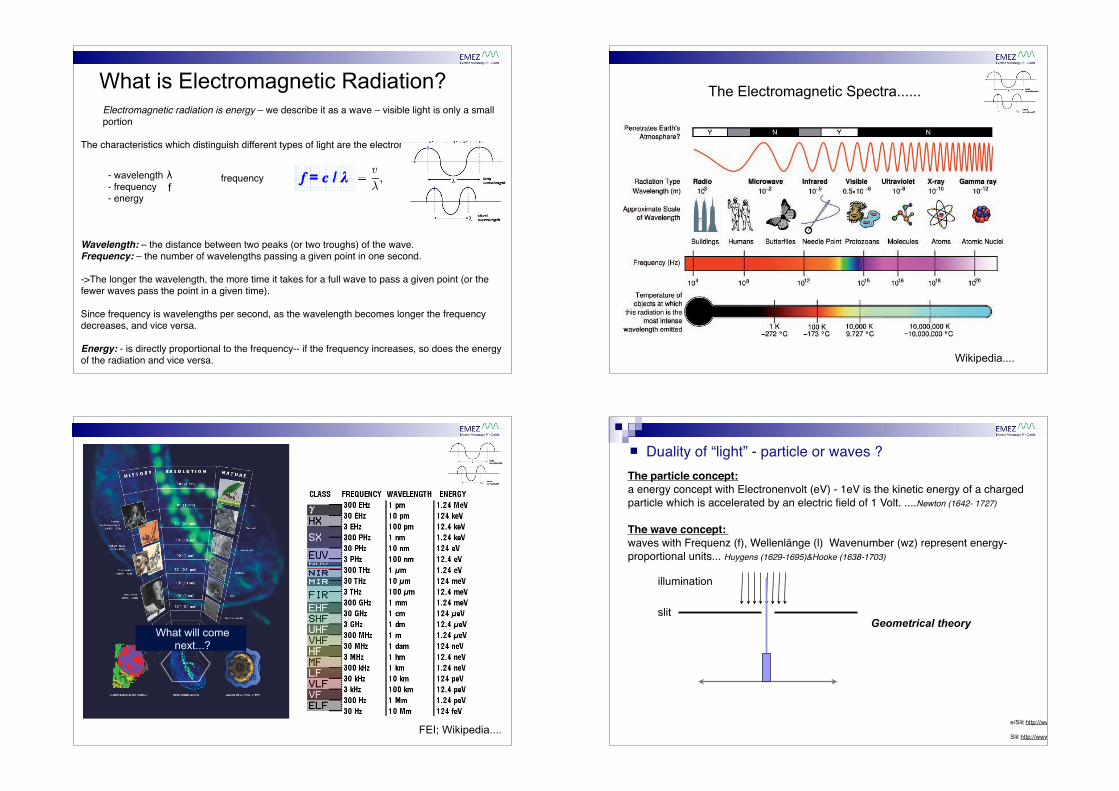
The Electromagnetic Spectra......
FEI; Wikipedia....
What will come next...?
! Duality of “light” - particle or waves ?The particle concept:a energy concept with Electronenvolt (eV) - 1eV is the kinetic energy of a charged particle which is accelerated by an electric field of 1 Volt. ....Newton (1642- 1727)
The wave concept: waves with Frequenz (f), Wellenlänge (l) Wavenumber (wz) represent energy-proportional units... Huygens (1629-1695)&Hooke (1638-1703)
Experimentalx particles
Experimentalmany particles
slit
illumination
-> diffraction of “light” => wave (bending around corners)
! Duality of “light” - particle or waves ?The particle concept:a energy concept with Electronenvolt (eV) - 1eV is the kinetic energy of a charged particle which is accelerated by an electric field of 1 Volt. ....Newton (1642- 1727)
The wave concept: waves with Frequenz (f), Wellenlänge (l) Wavenumber (wz) represent energy-proportional units... Huygens (1629-1695)&Hooke (1638-1703)
Experimentalmany particles
slit
illumination
-> diffraction of “light” => wave (bending around corners)
Geometrical theory
Unbelievable....... ....also particles e.g. electrons/ He-ions do it if not observed
http://www.youtube.com/watch?v=oxknfn97vFE
! Duality of “light” - particle or waves.....The particle concept: a energy concept with Electronenvolt (eV) - 1eV is the kinetic energy of a charged particle which is accelerated by an electric field of 1 Volt.
-> “classical ray optics”
The wave concept: waves with frequency (f), wavelength (!) wave-number (wz) represent energy-proportional units......
-> “wave optics” (wave front and Schrödinger eq.)
(h...PLANCKʼs constant) directly relates the energy with the frequency of a “ray” Quantum Theory (Planck & Einstein)
The relation between the wavelength (λ) of a particle of mass, m, moving at a velocity, v, is given by the DeBroglie wave equation:
! Not one wave but plane waves are used for imaging
In the physics of wave propagation, a plane wave is a constant-frequency wave whose wavefronts (surfaces of constant phase) are infinite parallel planes of constant amplitude normal to the phase velocity vector.
Mathematically, a plane wave is a wave of the following form:
where i is the imaginary unit, k is the wave vector, ω is the angular frequency, and A is the (complex) amplitude.
-> two dimensional sinusoids have a frequency, phase, amplitude and direction!
(This becomes of interest when Fourier Transformations (FFT) are used to analyze images (Amplitude in real part, Phase in imaginary part))
Not one wave but plane waves are used for imaging
! Conversion of energy-> wavelength-> wave-number.....
The wavelength of light is via the speed of light (c) linked to the frequency (general: ) => λ = c / f
The so called wave-number is the reciprocal of the wavelength (wz, n) n = 1 /λ (n Wave number usually in cm-1)
-> E = h * c * n Conversion from one to the other unit:
λ [µm] = 10ʼ000 / n [cm-1] ; λ [nm] = 10ʼ000ʼ000 / n [cm-1]
f [Hz] = 3 * 1010 * n [cm-1]
E [eV] = 1 / 8065,5 * n [cm-1] http://www.cactus2000.de/de/unit/masswav.shtml
The wavelength of light is via the speed of light (c) linked to the frequency (general: ) =>
Imaging: Principle “Information transfer chain”
To image a certain structure the used wavelength should be in the range of the structure detail to be
imaged... if not the wavelength can not interact linearly with the object...
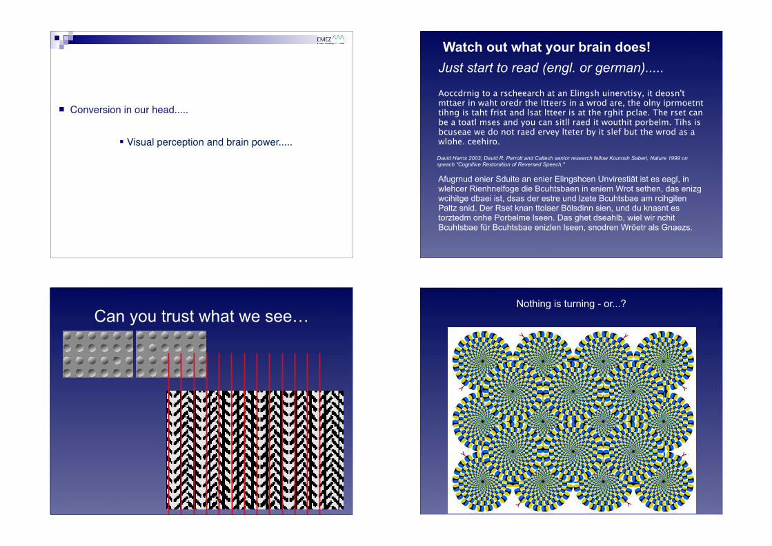
! Conversion in our head.....
" Visual perception and brain power.....
Watch out what your brain does! Just start to read (engl. or german).....
Aoccdrnig to a rscheearch at an Elingsh uinervtisy, it deosn't mttaer in waht oredr the ltteers in a wrod are, the olny iprmoetnt tihng is taht frist and lsat ltteer is at the rghit pclae. The rset can be a toatl mses and you can sitll raed it wouthit porbelm. Tihs is bcuseae we do not raed ervey lteter by it slef but the wrod as a wlohe. ceehiro.
David Harris 2003; David R. Perrott and Caltech senior research fellow Kourosh Saberi, Nature 1999 on speach "Cognitive Restoration of Reversed Speech,"
Afugrnud enier Sduite an enier Elingshcen Unvirestiät ist es eagl, in wlehcer Rienhnelfoge die Bcuhtsbaen in eniem Wrot sethen, das enizg wcihitge dbaei ist, dsas der estre und lzete Bcuhtsbae am rcihgiten Paltz snid. Der Rset knan ttolaer Bölsdinn sien, und du knasnt es torztedm onhe Porbelme lseen. Das ghet dseahlb, wiel wir nchit Bcuhtsbae für Bcuhtsbae enizlen lseen, snodren Wröetr als Gnaezs.
Can you trust what we see!Nothing is turning - or...?
.! an nothing is moving! Do we trust what we see!Illusionen!
What do you see in this image..........?
Children see onlyDolphins
...what did you see???
Sandro Del-Prete
“A world were youreyes deceive you”
http://www.illusoria.com
Photon Electron (Boson/Eichboson) (Fermion/Lepton)
Charge: 0
Mass: 0
RestingEnergy: eV
ComptonWavelength: -
Spin: 1
Radius: -
Wavelength 300nm-1600nm in use today...
Photon........ Properties:
Imaging: “Information transfer chain”
“Microscope...”
Information transfer......
! Imaging Mode: Light Microscopy (LM)
! Full field illumination (Bright field, Phase contrast, Differential Interference Contrast, Fluorescence, Polarisation, Spectroscopy...)
! -> parallel light is exposed to the whole specimen! -> thickness of specimen is limiting resolution ! -> fast light exposure possible! -> projection/reflexion images of the exposed area
! Scanning mode (Reflexion, Fluorescence, Spectroscopy - Raman! -> focused light spot is scanned through the specimen! -> scanning in x,y and z possible! -> thickness not so critical! -> slow for large image area! -> pixel by pixel images
Light Microscope (LM)Illumination types....
2007; Jyoti K Jaiswa & Sanford M Simon
Wide field &confocal illumination
TIRF illumination
F-actin(ø 9nm)
Microtubules(ø 25nm)
http://www.aqua-fish.net/
Motile cells (Fish keratocyte) - Fluorescence LM...(Resolution or Seeing....?)
! Imaging Mode: Light Microscopy (LM)! Probe: # # bundle of light or focused spot of light! Wavelength: # 300-1000nm