Imaging biomarkers in prediction of the blood loss during nephrectomy due to renal cell carcinoma after embolization of renal artery: application of the apparent diffusion coefficient
AbstractRenal artery embolization (RAE) is used for preparation of patients with renal cell carcinoma (RCC) of large size or significant vascularization to surgical treat-ment. Currently there is no accurate method of prediction of intraoperative blood loss in patients with RCC after RAE, that may give possibility for more ad-equate pre – and postoperative patient management and to define the indica-tions for re-embolization. The goal of the study was to evaluate the possibility of application of apparent diffusion coefficient (ADC) for the prediction of the esti-mated intraoperative blood loss (EIBL) during the open radical nephrectomy in patients with RCC and after RAE. The study enrolled 35 patients (main group, 20 males and 15 females) with solid RCC according to clinical and radiologic data and with the indications to selective RAE with subsequent radical nephrecto-my. In all patients with RCC in the same day or day before RAE and 7 days after the RAE MRI with additional DWI sequence with b-value=0.800 was performed. Subsequent measuring of the ADC over the tumor region was done in all cases.
Yulian Mytsyk , Yuriy Borys, Serhiy Pasichnyk, Dmytro Vorobets, Bohdan Borys, Volodymyr Dmytriv, Roman Zahorujko, Oleh Mytsyk, Natalia Chernova
Danylo Halytsky Lviv National Medical University, Department of Urology, Lviv, Ukraine
European Journal of Medical Technologies 2019; 2(23): 1-12
BackgroundThe renal cell carcinoma (RCC) accounts for approx-imately 3.7% of all adult malignancies and more than 90 % of neoplasm arising from kidney [1]. The «gold standard» for localized and locally advanced RCC treatment is still nephrectomy. During the last decade renal artery embolization (RAE) is used as prepara-tion of patients with big or/and hypervascularized RCC’s for surgical treatment [2]. The main advan-tages of RAE before nephrectomy are: reduction of tumor clots` size (that facilitate its removal), blood loss reduction during surgery, alleviation of dissec-tion because of edema development. RAE is usually performed 7-10 days before surgery. The adverse side of RAE is postinfarction syndrome, which appear as pain in the iliac region, nausea and fever, this syn-drome occur in ¾ of patients. Some authors observe significant blood loss during nephrectomy after RAE, perhaps it is caused by the incomplete schematiza-tion of tumor tissues. Literature data about efficiency of RAE performed as preparation of patients with RCC to nephrectomy are often contradictory, espe-cially concerning estimated intraoperative blood loss (EIBL) volume and amount of blood transfusion, op-erative time, complications and survival rates [7, 13]. Thus, according to Schwartz and co-authors EIBL volume during radical nephrectomy in patients with
RCC after RAE with median tumor size of 11.2 cm was variable and ranged from 100 to 5000 ml [11]. In another new research conducted by Ramaswamy, was established, that blood transfusion volume after radical nephrectomy in patients with RCC after total RAE also was also significantly variable and ranged from 222 to 1050 ml [10]. At the same time, accord-ing to Suad and co-authors, medium volumes of EIBL and intraoperative blood transfusion amounted 300 ml and 250 ml accordingly [5].
Currently there is no accurate method of predic-tion of EIBL volume during nephrectomy in patients with RCC after RAE, that may give possibility for more adequate treatment planning and postoperative patient management and to define the indications for re-embolization. In the last years the attention of re-searchers was focused on investigation of MRI and its modalities as diffusion weighted imaging (DWI) to estimate the efficiency of arterial embolization in treatment of benign and malignant tumors, as uter-ine fibroid and leiomyoma, hepatocellular cancer and breast cancer [3, 6, 9, 12]. DWI is the MRI sequence, which uses strong bipolar gradients to enhance sen-sitivity to thermally induced Brownian motion of hydrogen molecules, that allows to measure molecu-lar diffusion in tissues in vivo and it is important for profound characteristic of neoplasm [4]. However, all conducted studies were directed toward estimating
ADC values of the normal renal parenchyma for control were achieved during the examination of 15 healthy volunteers. In all patients with RCC 7-8 days after the RAE open radical nephrectomy with simultaneous EIBL measurement was executed. In patients with EIBL less than 500 ml and with no episodes of the he-motransfusions mean ADC value decreased by 18.4-31.9% in comparison with initial ADC value. In patients with EIBL more than 500 ml (with hemotransfu-sions) mean ADC value increased by 4.91-65.64% compared to baseline value. In main group of patients in whom no hemotransfusions were required in post-op period (n=29, 82.86%) there was significant (р<0.05) difference in mean ADC values before and after RAE (decrease by 20.25%): 1.63±0.31×10⁻³ mm2/s vs 1.30±0.19×10⁻³ mm2/s. In main group patients with hemotransfusions in post-op period this value increased by 28.83%: 1.63±0.31×10⁻³ mm2/s vs 2.10±0.47×10⁻³ mm2/s (р<0.05). Conclusions. Application of MRI and its imag-ing biomarker ADC can be valuable clinical instrument for prediction of the EIBL volume during the open radical nephrectomy in patients with RCC after RAE and need of the hemotransfusion in postoperative period.
EJMT 2(23) 2019 • European Journal of Medical Technologies
the efficiency of arterial embolization, which was performed as a disease palliation but not during preparation to surgery.
In our previous work we investigated the efficiency of DWI MRI and its quantitative parameter – appar-ent diffusion coefficient (ADC) as imaging biomark-ers in differential diagnostic of solid and cystic RCCs, benign renal tumors, benign cysts and abscesses [8]. We achieved statistically significant difference in mean ADC values of RCC and of the healthy renal parenchyma. We observed significant restriction of diffusion of hydrogen molecules in regions of solid neoplasms and it’s increase in renal cysts and in ab-scesses in comparison with normal kidney paren-chyma. Taking into account that tissue density affects diffusion of hydrogen molecules in them and that ADC consists of 3 main components – intracellular (inside the cytoplasm), extracellular (in the intersti-tial liquid, in blood and lymph vessels) and diffusion between intra – and extracellular enviroments, we suggested that DWI and ADC may be used as imag-ing biomarkers for evaluation of changes, which oc-curs in RCC tissues after RAE and for prediction of intraoperative blood volume loss in patients in whom nephrectomy will be executed.
GoalThe goal of the study was to evaluate the possibility of application of ADC for prediction of the EIBL during the open radical nephrectomy in patients with RCC and after RAE.
Materials and methodsThe study was allowed by Ethics Committee and conducted on the basis of the Departments of Urol-ogy and Radiology of Danylo Halytsky Lviv National Medical University, Department of Interventional Radiology of Lviv Emergency Hospital, Center of Endourology of Lviv Clinical Railway Hospital and at the Medical Center “Euroclinic”, Lviv, during 2013-2017 years.
The main group enrolled 35 patients (20 males and 15 females) with solid RCC according to clinical and radiological data and with the indications to selec-tive RAE with subsequent radical nephrectomy. The age of the patients ranged from 52 to 70 years old, the mean age was 62±6.3 years. Tumor size ranged from 7.3 to 11.4 cm, the mean size was 8.6±3.8 cm in the greatest dimension. Before surgery males had the level of hematocrit of 38-49% and females of 33-43%.
In all patients with RCC in the same day or day before RAE abdominal MRI using 1.5 T scanner (Signa HDxt, General Electric, USA) and 8-chan-nel coil was performed. In all cases was applied same standardized scanning protocol (General Electric), which additionally included DWI sequence with fol-lowing parameters: repeat time (TR)=12000 ms, echo time (TE)=90 ms, field of view (FOV)=40x40 cm, matrix=200 x 192, number of excitations (NEX)=3, bandwidth= 250 kHz, diffusion direction= slice, slice thickness= 6,0 mm, interscan gap = 1,0 mm with b-value=0.800 mm2/s, acquisition time=17 sec. DWI was conducted before contrast media administration, using single-shot echo-planar imaging sequence with parallel imaging technique and fat saturation during one breath-hold. The mean duration of MRI exami-nation was 35 minutes.
Selective RAE was performed under fluoroscopic guidance using the following method: under local anesthesia (0.5 % Novocaine) radial artery was punc-tured by Seldinger and after that introducer (6 Fr) was inserted. Selective angiography of right renal artery was performed using MR-catheter and injec-tion of 80 ml of contrast (Ultratwist-370). After that one COOK spiral and three Gianturco spirals were alternately introduced (all spirals MRI-compatible), achieving total RAE. After RAE puncture area was bandaged with compression for 6 hours. The mean duration of the procedure was 120 minutes. Re-RAE wasn’t performed in any case.
In all patients with RCC 7-8 days after selective RAE open radical nephrectomy with further patho-logic verification of diagnosis was executed: in 100% of cases RCC diagnosis was confirmed. Measurement of EIBL was performed in all patients according to the following method: it was accepted, that 1 ml of
EJMT 2(23) 2019 • European Journal of Medical Technologies
blood weigh is 1 g; swabs saturated with blood were weighted and then the mass of blank swabs from the same lot were subtracted; during weighing capacities for blood the mass of blank capacities were subtract-ed; blood volume, which got on surgical sheet, under body of the patient was assessed; the volume of the solution for irrigation was taken into account and it was subtracted from the general volume of EIBL.
Control MRI was performed in all patients 7 days after nephrectomy, which on purpose to reduce fi-nancial and temporal expense, included only 2 se-quences: coronal T2-weighted single shot fast spin echo (SSFSE) and DWI with identical to above men-tioned parameters. The mean duration of the exami-nation was 10 minutes.
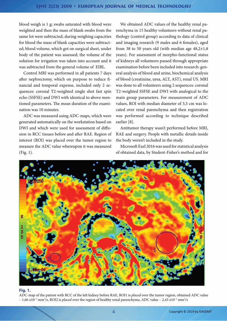
ADC was measured using ADC-maps, which were generated automatically on the workstation based on DWI and which were used for assessment of diffu-sion in RCC tissues before and after RAE. Region of interest (ROI) was placed over the tumor region to measure the ADC value whereupon it was measured (Fig. 1).
We obtained ADC values of the healthy renal pa-renchyma in 15 healthy volunteers without renal pa-thology (control group) according to data of clinical and imaging research (9 males and 6 females), aged from 38 to 50 years old (with median age 48,2±1,8 years). For assessment of morpho-functional status of kidneys all volunteers passed through appropriate examination before been included into research: gen-eral analysis of blood and urine, biochemical analysis of blood (creatinine, urea, ALT, AST), renal US. MRI was done to all volunteers using 2 sequences: coronal T2-weighted SSFSE and DWI with analogical to the main group parameters. For measurement of ADC values, ROI with median diameter of 3,5 cm was lo-cated over renal parenchyma and then registration was performed according to technique described earlier [8].
Antitumor therapy wasn’t performed before MRI, RAE and surgery. People with metallic details inside the body weren’t included in the study.
Microsoft Exel 2016 was used for statistical analysis of obtained data, by Student-Fisher’s method and for
Fig. 1. ADC-map of the patient with RCC of the left kidney before RAE, ROI1 is placed over the tumor region, obtained ADC value – 1,66 x10⁻³ mm2/s, ROI2 is placed over the region of healthy renal parenchyma, ADC value – 2,43 x10⁻³ mm2/s
EJMT 2(23) 2019 • European Journal of Medical Technologies
calculation of Pearson correlation coefficient. Value p<0.05 considered as a statistically significant result.
Results and discussionDuring the open nephrectomy in patients with RCC after RAE, EIBL varied from 158 to 1250 ml, the mean volume was 457,3±260,52 ml. Depending on the EIBL all patients of the main group were separated into 4 different categories: І category – EIBL – 0-250 ml (n=11), ІІ category – EIBL 250-500 ml (n=14), ІІІ category – EIBL 500-1000 ml (n=8) and IV category – EIBL more than 1000 ml (n=2). Medium values of EIBL for each category were: І – 216,55±34,45 ml, ІІ – 387,29±64,19 ml, ІІІ – 669,36±118,23 ml, IV – 1182,5±95,46 ml.
We established, that medium ADC value of the healthy renal parenchyma of volunteers was 2,47±0,12×10⁻³ mm2/s. In a result of analysis of the ob-tained ADC data we obtained statistically significant (р<0.05) difference between the mean ADC values in the main group before and after RAE and in control group: 1,63±0,31×10⁻³ mm2/s vs 1,44±0,40×10⁻³ vs
2,47±0,12×10⁻³ mm2/s accordingly. We observed di-rect correlation between the values of EIBL volume and ADC in patients after RAE, Pearson correlation coefficient was r=0,96. In patients of І and ІІ catego-ries with EIBL volume less than 500 ml and with no episodes of hemotransfusions mean ADC value de-creased by 18.4-31.9% compared to baseline value, because of bigger infarct areas of the tumor tissues, decrease of microcirculation and as a result the re-striction of hydrogen molecules diffusion. As op-posed to that in patients of ІІІ and IV categories with EIBL volume more than 500 ml and in some cases with hemotransfusions mean ADC value increased by 4.91-65.64% compared to baseline value. The in-terconnection between the increase of this ADC val-ue and EIBL volume in the IV category of the patients can be explained with expressed oedema of the renal tissue near infarct area, necrotic process and defi-cient decrease of microcirculation in tumor area. All of this together conduced the increase of molecular diffusion in tumor tissues and possibly as a result of it the increase of bleeding during surgery. The detailed description of EIBL and ADC values of the patients with RCC before and after RAE presented in table 1.
Category of patients/subgroup
Mean ADC before RAE, ×10−3 mm2/s
Mean ADC after RAE, ×10−3 mm2/s
Mean EIBL, (range), ml
Category І, EIBL 0-250 ml(n=11)
1.63±0.31 1.11±0.10 216.55±34.45(158-250)
Category ІI, EIBL 250-500 ml(n=14)
1.33±0.08 387.29±64.19(275-487)
Category ІII, EIBL 500-1000 ml(n=8)
1.71±0.12 669.36±118.23(510-823)
Category ІV, EIBL >1000 ml(n=2)
2.70±0.06 1182.5±95.46(1115-1250)
Patients, in whom no hemotransfusions were required(n=29)
1.30±0.19* 351.33±119.75(158-570)
Patients, who had hemotransfusion(n=6)
2.10±0.47* 899.0±226.45(720-1250)
Table 1. Mean ADC values of the main group of patients before and after RAE and EIBL volume
*comparison of groups between each other (p<0.05)
EJMT 2(23) 2019 • European Journal of Medical Technologies
Analysis of the acquired data showed significant (р<0.05) difference in in mean ADC values in sub-group of patients in whom no hemotransfusions were required in post-op period (n=27, 82.86%) in comparison with baseline mean ADC value: 1.30±0.19×10⁻³ mm2/s vs 1.63±0.31×10⁻³ mm2/s ac-cordingly (decrease by 20.25%). In sungroup of pa-tients with hemotransfusions in post-op period (n=6, 17,14 %), the mean ADC value increased by 28.83% compared to baseline value: 1.63±0.31×10⁻³ mm2/s vs 2.10±0.47×10⁻³ mm2/s, such difference was statis-tically significant (р<0.05).
ConclusionsStrong direct correlation was observed between the volume of EIBL during open radical nephrectomy and ADC values in patients with RCC after RAE. Ap-plication of MRI and its imaging biomarker ADC can be valuable clinical instrument for prediction of the EIBL volume during the open radical nephrectomy in patients with RCC after RAE and need of the he-motransfusion in postoperative period.
Rak v Ukraini, 2014–2015. Bul. Nats. cancer-reje-stru Ukrainy ą17. Kyiv 2015, pp. 56–57.
2. Alan J. Wein, Louis R. Kavoussi, Alan W. Partin. Campbell-Walsh Urology, 11th Edition. Elsevier 2016, pp. 1414–1416.
3. Buijs Manon, Ihab R. Kamel, Josephina A. Vossen, Christos S. Georgiades, Kelvin Hong. Assessment of Metastatic Breast Cancer Response to Chemo-embolization with Contrast Agent Enhanced and Diffusion-Weighted MR Imaging. Journal of Va-scular and Interventional Radiology 2007; 8(18): 957–963.
4. Derek K. Jones. Diffusion MRI: Theory, Methods, and Applications. Oxford University Press USA. 2010, p. 132.
5. Jaganjac Suad, L. Schefe, Edin Avdagić, Hajrudin Spahović, Mustafa Hiros. Preoperative Kidney Tumor Embolization as Procedure for Therapy of Advanced Kidney Cancer. Journal of the Society for Medical Informatics of Bosnia & Herzegovina 2014; 5(22): 302–305.
6. Lee Mu Sook, Man Deuk Kim, Dae Chul Jung, My-ungsu Lee, Jong Yun Won, Apparent Diffusion Coefficient of Uterine Leiomyoma as a Predictor of the Potential Response to Uterine Artery Em-bolization. Journal of Vascular and Interventional Radiology 2013; 9(24): 1361–1365.
7. Muller Arnaud, and Olivier Rouvičre. Renal Artery Embolization—indications, Technical Approaches and Outcomes., Nature Reviews Nephrology 2015; 5(11): 288–301.
8. Mytsyk Y., Dutka, I., Borys, Y. et al Renal cell carci-noma: applicability of the apparent coefficient of the diffusion-weighted estimated by MRI for improving their differential diagnosis, histologic subtyping, and differentiation grade. Int Urol Ne-phrol., 2016. published online ahead of print. P. 1–10. doi:10.1007/s11255-016-1460-3.
9. Noda Yoshifumi, Masayuki Kanematsu, Satoshi Goshima, Hiroshi Kondo, Haruo Watanabe. Predic-tion of Early Response to Uterine Artery Emboliza-tion in Fibroids: Value of MR Signal Intensity Ratio., Magnetic Resonance Imaging 2015; 1(33): 51–55.
10. Ramaswamy S. Raja, Michael D. Darcy. Arterial Em-bolization for the Treatment of Renal Masses and Traumatic Renal Injuries., Techniques in Vascular & Interventional Radiology 2016; 3(19): 203–210.
11. Schwartz Michael J., Eric B. Smith, David W. Trost, and E. Darracott Vaughan. Renal Artery Emboliza-tion: Clinical Indications and Experience from over 100 Cases., BJU International 2007; 4(99): 881–886.
12. Vandecaveye Vincent, Katrijn Michielsen, Frede-rik De Keyzer, Wim Laleman, Mina Komuta. Che-moembolization for Hepatocellular Carcinoma: 1-Month Response Determined with Apparent Diffusion Coefficient Is an Independent Predictor of Outcome. Radiology 2014; 3(270): 747–757.
13. Zelens’kyĭ R.O. Evaluation of vascular bed before and after embolization of renal artery for renal cancer. Klinichna Khirurhiia 2015; 1: 32–34.