Imaging in Sarcoidosis Hilario Nunes, M.D., 1 Pierre-Yves Brillet, M.D., 2 Dominique Valeyre, M.D., 1 Michel W. Brauner, M.D., 2 and Athol U. Wells, M.D. 3 ABSTRACT Sarcoidosis is a multisystemic granulomatous disease of unknown etiology that may involve virtually any organ. Pulmonary involvement predominates, but sarcoidosis can involve multiple organs, with or without concomitant lung involvement. Aberrations on chest radiographs are present in more than 90% of patients with sarcoidosis. Bilateral hilar lymphadenopathy, with or without lung parenchymal infiltrates, is typical but a wide range of chest radiographic patterns may be observed. This article discusses the characteristic chest radiographic features of sarcoidosis and the prognostic value of the radiographic staging classification as espoused by Scadding more than 4 decades ago. Thin-section high- resolution computed tomographic (HRCT) scans more clearly elucidate the intrathoracic lesions observed in sarcoidosis and may discriminate active inflammation from end-stage fibrosis. Although HRCT is not necessary to manage all cases of sarcoidosis, HRCT may be invaluable in selected patients with stage II or III sarcoidosis to discriminate alveolitis (which may be amenable to therapy) from fibrosis. Additionally, radionuclide techniques may have a role in extrapulmonary sarcoidosis (particularly when central nervous system or cardiac involvement is suspected). We review the salient features and role of magnetic resonance imaging and diverse radionuclide techniques to diagnose or follow selected cases of extrapulmonary sarcoidosis. KEYWORDS: Sarcoidosis, chest radiographs, computed tomographic (CT) scans, magnetic resonance imaging (MRI), radionuclide scans Sarcoidosis is a multisystemic granulomatous disease of unknown etiology. 1–3 Multiple clinical phe- notypes are observed as judged by the mode of presen- tation, pattern of organ involvement, disease duration, and severity. Sarcoidosis primarily affects the lungs and the lymphatic system in more than 90% of patients but virtually no organ is immune from the disease. 1–3 The diagnosis of sarcoidosis is based upon the association of compatible clinicoradiological findings, histological demonstration of noncaseating granulomas, and exclu- sion of other granulomatous disorders. 1 However, the clinical and radiological expression of the disease is remarkably variable, with findings sometimes pathogno- monic but often merely suggestive or markedly atypical. The radiological differential diagnosis is often broad, and because histological confirmation is sometimes elu- sive, a confident diagnosis often requires the amalgama- tion of information from imaging and other tests. Moreover, chest radiographic findings make a major contribution to prognostic evaluation. The course and prognosis of sarcoidosis are highly variable. 1–3 Although spontaneous recovery occurs in nearly two thirds of cases, therapy is necessary in one third of cases, and some patients experience a prolonged and severe course. 1 Services de Pneumologie; 2 Radiologie, Ho ˆ pital Avicenne, Assistance Publique-Ho ˆ pitaux de Paris, Universite ´ Paris, Bobigny, France; 3 Interstitial Lung Disease Unit, Royal Brompton Hospital, London, United Kingdom. Address for correspondence and reprint requests: Hilario Nunes, M.D., Service de Pneumologie, Ho ˆpital Universitaire Avicenne, 125 rue de Stalingrad, 93009 Bobigny, France. E-mail: [email protected]. Sarcoidosis: Evolving Concepts and Controversies; Guest Edi- tors, Marc A. Judson, M.D., Michael C. Iannuzzi, M.D. Semin Respir Crit Care Med 2007;28:102–120. Copyright # 2007 by Thieme Medical Publishers, Inc., 333 Seventh Avenue, New York, NY 10001, USA. Tel: +1(212) 584-4662. DOI 10.1055/s-2007-970336. ISSN 1069-3424. 102
Michel W. Brauner, M.D.,2 and Athol U. Wells, M.D.3
ABSTRACT
Sarcoidosis is a multisystemic granulomatous disease of unknown etiology thatmay involve virtually any organ. Pulmonary involvement predominates, but sarcoidosis caninvolve multiple organs, with or without concomitant lung involvement. Aberrations onchest radiographs are present in more than 90% of patients with sarcoidosis. Bilateral hilarlymphadenopathy, with or without lung parenchymal infiltrates, is typical but a wide rangeof chest radiographic patterns may be observed. This article discusses the characteristicchest radiographic features of sarcoidosis and the prognostic value of the radiographicstaging classification as espoused by Scadding more than 4 decades ago. Thin-section high-resolution computed tomographic (HRCT) scans more clearly elucidate the intrathoraciclesions observed in sarcoidosis and may discriminate active inflammation from end-stagefibrosis. Although HRCT is not necessary to manage all cases of sarcoidosis, HRCT maybe invaluable in selected patients with stage II or III sarcoidosis to discriminate alveolitis(which may be amenable to therapy) from fibrosis. Additionally, radionuclide techniquesmay have a role in extrapulmonary sarcoidosis (particularly when central nervous system orcardiac involvement is suspected). We review the salient features and role of magneticresonance imaging and diverse radionuclide techniques to diagnose or follow selected casesof extrapulmonary sarcoidosis.
magnetic resonance imaging (MRI), radionuclide scans
Sarcoidosis is a multisystemic granulomatousdisease of unknown etiology.1–3 Multiple clinical phe-notypes are observed as judged by the mode of presen-tation, pattern of organ involvement, disease duration,and severity. Sarcoidosis primarily affects the lungs andthe lymphatic system in more than 90% of patients butvirtually no organ is immune from the disease.1–3 Thediagnosis of sarcoidosis is based upon the association ofcompatible clinicoradiological findings, histologicaldemonstration of noncaseating granulomas, and exclu-sion of other granulomatous disorders.1 However, theclinical and radiological expression of the disease is
remarkably variable, with findings sometimes pathogno-monic but often merely suggestive or markedly atypical.The radiological differential diagnosis is often broad,and because histological confirmation is sometimes elu-sive, a confident diagnosis often requires the amalgama-tion of information from imaging and other tests.Moreover, chest radiographic findings make a majorcontribution to prognostic evaluation. The course andprognosis of sarcoidosis are highly variable.1–3 Althoughspontaneous recovery occurs in nearly two thirds of cases,therapy is necessary in one third of cases, and somepatients experience a prolonged and severe course.
1Services de Pneumologie; 2Radiologie, Hopital Avicenne, AssistancePublique-Hopitaux de Paris, Universite Paris, Bobigny, France;3Interstitial Lung Disease Unit, Royal Brompton Hospital, London,United Kingdom.
Address for correspondence and reprint requests: HilarioNunes, M.D., Service de Pneumologie, Hopital UniversitaireAvicenne, 125 rue de Stalingrad, 93009 Bobigny, France. E-mail:
tors, Marc A. Judson, M.D., Michael C. Iannuzzi, M.D.Semin Respir Crit Care Med 2007;28:102–120. Copyright #
2007 by Thieme Medical Publishers, Inc., 333 Seventh Avenue,New York, NY 10001, USA. Tel: +1(212) 584-4662.DOI 10.1055/s-2007-970336. ISSN 1069-3424.
102
Adverse prognostic determinants applicable to smallgroups of patients include onset of the disease after theage of 40, neurosarcoidosis, cardiac involvement, lupuspernio, sinonasal involvement, chronic uveitis, chronichypercalcemia, nephrocalcinosis, and bone involve-ment.1–3 By contrast, chest radiograph staging is relevantto most patients, and in the remaining cases with a majorextrapulmonic pattern of disease, imaging procedures atother sites are usually central to prognostic evaluationand the formulation of management.
This article reviews our current knowledge andthe emerging concepts in the imaging of sarcoidosis,with a particular focus on pulmonary sarcoidosis and abroader review of other selected sites of involvement.The article describes both typical and atypical featuresand discusses the role of imaging as a diagnostic andprognostic tool in the management of patients withsarcoidosis.
PULMONARY SARCOIDOSIS—CHESTRADIOGRAPHDespite the progress in new imaging technologies, con-ventional chest radiograph continues to have a crucialrole for the diagnosis, prognosis, and follow-up ofsarcoidosis. The chest radiograph is abnormal at somepoint in more than 90% of patients1–5 and is often thefirst investigation to suggest the diagnosis. Up to 60% ofpatients present with asymptomatic chest radiographicabnormalities.2,3
Radiographic Staging
More than 4 decades ago, Scadding classified poster-oanterior (PA) chest radiographic findings as stage 0 (anormal radiograph), stage I [bilateral hilar lymphaden-opathy (BHL), variably associated with paratrachealadenopathy] (Fig. 1), stage II (BHL accompanied byparenchymal infiltration) (Fig. 2), stage III (parenchy-mal infiltration without BHL) (Fig. 3) and stage IV(parenchymal infiltration with overt pulmonary fibrosis)(Fig. 4).1,6 The frequency of each stage at presentation isreported as stage 0: 8 to 16%, stage I: 25 to 65%, stage II:14 to 49%, stage III: �10%, stage IV: �5%.2
Radiographic Features
Sarcoid lymphadenopathy is typically bilateral, symmet-rical, and noncompressive.3,7–9 The most characteristicfeature is BHL, noted in 50 to 80%. In patients withthoracic lymphadenopathy, BHL is present in over 95%of cases, with enlargement of right paratracheal andaortic-pulmonic window lymph nodes also common(> 70%). Subcarinal (21%), anterior mediastinal (16%),and posterior mediastinal (2%) involvement are lessfrequent.9 Lone paratracheal, subcarinal, or mediastinal
enlarged lymph nodes without BHL increases the like-lihood of an alternative diagnosis, particularly lym-phoma. Lymph node size ranges from minimal tomassive and tends to be largest at presentation, withgradual diminution leading, in a majority of cases, tocomplete regression within 2 years. In long-standingcases nodal eggshell calcification may be observed.
Parenchymal infiltration is noted in 25 to 50% ofpatients with sarcoidosis.3 It is usually bilateral andsymmetrical with a patent predominance for centralregions and upper lobes. The pattern of infiltration istypically micronodular or reticulomicronodular.2,3 Otherwell-recognized radiographic features, including focalalveolar opacities and ground-glass opacities, are lessfrequent. In progressive disease, radiological evidenceof fibrosis becomes increasingly prominent, includingarchitectural distortion, upper lobe volume loss withhilar retraction, masses, coarse linear bands, and bul-lae.2,3 Findings are atypical in�20% of cases10,11 and aremore frequent over the age of 50.12
Either the pattern or the distribution of diseasemay be abnormal.10,11 Opacities may be basal or con-fined to part or all of one lung. Among a multiplicity of
Figure 1 Radiographic stage I. The posteroanterior film showsbilateral noncompressive, predominantly right-sided hilar lympha-denopathy with clear lung fields.
Figure 2 Radiographic stage II. The posteroanterior film showssymmetrical bilateral hilar lymphadenopathy associated withparenchymal infiltration.
IMAGING IN SARCOIDOSIS/NUNES ET AL 103
atypical patterns, the most frequent are multiple large,round nodules and alveolar consolidations, named ‘‘nod-ular’’13–15 or ‘‘alveolar sarcoidosis’’16; diffuse ground-glassopacities17; tumorlike opacities15; cavitation4,18; pleuralinvolvement, including effusion,19 pleural thickening orcalcification, and pneumothorax20; and atelectases.21,22
These features may occur in isolation but are oftenadmixed with more typical abnormalities.10,11 Radiolog-ical atypical presentations and complications will bediscussed in the section on computed tomography (CT)because CT tends to be especially helpful when the chestradiographic diagnosis is not immediately apparent.
Diagnostic Role of Chest Radiography
In the absence of histological confirmation, clinical and/or radiological features may be diagnostic in stage I
(reliability of 98%) or stage II (89%), but are less accuratefor patients with stage III (52%) or stage 0 (23%)disease.1 Other important causes of BHL, all muchless frequent than sarcoidosis, are infection (fungal ormycobacterial) and malignancy (lymphoma, broncho-genic carcinoma or extrathoracic carcinoma). In a largereview by Winterbauer et al, symmetrical BHL was themode of presentation in only 3.8% of lymphomas, 0.8%of bronchogenic carcinomas, and 0.2% of extrathoraciccarcinomas.8 Asymptomatic BHL, in association with anunremarkable physical examination or acute symptoms(i.e., uveitis, polyarthritis, or erythema nodosum), wasstrongly indicative of sarcoidosis. BHL indicated malig-nancy when associated with anemia, a pleural effusion oranterior mediastinal mass, peripheral lymphadenopathy,or hepatosplenomegaly.8 Thus histological confirmationcan reasonably be viewed as superfluous in many patientswith stage I disease, provided that disease resolution israpid and spontaneous.1
Prognostic Role of Chest Radiography
The purely descriptive nature of chest radiographicstaging should be stressed. In individual cases, findingsdo not, in themselves, reliably discriminate betweenactive inflammation and fibrosis, but they do identifymajor prognostic differences.1–3,23–25 Spontaneous reso-lution occurs in 55 to 90% of patients with stage I, 40 to70% with stage II, 10 to 20% with stage III, and does notoccur with stage IV disease.1–3,23–25 More than 85% ofspontaneous remissions occur within the first 2 years andare very seldom followed by relapse. Regardless of theinitial stage, the likelihood of remission is reduced after2 years,24 but changes in stage are highly variable. In alarge patient cohort, only 9% of stage I patients hadprogressed to stage II (and 1.6% to stages III or IV) at5 years, with only 5.5% of stage II patients progressing tostage IV.25 Stage I persists after 5 years in up to 10% ofcases, but this is not necessarily indicative of ongoingdisease activity.1–3
Pulmonary function tests (PFTs) are abnormal in20%of patients with stage I and 40 to 70%of patients withstage II, III, or IV, but functional impairment does notcorrelate well with chest radiographic changes.1 Treat-ment is usually introduced because of symptoms or func-tional impairment but radiographic findings per se mayalso prompt therapeutic intervention.26–28 A prospectiverandomized study conducted by the British ThoracicSociety provides some support for long-term corticoste-roid therapy in asymptomatic patients with radiographicabnormalities present for at least 12 months.26 In anotherplacebo-controlled study, patients with newly detectedstage II or III disease and normal lung function indiceswere treated with oral prednisolone for 3 months, fol-lowed by inhaled budesonide for 15 months.27 Radio-graphic benefits were maintained 5 years after cessation
Figure 3 Radiographic stage III. The posteroanterior filmshows parenchymal infiltration without hilar lymphadenopathy,but no obvious fibrotic changes. The parenchymal shadowing ismicronodular and has a predilection for themid and upper parts ofthe lungs.
Figure 4 Radiographic stage IV. The posteroanterior filmshows predominantly upper lobe fibrotic abnormalities with hilarretraction, coarse fibrotic masses (mostly on the left), and bibasalcompensatory emphysema (secondary to the loss of upper lobevolume).
104 SEMINARS IN RESPIRATORY AND CRITICAL CARE MEDICINE/VOLUME 28, NUMBER 1 2007
of therapy but functional differences were small.28 Itremains uncertain whether modest improvements inasymptomatic patients justify the morbidity associatedwith treatment.
A further advantage of serial chest radiography isthe detection of sarcoidosis complications, includingpneumothorax, aspergilloma, bronchial stenosis, andpulmonary malignancy. In this regard, chest radiographyoften serves to prompt CT evaluation.
CHEST COMPUTED TOMOGRAPHY
Standard CT Scanning Protocols
HRCT is now widely accepted as the imaging referencestandard in the evaluation of diffuse infiltrative lungdiseases (DLDs).29 HRCT is undoubtedly superior toconventional CT in delineating the distribution andpattern of pulmonary interstitial lesions. Collimationwidths of 1 to 1.5 mm provide optimal signal. Thus, inroutine diagnostic protocols, 1 to 1.5 mm thin slices areacquired at 10 mm spaced intervals from the apices to thebases at full inspiration.29 For lung analysis, imagereconstruction with a high-spatial-frequency algorithmenhances the definition of fine parenchymal detail.29
Additional sections at end expiration may show airtrapping in patients with small airways obstruction.Administration of contrast agents can be useful to betterdiscern lymphadenopathy and in patients with vascularcomplications, including pulmonary hypertension (PH).Recently, the advent of multidetector CT (MDCT),combining helical volumetric acquisitions and thin slicethickness (0.6 to 1.25 mm), has allowed CT to image theentire lung during a single breath-hold. Postprocessingtechniques are likely to become increasingly useful,especially maximum and minimum intensity projections,which enhance the detection and analysis of micro-nodular structures and areas of increased and reduceddensity.30 However, MDCT acquisitions increase theradiation burden and the routine use of these newtechniques has not been validated in young patientswith sarcoidosis.
Indications for Chest CT
The role of CT in the diagnosis and management ofsarcoidosis continues to be debated. The diagnosticaccuracies of CT and chest radiography, with or withoutclinical information, have been compared in severalstudies of patients with a variety of DLDs.31–36 Whena confident diagnosis can be made from typical clinicaland chest radiographic features, CT appears to addlittle,1–3,37 especially in stage I disease. By contrast,HRCT makes important diagnostic contributions inselected cases with stage II, III, and IV.1–3 Accordingto the American Thoracic Society (ATS)/European
Respiratory Society (ERS)/World Association of Sar-coidosis and Other Granulomatous Disorders (WA-SOG) expert consensus statement on sarcoidosis, CTcan be justified in the following circumstances: (1)atypical clinical and/or chest radiograph findings, (2) anormal chest radiograph but a clinical suspicion of thedisease, and (3) detection of complications of the lungdisease.1 CT findings may also discriminate betweenactive inflammation and irreversible fibrosis,38–44 withoccasional influence on therapeutic decisions. Further-more, numerous studies have explored correlations be-tween disease severity on CT and functionalimpairment.40,44–53 However, despite moderately strongfunctional morphological relationships in patient pop-ulations, HRCT makes only, at best, a small contribu-tion to management in most individuals, once thediagnosis is secure.
The CT features of sarcoidosis have been exten-sively depicted in many studies38–60 and have been thesubject of several reviews.61,62 The spectrum of diseaseon CT is extraordinarily variable. Several characteristicCT profiles have now been identified, but in many casesappearances are atypical and not strongly suggestive ofthe diagnosis.
Typical CT Features
THORACIC LYMPHADENOPATHY
Although not necessary in typical stage I disease, CT ismore sensitive in detecting enlarged lymph nodes than aPA chest x-ray.54,57 Hilar or mediastinal lymphadenop-athy is present on CT in 47 to 94% of patients withsarcoidosis, irrespective of radiographic staging.41,44,56–60 Lymph nodes are usually bilateral, most commonlywith right-sided predominance.57–60 The distribution ofthoracic lymphadenopathy, according to the ATS lymphnode map, has been evaluated in two studies.59,60 Themost commonly involved nodal stations, in decreasingorder of frequency, were 4R (right lower paratracheal),10R (right hilar), 7 (subcarinal), 5 (subaortic, i.e., aorto-pulmonic window), 11R (right interlobar), and 11L (leftinterlobar).59,60 The number of enlarged lymph nodesranged from zero to eight with a median of three, andtheir maximum short-axis diameter was � 10 mm in23% of cases and � 20 mm in 16% of cases.60 Althoughoccasionally present in sarcoidosis, the enlargement ofinternal mammary and pericardial lymph nodes requiresexclusion of lymphoma. Sarcoid lymph nodes are usuallynonnecrotic and noncompressive, with calcification fre-quent in longstanding disease. In a study of patientsknown to have had disease for up to 32 years, nodalcalcification (Fig. 5) was noted in 53%, with eggshellcalcification present in 9%.58 In a further report, calci-fication was identified at presentation in 20%, increasingto 44% within 4 years.41
IMAGING IN SARCOIDOSIS/NUNES ET AL 105
TYPICAL PARENCHYMAL LESIONS AND PATTERNS
HRCT identifies parenchymal structures lying below theresolution limits of chest radiography but can, nonethe-less, be normal in patients with histological proof of lunginvolvement.55 The cardinal parenchymal features ofsarcoidosis on HRCT are micronodules and nodules,central peribronchovascular thickening, alveolar or pseu-doalveolar consolidations, septal and nonseptal lines,ground-glass opacification, signs of lung architecturaldistortion, conglomerate masses, bronchiectasis, honey-combing or other types of cyst, emphysema, and thick-ening of the pleural surface.61,62 CT presentation isinfluenced by the radiographic stage and disease chron-icity. Moreover, multiple CT features or patterns may bevariously associated in individual patients and mayevolve over time either spontaneously or with ther-apy.39–41,44,47,53 Interestingly, in the study of Terasakiet al, CT findings did not differ materially betweensmokers and nonsmokers, except that emphysema wasmore extensive in smokers.52
Nodular Opacities Nodules are the hallmark of pul-monary sarcoidosis, seen in 80 to 100% of all patients atHRCT39–42,47,52–56 but less frequently in stage IV.44
Nodular opacities represent aggregates of granulomas,and CT appearances mirror the histological distributionof sarcoid granulomatous reaction.63 Nodules have beendivided into specific categories according to their size:micronodules < 3 mm; nodules: 3 to 10 mm; irregularmarginated nodules: 10 to 20 mm; and alveolar orpseudoalveolar nodules: > 20 mm in diameter.39,47
Most nodules are small, usually between 1 and 10 mmin diameter, and have irregular poorly circumscribedmargins (Fig. 6). Their distribution is widespread butthey tend to have a higher profusion around broncho-vascular structures and subpleurally, along the concavityof the chest wall, the mediastinum and/or the fissures,along the bronchovascular sheath and the interlobular
septa39,41,55 (Fig. 7). Irregularity or thickening of thebronchovascular bundles is widely cited as a secondcardinal sign of pulmonary sarcoidosis.39,44,47 Peribron-chovascular thickening often emanates from the hilarregions in an axial fashion and occasionally gives rise toluminal stenosis.56 Other diseases distributed alonglymphatic channels, such as lymphangitic carcinomatosisand lymphoma, are usually readily distinguished fromsarcoidosis on HRCT.64 Nodularity can result in afissural or bronchovascular ‘‘beaded’’ aspect, which iswidely accepted as virtually pathognomonic of sarcoido-sis (although never formally validated).
Small nodules can coalesce into larger nodulesover 10 mm in size (Fig. 8), which can very rarelycavitate. Alveolar or pseudoalveolar consolidation con-sists of large areas of homogeneous intense increasedattenuation causing obscuration of bronchovascular mar-gins, with or without an air bronchogram, seen in 12 to38% of patients.39,40,42,44,52,53,56 Recently, Nakatsu et alcoined the term sarcoid galaxy to describe a large nodule,usually with irregular boundaries, encircled by a rim of
Figure 6 High-resolution computed tomographic section show-ing widespread nodules more profuse subpleurally on the right.
Figure 5 Computed tomographic section showing focal calci-fication (arrows) of bilateral hilar and subcarinal lymph nodes.
Figure 7 High-resolution computed tomographic sectionshowing micronodules with a typical perilymphatic distribution(i.e., concentrated along the major fissure, the bronchovascularsheaths, and the interlobular septae). There is subtle irregularbeading of the fissure and of the polygonal interlobular septae(arrows).
106 SEMINARS IN RESPIRATORY AND CRITICAL CARE MEDICINE/VOLUME 28, NUMBER 1 2007
numerous tiny satellite nodules.65 In their series of 59patients with sarcoidosis, 16 (27%) had large nodules,and the ‘‘sarcoid galaxy’’ sign was found in all; sarcoidgalaxies were multiple in all but one case, most were 10to 20 mm in diameter and two contained cavitation.Histologically, a sarcoid galaxy represents innumerablecoalescent granulomas that are more concentrated in thecenter of the lesion.65 It is likely that sarcoid galaxylesions overlap with large nodules that were previouslynamed ‘‘irregular marginated nodules’’ or to sphericalalveolar or pseudoalveolar consolidation.62
Linear Opacities Nodules are the only HRCT abnor-mality in approximately one third of patients but aremore often associated with other patterns.40,47 Thick-ened interlobular septae or septal lines are frequent, witha wide reported prevalence range (26 to 89%),39–42,44,47,53,55 and are usually less profuse than nodules.Nonseptal lines are less frequent and encompass hilarperipheral lines (which are distinct from bronchovascularbundles) (Fig. 9), subpleural lines (which are curvilineardensities paralleling the pleura), and other translobularlines without a precise topography.39–42,44,47,53,55Fi-brotic lines are often irregular or distorted or both.Linear opacities are sometimes organized as a polygonalconfiguration in a reticular network, with or withoutground-glass44,47,55 (Fig. 10).
Ground-Glass Opacification Ground-glass opacifi-cation is defined as hazy areas of slightly increasedattenuation in which vessels and bronchi remain visible.The frequency of ground-glass has a wide reported range(16 to 83%).39–42,44,47,52,53,56 Ground-glass seems to bemore common at presentation than later in the diseasecourse.47 Although occasionally the predominant abnor-mality,47,48,50,51,53,66 ground-glass is usually multifocal
rather than diffuse. The histological significance ofground-glass attenuation is varied because granuloma-tous and fibrotic lesions alike can give rise to this HRCTfeature.63
Scarring and Fibrosis Lung architectural distortionincludes abnormal displacement of hila, fissures, bron-chi, or vessels and distortion of secondary pulmonarylobules. Lung architectural distortion is invariable instage IV disease,44,47 and, overall, is reported in 20 to50% of patients.39–41,47,56 Bronchi may be deformed,angulated, crossed, or stenosed.44 Bronchial distortion istypically shown by posterior displacement of the main orupper lobe bronchus. This sign, as well as distortion ofthe major fissure, is likely to indicate loss of lung volume,especially in the posterior segment of the upper lobe.44
Conglomerate masses are defined as large opacitiesgreater than 3 cm in diameter that often surround andencompass the bronchi and vessels (Fig. 11). Althoughsometimes difficult to identify, due to amalgamation
Figure 8 High-resolution computed tomographic sectionshowing poorly defined small nodules with larger nodules anddense consolidation at the lung periphery. There is narrowing ofthe middle lobe bronchus (arrow) and atelectasis due to right hilarlymphadenopathy. Bilateral pleural thickening is also present.
Figure 9 High-resolution computed tomographic sectionshowing a linear pattern, consisting of irregular, mainly hiloper-ipheral lines (arrows). Multiple calcified lymph nodes are alsopresent.
Figure 10 High-resolution computed tomographic sectionshowing septal reticulations, ground-glass attenuation, and peri-bronchovascular thickening, as well as bilateral hilar lymphadeno-pathy.
IMAGING IN SARCOIDOSIS/NUNES ET AL 107
with vascular structures, conglomerate masses are afrequent feature in stage IV disease (60%) and are usuallycentral and associated with bronchial distortion.44 Peri-hilar conglomerate masses may be indistinguishablefrom lymphadenopathy.44
Traction bronchiectasis, honeycombing, othertypes of cystic destruction, paracicatricial emphysema,and bullae are relatively rare in sarcoidosis, althoughmore frequent in chronic fibrotic disease. Abehsera et al,in a study of 80 patients with stage IV disease, identifiedbronchiectasis in 40% and honeycombing in 32%(Fig. 12), with a lower prevalence of cystic abnormalities(19%) and emphysema (12%).44 Three distinct patternsof distribution were recognized. A bronchial distortionpattern (47%), with or without coexistence masses, wasusually central. A honeycombing pattern (29%) waslargely peripheral and often occurred predominantly inthe upper lobes. A linear pattern (24%) was mainlydiffuse.44 Nodules were less evident in association witha honeycombing pattern, whereas peribronchovascularthickening commonly accompanied bronchial distor-
tion.44 In the serial HRCT study of Akira et al,ground-glass and consolidation tended to evolve intofibrotic disease, with significant increases in bronchialdistortion, honeycombing, traction bronchiectasis, em-physema, and parenchymal bands,53 in variable combi-nations (Fig. 13).
Airways Involvement Air trapping manifests onHRCT as areas of decreased attenuation, essentially ina mosaic pattern, containing pulmonary vessels of re-duced caliber. Air trapping is often obvious on inspir-atory images and is enhanced on expiration. Since firstreported by Gleeson et al,67 increasing attention hasbeen devoted to air trapping in sarcoidosis.48–50,52,68–70
Subsequent studies suggested that expiratory air trap-ping was common on HRCT, being identified in 83.3 to98% of patients,48–50,52,70 and sometimes providing thesole CT evidence of pulmonary sarcoidosis.50,67 Theprevalence and extent of decreased attenuation do notvary according to the chest radiographic stage,48 thepredominant inspiratory CT pattern,50 or smoking sta-tus.52 It has been suggested that air trapping reflectssmall-airway obstruction by peribronchiolar or intra-luminal granulomas. However, it should be stressedthat expiratory air trapping is a nonspecific HRCTsign that is not infrequent in healthy subjects71; thusthe clinical significance of limited mosaic attenuation isoften difficult to interpret.
Bronchomalacia in sarcoidosis has recently beenreported in a small series based on volumetric expiratoryHRCT images.70 Lenique et al investigated bronchialappearances on HRCT in 60 patients with sarcoidosisand found that increased bronchial wall thickness andother luminal abnormalities were present in 65% and23%, respectively, with a positive association betweenbronchial abnormalities and the presence of mucosalgranulomas.56
Figure 11 High-resolution computed tomographic sectionshowing deformation and angulation of bronchi (arrow); withposterior displacement of the main and upper lobe bronchi (dueto segmental volume loss), fibrotic conglomerate masses, andenlargement of the main pulmonary artery.
Figure 13 High-resolution computed tomographic sectionshowing a combination of bronchial distortion and linear opacitiesin the left lung and bullae in the right upper lobe. There is also leftpleural thickening (arrow).
108 SEMINARS IN RESPIRATORY AND CRITICAL CARE MEDICINE/VOLUME 28, NUMBER 1 2007
Atypical CT Features
ATYPICAL THORACIC LYMPHADENOPATHY
Lymphadenopathy may be unilateral or localized in anunusual site. Extrinsic compression by enlarged adjacentlymph nodes and/or mediastinal fibrosis is reported in ahandful of cases, with compression or distortion ofbronchi,56,72 large pulmonary arteries,73 the superiorvena cava,74 the esophagus,75 the left recurrent nerve,76
or the thoracic duct.77
ATYPICAL PARENCHYMAL LESIONS AND PATTERNS
‘‘Nodular’’ or ‘‘Alveolar’’ Sarcoidosis A predomi-nance of multiple large nodules or an alveolar or pseu-doalveolar consolidation (‘‘nodular’’13–15 or ‘‘alveolar’’sarcoidosis16 on chest x-ray) can mimic organizingpneumonia or malignancy. The presentation is acuteand the prognosis is excellent. Johkoh et al, in a CTevaluation of 10 patients with this appearance, reportedhomogeneous opacities between 10 and 40 mm indiameter, located along the bronchovascular bundles orsubpleurally pleural and characterized by ill-definedcontours and many small nodules in the surroundinglung.78 An air bronchogram was sometimes present, andother CT abnormalities considered typical of sarcoidosiswere usually present.
Necrotizing Sarcoid Granulomatosis Necrotizingsarcoid granulomatosis (NSG) is characterized histolog-ically by a sarcoid-like granulomatous reaction withvasculitis (involving both arteries and veins) and non-caseating necrosis.79 NSG and ‘‘nodular’’/‘‘alveolar’’ sar-coidosis share many clinical and radiological features butappear to be separate entities, nonetheless. In a recentstudy of 14 patients with NSG, the most common CTfeature was alveolar opacities, seen in seven cases (50%),and these were mainly subpleural without predilectionfor any single lobar site; other variable findings includeda bronchogram, a solitary nodule, bilateral/multiplenodules, cavitation, bilateral hilar or mediastinal aden-opathy, and pleural thickening.80
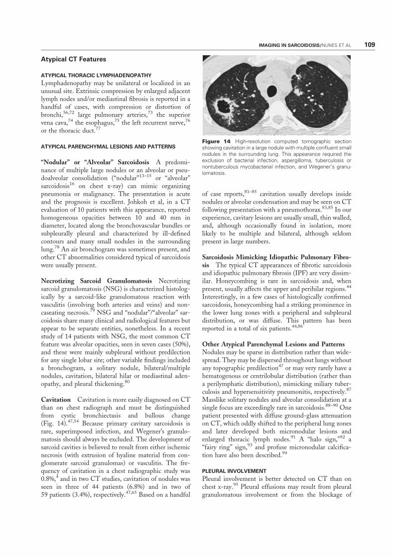
Cavitation Cavitation is more easily diagnosed on CTthan on chest radiograph and must be distinguishedfrom cystic bronchiectasis and bullous change(Fig. 14).47,54 Because primary cavitary sarcoidosis israre, superimposed infection, and Wegener’s granulo-matosis should always be excluded. The development ofsarcoid cavities is believed to result from either ischemicnecrosis (with extrusion of hyaline material from con-glomerate sarcoid granulomas) or vasculitis. The fre-quency of cavitation in a chest radiographic study was0.8%,4 and in two CT studies, cavitation of nodules wasseen in three of 44 patients (6.8%) and in two of59 patients (3.4%), respectively.47,65 Based on a handful
of case reports,81–85 cavitation usually develops insidenodules or alveolar condensation and may be seen on CTfollowing presentation with a pneumothorax.83,85 In ourexperience, cavitary lesions are usually small, thin walled,and, although occasionally found in isolation, morelikely to be multiple and bilateral, although seldompresent in large numbers.
Sarcoidosis Mimicking Idiopathic Pulmonary Fibro-sis The typical CT appearances of fibrotic sarcoidosisand idiopathic pulmonary fibrosis (IPF) are very dissim-ilar. Honeycombing is rare in sarcoidosis and, whenpresent, usually affects the upper and perihilar regions.44
Interestingly, in a few cases of histologically confirmedsarcoidosis, honeycombing had a striking prominence inthe lower lung zones with a peripheral and subpleuraldistribution, or was diffuse. This pattern has beenreported in a total of six patients.44,86
Other Atypical Parenchymal Lesions and PatternsNodules may be sparse in distribution rather than wide-spread. They may be dispersed throughout lungs withoutany topographic predilection47 or may very rarely have ahematogenous or centrilobular distribution (rather thana perilymphatic distribution), mimicking miliary tuber-culosis and hypersensitivity pneumonitis, respectively.87
Masslike solitary nodules and alveolar consolidation at asingle focus are exceedingly rare in sarcoidosis.88–90 Onepatient presented with diffuse ground-glass attenuationon CT, which oddly shifted to the peripheral lung zonesand later developed both micronodular lesions andenlarged thoracic lymph nodes.91 A ‘‘halo sign,’’92 a‘‘fairy ring’’ sign,93 and profuse micronodular calcifica-tion have also been described.94
PLEURAL INVOLVEMENT
Pleural involvement is better detected on CT than onchest x-ray.95 Pleural effusions may result from pleuralgranulomatous involvement or from the blockage of
Figure 14 High-resolution computed tomographic sectionshowing cavitation in a large nodule with multiple confluent smallnodules in the surrounding lung. This appearance required theexclusion of bacterial infection, aspergilloma, tuberculosis ornontuberculous mycobacterial infection, and Wegener’s granu-lomatosis.
IMAGING IN SARCOIDOSIS/NUNES ET AL 109
interlobular septal lymphatic channels by granulomas.Apparent pleural thickening often represents inwardretraction of extrathoracic soft tissue and extrapleuralfat, rather than a true pleural abnormality.62 Pleuralinvolvement is rare in sarcoidosis, with pleural effusionsobserved in less than 5% on chest radiography.10,11 InCT studies, pleural thickening surface is present in 11 to27% of patients.41,47,54,56 However, in the recent studyof Szwarcberg et al the frequency of pleural involvementreached 41%, including thickening in 32.8% of cases,mainly associated with pulmonary fibrosis, and a limitedeffusion in 8.2% of cases.95
Diagnostic Role of CT
The diagnostic superiority of CT over chest radiographyin DLD has been clearly demonstrated.31–36 Whenstudied populations were grouped, the overall diagnosticsensitivity of CT for pulmonary sarcoidosis was 78% asopposed to 70% for chest radiography.61,62 Grenier et alevaluated supplementary information yielded by CTafter taking into account clinical data and radiographicfindings.35 Computer-aided diagnoses were made byapplying a Bayesian model in a large population of 308patients with DLDs, including 121 with pulmonarysarcoidosis. For sarcoidosis, the overall sensitivity (uti-lizing two observers) was 76 to 83% with clinical dataalone, 80 to 88% with clinical and radiographic findings,and 85 to 90% with the addition of CT. The percentagesof correct diagnosis with a high level of confidence wererespectively 33 to 42%, 52 to 76%, and 78 to 80%.35 Thisrelatively small difference emphasizes the fact that CTadds little when clinical and radiographic presentation istypical of sarcoidosis, understates the true diagnosticvalue of CT in other, less straightforward cases.
In atypical variants, CT often increases the diag-nostic likelihood of sarcoidosis, especially when unusualfeatures are associated with more characteristic patterns:atypical CT features seen in isolation makes the diseasedifficult to diagnose.
Thoracic lymphadenopathy on CT is usual insarcoidosis but is not a specific CT feature. Niimi et alfound that lymphadenopathy was also common in colla-gen vascular disease (70%), IPF (67%), extrinsic allergicalveolitis (53%), and organizing pneumonia (36%).However, the number of enlarged nodes was higher insarcoidosis than in other DLDs (mean 3.2 vs 1.2), and allpatients with nodes � 20 mm in short-axis diameter hadsarcoidosis.60
Conversely, the pattern and distribution of lymphnode calcification may be an important ancillary diag-nostic sign on CT to distinguish between sarcoidosis andan alternative DLD associated with. a previous episodeof tuberculosis. Gawne-Caine and Hansell showed thatthe mean diameter of calcified nodes was significantlylarger in sarcoidosis (12 mm vs 7 mm); calcium deposi-
tion was more commonly focal in sarcoidosis (58% vs23%) and diffuse in tuberculosis (27% vs 62%), and,when present, hilar nodal calcification was more likely tobe bilateral in sarcoidosis than in tuberculosis (65% vs8%).58
CT FOR NORMAL CHEST RADIOGRAPH BUT CLINICAL
SUSPICION OF SARCOIDOSIS
Ascertaining the diagnosis of sarcoidosis is sometimesproblematic when sarcoidosis is confined to one organ.CT may facilitate the diagnosis when standard diagnos-tic tests are negative or equivocal (as is commonly thecase in uveitis) or when a biopsy of an extrapulmonic siteis considered too risky (as in central nervous systeminvolvement). In one study of uveitis, CT was especiallyuseful in elderly women with normal chest radiographicappearances.96
CT AND THE DETECTION OF COMPLICATIONS
The value of CT in the detection of complications hasnever been formally quantified but is amply validated byclinical experience, especially when there is unexplainedworsening of respiratory symptoms, hemoptysis, dispro-portionate impairment of PFTs or airflow obstruction,uncertain chest radiographic abnormalities, aspergil-loma, or PH.1,97
Aspergilloma Mycetoma formation is a frequent com-plication of stage IV disease, particularly in patients withadvanced fibrocystic or cavitary disease.5,98 Aspergillomais more readily visualized using CT, which generallyreveals a cavity containing a well-defined homogeneousnodule, with or without a characteristic air crescent(Fig. 15).99 Fungus balls can be mobile, as judged bymovement within a cavity with a change from the supineto the prone position. Aspergillomas are often associatedwith thickening of the cavity wall and adjacent pleura,which is sometimes the earliest sign before any visible
Figure 15 High-resolution computed tomographic sectionshowing an aspergilloma (arrow) in a cavity with a characteristicair crescent.
110 SEMINARS IN RESPIRATORY AND CRITICAL CARE MEDICINE/VOLUME 28, NUMBER 1 2007
changes are seen within the cavity.99 Aspergilloma maybe multiple and bilateral, and this is best appreciated byCT and imperative to recognize before surgery.
Pulmonary Hypertension PH is not uncommon inadvanced disease100 and is usually attributed to thedestruction of the distal capillary bed by fibrotic processor to chronic hypoxemia or both. However, it has alsobeen reported in the early stages of the disease withmarginal or no pulmonary involvement,73 suggestingalternative pathogenetic mechanisms, including extrinsiccompression of large pulmonary arteries (Figs. 16 and17) by enlarged lymph nodes or mediastinal fibro-sis101,102; specific granulomatous vascular involve-ment,101,103 which sometimes simulates secondarypulmonary veno-occlusive disease104; and pulmonaryvasoconstriction by vasoactive factors.105 Contrast-en-hanced CT may raise the possibility of PH when themain pulmonary artery is larger than 30 mm in diameterand may also help to identify underlying mechanisms,73
but pulmonary angiography is often required to confirmcompression by extravascular structures.73
Bronchial Stenosis Symptomatic bronchial stenosis israre and may develop at any stage of the disease.56,72
Obstruction may be the consequence of endobronchialgranulomatous involvement, external compression byenlarged lymph nodes, or severe architectural distortion.For anatomical reasons, the right middle lobe is mostoften affected.72 Apart from showing atelectasis, CT issometimes useful in determining the extent and natureof bronchial stenosis.56
Prognostic Role of CT
Correlations between CT findings and classic markers ofsarcoidosis activity, including serum angiotensin-con-verting enzyme (ACE) levels, bronchoalveolar lavage
findings, and gallium scan signal, have been inconclusiveor discrepant.31,38,40,42,43,47 Much more has been learnedfrom studies of the reversibility of CT features (with orwithout treatment) on serial scanning.39–41,53 Architec-tural distortion, traction bronchiectasis, honeycombing,and bullae are consistently irreversible. Micronodules,nodules, peribronchovascular thickening, and consolida-tion are wholly or partially reversible in most (but not all)cases. The evolution of ground-glass attenuation andlinear opacities is more variable. Ground-glass opacitiesmay steady, worsen, or improve over time, with orwithout treatment,39–41,53 reflecting the fact that groundglass may represent either granulomas or fine fibro-sis.63,66 As in other DLDs, a coarse texture or concom-itant traction bronchiectasis increases the likelihood ofunderlying fibrosis.66 Similarly, septal thickening fromintense granulomatous infiltration is likely to reverse, butother linear opacities are more likely to be fibrotic.
Discrimination between active inflammation andirreversible fibrosis with CT is appealing in theory butseldom employed in practice, perhaps because decisionson the need for treatment are straightforward in mostcases. However, CT may occasionally be helpful whenthe decision to initiate or continue potentially toxictreatment is marginal. For instance, in stage IV diseasea trial of therapy might be warranted if a potentiallyreversible component is visible on CT.44
CORRELATIONS BETWEEN CT AND PULMONARY
FUNCTION TESTS
Correlations between CT findings and PFTs have beenexamined in many studies40,44–53 with variable results,
Figure 16 Enhanced computed tomographic section from apatient with moderate pulmonary hypertension showing extrinsiccompression of the main right artery by lymphadenopathy (ar-row). There is prestenotic enlargement of the right pulmonaryartery.
Figure 17 Pulmonary angiogram from a patient with moderatepulmonary hypertension confirming the presence of concentricstenosis of the intermediary pulmonary artery consistent withextrinsic compression (arrow).
IMAGING IN SARCOIDOSIS/NUNES ET AL 111
probably reflecting the diversity of CT scoring systems.In most reports, the degree of functional impairment, asassessed by gas transfer levels and spirometric volumes, isusually linked to an overall CT score,40,44–53 but mostcorrelations are weak45–47,51 and CT is not consistentlysuperior to chest radiography.45–47,51 Hansell demon-strated that the extent of reticular abnormalities wasstrongly and independently associated with several in-dices of airflow obstruction.29 Abehsera et al. showedthat in stage IV disease, bronchial distortion was asso-ciated with lower expiratory airflow rates, honeycombingwas associated with restriction and lower diffusing ca-pacity for carbon monoxide (DLCO), but functionalimpairment was relatively minor when nondestructivelinear abnormalities predominated.44 Air trapping isassociated with evidence of small-airway obstruction,including maximal midrespiratory flow rates at 25 to75% of the vital capacity (MMEF25–75), residual vol-ume (RV), and/or RV/total lung capacity (TLC),48–50
although this was not the case in one report.52
OTHER IMAGING TECHNIQUESMagnetic resonance imaging (MRI) is seldom useful inevaluation of the lung parenchyma because of imaginglimitations, due mainly to the composition of lung tissueand physiological motion (cardiac pulsation and respi-ration). Despite these inherent pitfalls, several authorshave studied thoracic MRI in sarcoidosis106,107 or DLDat large.108–111 All studies but one used unenhanced T1-and T2-weighted MRI106–110 with unconvincing re-sults,112 and MRI was inferior to HRCT.109 Morerecently Gaeta et al first used gadolinium-enhancedthoracic MRI in a DLD cohort including 10 patientswith sarcoidosis111 and showed enhancement of pulmo-nary lesions in five of seven patients with active disease.However, it appears highly unlikely that MRI willsupplant HRCT in the foreseeable future.
The lung clearance of inhaled technetium-99mlabeled diethylene triamine penta-acetic acid (99mTc-DTPA) has been studied in pulmonary sarcoidosis.113–120 An increased rate of lung clearance has been asso-ciated with disease activity in some studies113,114 but notin others,115,118 and with functional deterioration.116
DTPA clearance sometimes returns to normal withcorticosteroid therapy.116,118 However, the role of thistest in the management of sarcoid patients remainslargely speculative.
CENTRAL NERVOUS SYSTEMSARCOIDOSISCentral nervous system (CNS) involvement occurs inless than 10% of patients with sarcoidosis.1,5,120
Although any part of the CNS can be affected, sarcoi-dosis has a strong predilection for the base of the skull,
hypothalamus, pituitary gland, and optic chiasma.112,121
The development of neurosarcoidosis is primarily lepto-meningeal and vascular (Fig. 18) in nature. It may followdisruption of the leptomeningeal blood–brain barrier,permitting the spread of the granulomatous process tothe brain parenchyma along the so-called perivascular orVirchow-Robin spaces that accompany the penetratingarteries up to the capillaries.112,121 As a consequence, twotypes of lesion are observed: coalescent intraparenchymalgranulomas and small ischemic foci.107,108 Imaging ispivotal for the diagnosis of CNS sarcoidosis, especially inpatients without systemic evidence of disease, becauseCNS biopsy is seldom justifiable.
MRI
Contrast-enhanced brain CT is insensitive, with normalappearances in up to a third of patients with definiteCNS sarcoidosis, especially when lesions are confined tothe cranial or peripheral nerves or brainstem.62 CNSsarcoidosis may still be imperceptible at MRI in somepatients with cranial nerve or neuroendocrine symp-toms122 but is clearly more sensitive than CT.112,121,123
Thus gadolinium-enhanced MRI is the diagnostic in-vestigation of choice in suspected CNS sarcoidosis.
Leptomeningeal involvement, the most frequentfeature, may not be apparent with nonenhancedMRI.124,125 It typically manifests as thickening andenhancement of leptomeninges on T1-weighted images,usually at the base of the skull. The abnormal signal maybe diffuse or nodular112,121 and is indistinguishable fromabnormalities seen in tuberculosis or malignancy.112,121
Focal dural masses may also mimic meningio-ma.112,121Intraparenchymal involvement is usually evidenton MRI as multifocal periventricular, subcortical, ordeep white matter lesions (Fig. 19).112,121 These
Figure 18 Axial contrast-enhanced T1-weighted brain mag-netic resonance image showing a typical multifocal enhancementafter gadolinium in the periventricular zone and along the pene-trating arteries.
112 SEMINARS IN RESPIRATORY AND CRITICAL CARE MEDICINE/VOLUME 28, NUMBER 1 2007
abnormalities are nonspecific and may be identical tothose seen in vascular disease or multiple sclerosis.112,121
Multiple or solitary space-occupying lesions, usuallywithout central necrosis, are not infrequent and maysimulate malignancy due to extensive surroundingedema.112,121 Intraparenchymal lesions are frequentlyclustered near regions of leptomeningeal involvement.Recent and active inflammatory lesions usually enhanceafter gadolinium injection on T1-weighted images,whereas irreversible lesions do not enhance and typicallydisplay hyperintense signal on T2-weighted image-s.122,126,127Hypothalamus and pituitary infiltration(Fig. 20) ranges from a thickening of pituitary infundib-
ulum, indistinguishable from findings in Langerhans’cell hystiocytosis, to an intra- or suprasellar mas-s.112,121Intramedullary lesions manifest as a fusiform en-largement of the spinal cord in the cervical or upperthoracic level (Fig. 21), appearing in weighted imagesand after gadolinium injection, depending upon thestage of disease stage.112,121,128 Neurological complica-tions, such as an obstructive or communicating hydro-cephalus, may also be disclosed by MRI.121 Despite thehigh frequency of histological vasculitis in CNS sarcoi-dosis, large brain infarcts are rare.121
Unfortunately, MRI manifestations are not path-ognomonic of sarcoidosis.127,129 However, the enhance-ment of intraparenchymal lesions and the coexistence ofleptomeningeal enhancement are highly suggestive in anappropriate clinical context.112 MRI may also have aprognostic role: lesions enhancing with gadolinium aremore likely to regress with therapy.122,126,127 SequentialMRI is sometimes useful in monitoring patients withCNS sarcoidosis,126,127 but symptomatic improvementoften correlates poorly with MRI change,122 especially inspinal cord disease.130
CARDIAC SARCOIDOSISClinical evidence of cardiac involvement is present in�5% of sarcoidosis patients,1,5,131 although cardiac ab-normalities are more prevalent in Japan.1,131 Histologi-cally, the myocardium is most frequently involved, butgranulomas may also be found in the pericardium,myocardium, and endocardium. The predominant sitesof myocardial involvement, in decreasing order of fre-quency, are left ventricular free wall and papillary
Figure 20 Midsagittal contrast-enhanced T1-weighted brainmagnetic resonance image demonstrating a gadolinium-enhan-cing mass of the floor of the third ventricle and hypothalamus(arrow).
Figure 21 Sagittal T1-weighted magnetic resonance image ofthe spinal cord showing an intramedullary gadolinium-enhancingmass responsible for cordal enlargement (arrow).
Figure 19 Coronal proton density-weighted brain magneticresonance image demonstrating hyperintense deepwhitematterlesions that did not enhance after gadolinium and infiltrativelesions in the chiasmatic area (arrow).
IMAGING IN SARCOIDOSIS/NUNES ET AL 113
muscles, the intraventricular septum, the right ventric-ular wall, the right atrium, and the left atrium.131 Thediagnostic evaluation of cardiac involvement is undeni-ably one of the most challenging management issues insarcoidosis. No diagnostic reference standard exists be-cause endomyocardial biopsy is invasive and lacks sensi-tivity as a result of sampling error. Official guidelinesfrom the Japanese Ministry of Health and Welfareinclude the results of histological analysis, electrocardio-gram (ECG), echocardiography, thallium-201 (201Tl) ortechnetium-99m (99mTc) myocardial scintigraphy orgallium-67-citrate (67Ga) scan, and cardiac catheteriza-tion.131 However, these guidelines have never beenprospectively validated and they predate the advent ofnewer techniques such as 67Ga plus 99mTc-MIBI132 andsingle-photon emission computed tomography.132,133
Moreover, there is emerging evidence that cardiacMRI (CMR)112,131,134–141 and fluorine 18 fluorodeox-yglucose (18FDG) positron emission tomography(PET)142–144 may be clinically useful.
Cardiac MRI
CMR has a unique ability to provide detailed anatomicalinformation and, also, to quantify global and regionalventricular function by means of cine sequen-ces.112,131,134 CMR typically shows zones of thinningand segmental myocardial wall motion abnormalitieswith increased signal intensity (Fig. 22), more pro-nounced on T2-weighted images because of associatededema, and variably enhancing with gadoli-nium.112,131,134 The pattern of enhancement followsthe histological distribution of granulomas and differs
from that seen in ischemic disease.112,131,134 The sen-sitivity and specificity of CMR remain uncertain. Sme-dema et al evaluated the accuracy of CMR in a series of58 patients with suspected cardiac sarcoidosis, using theJapanese guidelines131 as a ‘‘gold-standard,’’ and founda sensitivity and specificity of 100% and 78%, respec-tively.139 In a further study, Smedema et al comparedCMR with standard assessment in 55 patients withpulmonary sarcoidosis who were investigated becauseof cardiac symptoms or for screening purposes.141
CMR was abnormal in all 13 patients diagnosed withcardiac sarcoidosis by the standard assessment anddetected six additional patients in whom ECG, ambu-latory ECG, Doppler-echocardiography, and 201Tlscintigraphy were wholly normal. Thus CMR maydetect early cardiac involvement when a conventionalapproach fails to do so.137 However, the clinical rele-vance of limited CMR abnormalities was difficult toquantify as patient follow-up, though short, was un-eventful.141 The extent of enhancement correlated withdisease severity, especially ventricular dilatation, func-tional impairment (Fig. 23) and the presence of ven-tricular arrhythmias.141
Serial studies show that enhanced areas markedlyregress or normalize completely with corticosteroidtherapy135,137 in parallel with clinical improvement.137
The major drawback of CMR is that it cannot be usedin patients with cardiac implantable devices, which arefrequently required in cardiac sarcoidosis.112,131 Fur-ther systematic multicenter studies are required toconstruct a clinically useful diagnostic algorithm forCMR in the detection and management of cardiacsarcoidosis.
Figure 22 Delayed enhanced cardiac magnetic resonance imaging [short axis view (a)] demonstrating a transmural late enhancementof the anterolateral area (long arrow) and a subepicardial late enhancement of the inferoseptal area (short arrow). Single photon emissioncomputed tomography [short axis (b) and vertical long axis (c) views] demonstrates perfusion abnormalities in the same areas.
114 SEMINARS IN RESPIRATORY AND CRITICAL CARE MEDICINE/VOLUME 28, NUMBER 1 2007
18FDG PET18FDG PET has been compared with 67Ga,142–144201Tl,142 and 99mTc-MIBI scintigraphy143,144 in patientswith cardiac sarcoidosis according to the Japaneseguidelines. The sensitivity of 18FDG PET was 100%,which was higher than that of the other methods.128,129
By contrast, its specificity was slightly lower at 81.5 to90.9%. Yamagishi et al showed that 18FDG uptakeseither diminished in size and density or disappearedunder corticosteroids.142 Further studies are still neededbefore any definite statement can be made on the exactrole of 18FDG PET.
Other Imaging Techniques
Other imaging techniques have been tested in cardiacsarcoidosis but data are sporadic: myocardial scintig-raphy with the use of iodine-123-labeled 15-(p-iodo-phenyl)-3R,S-methylpentadecanoic acid,145 a creativetechnique of ultrasonic tissue characterization, whichnoninvasively measures acoustic properties of themyocardium,146 and contrast-enhanced mutlisliceCT.147
SPECIFIC IMAGING TECHNIQUES
67Ga Scanning
The sensibility of 67Ga scan in detecting pulmonarysarcoidosis ranges from 60 to 90%.112 The character-
istic appearances of 67Ga uptake in sarcoidosis has beenreferred to as the ‘‘lambda’’ and ‘‘panda’’ patterns.112,148
The lambda pattern consists of bilateral symmetricaluptake in the parahilar and infrahilar lymph nodes,together with right paratracheal lymph node uptake,and may be observed even when the chest radiograph isnormal. Symmetrical uptake in the parotid, lacrimal,and salivary glands with normal nasopharyngeal captureproduces a picture similar to the face of a panda.112,148
The ‘‘lambda plus panda’’ pattern, and the pandapattern coupled with bilateral hilar lymphadenopathyor bilateral parenchymal infiltration on chest radiogra-phy, are believed to be highly specific for sarcoidosis.However, a panda pattern in isolation lacks specificity,occurring as in other clinical contexts including lym-phoma.112,148–150 It has been argued that the presenceof the lambda plus panda pattern may obviate a tissuediagnosis.1
Uptake of 67Ga is seen in a variety of extrapulmo-nary sites,151 in addition to normal uptake in the liverand spleen, but the low sensitivity and specificity ofabnormal uptake limits the value of 67Ga scintigraphyin the evaluation of specific organ involvement. Theusefulness of 67Ga scan as an index of activity has beenexamined in several studies,152–157 but despite correla-tions with serum ACE levels154–156 and bronchoalveolarlavage findings,152,153,157 the results have been incon-clusive or conflicting.112 Similarly, 67Ga scan is notbelieved to be useful in prognostic evaluation or mon-itoring.112
Figure 23 Three-dimensional breath-hold cardiac magnetic resonance (short axis) in diastole (a) and systole (b) demonstrating septalthickening and functional impairment in the inferoseptal area (arrows) with corresponding perfusion abnormalities at single photonemission computed tomography [short axis (c) and vertical long axis (d) views].
IMAGING IN SARCOIDOSIS/NUNES ET AL 115
18FDG PET
Recently, several reports on 18FDG PET in sarcoidosishave appeared. 18FDG is taken up in several sites insarcoidosis but the sensitivity and specificity of abnormalsignal is uncertain. 18FDG may be most useful indiagnosing cardiac sarcoidosis.142–144 Brudin et al com-pared regional pulmonary 18FDG metabolism withPFTs, chest radiography, and serum ACE (SACE)levels, initially and after corticosteroid therapy, in sevenpatients with sarcoidosis.158 The degree of 18FDGuptake correlated well with SACE levels and diseaseextent at different stages and normalized during treat-ment.158 Yamada et al evaluated the uptake of 18FDGand carbon-11 labeled methionine in 31 patients withintrathoracic sarcoidosis lymphadenopathy. By using theratio of 18FDG to methionine in pretreatment evalua-tion, they were able to predict posttherapy improvementas assessed clinically and by chest radiography. In the18FDG dominant group (ratio � 2), the response ratewas 78% but in the methionine dominant group (ratio< 2) it dropped to 38%.159 Enthusiasm for 18FDG PETshould be tempered by concerns over its radiation doseand cost.
REFERENCES
1. Statement on sarcoidosis. Joint Statement of the AmericanThoracic Society (ATS), the European Respiratory Society(ERS) and the World Association of Sarcoidosis and OtherGranulomatous Disorders (WASOG) adopted by the ATSBoard of Directors and by the ERS Executive Committee,February 1999. Am J Respir Crit Care Med 1999;160:736–755
2. Nunes H, Soler P, Valeyre D. Pulmonary sarcoidosis.Allergy 2005;60:565–582
4. Mayock RL, Bertrand P, Morrison CE, Scott JH.Manifestations of sarcoidosis. Analysis of 145 patients, witha review of nine series selected from the literature. Am JMed 1963;35:67–89
5. Johns CJ, Michele TM. The clinical management ofsarcoidosis: a 50-year experience at the John HopkinsHospital. Medicine (Baltimore) 1999;78:65–111
6. Scadding JG. Prognosis of intrathoracic sarcoidosis inEngland. Br Med J 1961;2:1165–1172
7. Mana J, Gomez-Vaquero C, Montero A, et al. Lofgren’ssyndrome revisited: a study of 186 patients. Am J Med1999;107:240–245
8. Winterbauer RH, Belic N, Moores KD. Clinical interpre-tation of bilateral hilar adenopathy. Ann Intern Med 1973;78:65–71
9. Bein ME, Putman CE, McLoud TC, Mink JH. Areevaluation of intrathoracic lymphadenopathy in sarcoido-sis. AJR Am J Roentgenol 1978;131:409–413
10. Littner MR, Schachter EN, Putman CE, et al. The clinicalassessment of roentgenographically atypical pulmonarysarcoidosis. Am J Med 1977;62:361–368
14. Onal E, Lopata M, Lourenco RV. Nodular pulmonarysarcoidosis: clinical, roentgenographic, and physiologiccourse in five patients. Chest 1977;72:296–300
15. Romer FK. Sarcoidosis with large nodular lesions simulatingpulmonary metastases: an analysis of 126 cases of intra-thoracic sarcoidosis. Scand J Respir Dis 1977;58:11–16
16. Battesti JP, Saumon G, Valeyre D, et al. Pulmonarysarcoidosis with an alveolar radiographic pattern. Thorax1982;37:448–452
17. Tazi A, Desfemmes-Baleyte T, Soler P, et al. Pulmonarysarcoidosis with a diffuse ground-glass pattern on the chestradiograph. Thorax 1994;49:793–797
18. Rohatgi PK, Schwab LE. Primary acute pulmonarycavitation in sarcoidosis. AJR Am J Roentgenol 1980;134:1199–1203
19. Sharma OP, Gordonson J. Pleural effusion in sarcoidosis: areport of six cases. Thorax 1975;30:95–101
20. Soskel N, Sharma OP. Pleural involvement in sarcoidosis:case presentation and detailed review of the literature.Semin Respir Med 1992;13:492–514
21. Mendelson DS, Norton K, Cohen BA, et al. Bronchialcompression: an unusual manifestation of sarcoidosis.J Comput Assist Tomogr 1983;7:892–894
22. Munt PW. Middle lobe atelectasis in sarcoidosis. Am RevRespir Dis 1973;108:357–360
23. Siltzbach LE, James DG, Neville E, et al. Course andprognosis of sarcoidosis around the world. Am J Med 1974;57:847–852
24. Romer FK. Presentation of sarcoidosis and outcome ofpulmonary changes. Dan Med Bull 1982;29:27–32
25. Hillerdal G, Nou E, Osterman K, et al. Sarcoidosis:epidemiology and prognosis: a 15-year European study.Am Rev Respir Dis 1984;130:29–32
26. Gibson GJ, Prescott RJ, Muers MF, et al. British ThoracicSociety Sarcoidosis study: effects of long term corticosteroidtreatment. Thorax 1996;51:238–247
27. Pietinalho A, Tukiainen P, Haahtela T, et al. Oralprednisolone followed by inhaled budesonide in newlydiagnosed pulmonary sarcoidosis: a double-blind, placebo-controlled multicenter study. Finnish Pulmonary SarcoidosisStudy Group. Chest 1999;116:424–431
28. Pietinalho A, Tukiainen P, Haahtela T, et al. Earlytreatment of stage II sarcoidosis improves 5-year pulmonaryfunction. Chest 2002;121:24–31
29. Hansell DM. High-resolution CT of diffuse lung disease:value and limitations. Radiol Clin North Am 2001;39:1091–1113
30. Beigelman-Aubry C, Hill C, Guibal A, Savatovsky J,Grenier PA. Multi-detector row CT and postprocessingtechniques in the assessment of diffuse lung disease.Radiographics 2005;25:1639–1652
31. Bergin CJ, Coblentz CL, Chiles C, et al. Chronic lungdiseases: specific diagnosis by using CT. AJR Am JRoentgenol 1989;152:1183–1188
116 SEMINARS IN RESPIRATORY AND CRITICAL CARE MEDICINE/VOLUME 28, NUMBER 1 2007
32. Mathieson JR, Mayo JR, Staples CA, et al. Chronic diffuseinfiltrative lung disease: comparison of diagnostic accuracyof CT and chest radiography. Radiology 1989;171:111–116
33. Padley SP, Hansell DM, Flower CD, et al. Comparativeaccuracy of high-resolution computed tomography and chestradiography in the diagnosis of chronic diffuse infiltrativelung disease. Clin Radiol 1991;44:222–226
34. Grenier P, Valeyre D, Cluzel P, et al. Chronic diffuseinterstitial lung disease: diagnostic value of chest radiog-raphy and high-resolution CT. Radiology 1991;179:123–132
35. Grenier P, Chevret S, Beigelman C, et al. Chronic diffuseinfiltrative lung disease: determination of the diagnosticvalue of clinical data, chest radiography, and CT andBayesian analysis. Radiology 1994;191:383–390
36. Nishimura K, Izumi T, Kitaichi M, et al. The diagnosticaccuracy of high-resolution computed tomography in diffuseinfiltrative lung diseases. Chest 1993;104:1149–1155
37. Mana J, Teirstein AS, Mendelson DS, et al. Excessivethoracic computed tomographic scanning in sarcoidosis.Thorax 1995;50:1264–1266
38. Lynch DA, Webb WR, Gamsu G, et al. Computedtomography in pulmonary sarcoidosis. J Comput AssistTomogr 1989;13:405–410
39. Brauner MW, Lenoir S, Grenier P, et al. Pulmonarysarcoidosis: CT assessment of lesion reversibility. Radiology1992;182:349–354
40. Remy-Jardin M, Giraud F, Remy J, et al. Pulmonarysarcoidosis: role of CT in the evaluation of disease activityand functional impairment and in prognosis assessment.Radiology 1994;191:675–680
42. Leung AN, Brauner MW, Caillat-Vigneron N, et al.Sarcoidosis activity: correlation of HRCT findings withthose of 67Ga scanning, bronchoalveolar lavage, and serumangiotensin-converting enzyme assay. J Comput AssistTomogr 1998;22:229–234
43. Oberstein A, von Zitzewitz H, Schweden F, Muller-Quernheim J. Noninvasive evaluation of the inflammatoryactivity in sarcoidosis with high-resolution computedtomography. Sarcoidosis Vasc Diffuse Lung Dis 1997;14:65–72
44. Abehsera M, Valeyre D, Grenier P, et al. Sarcoidosis withpulmonary fibrosis: CT patterns and correlation withpulmonary function. AJR Am J Roentgenol 2000;174:1751–1757
45. Bergin CJ, Bell DY, Coblentz CL, et al. Sarcoidosis:correlation of pulmonary parenchymal pattern at CT withresults of pulmonary function tests. Radiology 1989;171:619–624
46. Muller NL, Mawson JB, Mathieson JR, et al. Sarcoidosis:correlation of extent of disease at CT with clinical,functional, and radiographic findings. Radiology 1989;171:613–618
47. Brauner MW, Grenier P, Mompoint D, et al. Pulmonarysarcoidosis: evaluation with high-resolution CT. Radiology1989;172:467–471
48. Hansell DM, Milne DG, Wilsher ML, et al. Pulmonarysarcoidosis: morphologic associations of airflow obstructionat thin-section CT. Radiology 1998;209:697–704
49. Davies CW, Tasker AD, Padley SP, et al. Air trapping insarcoidosis on computed tomography: correlation with lungfunction. Clin Radiol 2000;55:217–221
50. Magkanas E, Voloudaki A, Bouros D, et al. Pulmonarysarcoidosis: correlation of expiratory high-resolution CTfindings with inspiratory patterns and pulmonary functiontests. Acta Radiol 2001;42:494–501
51. Drent M, De Vries J, Lenters M, et al. Sarcoidosis:assessment of disease severity using HRCT. Eur Radiol2003;13:2462–2471
52. Terasaki H, Fujimoto K, Muller NL, et al. Pulmonarysarcoidosis: comparison of findings of inspiratory andexpiratory high-resolution CT and pulmonary function testsbetween smokers and nonsmokers. AJR Am J Roentgenol2005;185:333–338
53. Akira M, Kozuka T, Inoue Y, Sakatani M. Long-termfollow-up CT scan evaluation in patients with pulmonarysarcoidosis. Chest 2005;127:185–191
54. Hamper UM, Fishman EK, Khouri NF, et al. Typical andatypical CT manifestations of pulmonary sarcoidosis.J Comput Assist Tomogr 1986;10:928–936
55. Muller NL, Kullnig P, Miller RR. The CT findings ofpulmonary sarcoidosis: analysis of 25 patients. AJR Am JRoentgenol 1989;152:1179–1182
56. Lenique F, Brauner MW, Grenier P, et al. CT assessmentof bronchi in sarcoidosis: endoscopic and pathologiccorrelations. Radiology 1995;194:419–423
57. Sider L, Horton ES Jr. Hilar and mediastinal adenopathy insarcoidosis as detected by computed tomography. J ThoracImaging 1990;5:77–80
58. Gawne-Cain ML, Hansell DM. The pattern and distri-bution of calcified mediastinal lymph nodes in sarcoidosisand tuberculosis: a CT study. Clin Radiol 1996;51:263–267
59. Patil SN, Levin DL. Distribution of thoracic lymphaden-opathy in sarcoidosis using computed tomography. J ThoracImaging 1999;14:114–117
60. Niimi H, Kang EY, Kwong JS, et al. CT of chronicinfiltrative lung disease: prevalence of mediastinal lympha-denopathy. J Comput Assist Tomogr 1996;20:305–308
61. Wells A. High resolution computed tomography insarcoidosis: a clinical perspective. Sarcoidosis Vasc DiffuseLung Dis 1998;15:140–146
62. Lynch JP III. Computed tomographic scanning in sarcoi-dosis. Semin Respir Crit Care Med 2003;24:393–418
63. Nishimura K, Itoh H, Kitaichi M, et al. Pulmonarysarcoidosis: correlation of CT and histopathologic findings.Radiology 1993;189:105–109
64. Honda O, Johkoh T, Ichikado K, et al. Comparison ofhigh resolution CT findings of sarcoidosis, lymphoma,and lymphangitic carcinoma: is there any difference ofinvolved interstitium? J Comput Assist Tomogr 1999;23:374–379
65. Nakatsu M, Hatabu H, Morikawa K, et al. Large coalescentparenchymal nodules in pulmonary sarcoidosis: ‘‘sarcoidgalaxy’’ sign. AJR Am J Roentgenol 2002;178:1389–1393
66. Remy-Jardin M, Giraud F, Remy J, et al. Importance ofground-glass attenuation in chronic diffuse infiltrative lungdisease: pathologic-CT correlation. Radiology 1993;189:693–698
67. Gleeson FV, Traill ZC, Hansell DM. Evidence ofexpiratory CT scans of small-airway obstruction in sarcoi-dosis. AJR Am J Roentgenol 1996;166:1052–1054
69. Fazzi P, Sbragia P, Solfanelli S, et al. Functionalsignificance of the decreased attenuation sign on expiratoryCT in pulmonary sarcoidosis: report of four cases. Chest2001;119:1270–1274
70. Nishino M, Kuroki M, Roberts DH, et al. Bronchomalaciain sarcoidosis: evaluation on volumetric expiratory high-resolution CT of the lung. Acad Radiol 2005;12:596–601
71. Dalal PU, Hansell DM. High-resolution computed tomog-raphy of the lungs: the borderlands of normality. Eur Radiol2006;16:771–780
72. Aye M, Campbell AP, Greenstone MA. An unusual case oflobar collapse. Chest 2002;122:1465–1466
73. Nunes H, Humbert M, Capron F, et al. Pulmonaryhypertension associated with sarcoidosis: mechanisms,haemodynamics and prognosis. Thorax 2006;61:68–74
74. Narayan D, Brown L, Thayer JO. Surgical management ofsuperior vena caval syndrome in sarcoidosis. Ann ThoracSurg 1998;66:946–948
75. Cappell MS. Endoscopic, radiographic, and manometricfindings in dysphagia associated with sarcoid due to extrinsicesophageal compression from subcarinal lymphadenopathy.Am J Gastroenterol 1995;90:489–492
76. Tobias JK, Santiago SM, Williams AJ. Sarcoidosis as acause of left recurrent laryngeal nerve palsy. Arch Otolar-yngol Head Neck Surg 1990;116:971–972
77. Jarman PR, Whyte MK, Sabroe I, Hughes JM. Sarcoidosispresenting with chylothorax. Thorax 1995;50:1324–1325
78. Johkoh T, Ikezoe J, Takeuchi N, et al. CT findings in‘‘pseudoalveolar’’ sarcoidosis. J Comput Assist Tomogr1992;16:904–907
79. Liebow AA, The J. Burns Amberson lecture: pulmonaryangiitis and granulomatosis. Am Rev Respir Dis 1973;108:1–18
80. Quaden C, Tillie-Leblond I, Delobbe A, et al. Necrotisingsarcoid granulomatosis: clinical, functional, endoscopicaland radiological evaluations. Eur Respir J 2005;26:778–785
81. Mackie B, Humphrey DA, Flannery MT. Atypicalsarcoidosis: a case report and literature review. Am J MedSci 2006;331:277–279
82. Ozseker ZF, Yilmaz A, Bayramgurler B, et al. Cavitarysarcoidosis: analysis of two cases. Respirology 2002;7:289–291
83. Froudarakis ME, Bouros D, Voloudaki A, et al. Pneumo-thorax as a first manifestation of sarcoidosis. Chest1997;112:278–280
84. Ichikawa Y, Fujimoto K, Shiraishi T, et al. Primary cavitarysarcoidosis: high-resolution CT findings. AJR Am JRoentgenol 1994;163:745
86. Padley SP, Padhani AR, Nicholson A, et al. Pulmonarysarcoidosis mimicking cryptogenic fibrosing alveolitis onCT. Clin Radiol 1996;51:807–810
87. Gruden JF, Webb WR, Naidich DP, McGuinness G.Multinodular disease: anatomic localization at thin-sectionCT-multireader evaluation of a simple algorithm. Radiology1999;210:711–720
88. Tsiodras S, Eiger G, Guttentag A, Lippmann M.Sarcoidosis presenting as unilateral alveolar consolidation.Am J Med Sci 1997;314:346–347
89. Gotway MB, Tchao NK, Leung JWT, et al. Sarcoidosispresenting as an enlarging solitary pulmonary nodule.J Thorac Imaging 2001;16:117–122
90. Judson MA, Uflacker R. Treatment of a solitary pulmonarysarcoidosis mass by CT-guided direct intralesional injectionof corticosteroid. Chest 2001;120:316–317
91. Ito A, Fujino M, Isada A, et al. Retrograde radiographicdevelopment in pulmonary sarcoidosis. Intern Med2006;45:819–822
92. Marten K, Rummeny EJ, Engelke C. The CT halo: a newsign in active pulmonary sarcoidosis. Br J Radiol 2004;77:1042–1045
93. Marlow TJ, Krapiva PI, Schabel SI, et al. The ‘‘fairy ring’’: anew radiographic finding in sarcoidosis. Chest 1999;115:275–276
95. Szwarcberg JB, Glajchen N, Teirstein AS. Pleural involve-ment in chronic sarcoidosis detected by thoracic CTscanning. Sarcoidosis Vasc Diffuse Lung Dis 2005;22:58–62
96. Kaiser PK, Lowder CY, Sullivan P, et al. Chest compu-terized tomography in the evaluation of uveitis in elderlywomen. Am J Ophthalmol 2002;133:499–505
101. Battesti J-P, Georges R, Basset F, Saumon G. Chronic corpulmonale in pulmonary sarcoidosis. Thorax 1978;33:76–84
102. Damuth TE, Bower JS, Cho K, Dantzker DR. Majorpulmonary artery stenosis causing pulmonary hypertensionin sarcoidosis. Chest 1980;78:888–891
103. Smith LJ, Lawrence JB, Katzenstein AL. Vascular sarcoi-dosis: a rare cause of pulmonary hypertension. Am J MedSci 1983;285:38–44
104. Hoffstein V, Ranganathan N, Mullen JBM. Sarcoidosissimulating pulmonary veno-occlusive disease. Am RevRespir Dis 1986;134:809–811
105. Barst RJ, Ratner SJ. Sarcoidosis and reactive pulmonaryhypertension. Arch Intern Med 1985;145:2112–2114
106. Craig DA, Colletti PM, Ratto D, et al. MRI findings inpulmonary sarcoidosis. Magn Reson Imaging 1988;6:567–573
107. Mendelson DS, Gray CE, Teirstein AS. Magnetic reso-nance findings in sarcoidosis of the thorax. Magn ResonImaging 1992;10:523–529
108. McFadden RG, Carr TJ, Wood TE. Proton magneticresonance imaging to stage activity of interstitial lungdisease. Chest 1987;92:31–39
109. Muller NL, Mayo JR, Zwirewich CV. Value of MR imagingin the evaluation of chronic infiltrative lung diseases:comparison with CT. AJR Am J Roentgenol 1992;158:1205–1209
118 SEMINARS IN RESPIRATORY AND CRITICAL CARE MEDICINE/VOLUME 28, NUMBER 1 2007
110. Primack SL, Mayo JR, Hartman TE, et al. MRI ofinfiltrative lung disease: comparison with pathologic find-ings. J Comput Assist Tomogr 1994;18:233–238
111. Gaeta M, Blandino A, Scribano E, et al. Chronic infiltrativelung diseases: value of gadolinium-enhanced MRI in theevaluation of disease activity–early report. Chest 2000;117:1173–1178
112. Mana J. Magnetic resonance imaging and nuclear imagingin sarcoidosis. Curr Opin Pulm Med 2002;8:457–463
113. Jacobs MP, Baughman RP, Hughes J, Fernandez-Ulloa M.Radioaerosol lung clearance in patients with active pulmo-nary sarcoidosis. Am Rev Respir Dis 1985;131:687–689
114. Dusser DJ, Collignon MA, Stanislas-Leguern G, et al.Respiratory clearance of 99mTc-DTPA and pulmonaryinvolvement in sarcoidosis. Am Rev Respir Dis 1986;134:493–497
115. Thunberg S, Larsson K, Eklund A, Blaschke E. 99mTc-DTPA clearance measured by a dual head gamma camera inhealthy subjects and patients with sarcoidosis: studies ofreproducibility and relation to bronchoalveolar lavagefindings. Eur J Nucl Med 1989;15:71–77
116. Chinet T, Dusser D, Labrune S, et al. Lung functiondeclines in patients with pulmonary sarcoidosis andincreased respiratory epithelial permeability to 99mTc-DTPA. Am Rev Respir Dis 1990;141:445–449
117. Bradvik I, Wollmer P, Evander E, et al. Different kinetics oflung clearance of technetium-99m labelled diethylenetriamine penta-acetic acid in patients with sarcoidosis andsmokers. Eur J Nucl Med 1994;21:1218–1222
118. Bradvik I, Wollmer P, Evander E, et al. One year follow-upof lung clearance of 99mTc-diethylene triamine penta-aceticacid and disease activity in sarcoidosis. Sarcoidosis VascDiffuse Lung Dis 2000;17:281–287
119. Bradvik I, Wollmer P, Evander E, et al. Kinetics of lungclearance of 99mTc-DTPA in smoking patients withsarcoidosis compared to healthy smokers. Respir Med 2002;96:317–321
120. Hoitsma E, Faber CG, Drent M, Sharma OP. Neuro-sarcoidosis: a clinical dilemma. Lancet Neurol 2004;3:397–407
121. Smith JK, Matheus MG, Castillo M. Imaging manifes-tations of neurosarcoidosis. AJR Am J Roentgenol 2004;182:289–295
122. Christoforidis GA, Spickler EM, Recio MV, Mehta BM.MR of CNS sarcoidosis: correlation of imaging features toclinical symptoms and response to treatment. AJNR Am JNeuroradiol 1999;20:655–669
123. Hayes WS, Sherman JL, Stern BJ, et al. MR and CTevaluation of intracranial sarcoidosis. AJR Am J Roentgenol1987;149:1043–1049
124. Sherman JL, Stern BJ. Sarcoidosis of the CNS: comparisonof unenhanced and enhanced MR images. AJR Am JRoentgenol 1990;155:1293–1301
125. Seltzer S, Mark AS, Atlas SW. CNS sarcoidosis: evaluationwith contrast-enhanced MR imaging. AJNR Am J Neuro-radiol 1991;12:1227–1233
126. Dumas JL, Valeyre D, Chapelon-Abric C, et al. Centralnervous system sarcoidosis: follow-up at MR imagingduring steroid therapy. Radiology 2000;214:411–420
127. Lexa FJ, Grossman RI. MR of sarcoidosis in the head andspine: spectrum of manifestations and radiographic responseto steroid therapy. AJNR Am J Neuroradiol 1994;15:973–982
128. Junger SS, Stern BJ, Levine SR, et al. Intramedullary spinalsarcoidosis: clinical and magnetic resonance imaging char-acteristics. Neurology 1993;43:333–337
130. Koike H, Misu K, Yasui K, et al. Differential response tocorticosteroid therapy of MRI findings and clinical mani-festations in spinal cord sarcoidosis. J Neurol 2000;247:544–549
131. Doughan AR, Williams BR. Cardiac sarcoidosis. Heart2006;92:282–288
132. Nakazawa A, Ikeda K, Ito Y, et al. Usefulness of dual 67Gaand 99mTc-sestamibi single-photon-emission CT scanningin the diagnosis of cardiac sarcoidosis. Chest 2004;126:1372–1376
133. Barneveld PC, van Leeuwen C, van Isselt JW. Scintigraphicdemonstration of myocardial sarcoidosis: the added value ofsingle-photon emission computed tomography. J NuclCardiol 1997;4:256–257
134. Vignaux O. Cardiac sarcoidosis: spectrum of MRI features.AJR Am J Roentgenol 2005;184:249–254
135. Shimada T, Shimada K, Sakane T, et al. Diagnosis ofcardiac sarcoidosis and evaluation of the effects of steroidtherapy by gadolinium-DTPA-enhanced magnetic reso-nance imaging. Am J Med 2001;110:520–527
136. Vignaux O, Dhote R, Duboc D, et al. Detection ofmyocardial involvement in patients with sarcoidosis applyingT2-weighted, contrast-enhanced, and cine magnetic reso-nance imaging: initial results of a prospective study.J Comput Assist Tomogr 2002;26:762–767
137. Vignaux O, Dhote R, Duboc D, et al. Clinical significanceof myocardial magnetic resonance abnormalities in patientswith sarcoidosis: a 1-year follow-up study. Chest 2002;122:1895–1901
138. Skold CM, Larsen FF, Rasmussen E, et al. Determinationof cardiac involvement in sarcoidosis by magnetic resonanceimaging and Doppler echocardiography. J Intern Med2002;252:465–471
139. Smedema JP, Snoep G, van Kroonenburgh MP, et al.Evaluation of the accuracy of gadolinium-enhanced cardi-ovascular magnetic resonance in the diagnosis of cardiacsarcoidosis. J Am Coll Cardiol 2005;45:1683–1690
140. Tadamura E, Yamamuro M, Kubo S, et al. Effectiveness ofdelayed enhanced MRI for identification of cardiacsarcoidosis: comparison with radionuclide imaging. AJRAm J Roentgenol 2005;185:110–115
141. Smedema JP, Snoep G, van Kroonenburgh MP, et al. Theadditional value of gadolinium-enhanced MRI to standardassessment for cardiac involvement in patients withpulmonary sarcoidosis. Chest 2005;128:1629–1637
142. Yamagishi H, Shirai N, Takagi M, et al. Identification ofcardiac sarcoidosis with (13)N-NH(3)/(18)F-FDG PET.J Nucl Med 2003;44:1030–1036
143. Okumura W, Iwasaki T, Toyama T, et al. Usefulness offasting 18F-FDG PET in identification of cardiac sarcoi-dosis. J Nucl Med 2004;45:1989–1998
144. Ishimaru S, Tsujino I, Takei T, et al. Focal uptake on 18F-fluoro-2-deoxyglucose positron emission tomographyimages indicates cardiac involvement of sarcoidosis. EurHeart J 2005;26:1538–1543
145. Kaminaga T, Takeshita T, Yamauchi T, et al. The role ofiodine-123-labeled 15-(p-iodophenyl)-3R,S-methylpenta-decanoic acid scintigraphy in the detection of local
IMAGING IN SARCOIDOSIS/NUNES ET AL 119
myocardial involvement of sarcoidosis. Int J Cardiol2004;94:99–103
146. Hyodo E, Hozumi T, Takemoto Y, et al. Early detection ofcardiac involvement in patients with sarcoidosis by a non-invasive method with ultrasonic tissue characterisation.Heart 2004;90:1275–1280
147. Kanao S, Tadamura E, Yamamuro M, et al. Demonstrationof cardiac involvement of sarcoidosis by contrast-enhancedmultislice computed tomography and delayed-enhancedmagnetic resonance imaging. J Comput Assist Tomogr2005;29:745–748
148. Sulavik SB, Spencer RP, Weed DA, et al. Recognition ofdistinctive patterns of gallium-67 distribution in sarcoidosis.J Nucl Med 1990;31:1909–1914
149. Israel HL, Albertine KH, Park CH, Patrick H. Whole-body gallium 67 scans: role in diagnosis of sarcoidosis. AmRev Respir Dis 1991;144:1182–1186
150. Sulavik SB, Spencer RP, Palestro CJ, et al. Specificity andsensitivity of distinctive chest radiographic and/or 67Gaimages in the noninvasive diagnosis of sarcoidosis. Chest1993;103:403–409
151. Sulavik SB, Spencer RP, Weed DA, et al. Recognition ofdistinctive patterns of gallium-67 distribution in sarcoidosis.J Nucl Med 1990;31:1909–1914
152. Line BR, Hunninghake GW, Keogh BA, et al. Gallium-67scanning to stage the alveolitis of sarcoidosis: correlationwith clinical studies, pulmonary function tests and bron-choalveolar lavage. Am Rev Respir Dis 1981;123:440–446
153. Fajman WA, Greenwald LV, Staton G, et al. Assessing theactivity of sarcoidosis: quantitative 67Ga-citrate imaging.AJR Am J Roentgenol 1984;142:683–688
154. Alberts C, van der Schoot JB, van Daatselaar JJ, et al. 67Gascintigraphy, serum lysozyme and angiotensin-convertingenzyme in pulmonary sarcoidosis. Eur J Respir Dis 1983;64:38–46
155. Beaumont D, Herry JY, Sapene M, et al. Gallium-67 in theevaluation of sarcoidosis: correlations with serum angioten-sin-converting enzyme and bronchoalveolar lavage. Thorax1982;37:11–18
156. Kohn H, Klech H, Kummer F, Mostbeck A. Assessment ofactivity in Sarcoidosis: sensitivity and specificity of 67-Gallium scintigraphy, serum ACE levels, chest roentgen-ography and blood lymphocyte subpopulations. Chest 1982;82:732–738
157. Duffy GJ, Thirumurthi K, Casey M, et al. Semi-quantitative gallium-67 lung scanning as a measure of theintensity of alveolitis in pulmonary sarcoidosis. Eur J NuclMed 1986;12:187–191
158. Brudin LH, Valind SO, Rhodes CG, et al. Fluorine-18deoxyglucose uptake in sarcoidosis measured with posi-tron emission tomography. Eur J Nucl Med 1994;21:297–305
159. Yamada Y, Uchida Y, Tatsumi K, et al. Fluorine-18-fluorodeoxyglucose and carbon-11-methionine evaluation oflymphadenopathy in sarcoidosis. J Nucl Med 1998;39:1160–1166
120 SEMINARS IN RESPIRATORY AND CRITICAL CARE MEDICINE/VOLUME 28, NUMBER 1 2007