Proc. Natl. Acad. Sci. USA Vol. 81, pp. 7599-7603, December 1984 Medical Sciences Imaging of hepatic low density lipoprotein receptors by radionuclide scintiscanning in vivo (123I-labeled lipoproteins/Watanabe-heritable hyperlipidemic rabbits/regulation of hepatic lipoprotein receptors/familial hypercholesterolemia) MANFRED HUETTINGER*, JAMES R. CORBETTt, WOLFGANG J. SCHNEIDER*, JAMES T. WILLERSONI, MICHAEL S. BROWN*t, AND JOSEPH L. GOLDSTEIN*: Departments of *Molecular Genetics, tRadiology, and Unternal Medicine, University of Texas Health Science Center at Dallas, Southwestern Medical School, Dallas, TX 75235 Contributed by Michael S. Brown, August 8, 1984 ABSTRACT The low density lipoprotein (LDL) receptor mediates the cellular uptake of plasma lipoproteins that are derived from very low density lipoproteins (VLDL). Most of the functional LDL receptors in the body are located in the liver. Here, we describe a radionuclide scintiscanning tech- nique that permits the measurement of LDL receptors in the livers of intact rabbits. '231-labeled VLDL were administered intravenously, and scintigraphic images of the liver and heart were obtained at intervals thereafter. In seven normal rabbits, radioactivity in the liver increased progressively between 1 and 20 min after injection, while radioactivity in the heart (reflect- ing that in plasma) decreased concomitantly. In Watanabe- heritable hyperlipidemic rabbits, which lack LDL receptors on a genetic basis, there was little uptake of 1231-labeled VLDL into the liver and little decrease in cardiac radioactivity during this interval. These findings demonstrate that the LDL recep- tor is necessary for the hepatic uptake of VLDL-derived lipo- proteins in the rabbit. Two conditions that diminish hepatic LDL receptor activity, cholesterol-feeding and prolonged fast- ing, also reduced the uptake of 123I-labeled VLDL in the liver as measured by scintiscanning. The data suggest that radionu- clide scintiscanning can be used as a noninvasive method to quantify the number of LDL receptors expressed in the liver in vivo. Low density lipoprotein (LDL) receptors mediate the catab- olism of two atherogenic plasma lipoproteins, intermediate density lipoproteins (IDL) and LDL (reviewed in ref. 1). Projecting outward from the surfaces of cells in the liver and other organs, these receptors bind plasma IDL and LDL and mediate their cellular uptake and delivery to lysosomes, where the lipoproteins are degraded. IDL and LDL are both derived from very low density lipoproteins (VLDL), which are triacylglycerol-rich lipoproteins that are secreted by the liver. In extrahepatic tissues, the triacylglycerols of VLDL are hydrolyzed by lipoprotein lipase and the VLDL are con- verted to cholesterol-rich IDL. Most of the IDL are removed rapidly from plasma by binding to LDL receptors in the liv- er. Some of the IDL particles escape hepatic uptake, under- go furt er lipolysis in the circulation, and become LDL. Eventtally, most of the LDL particles are also cleared from plasma by binding to LDL receptors in liver and in other tissues. The liver takes up IDL much more rapidly than LDL because IDL contains a protein, apoprotein E, that binds to LDL receptors with higher affinity than the apoprotein B of LDL. When the number of LDL receptors is diminished, either as a result of defects in the gene encoding the receptor or by environmental factors that suppress production of receptors, IDL is not cleared normally by the liver and is converted to LDL in increased amounts (1). Catabolism of LDL is also slowed. Because of the combined overproduction and under- catabolism, the level of LDL in plasma rises and atheroscle- rosis is accelerated. For this reason, it is important to devel- op methods to assess the number of LDL receptors operat- ing in vivo. In experimental animals (2-5) and in man (6-8), the total number of LDL receptors in the body has been estimated by measuring the rate of disappearance of 125I-labeled LDL from the circulation. In animals, the number of receptors in various tissues can be estimated by removal of organs after intravenous administration of radiolabeled lipoproteins and measurement of the radioactive lipoprotein that has been taken up by those organs. Such studies have revealed that the vast bulk of LDL receptors in animals are in the liver (4, 9, 10) and that these hepatic receptors are subject to regula- tion (2, 5, 10-13). In rabbits, LDL receptor activity is sup- pressed by high cholesterol diets (11), by diets composed of wheat starch and casein (12), and by prolonged fasting (13). Conversely, LDL receptor activity is stimulated by agents that lower the cholesterol content of liver, such as bile acid- binding resins and inhibitors of cholesterol synthesis (2, 8). A method for the noninvasive measurement of LDL re- ceptor activity in liver would be desirable, initially for ex- perimental studies in animals, and eventually for clinical studies in humans. The possibility of such measurement emerges from recent advances in scintiscanning and comput- er techniques and in the availability of low-energy isotopes (14). Theoretically, these methods should allow quantitative estimates in vivo of the receptor-mediated uptake of radiola- beled lipoproteins in the liver. To this end, we administered 123I-labeled VLDL intrave- nously to rabbits and measured the uptake of the lipoprotein in livers of these animals by radionuclide scintiscanning. We have chosen VLDL as a ligand because this lipoprotein is converted in the bloodstream to IDL, which enters the liver rapidly, due to its content of apoprotein E (15, 16). In earlier studies, we showed that, in normal rabbits, 32% of intrave- nously administered 125I-labeled VLDL radioactivity was found in the liver 30 min after injection (15). Evidence that this uptake occurred by a receptor-mediated mechanism was confirmed by comparative studies in Watanabe-heritable hy- perlipidemic (WHHL) rabbits, which are homozygous for a genetic defect in the LDL receptor (1, 15, 17). When 1251_ labeled VLDL was injected intravenously into WHHL rab- bits, the uptake of the lipoprotein into the liver was <10% of that observed in normal rabbits, owing to the lack of func- tional LDL receptors (1, 15). In this report, we show that Abbreviations: IDL, intermediate density lipoproteins; LDL, low density lipoproteins; VLDL, very low density lipoproteins; WHHL rabbits, Watanabe-heritable hyperlipidemic rabbits. 7599 The publication costs of this article were defrayed in part by page charge payment. This article must therefore be hereby marked "advertisement" in accordance with 18 U.S.C. §1734 solely to indicate this fact.
Transcript
Proc. Natl. Acad. Sci. USAVol. 81, pp. 7599-7603, December 1984Medical Sciences
Imaging of hepatic low density lipoprotein receptors byradionuclide scintiscanning in vivo
(123I-labeled lipoproteins/Watanabe-heritable hyperlipidemic rabbits/regulation of hepatic lipoprotein receptors/familialhypercholesterolemia)
MANFRED HUETTINGER*, JAMES R. CORBETTt, WOLFGANG J. SCHNEIDER*, JAMES T. WILLERSONI,MICHAEL S. BROWN*t, AND JOSEPH L. GOLDSTEIN*:Departments of *Molecular Genetics, tRadiology, and Unternal Medicine, University of Texas Health Science Center at Dallas, Southwestern Medical School,Dallas, TX 75235
Contributed by Michael S. Brown, August 8, 1984
ABSTRACT The low density lipoprotein (LDL) receptormediates the cellular uptake of plasma lipoproteins that arederived from very low density lipoproteins (VLDL). Most ofthe functional LDL receptors in the body are located in theliver. Here, we describe a radionuclide scintiscanning tech-nique that permits the measurement of LDL receptors in thelivers of intact rabbits. '231-labeled VLDL were administeredintravenously, and scintigraphic images of the liver and heartwere obtained at intervals thereafter. In seven normal rabbits,radioactivity in the liver increased progressively between 1 and20 min after injection, while radioactivity in the heart (reflect-ing that in plasma) decreased concomitantly. In Watanabe-heritable hyperlipidemic rabbits, which lack LDL receptors ona genetic basis, there was little uptake of 1231-labeled VLDLinto the liver and little decrease in cardiac radioactivity duringthis interval. These findings demonstrate that the LDL recep-tor is necessary for the hepatic uptake of VLDL-derived lipo-proteins in the rabbit. Two conditions that diminish hepaticLDL receptor activity, cholesterol-feeding and prolonged fast-ing, also reduced the uptake of 123I-labeled VLDL in the liveras measured by scintiscanning. The data suggest that radionu-clide scintiscanning can be used as a noninvasive method toquantify the number of LDL receptors expressed in the liver invivo.
Low density lipoprotein (LDL) receptors mediate the catab-olism of two atherogenic plasma lipoproteins, intermediatedensity lipoproteins (IDL) and LDL (reviewed in ref. 1).Projecting outward from the surfaces of cells in the liver andother organs, these receptors bind plasma IDL and LDL andmediate their cellular uptake and delivery to lysosomes,where the lipoproteins are degraded. IDL and LDL are bothderived from very low density lipoproteins (VLDL), whichare triacylglycerol-rich lipoproteins that are secreted by theliver. In extrahepatic tissues, the triacylglycerols of VLDLare hydrolyzed by lipoprotein lipase and the VLDL are con-verted to cholesterol-rich IDL. Most of the IDL are removedrapidly from plasma by binding to LDL receptors in the liv-er. Some of the IDL particles escape hepatic uptake, under-go furt er lipolysis in the circulation, and become LDL.Eventtally, most of the LDL particles are also cleared fromplasma by binding to LDL receptors in liver and in othertissues. The liver takes up IDL much more rapidly than LDLbecause IDL contains a protein, apoprotein E, that binds toLDL receptors with higher affinity than the apoprotein B ofLDL.When the number of LDL receptors is diminished, either
as a result of defects in the gene encoding the receptor or byenvironmental factors that suppress production of receptors,
IDL is not cleared normally by the liver and is converted toLDL in increased amounts (1). Catabolism of LDL is alsoslowed. Because of the combined overproduction and under-catabolism, the level of LDL in plasma rises and atheroscle-rosis is accelerated. For this reason, it is important to devel-op methods to assess the number of LDL receptors operat-ing in vivo.
In experimental animals (2-5) and in man (6-8), the totalnumber of LDL receptors in the body has been estimated bymeasuring the rate of disappearance of 125I-labeled LDLfrom the circulation. In animals, the number of receptors invarious tissues can be estimated by removal of organs afterintravenous administration of radiolabeled lipoproteins andmeasurement of the radioactive lipoprotein that has beentaken up by those organs. Such studies have revealed thatthe vast bulk of LDL receptors in animals are in the liver (4,9, 10) and that these hepatic receptors are subject to regula-tion (2, 5, 10-13). In rabbits, LDL receptor activity is sup-pressed by high cholesterol diets (11), by diets composed ofwheat starch and casein (12), and by prolonged fasting (13).Conversely, LDL receptor activity is stimulated by agentsthat lower the cholesterol content of liver, such as bile acid-binding resins and inhibitors of cholesterol synthesis (2, 8).A method for the noninvasive measurement of LDL re-
ceptor activity in liver would be desirable, initially for ex-perimental studies in animals, and eventually for clinicalstudies in humans. The possibility of such measurementemerges from recent advances in scintiscanning and comput-er techniques and in the availability of low-energy isotopes(14). Theoretically, these methods should allow quantitativeestimates in vivo of the receptor-mediated uptake of radiola-beled lipoproteins in the liver.To this end, we administered 123I-labeled VLDL intrave-
nously to rabbits and measured the uptake of the lipoproteinin livers of these animals by radionuclide scintiscanning. Wehave chosen VLDL as a ligand because this lipoprotein isconverted in the bloodstream to IDL, which enters the liverrapidly, due to its content of apoprotein E (15, 16). In earlierstudies, we showed that, in normal rabbits, 32% of intrave-nously administered 125I-labeled VLDL radioactivity wasfound in the liver 30 min after injection (15). Evidence thatthis uptake occurred by a receptor-mediated mechanism wasconfirmed by comparative studies in Watanabe-heritable hy-perlipidemic (WHHL) rabbits, which are homozygous for agenetic defect in the LDL receptor (1, 15, 17). When 1251_labeled VLDL was injected intravenously into WHHL rab-bits, the uptake of the lipoprotein into the liver was <10% ofthat observed in normal rabbits, owing to the lack of func-tional LDL receptors (1, 15). In this report, we show that
Abbreviations: IDL, intermediate density lipoproteins; LDL, lowdensity lipoproteins; VLDL, very low density lipoproteins; WHHLrabbits, Watanabe-heritable hyperlipidemic rabbits.
7599
The publication costs of this article were defrayed in part by page chargepayment. This article must therefore be hereby marked "advertisement"in accordance with 18 U.S.C. §1734 solely to indicate this fact.
7600 Medical Sciences: Huettinger et al.
radionuclide scintiscanning with 1231I-labeled VLDL as atracer can be used as a noninvasive method to quantify re-ceptor-mediated uptake of lipoproteins in the liver.
METHODSMaterials. We obtained carrier-free iodine-123 from
Crocker Nuclear Laboratory (University of California, Da-vis); Rompun (xylazine) and Vetalar (ketamine hydrochlo-ride) from Pioneer Veterinary Supply (Houston, TX); andshort catheters (Quick-Cath) from Travenol Laboratories(Deerfield, IL). All other materials were obtained as previ-ously described (15).
Rabbits. New Zealand White rabbits were purchased fromHickory Hill Rabbitry (Flint, TX). Homozygous WHHLrabbits were raised in Dallas (15). Animals were 3-36 monthsold (1.9-4.7 kg) at the time of study (Table 1). As indicated inTable 1, animals were fed either Purina rabbit chow (normaldiet) or a diet containing 0.2% cholesterol and 10% corn oil(0.2% cholesterol diet) (11) or were fasted for 3 weeks. Thefasted animals had access to tap water.
'231-Labeled VLDL. VLDL was prepared from plasma ofWHHL rabbits fed a standard diet without fasting (15). Onemilliliter of this preparation containing 2.5 mg of protein wasradiolabeled with 5 mCi (0.185 GBq) of 1231 by the iodinemonochloride method (18), after which bovine serum albu-min was added to a final concentration of 40 mg/ml. Freeiodine was removed by chromatography on a Sephadex G-25column (PD-10, Pharmacia) equilibrated in 150 mMNaCl/0.24 mM EDTA, pH 7.4, followed by dialysis (2 x 2hr) against 6 liters of the same buffer at 40C. Of the totalradioactivity in 123I-labeled VLDL, <15% was extractable
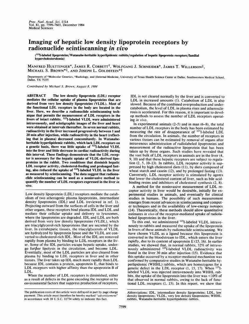
A Normal rabbit
into chloroform/methanol (2:1, vol/vol) and >97% was pre-cipitable by 10% (wt/vol) trichloroacetic acid.
Injection of 1231-Labeled VLDL into Animals. Six hours af-ter the iodination reaction, 47-279 x 106 cpm (-0.3 ml) of1231I-labeled VLDL was injected via a short catheter into themarginal ear vein of animals lightly anesthetized with Rom-pun (3 mg/kg of body weight) and Vetalar (25 mg/kg of bodyweight) to prevent movement during scanning.
Nuclear Imaging and Analysis. Images were obtained usinga standard field-of-view gamma camera (DynaMo, Picker In-ternational) equipped with a low-energy, general-purpose,parallel-hole collimator. Energy discrimination was providedby a 20% window centered on the 159 keV photopeak. Pixeldimensions were 2.2 x 2.2 mm. System resolution was mea-sured to be 2.5 mm full-width at half maximum amplitude onthe collimator face. Animals were positioned with the anteri-or torso on the collimator face and taped to the head of thecamera. Following equilibration for 1 min after intravenousinjection of 123I-labeled VLDL, an initial image was obtainedby acquiring counts for 4 min. Then, further images wereobtained over periods of 4 min each, starting at 10, 20, and 30min after injection. Images containing =100,000 counts eachwere stored on a dedicated nuclear medicine computer sys-tem (Technicare Model 450) at a digital resolution of 128 x128. Images were analyzed by manually assigning areas ofinterest over the heart and liver, and time vs. activity curveswere generated for each animal. The areas used for the cal-culations were maintained constant throughout a study.Other Assays. Protein was determined by the method of
Lowry et al. (19). Cholesterol levels were measured as de-scribed (6).
B WHHL rabbit
FIG. 1. Scintigraphic images of the liver and heart of a normal rabbit (A) and a WHHL rabbit (B) after intravenous injection of '23I-labeledVLDL. The normal rabbit (animal 3 in Table 1) and the WHHL rabbit (animal 11 in Table 1) each received 1.1 x 10⁸ cpm of 123I-labeled VLDLintravenously. Scintigrams were obtained at 1, 10, 20, and 30 min after injection as described in Methods. The heart is indicated by the arrowsand the liver is indicated by the dotted lines. Red indicates a higher concentration of radioactivity than green.
Proc. NatL Acad Sci. USA 81 (1984)
Proc. Natl. Acad Sci USA 81 (1984) 7601
RESULTS
Fig. 1 shows scintigraphic images of radioactivity in theheart and liver of a normal and of a WHHL rabbit at differenttimes after intravenous administration of 123I-labeled VLDL.In the normal animal, the amount of radioactivity in the heartwas highest at the initial time point (1 min after injection) anddeclined progressively thereafter, as evidenced by the de-creasing intensity of the heart image (Fig. 1A). The liver, onthe other hand, showed progressive accumulation of radio-activity from 1 min to 30 min after injection, as indicated bythe transition from a green to a red color. In the WHHLrabbit, these changes did not occur (Fig. 1B). The amount ofradioactivity in the heart declined only slightly and theamount of radioactivity in the liver did not increase duringthe course of the experiment. We interpret these findings toindicate that, at 1 min after injection, the radioactivity inboth heart and liver largely reflects extracellular 123I-labeledVLDL. The progressive increase of radioactivity in the liverand the progressive decrease in the heart after 1 min reflectthe receptor-mediated uptake of labeled lipoprotein into liv-er cells, with consequent reduction in the blood level.Table 1 summarizes the quantitative results of scintiscan-
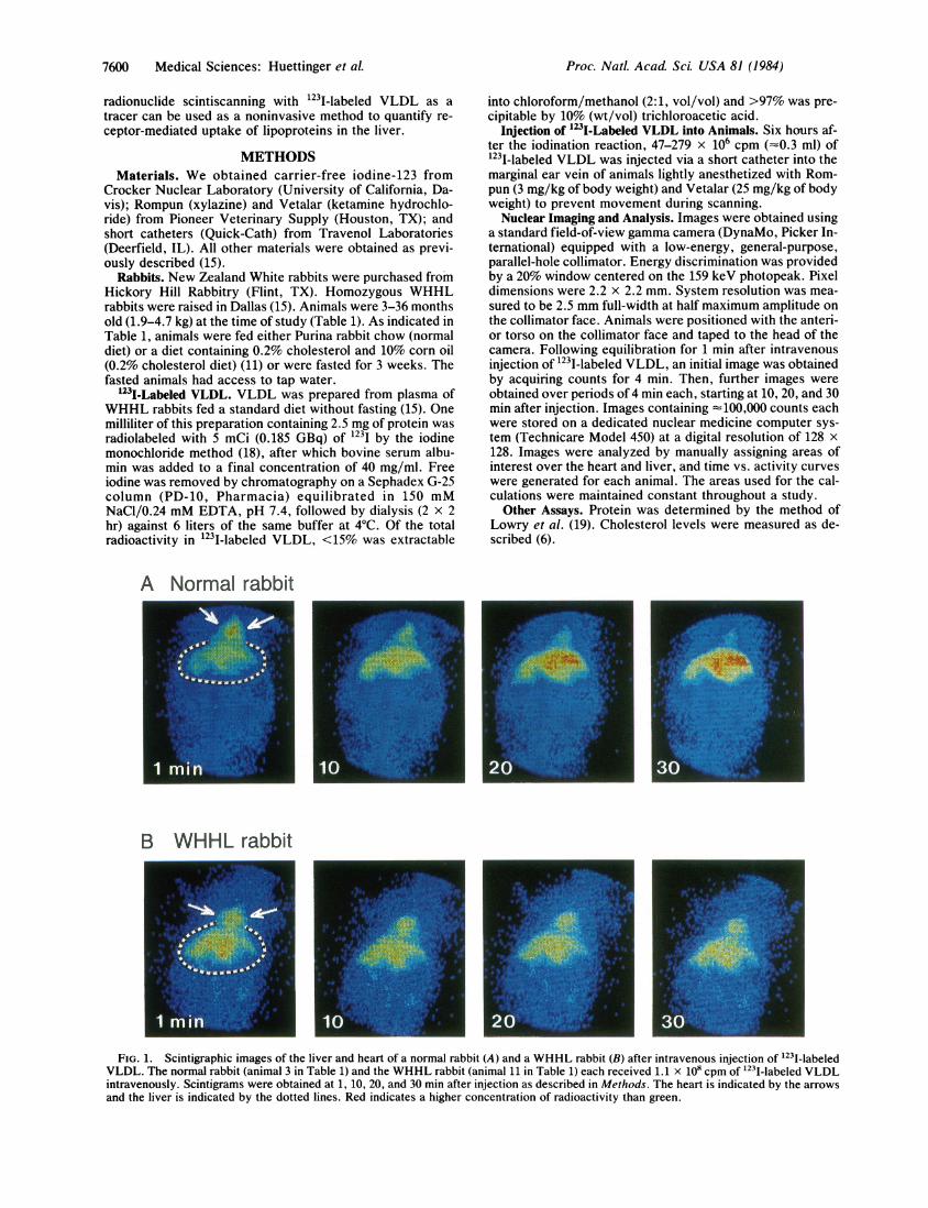
ning measurements of 123I-labeled VLDL in four groups ofrabbits. The radioactivity detected over the liver at the initialtime point (1 min after injection) varied within each group.This was due in part to differences in the specific radioactiv-ity of the injected 1231-labeled VLDL and in part to differ-ences in the anatomy of individual animals, which affects theefficiency of scintiscanning. Thus, to quantify the scinti-scanning data, we calculated the radioactivity at the 20-mintime point and expressed it as a fraction of the radioactivityat the 1-min time point. When the data were thus normal-
ized, consistent results were obtained [(b)/(a) ratios in Table1].
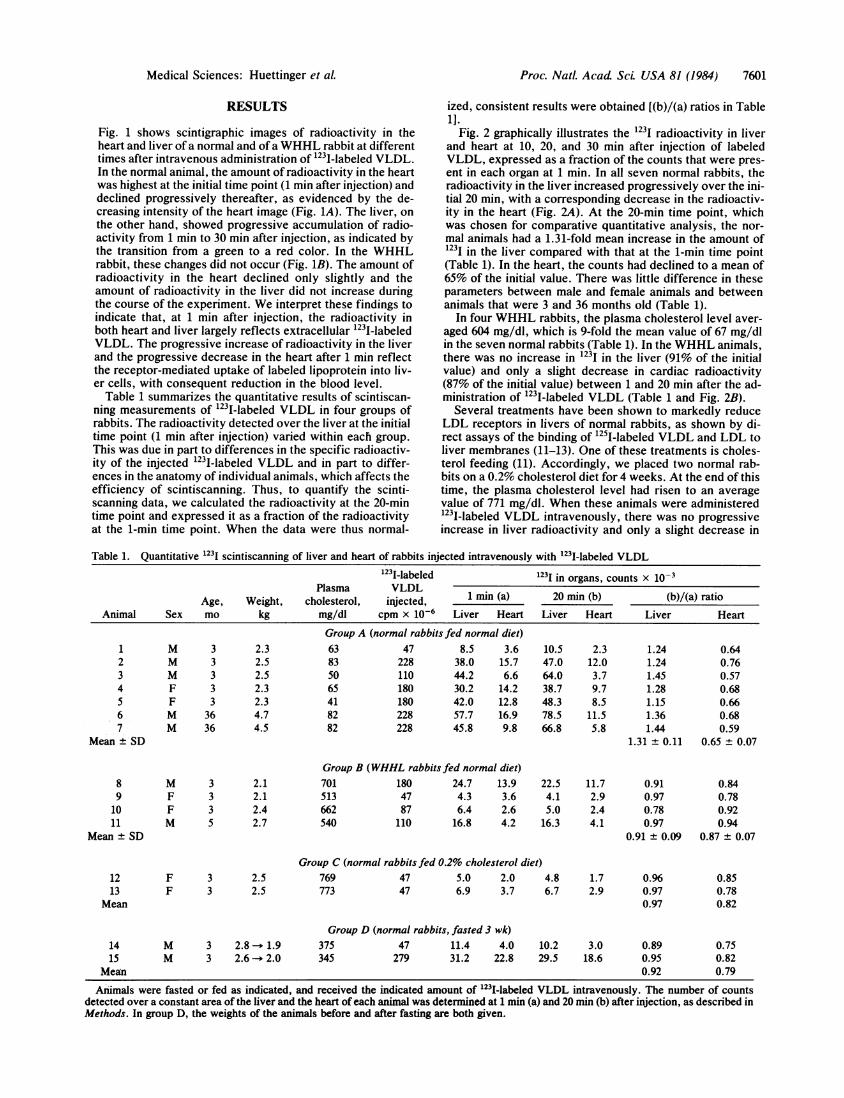
Fig. 2 graphically illustrates the 123I radioactivity in liverand heart at 10, 20, and 30 min after injection of labeledVLDL, expressed as a fraction of the counts that were pres-ent in each organ at 1 min. In all seven normal rabbits, theradioactivity in the liver increased progressively over the ini-tial 20 min, with a corresponding decrease in the radioactiv-ity in the heart (Fig. 2A). At the 20-min time point, whichwas chosen for comparative quantitative analysis, the nor-mal animals had a 1.31-fold mean increase in the amount of1231 in the liver compared with that at the 1-min time point(Table 1). In the heart, the counts had declined to a mean of65% of the initial value. There was little difference in theseparameters between male and female animals and betweenanimals that were 3 and 36 months old (Table 1).
In four WHHL rabbits, the plasma cholesterol level aver-aged 604 mg/dl, which is 9-fold the mean value of 67 mg/dlin the seven normal rabbits (Table 1). In the WHHL animals,there was no increase in 1231 in the liver (91% of the initialvalue) and only a slight decrease in cardiac radioactivity(87% of the initial value) between 1 and 20 min after the ad-ministration of 123I-labeled VLDL (Table 1 and Fig. 2B).
Several treatments have been shown to markedly reduceLDL receptors in livers of normal rabbits, as shown by di-rect assays of the binding of 1251-labeled VLDL and LDL toliver membranes (11-13). One of these treatments is choles-terol feeding (11). Accordingly, we placed two normal rab-bits on a 0.2% cholesterol diet for 4 weeks. At the end of thistime, the plasma cholesterol level had risen to an averagevalue of 771 mg/dl. When these animals were administered123I-labeled VLDL intravenously, there was no progressiveincrease in liver radioactivity and only a slight decrease in
Table 1. Quantitative 1231 scintiscanning of liver and heart of rabbits injected intravenously with "231-labeled VLDL123I-labeled 1231 in organs, counts x 10-3
Plasma VLDL1mn() 2 i b/a aiAge, Weight, cholesterol, injected, 1 mm (a) 20 mm (b) (b)/(a) ratio
Animal Sex mo kg mg/dl cpm x 10-6 Liver Heart Liver Heart Liver Heart
Group A (normal rabbits fed normal diet)1 M 3 2.3 63 47 8.5 3.6 10.5 2.3 1.24 0.642 M 3 2.5 83 228 38.0 15.7 47.0 12.0 1.24 0.763 M 3 2.5 50 110 44.2 6.6 64.0 3.7 1.45 0.574 F 3 2.3 65 180 30.2 14.2 38.7 9.7 1.28 0.685 F 3 2.3 41 180 42.0 12.8 48.3 8.5 1.15 0.666 M 36 4.7 82 228 57.7 16.9 78.5 11.5 1.36 0.687 M 36 4.5 82 228 45.8 9.8 66.8 5.8 1.44 0.59
Mean ± SD 1.31 ± 0.11 0.65 ± 0.07
Group B (WHHL rabbits fed normal diet)8 M 3 2.1 701 180 24.7 13.9 22.5 11.7 0.91 0.849 F 3 2.1 513 47 4.3 3.6 4.1 2.9 0.97 0.7810 F 3 2.4 662 87 6.4 2.6 5.0 2.4 0.78 0.9211 M 5 2.7 540 110 16.8 4.2 16.3 4.1 0.97 0.94
Mean ± SD 0.91 ± 0.09 0.87 ± 0.07
Group C (normal rabbits fed 0.2% cholesterol diet)12 F 3 2.5 769 47 5.0 2.0 4.8 1.7 0.96 0.8513 F 3 2.5 773 47 6.9 3.7 6.7 2.9 0.97 0.78
Mean 0.97 0.82
Group D (normal rabbits, fasted 3 wk)14 M 3 2.8 - 1.9 375 47 11.4 4.0 10.2 3.0 0.89 0.7515 M 3 2.6 2.0 345 279 31.2 22.8 29.5 18.6 0.95 0.82
Mean 0.92 0.79
Aiimals were fasted or fed as indicated, and received the indicated amount of I231-labeled VLDL intravenously. The number of countsdetected over a constant area of the liver and the heart of each animal was determined at 1 min (a) and 20 min (b) after injection, as described inMethods. In group D, the weights of the animals before and after fasting are both given.
Medical Sciences: Huettinger et aL
7602 Medical Sciences: Huettinger et al.
1.5 -[noiesteroiDiet
1.47
1.3 4 6
ql h~~I-U2 OrganZ'~ ~9Liver-Closed Symbols
expressedasthrtio te adoctviy n helier(coedsybos)orhert(oe smbls a te ndcae Heart- Open Symbols
0~~-1.1
7, 910,11,8@n14a15
0.8
05 I~ ~~~___ _ _ _ _ _ _ _ _ _
1 10 20 30 1 10 20 30 1 10 20 1 10 20 30Time after injection, min
FIG. 2. Quantitative scintiscanning of the liver and heart in rabbits at various times after injection of '231-labeled VLDL. The data areexpressed as the ratio of the radioactivity in the liver (closed symbols) or heart (open symbols) at the indicated time relative to that in the sameorgan at 1 min after injection. These data were obtained in the experiments described in Table 1; each number in the figure refers to thecorresponding animal in Table 1, and A-D correspond to groups A-D in the table. All animals were scanned at 1, 10, and 20 min; animals 2, 3, 6,7, 10, 11, and 15 were also scanned at 30 min.
cardiac radioactivity between 1 and 20 min after injection(Table 1 and Fig. 2C).Another treatment known to reduce hepatic LDL receptor
levels in rabbits is prolonged fasting (13). We subjected twonormal rabbits to a total fast for 3 weeks and then adminis-tered 123I-labeled VLDL. In these animals, the plasma cho-lesterol levels had risen to an average value of 360 mg/dl.Again, there was no progressive increase in radioactivity inthe liver and only a slight decrease in radioactivity in theheart over 20 min (Table 1 and Fig. 2D).
DISCUSSIONIn these experiments, we have applied scintiscanning tech-niques to the measurement of the uptake of VLDL-derivedlipoproteins by rabbit liver. Entry of 123I-labeled VLDL ra-dioactivity into the liver of normal rabbits was rapid. Thisuptake required the LDL receptor, since it did not occur inWHHL rabbits and was diminished by cholesterol feedingand by prolonged fasting, both of which are known to dimin-ish hepatic LDL receptor activity (10-13).
In the case of the WHHL rabbit, the failure to take up theI231-labeled VLDL is attributable exclusively to a lack ofLDL receptors, since a primary genetic deficiency of LDLreceptors has been demonstrated directly in these animals(reviewed in ref. 1). In the cholesterol-fed and the fasted rab-bits, the situation is somewhat more complicated. A dimin-ished production of LDL receptors has been demonstratedby in vitro membrane-binding assays, and this deficiency ini-tiates the buildup of IDL and LDL in plasma (11-13). As the
plasma levels of these lipoproteins rise, the remaining LDLreceptors become saturated, and the excess lipoproteinscompete with each other for the few receptors that are func-tional (11). Thus, when a radiolabeled ligand is injected intothe cholesterol-fed or fasted animals, the diminished uptakeis attributable to two factors: (i) a reduced number of recep-tors and (ii) saturation of the remaining receptors with en-dogenous ligands that compete with the radiolabeled ligand.The observed reduced uptake of the radioactive ligand im-plies that the receptors are the limiting factor in lipoproteinclearance, but these measurements alone cannot tell howmuch of the limitation is caused by a reduced number of re-ceptors and how much is caused by saturation of the remain-ing receptors.We do not know whether this scintiscanning technique can
be adapted to humans and whether it would be discriminat-ing enough to detect the 50% deficiency of LDL receptors inheterozygous familial hypercholesterolemia or to reveal thesubtle changes in hepatic LDL receptors that occur in re-sponse to diet and drug therapy. Humans, in general, seemto express fewer LDL receptors than do lower animal spe-cies such as rabbits (20). The clearance rate for 125I-labeledVLDL is much slower in humans (21) than in rabbits (15),and the percentage conversion of VLDL to LDL is higher inhumans. The clearance rate of LDL in humans (6) is alsomarkedly less than that in rabbits (3). A rapid rate of hepaticuptake is necessary for the scintiscanning method to workbecause the 123I-labeled-VLDL-derived radioactivity is re-leased from the liver within 60 min after the lipoprotein is
Proc. NatL Acad ScL USA 81 (1984)
Proc. Natl. Acad. Sci USA 81 (1984) 7603
taken up and digested in lysosomes. In rabbits, rapid degra-dation does not present a problem for the assay, since theuptake is rapid and the steady-state level of radioactivity inthe liver is relatively high. However, in humans the rate ofuptake of 123I-labeled VLDL may be so slow that a suffi-ciently high intracellular level of 1231 within the liver wouldnot be attained.
In this regard, it may be possible to use VLDL covalentlyattached to 123I-labeled tyramine-cellobiose as a ligand (22).When 125I-labeled tyramine-cellobiose is covalently attachedto LDL and the lipoprotein is taken up by the liver or otherorgans, the radioactive tyramine-cellobiose remains trappedin lysosomes and accumulates progressively (22). Since thelabeled tyramine-cellobiose is not excreted from the cells, itsuse should increase the sensitivity of the scintiscanning tech-nique quite markedly.Another possible approach for measurement of hepatic
LDL receptors in humans involves the use of radionuclide-labeled monoclonal antibodies directed against the LDL re-ceptor. Some of these antibodies bind to the receptor at asite that is different from the LDL binding site; their bindingis not inhibited competitively by LDL or other ligands forthe LDL receptor. After binding to the LDL receptor, theseantibodies are carried into the cell and delivered to lyso-somes, as is LDL (23). When such a monoclonal antibody islabeled with 1251 and injected into rabbits, most of the radio-activity is taken up rapidly in the liver in a process that de-pends on the antibody binding to the LDL receptor (24).
If administration of radiolabeled mouse monoclonal anti-bodies to humans can be shown to be a safe procedure, thenit should be feasible to use 1231-labeled monoclonal antibod-ies against the LDL receptor as probes for hepatic scinti-scanning. Such antibodies would overcome the difficulties ininterpretation that might result from saturation of the recep-tors by endogenous lipoproteins, and their rapid uptakewould also deliver sufficient radioactivity to the liver to pro-duce a highly sensitive and quantitative assay for LDL re-ceptors in intact humans.
We thank Laurie Ledbetter and Marvin Akers for excellent tech-nical assistance. This research was supported by grants from theNational Institutes of Health (HL20948 and HL17669) and the MossHeart Fund. M.H. is a recipient of a postdoctoral fellowship fromthe Max Kade Foundation, Inc. W.J.S. is an Established Investiga-tor of the American Heart Association.
1. Goldstein, J. L., Kita, T. & Brown, M. S. (1983) N. Engl. J.Med. 309, 288-295.
2. Kovanen, P. T., Bilheimer, D. W., Goldstein, J. L., Jaramillo,J. J. & Brown, M. S. (1981) Proc. Nati. Acad. Sci. USA 78,1194-1198.
3. Bilheimer, D. W., Watanabe, Y. & Kita, T. (1982) Proc. Natl.Acad. Sci. USA 79, 3305-3309.
4. Pittman, R. C., Carew, T. E., Attie, A. D., Witztum, J. L.,Watanabe, Y. & Steinberg, D. (1982) J. Biol. Chem. 257, 7994-8000.
5. Mahley, R. W., Hui, D. Y., Innerarity, T. L. & Weisgraber,K. H. (1981) J. Clin. Invest. 68, 1197-1206.
6. Bilheimer, D. W., Stone, N. J. & Grundy, S. M. (1979) J.Clin. Invest. 64, 524-533.
7. Shepherd, J., Bicker, S., Lorimer, A. R. & Packard, C. J.(1979) J. Lipid Res. 20, 999-1006.
8. Bilheimer, D. W., Grundy, S. M., Brown, M. S. & Goldstein,J. L. (1983) Proc. Natl. Acad. Sci. USA 80, 4124-4128.
9. Spady, D. K., Bilheimer, D. W. & Dietschy, J. M. (1983)Proc. Natl. Acad. Sci. USA 80, 3499-3503.
10. Brown, M. S. & Goldstein, J. L. (1983) J. Clin. Invest. 72,743-747.
11. Kovanen, P. T., Brown, M. S., Basu, S. K., Bilheimer, D. W.& Goldstein, J. L. (1981) Proc. Natl. Acad. Sci. USA 78,1396-1400.
12. Chao, Y.-S., Yamin, T.-T. & Alberts, A. W. (1982) J. Biol.Chem. 257, 3623-3627.
13. Stoudemire, J. B., Renaud, G., Shames, D. M. & Havel, R. J.(1984) J. Lipid Res. 25, 33-39.
14. Rellas, J. S., Corbett, J. R., Kulkarni, P., Morgan, C., De-vous, M. D., Buja, M., Bush, L., Parkey, R. W., Willerson,J. T. & Lewis, S. E. (1983) Am. J. Cardiol. 52, 1326-1332.
15. Kita, T., Brown, M. S., Bilheimer, D. W. & Goldstein, J. L.(1982) Proc. Natl. Acad. Sci. USA 79, 5693-5697.
16. Mahley, R. W. & Innerarity, T. L. (1983) Biochim. Biophys.Acta 737, 197-222.
17. Tanzawa, K., Shimada, Y., Kuroda, M., Tsujita, Y., Arai, M.& Watanabe, H. (1980) FEBS Lett. 118, 81-84.
18. Bilheimer, D. W., Eisenberg, S. & Levy, R. I. (1972) Biochim.Biophys. Acta 260, 212-221.
19. Lowry, 0. H., Rosebrough, N. J., Farr, A. L. & Randall,R. J. (1951) J. Biol. Chem. 193, 265-275.
20. Goldstein, J. L. & Brown, M. S. (1982) Clin. Res. 30, 417-426.21. Eisenberg, S., Bilheimer, D. W. & Levy, R. I. (1972) Biochim.
Biophys. Acta 280, 94-104.22. Pittman, R. C., Carew, T. E., Glass, C. K., Green, S. R.,
Taylor, C. A., Jr., & Attie, A. D. (1983) Biochem. J. 212, 791-800.
23. Beisiegel, U., Schneider, W. J., Goldstein, J. L., Anderson,R. G. W. & Brown, M. S. (1981) J. Biol. Chem. 256, 11923-11931.
24. Huettinger, M., Schneider, W. J., Ho, Y. K., Goldstein, J. L.& Brown, M. S. (1984) J. Clin. Invest. 74, 1017-1026.