70
Department of Radiology, Faculty of General Medicine, University of Szeged, Hungary Imaging of the lung
Department of Radiology, Faculty of General Medicine, University of Szeged, Hungary
Imaging of the lung

Department of Radiology, Faculty of General Medicine, University of Szeged, Hungary
Methods of examination
X-ray (radiography, fluoroscopy, tomosynthesis)
mediastinal contours and position
lung structure
– vessels
– bronchi
– interstitium
– alveoli
pleura, chest wall
diaphragm

Department of Radiology, Faculty of General Medicine, University of Szeged, Hungary
radiogram
fluoroscopy
Department of Radiology, Faculty of General Medicine, University of Szeged, Hungary
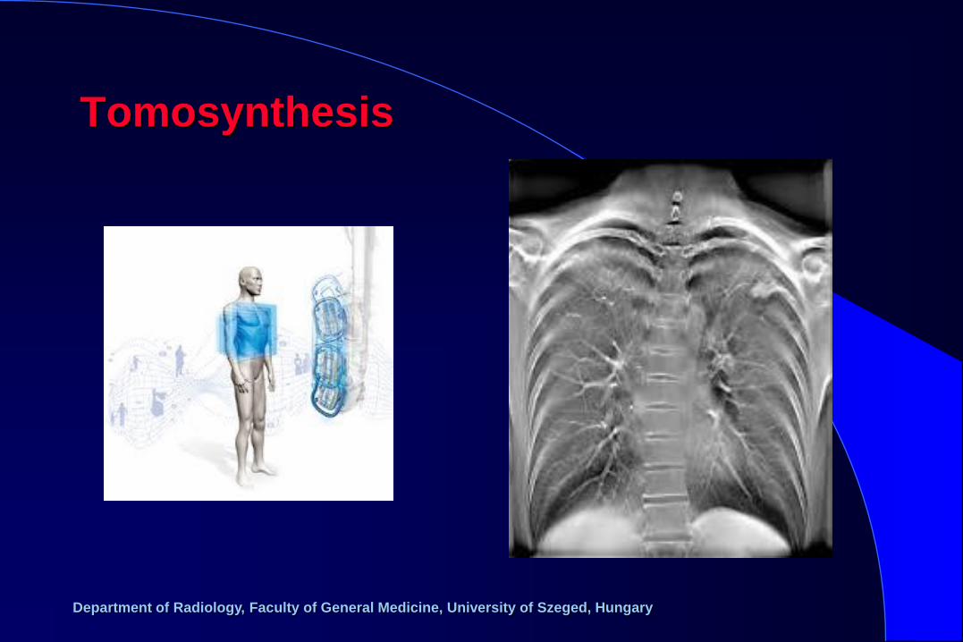
Tomosynthesis
Department of Radiology, Faculty of General Medicine, University of Szeged, Hungary
Methods of examination
Ultrasound (point of care ultrasound, PoCUS)
chest wall
pleura (fluid and gas)
diaphragm
consolidations close to lung surface

Department of Radiology, Faculty of General Medicine, University of Szeged, Hungary
PoCUS
normal ptx pleural effusion
Department of Radiology, Faculty of General Medicine, University of Szeged, Hungary
Methods of examination
Computed tomography
mediastinal structures
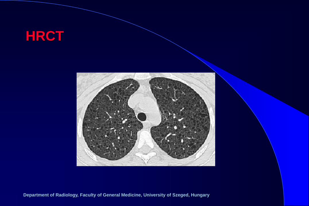
lung structure (high resolution CT = HRCT)
– vessels
– bronchi
– interstitium
– alveoli
pleura, chest wall
diaphragm
Department of Radiology, Faculty of General Medicine, University of Szeged, Hungary
HRCT

Department of Radiology, Faculty of General Medicine, University of Szeged, Hungary
Methods of examination
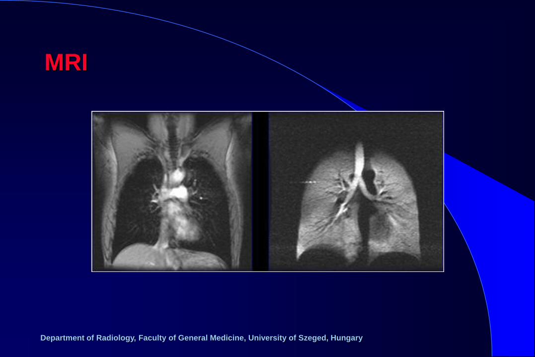
Magnetic resonance imaging
mediastinal structures
pleura, chest wall
diaphragm
solid lung lesions
Department of Radiology, Faculty of General Medicine, University of Szeged, Hungary
MRI

Department of Radiology, Faculty of General Medicine, University of Szeged, Hungary
Methods of examination
PET/CT
lesions with high FDG uptake

Department of Radiology, Faculty of General Medicine, University of Szeged, Hungary
PET-CT
Department of Radiology, Faculty of General Medicine, University of Szeged, Hungary
Methods of examination
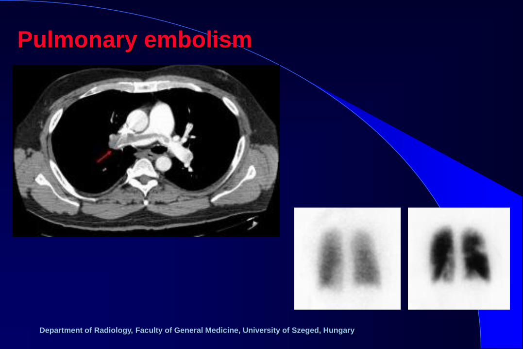
Ventillation/perfusion scintigraphy
– comparison of
patency of ventillation
patency of circulation
– „mismatch” = pulmonary embolism

Department of Radiology, Faculty of General Medicine, University of Szeged, Hungary
Ventilation-perfusion scintigraphy
Department of Radiology, Faculty of General Medicine, University of Szeged, Hungary
Methods of examination
Interventional procedures
sampling/drainage of fluid accumulations
sampling/ablation of solid lesions
visualisation/recanalisation/occlusion of vessels

Department of Radiology, Faculty of General Medicine, University of Szeged, Hungary
Lung biopsy

Department of Radiology, Faculty of General Medicine, University of Szeged, Hungary
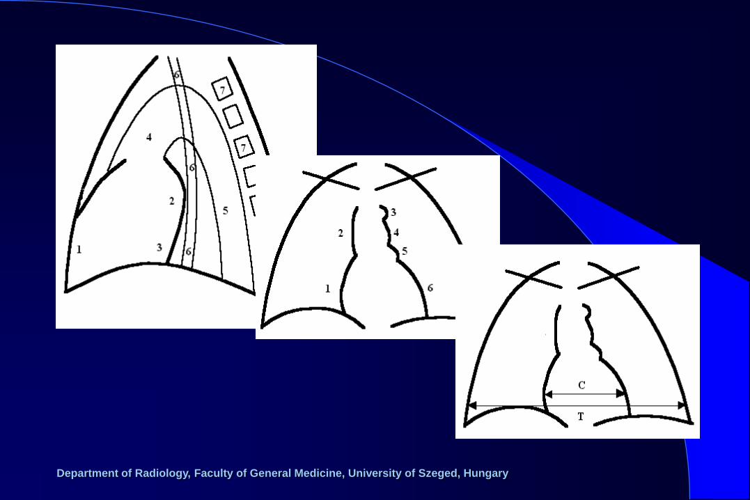
Anatomy

Department of Radiology, Faculty of General Medicine, University of Szeged, Hungary
Anatomy
Department of Radiology, Faculty of General Medicine, University of Szeged, Hungary
Basic radiographic changes of the lung
deformity
decreased transparency
(focal, diffuse) tumor, metastasis, consolidation,
atelectasia, edema, fibrosis, effusion,
callus, abscess
increased transparency
(focal, diffuse) emphysema, bulla, cyst, ptx, decreased
circulation
change of pattern
(vascular, interstitial)
Department of Radiology, Faculty of General Medicine, University of Szeged, Hungary
– alveolar
– interstitial
– ground-glass opacity
– air-trapping
– tree-in-bud
HRCT patterns of transparency
changes

Department of Radiology, Faculty of General Medicine, University of Szeged, Hungary
• congenital malformations
• physical injury (radiation, heat)
• chemical injury (toxic, irritative, allergic)
• inflammations (bacterial, tuberculotic, viral, fungal)
• degenerative
• autoimmune, granulomatous
• pulmonary embolism
• tumors (benign, primary malignant, metastatic)
Diseases of the lung:
Department of Radiology, Faculty of General Medicine, University of Szeged, Hungary
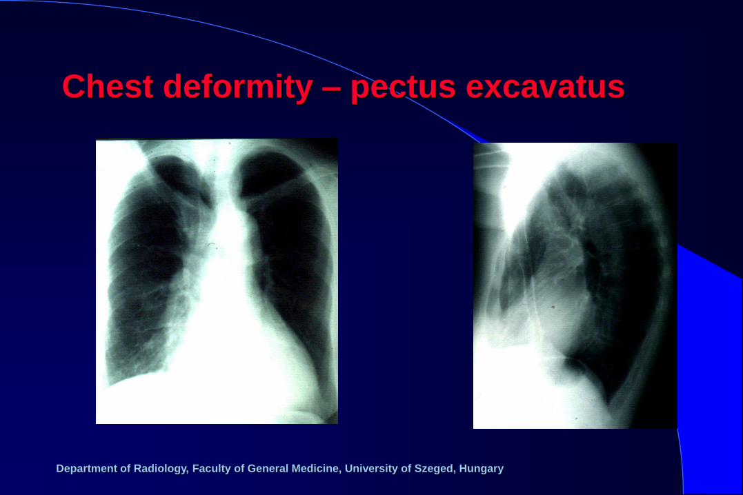
Chest deformity – pectus excavatus

Department of Radiology, Faculty of General Medicine, University of Szeged, Hungary
Chest deformity - scoliosis

Department of Radiology, Faculty of General Medicine, University of Szeged, Hungary
Elevated diaphragm

Department of Radiology, Faculty of General Medicine, University of Szeged, Hungary
Total opacification of the left hemithorax
Department of Radiology, Faculty of General Medicine, University of Szeged, Hungary
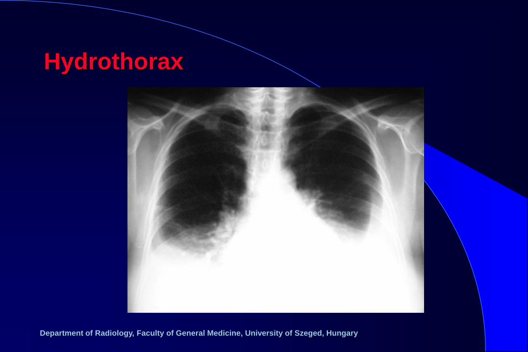
Hydrothorax
Department of Radiology, Faculty of General Medicine, University of Szeged, Hungary
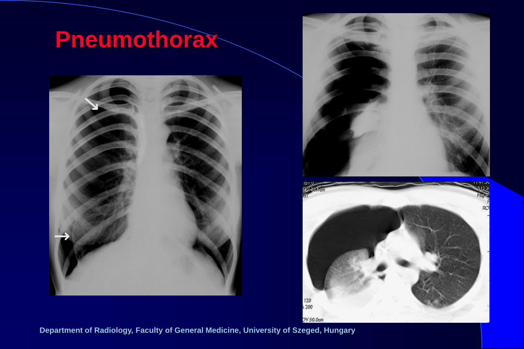
Pneumothorax
Department of Radiology, Faculty of General Medicine, University of Szeged, Hungary
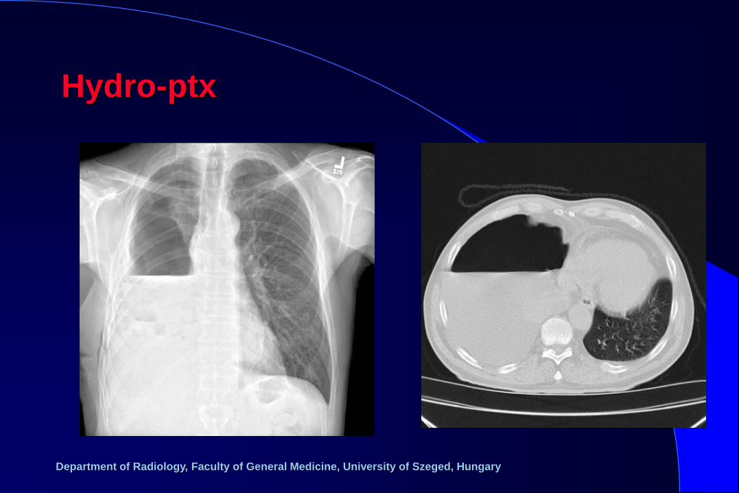
Hydro-ptx

Department of Radiology, Faculty of General Medicine, University of Szeged, Hungary
Irradiation pneumonitis
Department of Radiology, Faculty of General Medicine, University of Szeged, Hungary
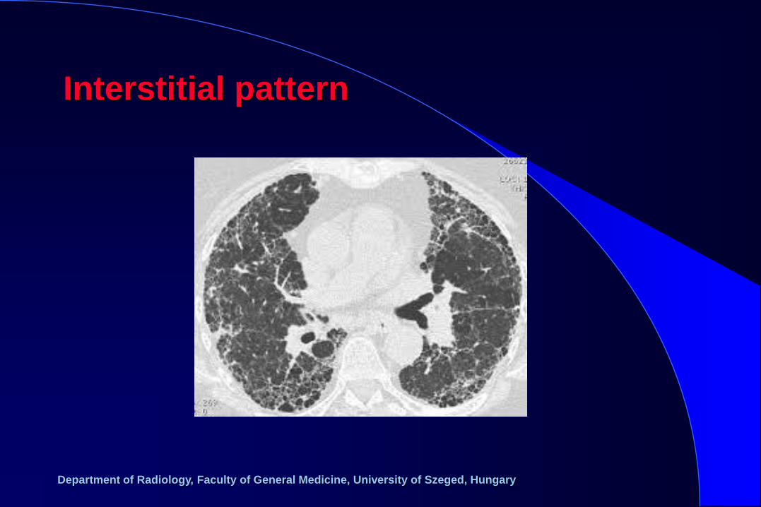
Interstitial pattern

Department of Radiology, Faculty of General Medicine, University of Szeged, Hungary
Alveolar pattern
Department of Radiology, Faculty of General Medicine, University of Szeged, Hungary
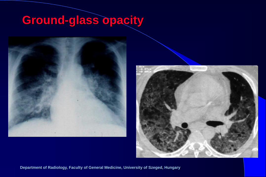
Ground-glass opacity

Department of Radiology, Faculty of General Medicine, University of Szeged, Hungary
Pneumonia

Department of Radiology, Faculty of General Medicine, University of Szeged, Hungary
Abscess
Department of Radiology, Faculty of General Medicine, University of Szeged, Hungary
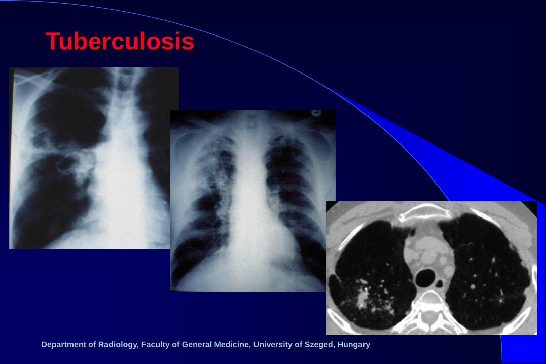
Tuberculosis

Department of Radiology, Faculty of General Medicine, University of Szeged, Hungary
Miliary TBC

Department of Radiology, Faculty of General Medicine, University of Szeged, Hungary
Focal decreased transparency

Department of Radiology, Faculty of General Medicine, University of Szeged, Hungary
Lung cancer

Department of Radiology, Faculty of General Medicine, University of Szeged, Hungary
Lung cancer

Department of Radiology, Faculty of General Medicine, University of Szeged, Hungary
Lung cancer

Department of Radiology, Faculty of General Medicine, University of Szeged, Hungary
Metastases
Department of Radiology, Faculty of General Medicine, University of Szeged, Hungary
Pulmonary embolism

Department of Radiology, Faculty of General Medicine, University of Szeged, Hungary
Pulmonary embolism – 24 hours later
Department of Radiology, Faculty of General Medicine, University of Szeged, Hungary
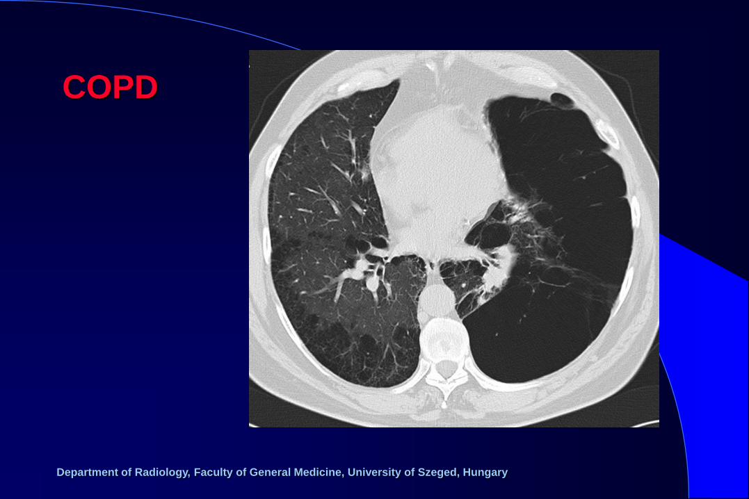
COPD
Department of Radiology, Faculty of General Medicine, University of Szeged, Hungary
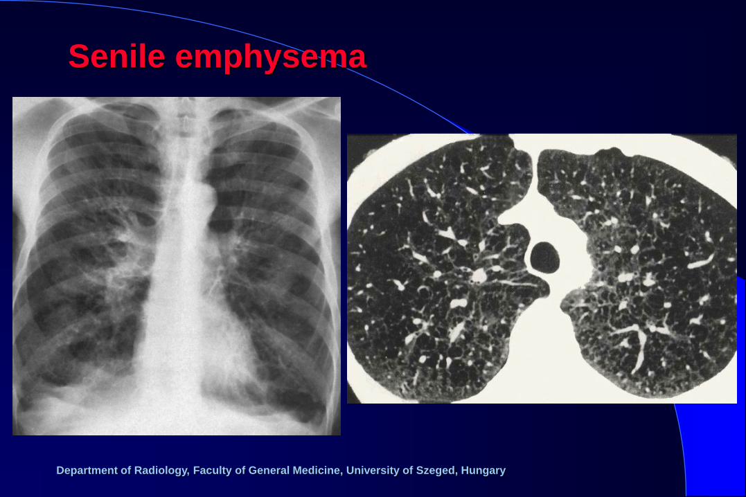
Senile emphysema
Department of Radiology, Faculty of General Medicine, University of Szeged, Hungary
Department of Radiology, Faculty of General Medicine, University of Szeged, Hungary
Imaging of mediastinal diseases
Department of Radiology, Faculty of General Medicine, University of Szeged, Hungary
Methods of examination
– x-ray
– ultrasound
– computed tomography
– magnetic resonance imaging
– interventional procedures
– scintigraphy
Department of Radiology, Faculty of General Medicine, University of Szeged, Hungary
• vascular dilatation / malposition
• thyroid enlargement
• thymus enlargement
• lymph node enlargement (inflammatory, benign
primary and secondary malignant)
• other space occupying lesions (neurogenic tumor,
cyst, dermoid, hiatus hernia)
• inflammation
• injury
Diseases of the mediastinum:
Department of Radiology, Faculty of General Medicine, University of Szeged, Hungary
Department of Radiology, Faculty of General Medicine, University of Szeged, Hungary
Department of Radiology, Faculty of General Medicine, University of Szeged, Hungary
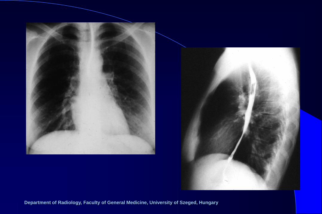
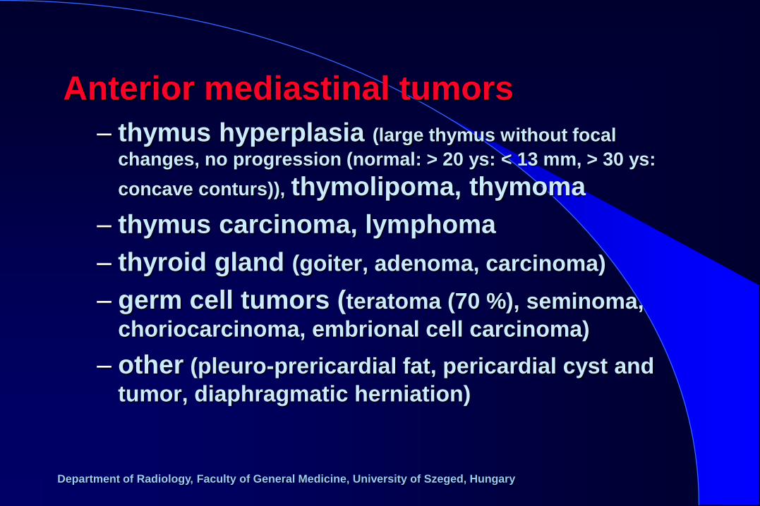
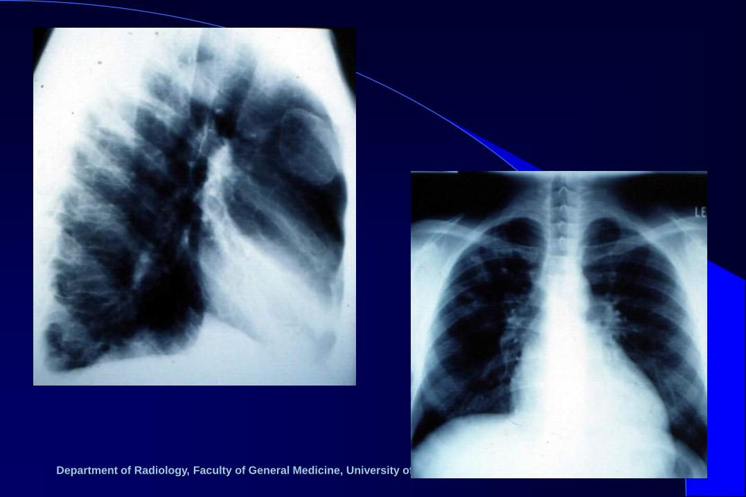
– thymus hyperplasia (large thymus without focal
changes, no progression (normal: > 20 ys: < 13 mm, > 30 ys:
concave conturs)), thymolipoma, thymoma
– thymus carcinoma, lymphoma
– thyroid gland (goiter, adenoma, carcinoma)
– germ cell tumors (teratoma (70 %), seminoma,
choriocarcinoma, embrional cell carcinoma)
– other (pleuro-prericardial fat, pericardial cyst and
tumor, diaphragmatic herniation)
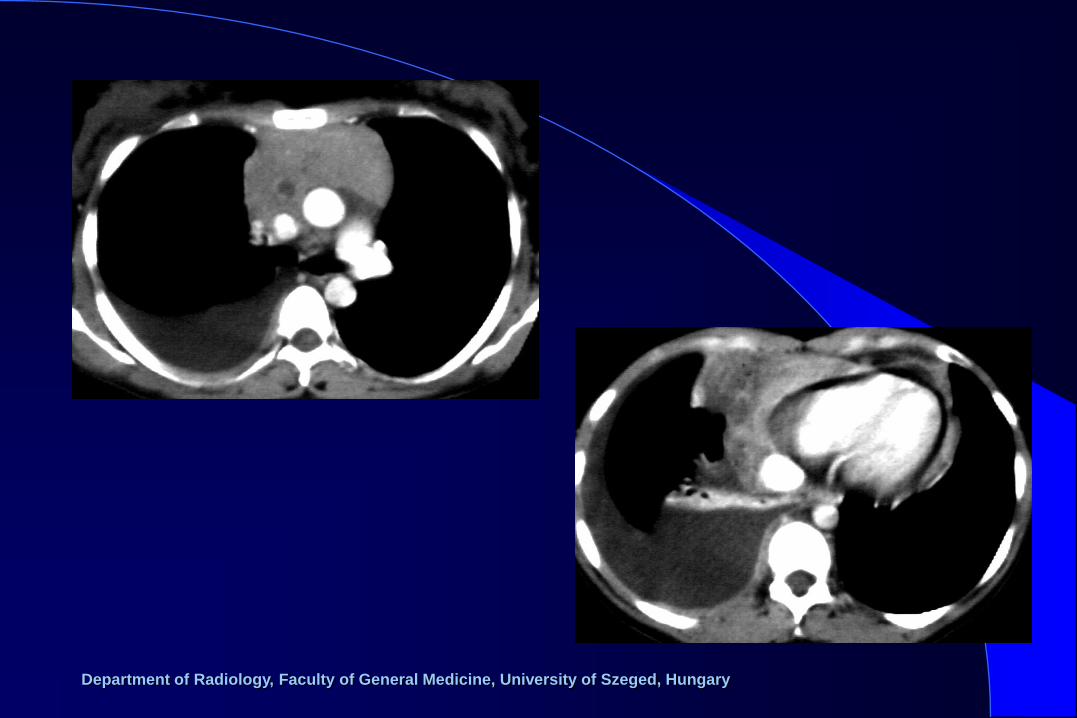
Anterior mediastinal tumors
Department of Radiology, Faculty of General Medicine, University of Szeged, Hungary
Department of Radiology, Faculty of General Medicine, University of Szeged, Hungary
Department of Radiology, Faculty of General Medicine, University of Szeged, Hungary
Department of Radiology, Faculty of General Medicine, University of Szeged, Hungary
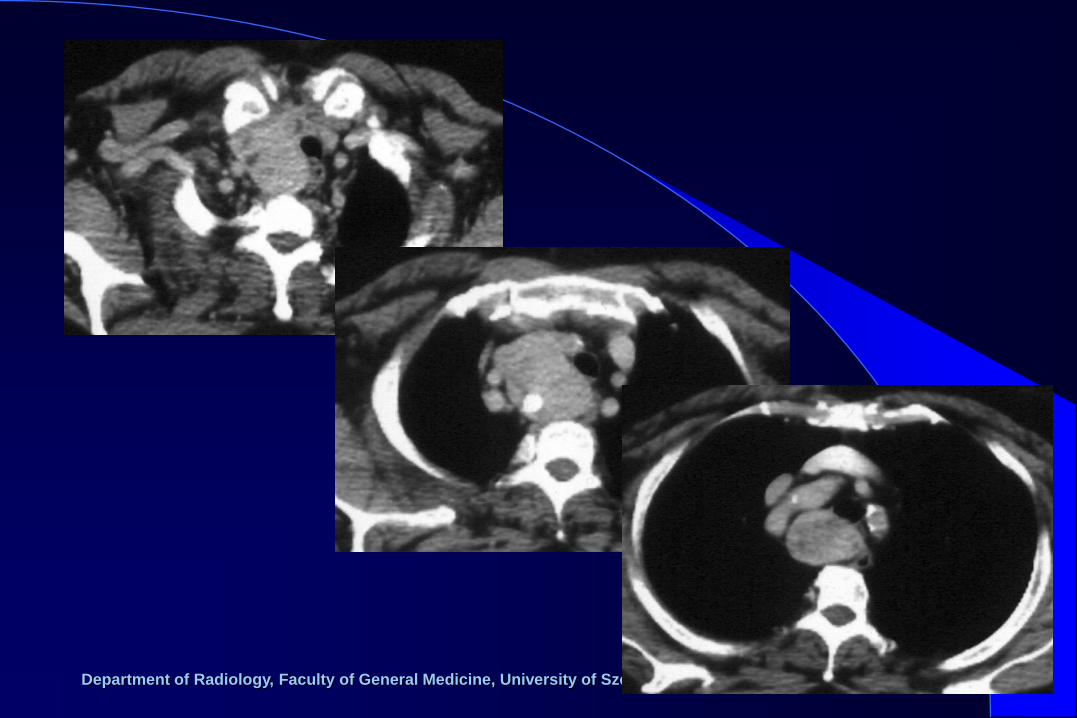
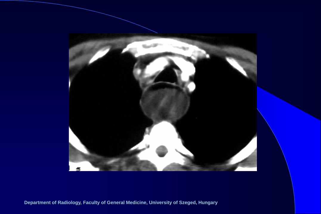
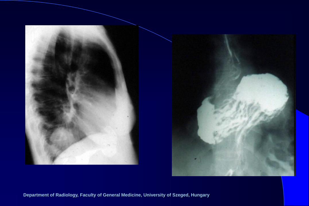
– esophagus (tumor, diverticulum, hiatus
hernia)
– bronchogenic cyst
– enterogenic cyst
Middle mediastinal masses
Department of Radiology, Faculty of General Medicine, University of Szeged, Hungary
Department of Radiology, Faculty of General Medicine, University of Szeged, Hungary
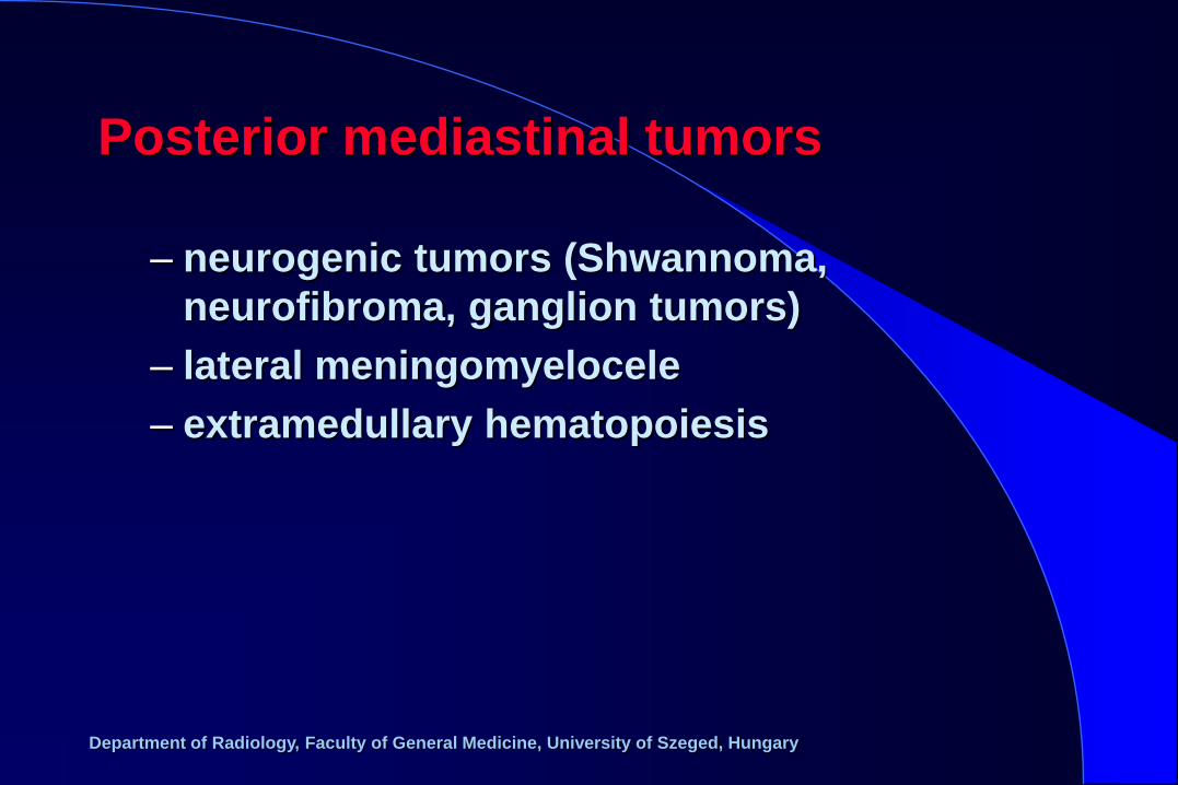
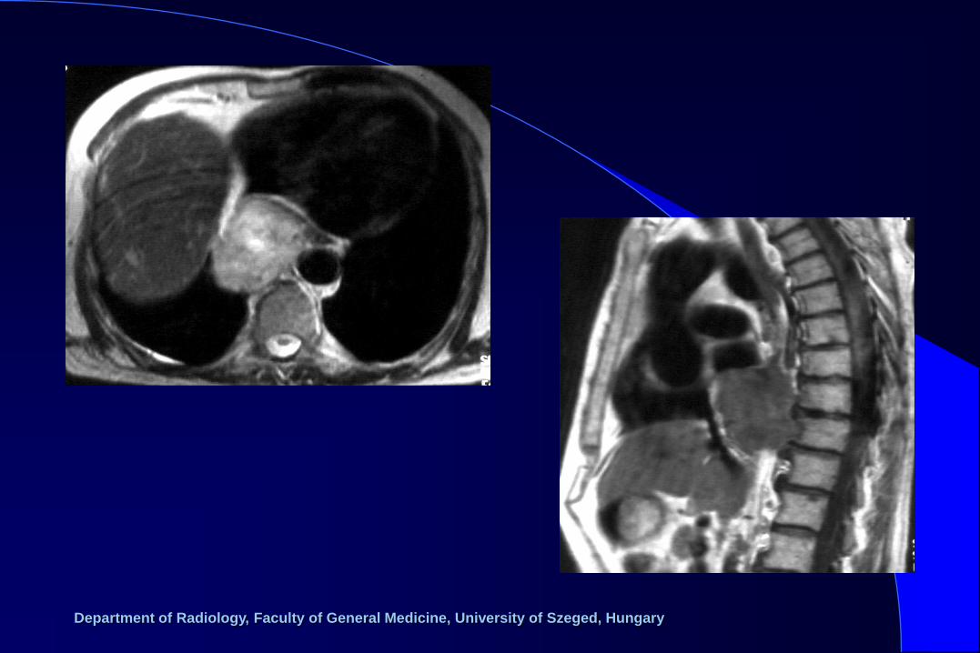
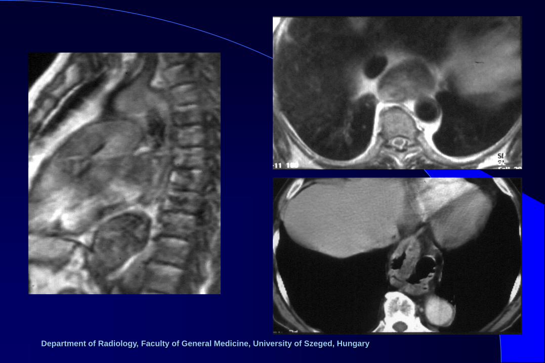
– neurogenic tumors (Shwannoma,
neurofibroma, ganglion tumors)
– lateral meningomyelocele
– extramedullary hematopoiesis
Posterior mediastinal tumors
Department of Radiology, Faculty of General Medicine, University of Szeged, Hungary
Department of Radiology, Faculty of General Medicine, University of Szeged, Hungary
Department of Radiology, Faculty of General Medicine, University of Szeged, Hungary
– malignant lymphoma (Hodgkin, non-Hodgkin)
– metastasis
– inflammation (TBC, virus, hystoplasma, etc.)
– silicosis
– sarcoidosis
– Castleman disease
Lymph node enlargement
Department of Radiology, Faculty of General Medicine, University of Szeged, Hungary
Department of Radiology, Faculty of General Medicine, University of Szeged, Hungary

Department of Radiology, Faculty of General Medicine, University of Szeged, Hungary
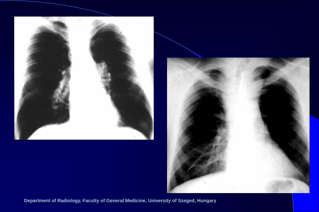
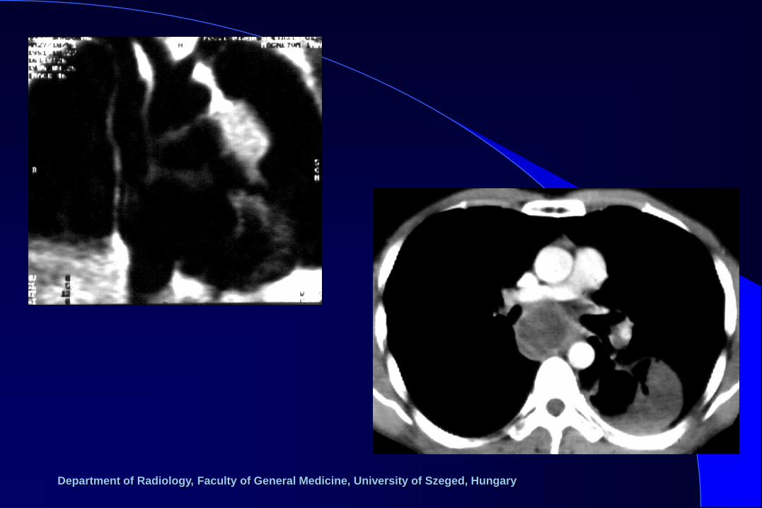
Vascular lesions
– variations (right aortis arch, vascular ring, etc.)
– aortic coarctation
– aortic dilatation high blood pressure, arteriosclerosis (ectasia)
aortic valve disease
aneurysma (fusiform, saccular, dissection) – sclerotic
– traumatic
– luetic aortitis
– Marfan sy.
– vein dilatation (VCS, v. azygos)

Department of Radiology, Faculty of General Medicine, University of Szeged, Hungary

Department of Radiology, Faculty of General Medicine, University of Szeged, Hungary
Department of Radiology, Faculty of General Medicine, University of Szeged, Hungary
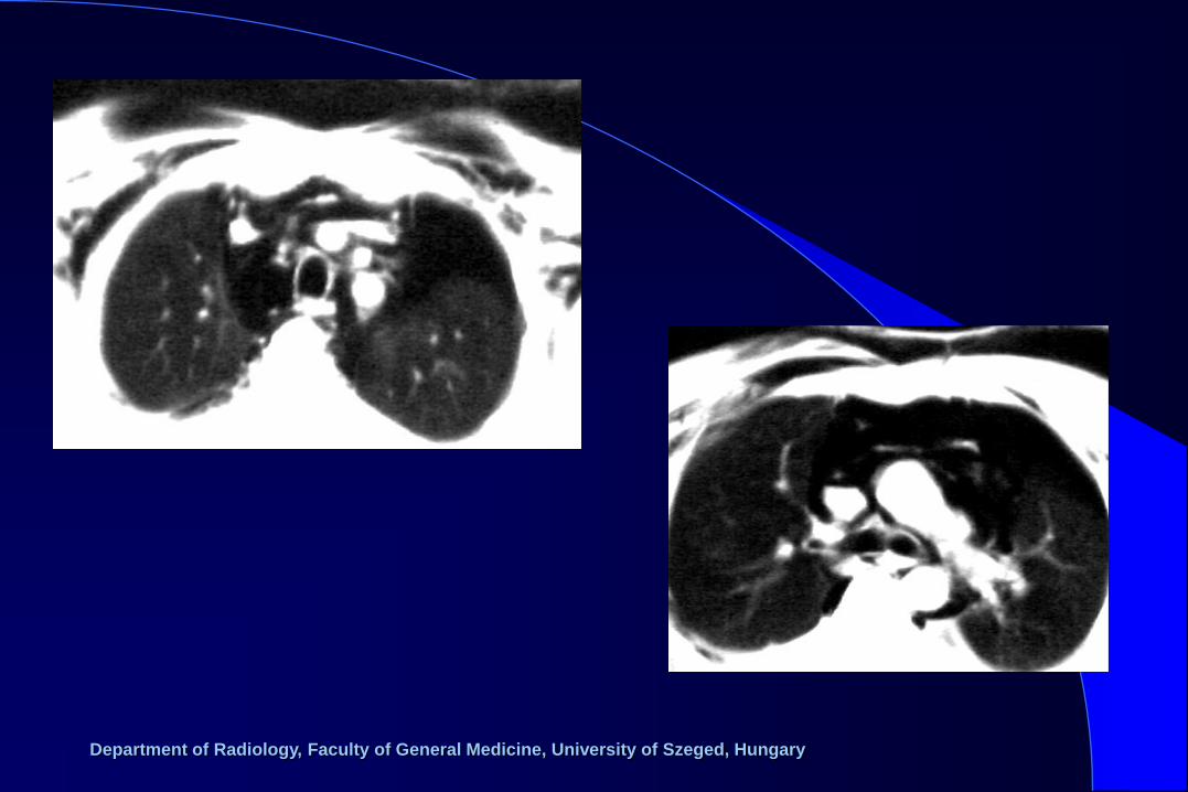
Mediastinal injuries
– pneumomediastinum asthma
barotrauma
ptx
tracheobronchial laceration
esophagus perforation, rupture
mediastinitis
intra-, retroperitoneal gas collection
– bleeding
– mediastinitis, abscess
Department of Radiology, Faculty of General Medicine, University of Szeged, Hungary

Department of Radiology, Faculty of General Medicine, University of Szeged, Hungary
Department of Radiology, Faculty of General Medicine, University of Szeged, Hungary


