Cardiovascular Research Foundation Cardiovascular Research Foundation New York, NY New York, NY Imaging Overview for Vulnerable Imaging Overview for Vulnerable Plaque: Data from IVUS Trial Plaque: Data from IVUS Trial and and An Introduction to VH An Introduction to VH - - IVUS IVUS Imgaging Imgaging Gary S. Gary S. Mintz Mintz , MD , MD
Transcript
Cardiovascular Research FoundationCardiovascular Research FoundationNew York, NYNew York, NY
Imaging Overview for Vulnerable Imaging Overview for Vulnerable Plaque: Data from IVUS TrialPlaque: Data from IVUS Trial
andandAn Introduction to VHAn Introduction to VH--IVUS IVUS
ImgagingImgagingGary S. Gary S. MintzMintz, MD, MD
•• Today, in reality, almost everything that we Today, in reality, almost everything that we currentlycurrently know about vulnerable plaque has come know about vulnerable plaque has come either from histopathology or from in vivo either from histopathology or from in vivo detection of plaque rupture or study of patients detection of plaque rupture or study of patients who present with acute coronary syndromes who present with acute coronary syndromes --NOT from prospective correlative studies or NOT from prospective correlative studies or prospective identification of vulnerable plaques prospective identification of vulnerable plaques before they rupture, rapidly progress, or before they rupture, rapidly progress, or thrombosethrombose..
•• To my knowledge, there are only three, To my knowledge, there are only three, retrospective IVUS studies relating lesion retrospective IVUS studies relating lesion findings to late events findings to late events –– and no trial dataand no trial data
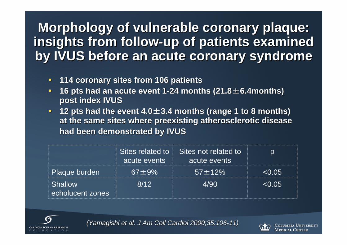
Morphology of vulnerable coronary plaque: Morphology of vulnerable coronary plaque: insights from followinsights from follow--up of patients examined up of patients examined by IVUS before an acute coronary syndromeby IVUS before an acute coronary syndrome
•• 114 coronary sites from 106 patients114 coronary sites from 106 patients•• 16 pts had an acute event 116 pts had an acute event 1--24 months (21.824 months (21.8±±6.4months) 6.4months)
post index IVUSpost index IVUS•• 12 pts had the event 4.012 pts had the event 4.0±±3.4 months (range 1 to 8 months) 3.4 months (range 1 to 8 months)
at the same sites where preexisting atherosclerotic disease at the same sites where preexisting atherosclerotic disease had been demonstrated by IVUShad been demonstrated by IVUS
((YamagishiYamagishi et al. J Am et al. J Am CollColl CardiolCardiol 2000;35:1062000;35:106--11)11)
Sites related to acute events
Sites not related to acute events
p
Plaque burden 67±9% 57±12% <0.05Shallow echolucent zones
8/12 4/90 <0.05
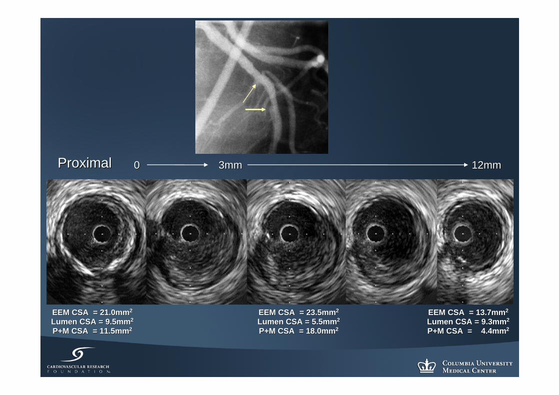
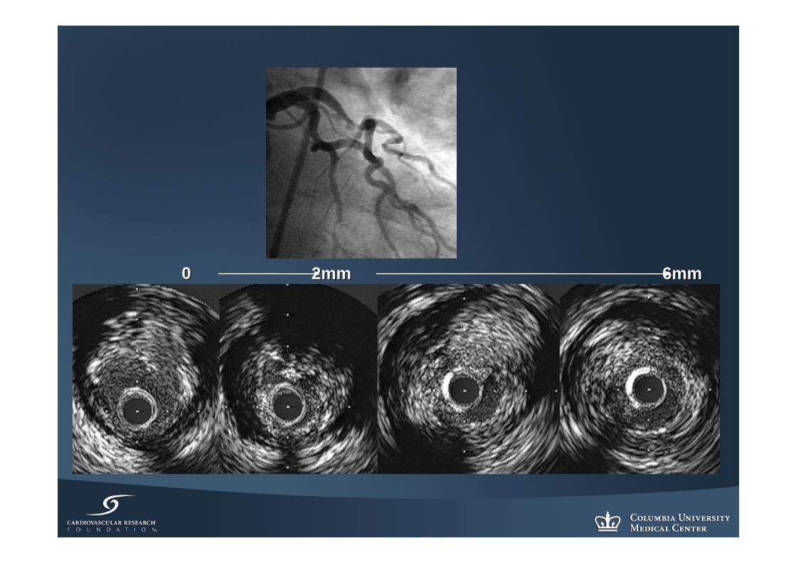
ProximalProximal 3mm3mm00 12mm12mm
EEM CSA = 21.0mmEEM CSA = 21.0mm22
Lumen CSA = 9.5mmLumen CSA = 9.5mm22
P+M CSA = 11.5mmP+M CSA = 11.5mm22
EEM CSA = 23.5mmEEM CSA = 23.5mm22
Lumen CSA = 5.5mmLumen CSA = 5.5mm22
P+M CSA = 18.0mmP+M CSA = 18.0mm22
EEM CSA = 13.7mmEEM CSA = 13.7mm22
Lumen CSA = 9.3mmLumen CSA = 9.3mm22
P+M CSA = 4.4mmP+M CSA = 4.4mm22
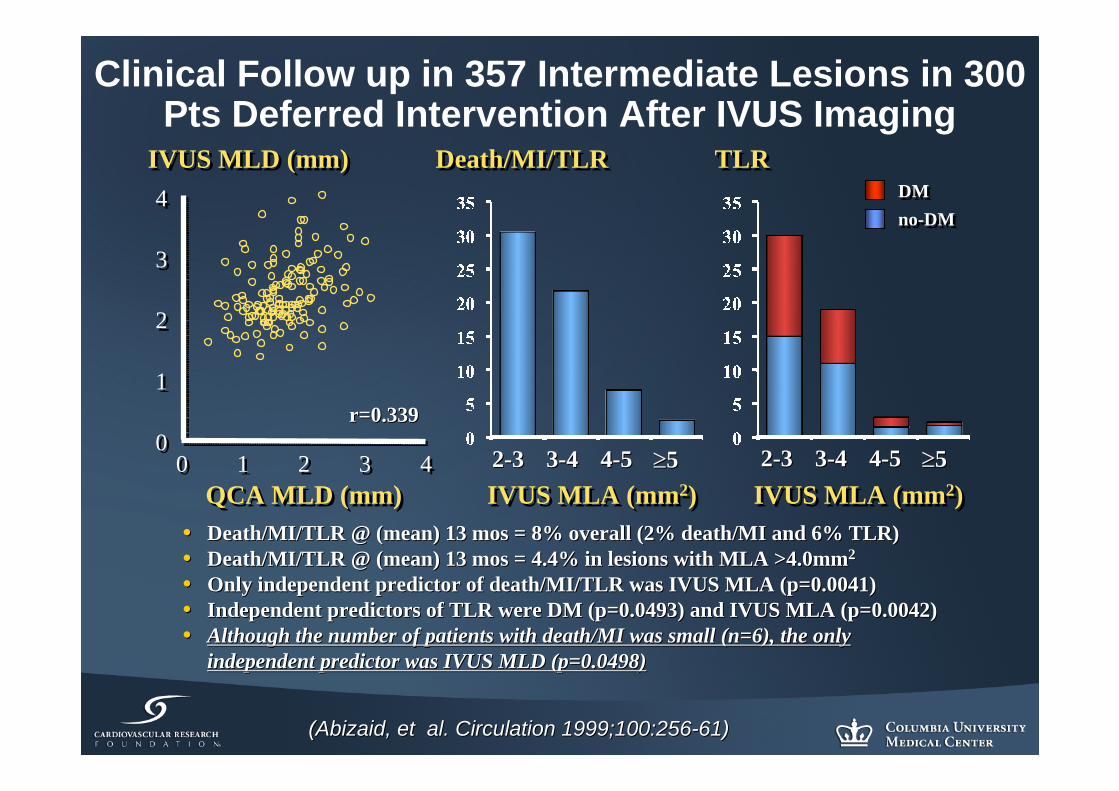
Clinical Follow up in 357 Intermediate Lesions in 300 Pts Deferred Intervention After IVUS Imaging
•• Death/MI/TLR @ (mean) 13 Death/MI/TLR @ (mean) 13 mosmos = 8% overall (2% death/MI and 6% TLR)= 8% overall (2% death/MI and 6% TLR)•• Death/MI/TLR @ (mean) 13 Death/MI/TLR @ (mean) 13 mosmos = 4.4% in lesions with MLA >4.0mm= 4.4% in lesions with MLA >4.0mm22
•• Only independent predictor of death/MI/TLR was IVUS MLA (p=0.004Only independent predictor of death/MI/TLR was IVUS MLA (p=0.0041)1)•• Independent predictors of TLR were DM (p=0.0493) and IVUS MLA (pIndependent predictors of TLR were DM (p=0.0493) and IVUS MLA (p=0.0042)=0.0042)•• Although the number of patients with death/MI was small (n=6), tAlthough the number of patients with death/MI was small (n=6), the only he only
independent predictor was IVUS MLD (p=0.0498)independent predictor was IVUS MLD (p=0.0498)
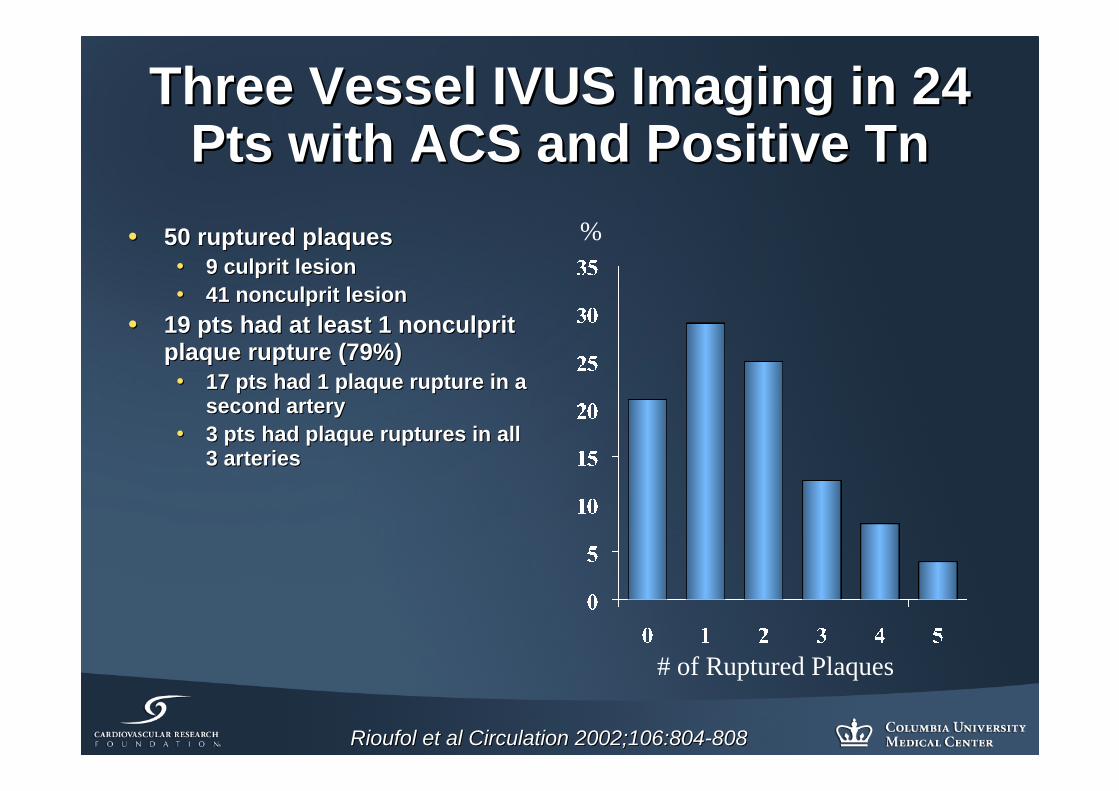
•• 19 pts had at least 1 nonculprit 19 pts had at least 1 nonculprit plaque rupture (79%)plaque rupture (79%)•• 17 pts had 1 plaque rupture in a 17 pts had 1 plaque rupture in a
second arterysecond artery•• 3 pts had plaque ruptures in all 3 pts had plaque ruptures in all
3 arteries3 arteries
Rioufol et al Circulation 2002;106:804Rioufol et al Circulation 2002;106:804--808808
%
# of Ruptured Plaques
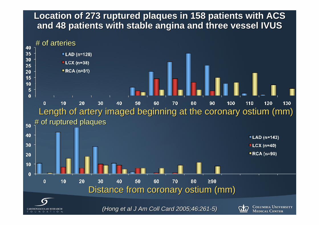
Location of 273 ruptured plaques in 158 patients with ACS Location of 273 ruptured plaques in 158 patients with ACS and 48 patients with stable angina and three vessel IVUSand 48 patients with stable angina and three vessel IVUS
Distance from coronary ostium (mm)Distance from coronary ostium (mm)
# of ruptured plaques# of ruptured plaquesLength of artery imaged beginning at the coronary ostium (mm)Length of artery imaged beginning at the coronary ostium (mm)
# of arteries# of arteries
(Hong et al J Am Coll Card 2005;46:261(Hong et al J Am Coll Card 2005;46:261--5)5)
Symptoms in 254 Symptoms in 254 patients with 300 patients with 300 plaque ruptures plaque ruptures in 257 arteriesin 257 arteries
11%11%11%11%
32%32%
46%46%
(Maehara et al (Maehara et al J Am Coll Cardiol 2002;40:904J Am Coll Cardiol 2002;40:904--10)10)
# of ruptured # of ruptured plaques per patient plaques per patient with stable angina with stable angina
((nn=113) =113)
(Hong et al (Hong et al Circulation 2004;110:928Circulation 2004;110:928--33)33)
The only independent predictor or a ruptured plaque in stable angina was diabetes mellitus (p=0.034, OR=2.553)
Comparison of Culprit & NonComparison of Culprit & Non--Culprit Rupture Sites in Culprit Rupture Sites in ACS Patients and Rupture Sites in NonACS Patients and Rupture Sites in Non--ACS PatientsACS Patients
Independent predictors of ACS were MLA and thrombus (both p=0.01Independent predictors of ACS were MLA and thrombus (both p=0.01))
Fuji et al. Circulation 2003;108:2473Fuji et al. Circulation 2003;108:2473--88
2mm2mm 6mm6mm00
Association of positive remodeling Association of positive remodeling and ACSand ACS
P=0.001P=0.001
(Schoenhagen et al. Circulation 2000;101:598(Schoenhagen et al. Circulation 2000;101:598--603)603) (Prati et al. Circulation 2003;107:2320(Prati et al. Circulation 2003;107:2320--5)5)
P=0.035P=0.035P=0.029P=0.029
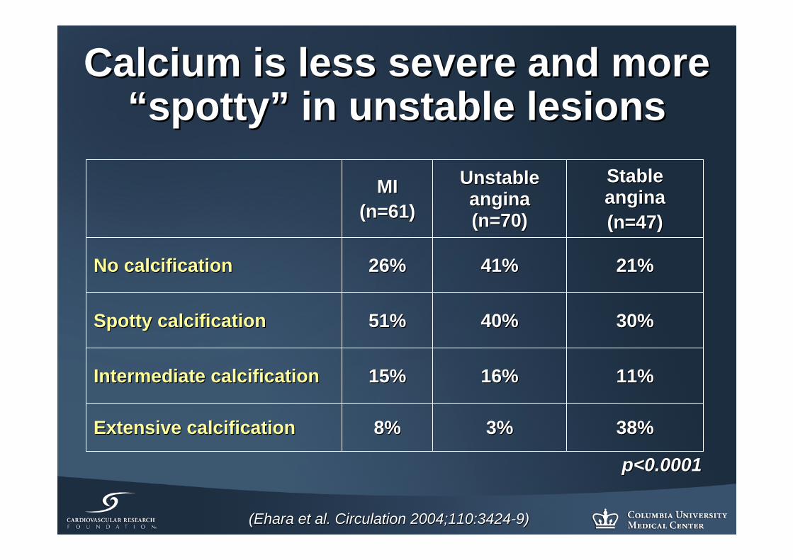
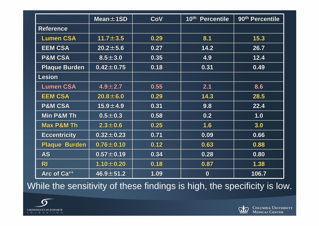
Calcium is less severe and more Calcium is less severe and more ““spottyspotty”” in unstable lesionsin unstable lesions
While the sensitivity of these findings is high, the specificity is low.
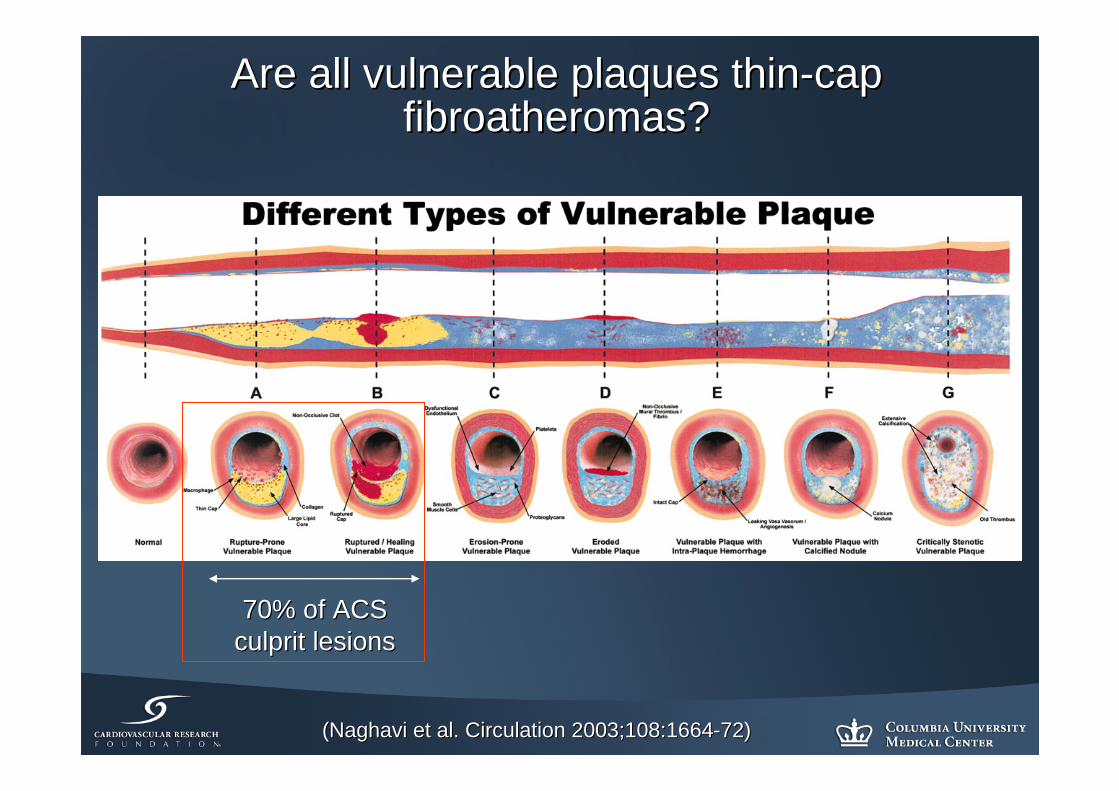
70% of ACS 70% of ACS culprit lesionsculprit lesions
(Naghavi et al. Circulation 2003;108:1664(Naghavi et al. Circulation 2003;108:1664--72)72)
Are all vulnerable plaques thinAre all vulnerable plaques thin--cap cap fibroatheromasfibroatheromas??
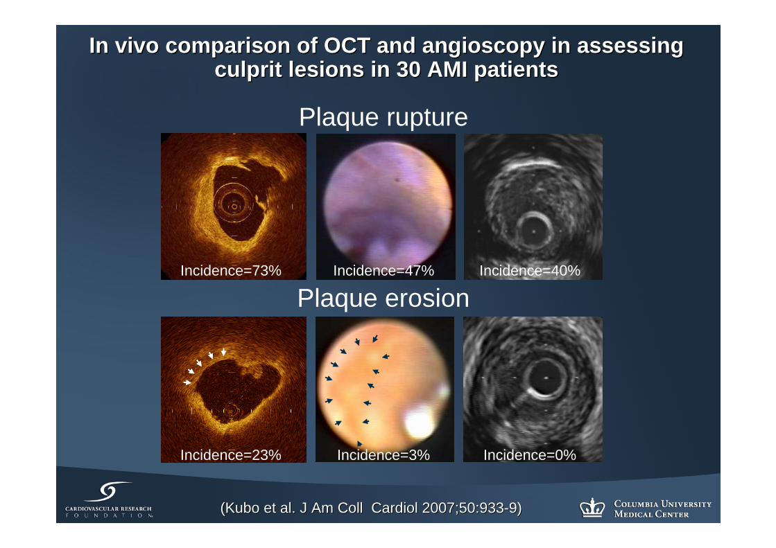
In vivo comparison of OCT and angioscopy in assessing In vivo comparison of OCT and angioscopy in assessing culprit lesions in 30 AMI patientsculprit lesions in 30 AMI patients
Plaque Erosion
Plaque rupture
Plaque erosion
(Kubo et al. J Am Coll Cardiol 2007;50:933(Kubo et al. J Am Coll Cardiol 2007;50:933--9)9)
Incidence=73% Incidence=47% Incidence=40%
Incidence=23% Incidence=3% Incidence=0%
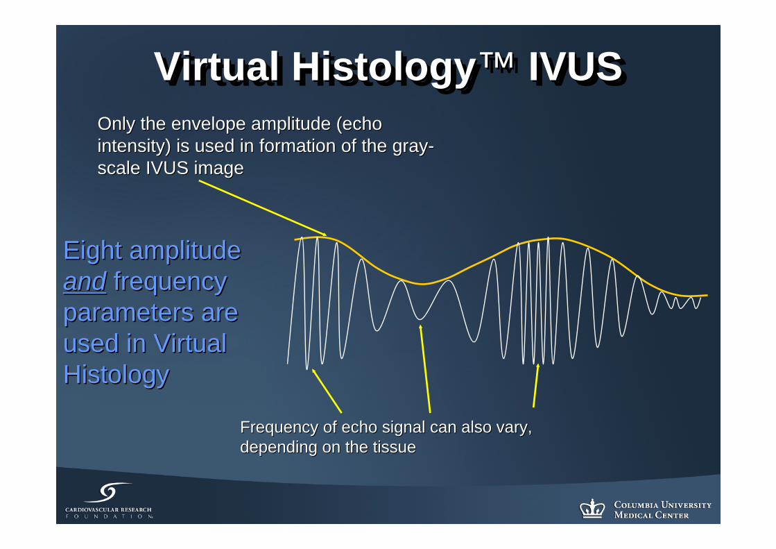
Virtual Histology™ IVUSVirtual HistologyVirtual Histology™™ IVUSIVUSOnly the envelope amplitude (echo Only the envelope amplitude (echo intensity) is used in formation of the grayintensity) is used in formation of the gray--scale IVUS imagescale IVUS image
Frequency of echo signal can also vary, Frequency of echo signal can also vary, depending on the tissuedepending on the tissue
Eight amplitude Eight amplitude andand frequency frequency parameters are parameters are used in Virtual used in Virtual HistologyHistology
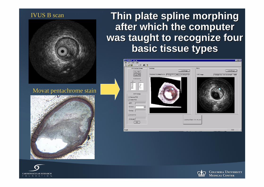
Thin plate spline morphing Thin plate spline morphing after which the computer after which the computer
was taught to recognize four was taught to recognize four basic tissue typesbasic tissue types
IVUS B scan IVUS B scan
Movat pentachrome stain Movat pentachrome stain
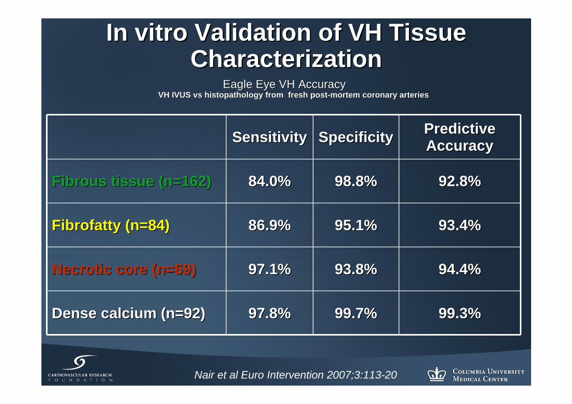
In vitro Validation of VH Tissue In vitro Validation of VH Tissue CharacterizationCharacterization
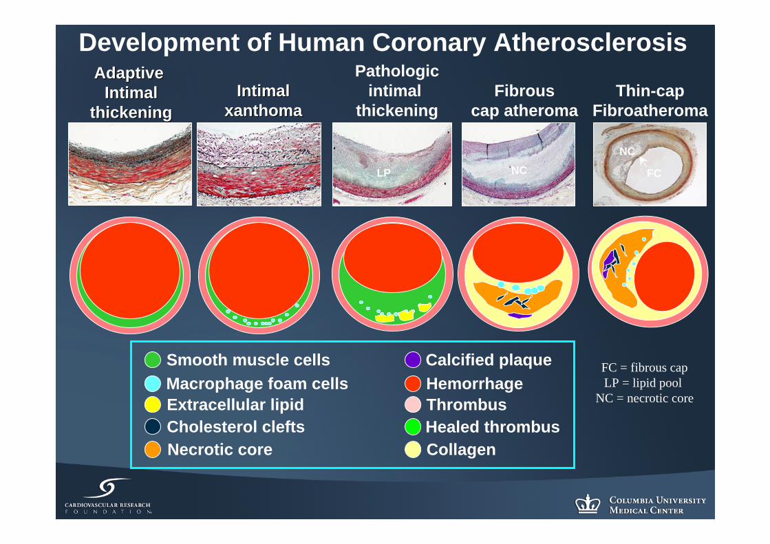
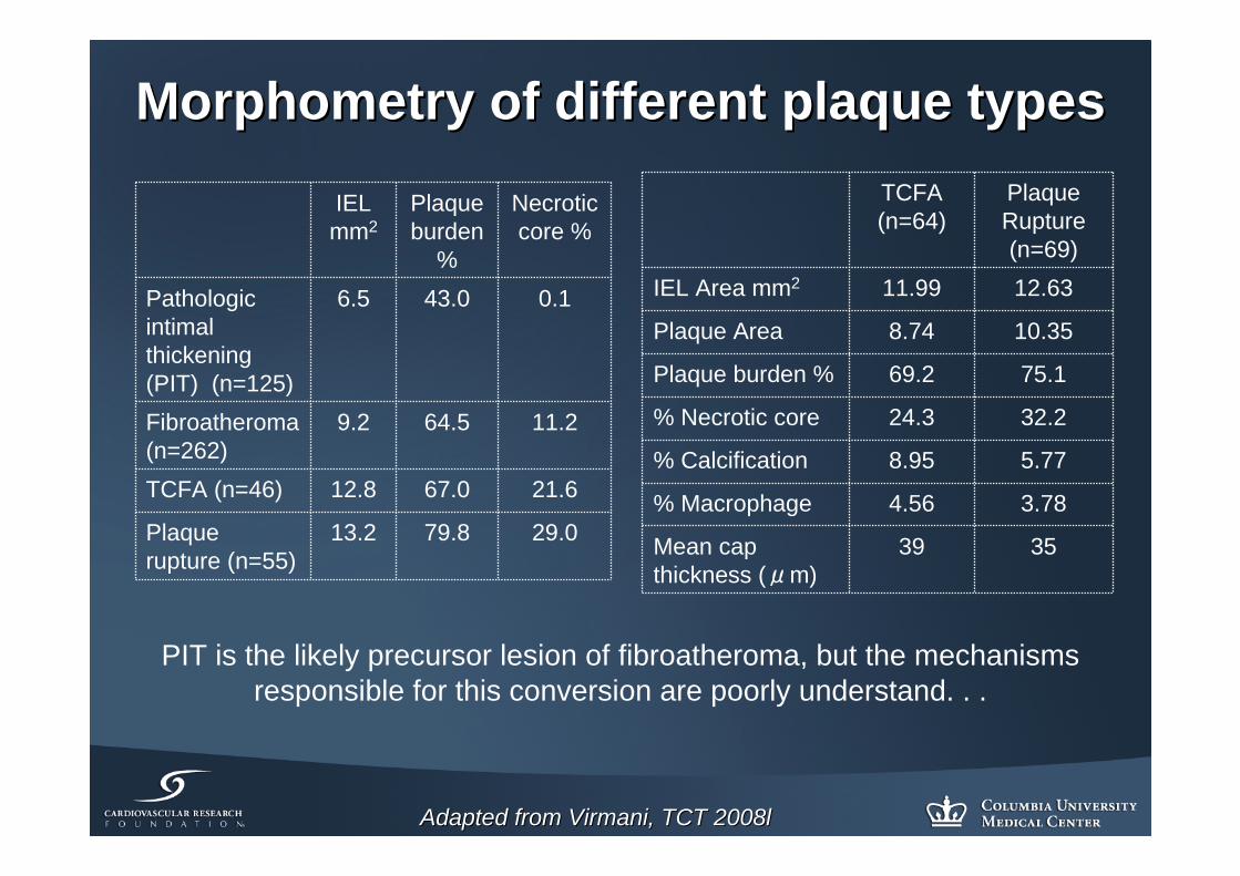
MorphometryMorphometry of different plaque typesof different plaque types
IEL mm2
Plaque burden
%
Necrotic core %
Pathologic intimal thickening (PIT) (n=125)
6.5 43.0 0.1
Fibroatheroma (n=262)
9.2 64.5 11.2
TCFA (n=46) 12.8 67.0 21.6
Plaque rupture (n=55)
13.2 79.8 29.0
Adapted from Virmani, TCT 2008lAdapted from Virmani, TCT 2008l
TCFA (n=64)
Plaque Rupture (n=69)
IEL Area mm2 11.99 12.63
Plaque Area 8.74 10.35
Plaque burden % 69.2 75.1
% Necrotic core 24.3 32.2
% Calcification 8.95 5.77
% Macrophage 4.56 3.78
Mean cap thickness (μm)
39 35
PIT is the likely precursor lesion of fibroatheroma, but the mechanisms responsible for this conversion are poorly understand. . .
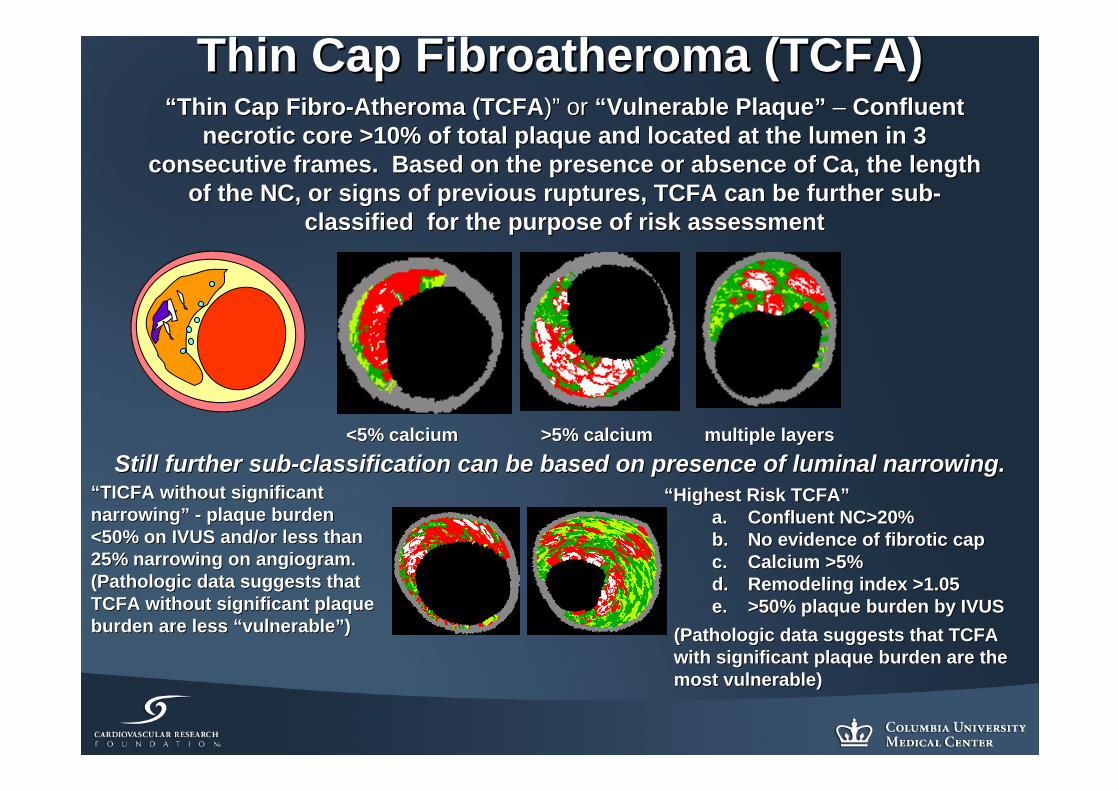
Thin Cap Fibroatheroma (TCFA)Thin Cap Fibroatheroma (TCFA)““Thin Cap FibroThin Cap Fibro--Atheroma (TCFAAtheroma (TCFA))”” or or ““Vulnerable PlaqueVulnerable Plaque”” –– Confluent Confluent
necrotic core >10% of total plaque and located at the lumen in 3necrotic core >10% of total plaque and located at the lumen in 3consecutive frames. Based on the presence or absence of Ca, theconsecutive frames. Based on the presence or absence of Ca, the length length
of the NC, or signs of previous ruptures, TCFA can be further suof the NC, or signs of previous ruptures, TCFA can be further subb--classified for the purpose of risk assessmentclassified for the purpose of risk assessment
““Highest Risk TCFAHighest Risk TCFA””a.a. Confluent NC>20%Confluent NC>20%b.b. No evidence of fibrotic capNo evidence of fibrotic capc.c. Calcium >5%Calcium >5%d.d. Remodeling index >1.05Remodeling index >1.05e.e. >50% plaque burden by IVUS>50% plaque burden by IVUS
““TICFA without significant TICFA without significant narrowingnarrowing”” -- plaque burden plaque burden <50% on IVUS and/or less than <50% on IVUS and/or less than 25% narrowing on angiogram. 25% narrowing on angiogram. (Pathologic data suggests that (Pathologic data suggests that TCFA without significant plaque TCFA without significant plaque burden are less burden are less ““vulnerablevulnerable””))
<5% calcium >5% calcium multiple layers<5% calcium >5% calcium multiple layersStill further subStill further sub--classification can be based on presence of luminal narrowing.classification can be based on presence of luminal narrowing.
(Pathologic data suggests that TCFA (Pathologic data suggests that TCFA with significant plaque burden are the with significant plaque burden are the most vulnerable)most vulnerable)
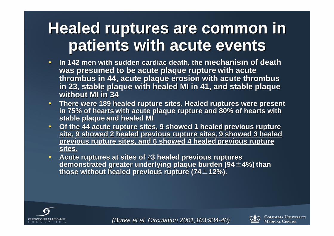
Healed ruptures are common in Healed ruptures are common in patients with acute eventspatients with acute events
•• In 142 men with sudden cardiac death, the In 142 men with sudden cardiac death, the mechanism of death mechanism of death was presumed to be acute plaque rupturewas presumed to be acute plaque rupture with acute with acute thrombus in 44, acute plaque erosion with acute thrombus thrombus in 44, acute plaque erosion with acute thrombus in 23, stable plaque with healed MI in 41, and stable plaque in 23, stable plaque with healed MI in 41, and stable plaque without MI in 34without MI in 34
•• There were 189 healed rupture sites. Healed ruptures were presenThere were 189 healed rupture sites. Healed ruptures were present t in 75% of heartsin 75% of hearts with acute plaque rupture and 80% of hearts with with acute plaque rupture and 80% of hearts with stable plaquestable plaque and healed MIand healed MI
•• Acute ruptures at sites of Acute ruptures at sites of ≥≥3 healed previous ruptures3 healed previous rupturesdemonstrated greater underlying plaque burden (94demonstrated greater underlying plaque burden (94±±4%)4%) than than those without healed previous rupture (74those without healed previous rupture (74±±12%).12%).
(Burke et al. Circulation 2001;103;934(Burke et al. Circulation 2001;103;934--40)40)
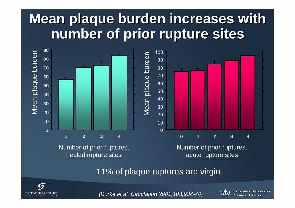
Mean plaque burden increases with Mean plaque burden increases with number of prior rupture sitesnumber of prior rupture sites
Mea
n pl
aque
bur
den
Number of prior ruptures, healed rupture sites
0
10
20
30
40
50
60
70
80
90
1 2 3 40
10
2030405060
7080
90100
0 1 2 3 4
Number of prior ruptures, acute rupture sites
11% of plaque ruptures are virgin
Mea
n pl
aque
bur
den
(Burke et al. Circulation 2001;103;934(Burke et al. Circulation 2001;103;934--40)40)
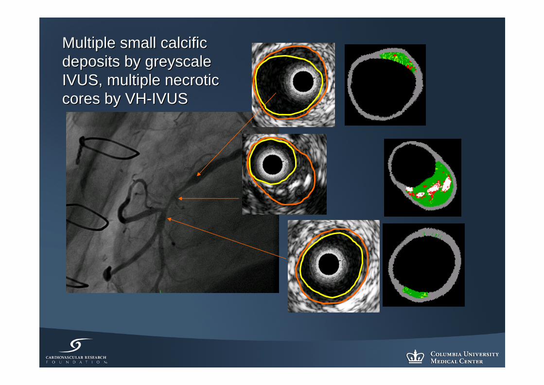
Multiple small calcific Multiple small calcific deposits by greyscale deposits by greyscale IVUS, multiple necrotic IVUS, multiple necrotic cores by VHcores by VH--IVUSIVUS
•• GreyscaleGreyscale IVUS findings are ubiquitous IVUS findings are ubiquitous in diffuse/advanced coronary in diffuse/advanced coronary atherosclerosis and, therefore, of atherosclerosis and, therefore, of limited ability to predict events.limited ability to predict events.
•• VHVH--IVUS criteria were based on IVUS criteria were based on presumptive presumptive histologichistologic evidence. But its evidence. But its ability to detect and assess the risk of a ability to detect and assess the risk of a specific lesion will depend NOT on specific lesion will depend NOT on correlation with histopathology, but on correlation with histopathology, but on the ability to predict future events. the ability to predict future events.
•• Perhaps PROSPECT will validate these Perhaps PROSPECT will validate these assumptions. Perhaps not.assumptions. Perhaps not.
Cardiovascular Research FoundationCardiovascular Research FoundationNew York, NYNew York, NY