57
Imaging with Impedance: Can We Guide Lung Ventilation? Andy Adler Systems and Computer Engineering, Carleton University, Ottawa, Canada

Imaging with Impedance: Can We Guide Lung Ventilation?
Andy AdlerSystems and Computer Engineering,
Carleton University,
Ottawa, Canada

Outline
• Imaging with Impedance– Electrical Impedance Tomography
• Lung monitoring
• Reconstruction of images– Data artefacts– Movement compensation– Total Variation
• EIDORS + Open Source Software

Electrode placement to monitor the lungs and heart
Source: eidors3d.sf.net/data_contrib/if-neonate-spontaneous

EIT: Block Diagram

Source: eidors3d.sf.net/tutorial/netgen/extrusion/thoraxmdl.shtml
IV
Current streamlines and voltage equipotentials
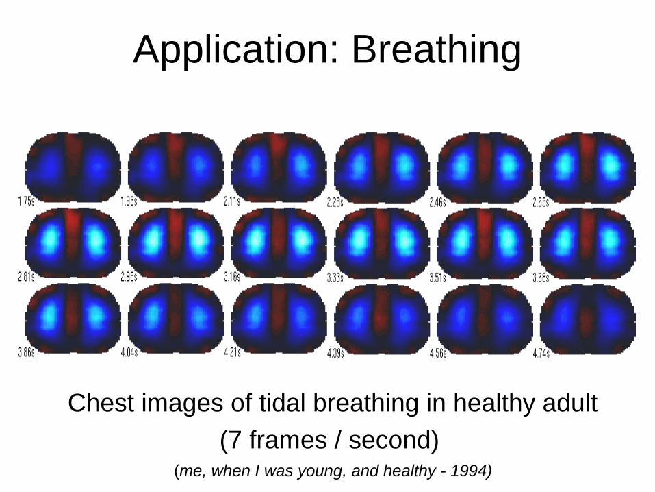
Application: Breathing
Chest images of tidal breathing in healthy adult
(7 frames / second) (me, when I was young, and healthy - 1994)
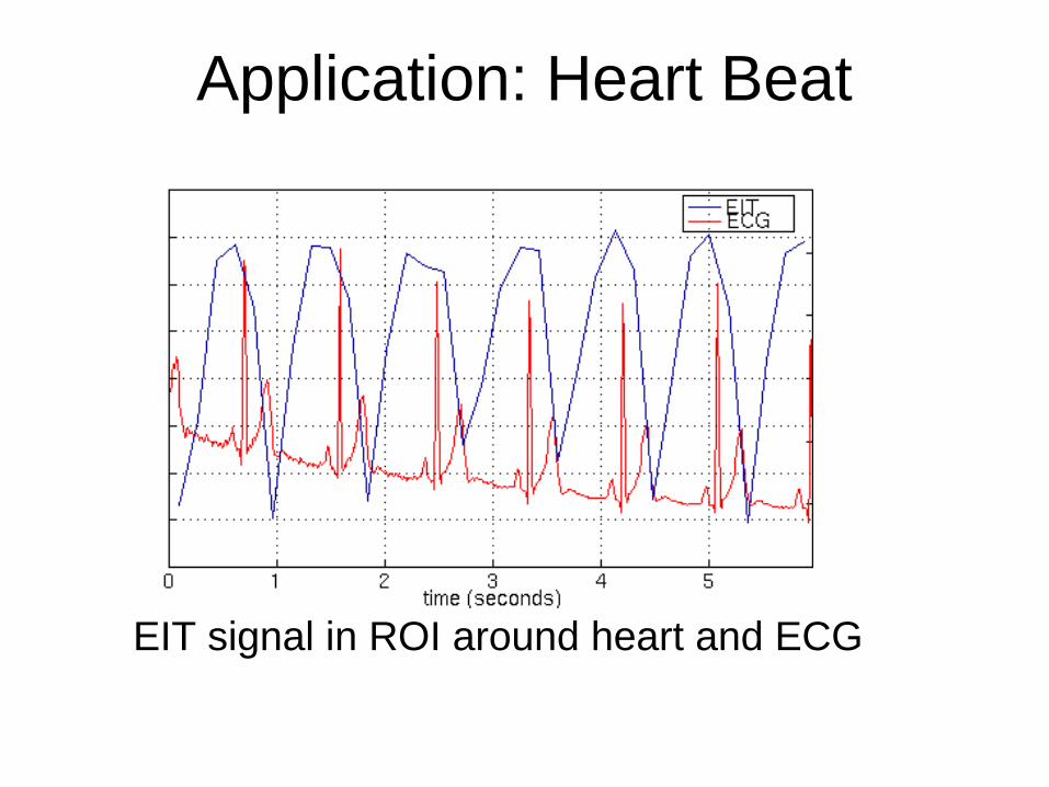
Application: Heart Beat
EIT signal in ROI around heart and ECG

Why image lungs? Respiratory Failure
Inadequate gas exchange by the respiratory system.Hypoxemia PaO2 < 60 mmHg or Hyercapnia PaCO2 > 45 mmHgCauses• Pulmonary dysfunction
– Asthma ,Emphysema , Chronic obstructive airway disease, Pneumonia , Pneumothorax, Hemothorax, Acute Respiratory Distress Syndrome (ARDS), Cystic Fibrosis
• Cardiac dysfunction – Pulmonary edema, Arrhythmia, Congestive heart failure, Valve
pathology Treatment
– Emergency treatment: cardiopulmonary resuscitation. – Treatment of the underlying cause– Mechanical ventilation
Ref: Wikipedia.org

Mechanical Ventilationused in acute settings (ICU). Often a life-saving technique, but has many complications
– pneumothorax, – airway injury, – alveolar damage,
Accordingly it is generally weaned off or to minimal settings as soon as possible. Ref: Wikipedia.org
Ref: healthlibrary.epnet.com/© 2009 Nucleus Medical Art, Inc.

Why image lungs?eg. Pneumonia
A: Normal chest x-ray B: Abnormal chest x-ray
shadowing from pneumonia in the right lung
Ref: Wikipedia.org
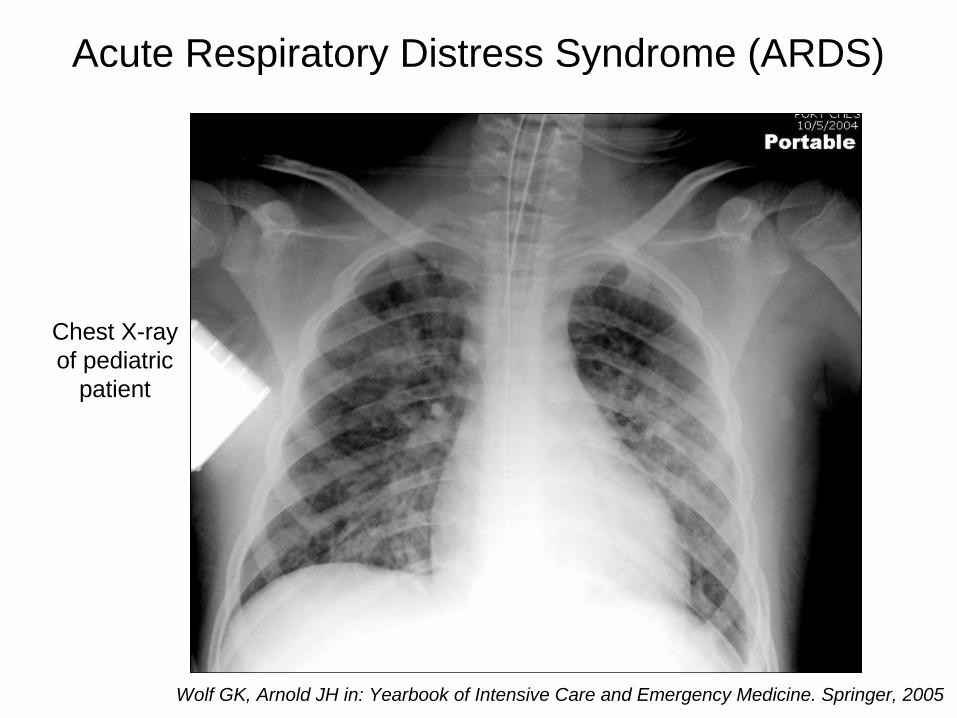
Acute Respiratory Distress Syndrome (ARDS)
Wolf GK, Arnold JH in: Yearbook of Intensive Care and Emergency Medicine. Springer, 2005
Chest X-rayof pediatric
patient
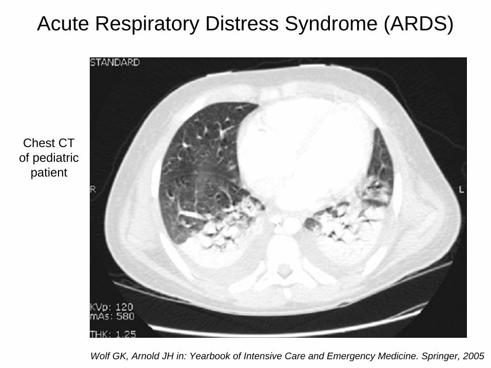
Acute Respiratory Distress Syndrome (ARDS)
Wolf GK, Arnold JH in: Yearbook of Intensive Care and Emergency Medicine. Springer, 2005
Chest CTof pediatric
patient
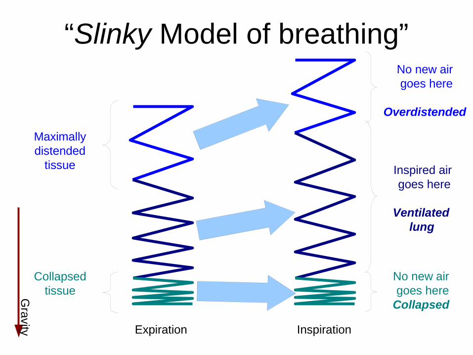
“Slinky Model of breathing”
Gravity
Maximallydistended
tissue
Collapsedtissue
Expiration Inspiration
No new air goes here
Overdistended
Inspired air goes here
Ventilated lung
No new air goes hereCollapsed

Regional ventilationImages from
Frerichs et al (2003) Intensive Care Med..
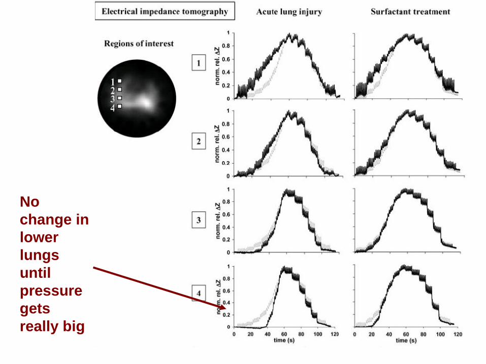
No change in lower lungs until pressuregets really big

EIT vs CT in ARDS
Data from pig study of EIT and CT Victorino JA et al (2004), Am J Respir Crit
Care Med
Show video

EIT in ARDS
Data from Gender: F, Age: 5.9 years, Weight: 20kg, Condition: Primary ARDS triggered by parainfluenza pneumonia.
GK Wolf, C Gómez-Laberge, JN Kheir, D Zurakowski, BK Walsh, A Adler, JH Arnold. Reversal of Dependent Lung Collapse Predicts Response to Lung Recruitment in Children with Early Acute Lung Injury Pediatr Crit Care Med, In Press 2012
Source: eidors3d.sf.net/data_contrib/cg-2012-ards-recruitment/
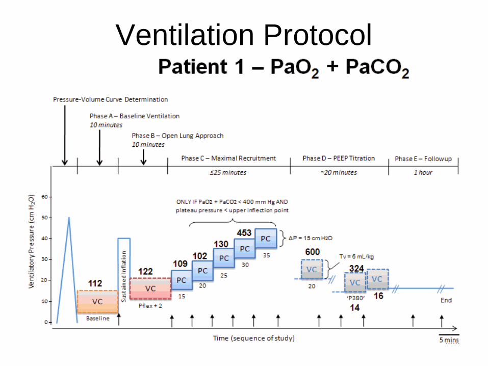
Ventilation Protocol

Patient #1: Lung opening and “optimal ventilation” images

Patient #1: Lung opening and “optimal ventilation” images
What can EIT tell us that is clinically useful?
EIT shows regional ventilation– Can a patient can be recruited?– Have we opened up the lungs?
EIT shows changes earlier than blood gas– PaO
2 responds slowly (LPF of blood)
– PaO2 responds only at high shunt fraction
– Can we control ventilation better with EIT?

Image Reconstruction
Linear difference imaging with pictures
• Total Variation
• Electrode Errors
• Electrode Movement
• Temporal Filtering
• GREIT
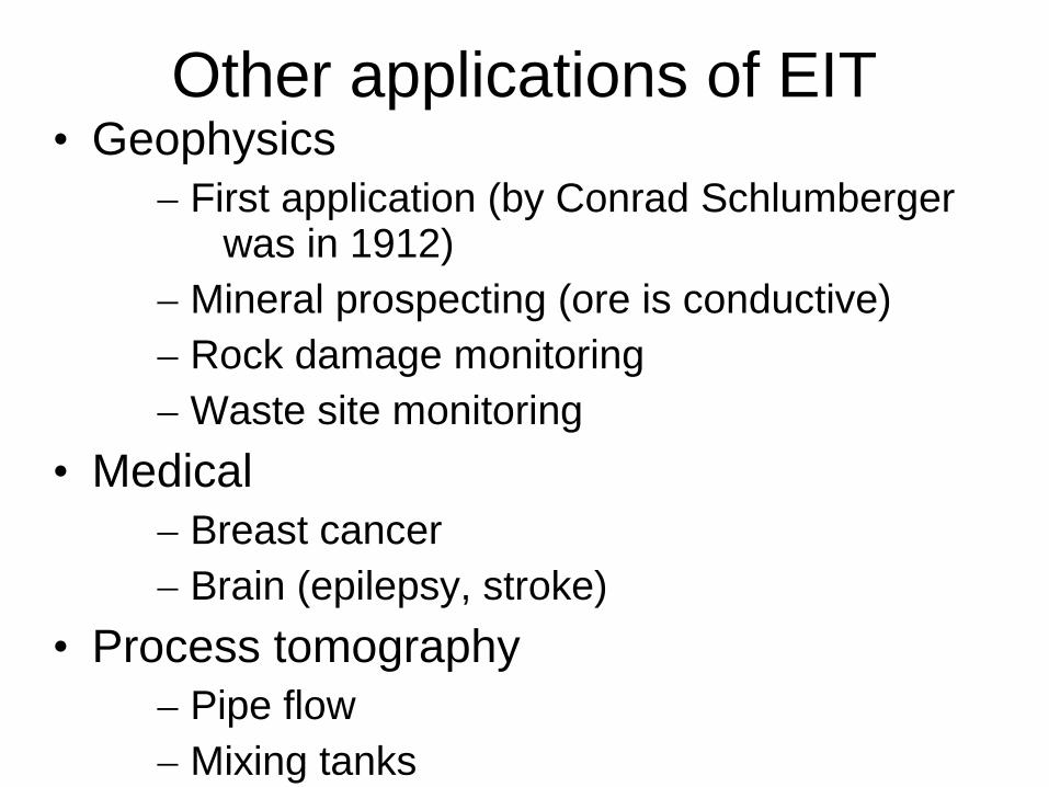
Other applications of EIT• Geophysics
– First application (by Conrad Schlumberger was in 1912)
– Mineral prospecting (ore is conductive)– Rock damage monitoring– Waste site monitoring
• Medical– Breast cancer– Brain (epilepsy, stroke)
• Process tomography– Pipe flow– Mixing tanks
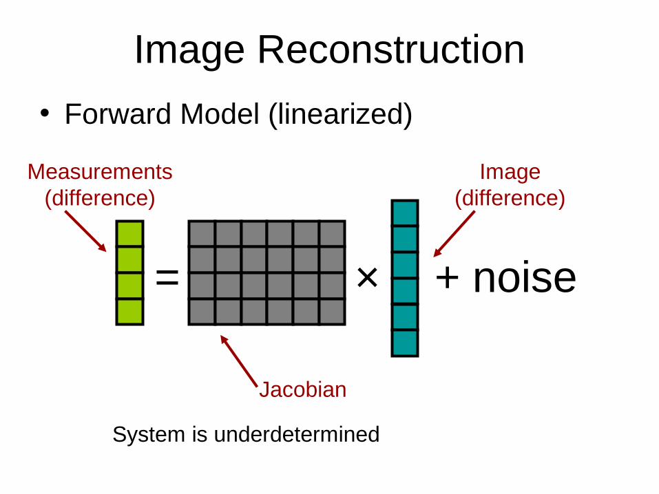
Image Reconstruction
• Forward Model (linearized)
= ×
Measurements(difference)
Image(difference)
Jacobian
System is underdetermined
+ noise

Image Reconstruction
Regularized linear Inverse Model
– ×
Norm weighted by measurement
accuracy
+ PenaltyFunction
2
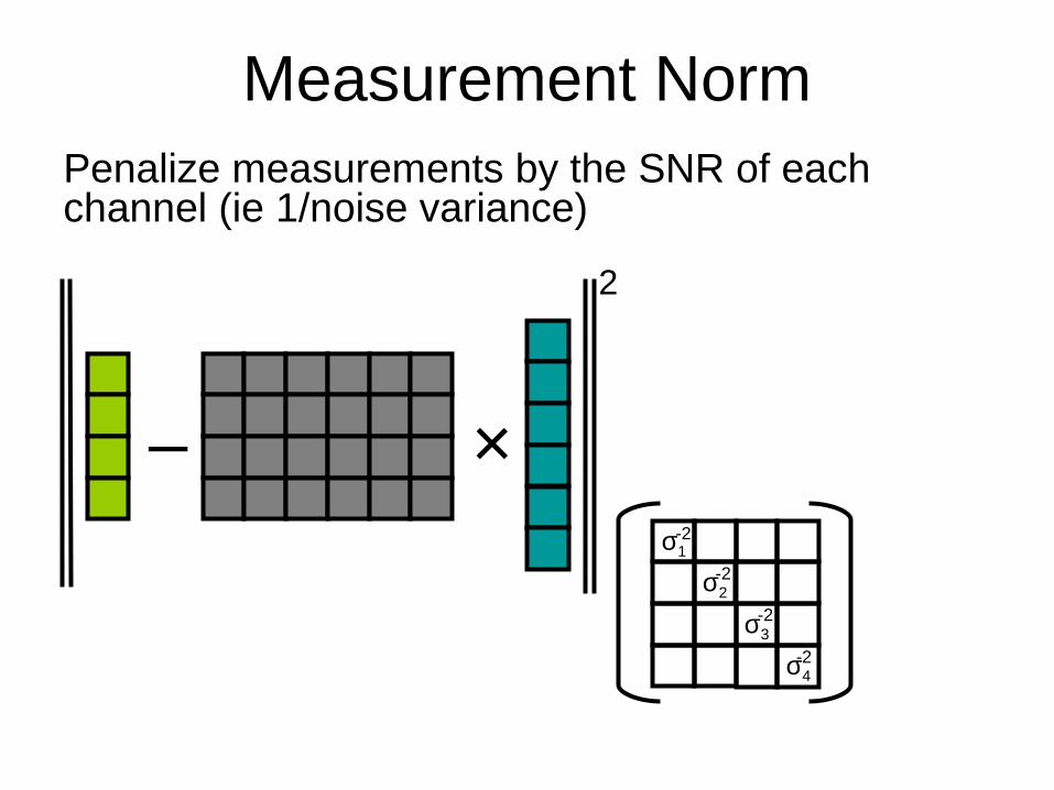
Measurement NormPenalize measurements by the SNR of each channel (ie 1/noise variance)
– ×
2
σ1
σ2
σ3
σ4
-2
-2
-2
-2

Image Reconstruction
Image Penalty Function
–
Zero forDifference EIT
=PenaltyFunction
Expectedimage
Norm weighted by “unlikelyhood” of image
2

Image Reconstruction
• Penalty functions: Image Amplitude
– Expectedimage
1
1
1
1
1
1
Tikhonov prior
2

Image Reconstruction
• Penalty functions: Image Smoothness
– Expectedimage
1
1
1
1
1
1
-½
-½
-½
-½
-½
-½
-½
-½
-½
-½
Laplacian prior
2

Total Variation
Image Penalty Function
–=PenaltyFunction
Expectedimage
Use 1-normSum( abs( . ))
1
TV penalty function does not prefer smooth to “blocky” images

Lung images with TV
Human(healthy)
Pig(ARDS)
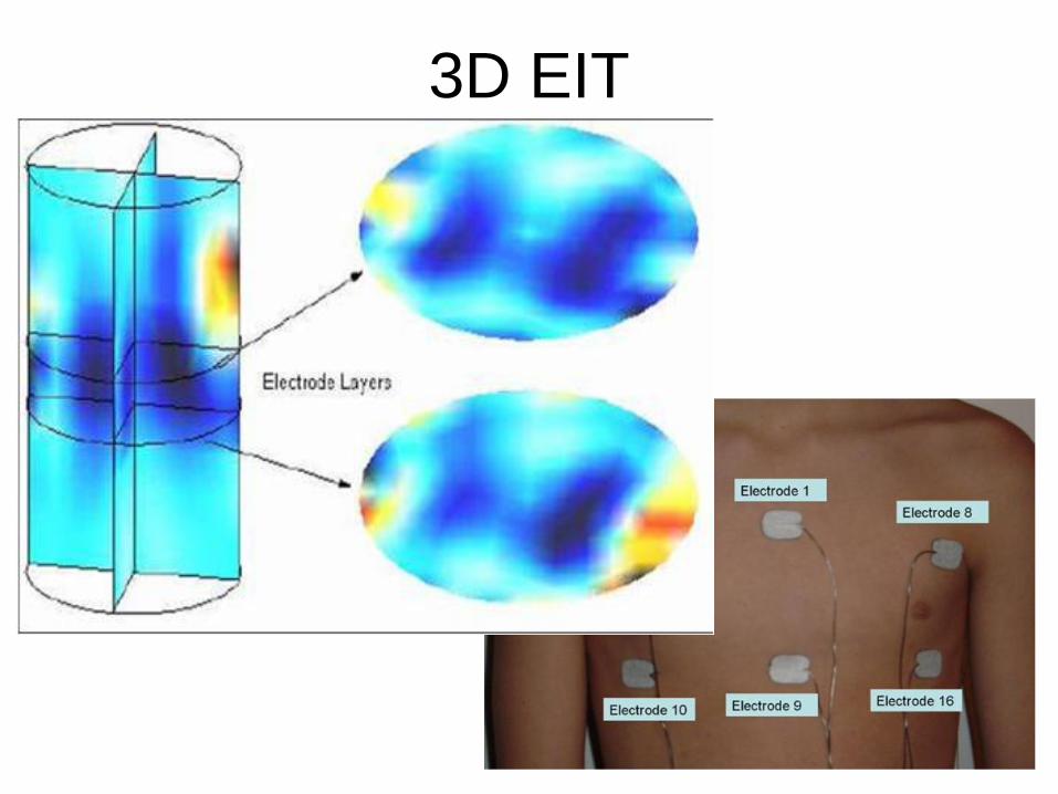
3D EIT
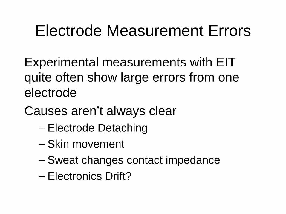
Electrode Measurement Errors
Experimental measurements with EIT quite often show large errors from one electrode
Causes aren’t always clear– Electrode Detaching– Skin movement– Sweat changes contact impedance– Electronics Drift?

Example of electrode errors
Images measured in anaesthetised, ventilated dogA. Image of 700 ml ventilation
B. Image of 100 ml saline instillation in right lung
C. Image of 700 ml ventilation and 100 ml saline
A B C
“Bad”Electrode

“Zero bad data” solution
“Traditional solution” (in the sense that I’ve done this)
– ×
2
σ1
σ2
σ3
σ4
-2
-2
-2
-2
Error HereReplace
With zero

Regularized imaging solution
Electrode errors are large measurement noise on affected electrode
– ×
2
σ1
σ2
σ3
σ4
-2
-2
-2
-2
Error Here
LowSNRhere
ReplaceWith zero

Correcting for errors. Results
A. Image of 700 ml ventilationB. Image of 100 ml saline instillation in right lungC. Image of 700 ml ventilation and 100 ml saline
A B C
“Bad”Electrode
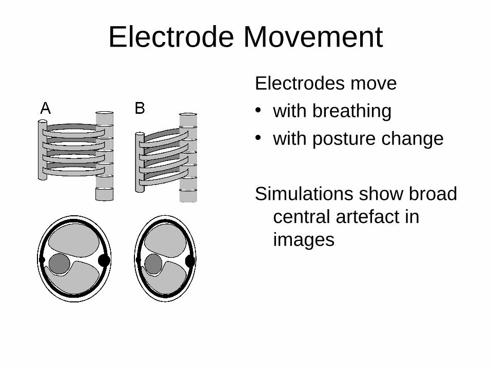
Electrode Movement
Electrodes move• with breathing• with posture change
Simulations show broad central artefact in images
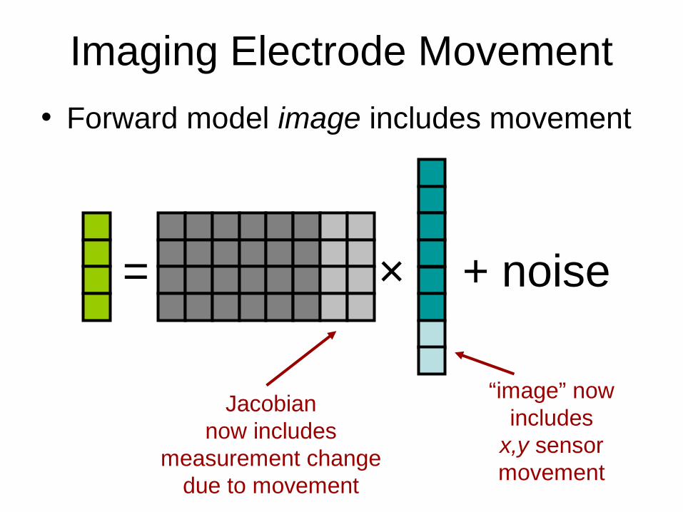
Imaging Electrode Movement
• Forward model image includes movement
= ×
Jacobiannow includes
measurement changedue to movement
+ noise
“image” nowincludes
x,y sensormovement

Image and movement
Penalty: Image and movement Smoothness
–Expected
image
1
1
1
1
1
1
-½
-½
-½
-½
-½
-½
-½
-½
-½
-½
2
Expectedmovement
“Unlikelyhood”of movement
“Unlikelyhood”of movement
and imageco-variance

Images of electrode movement
Simulation: tank with 3D deformation
y x
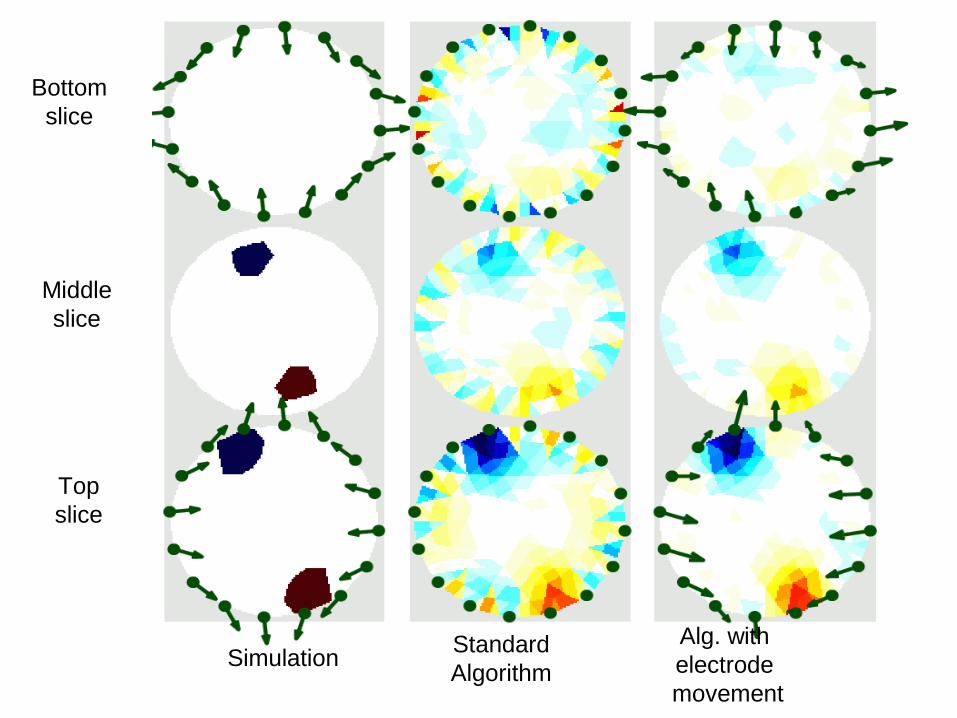
Bottomslice
Middleslice
Topslice
SimulationStandardAlgorithm
Alg. with electrode movement

EIT makes fast measurements. Can we use this fact?
……
0-1 +2 +n+1-2-n
past now future
=
Jacobian
…
Image sequenceMeasurement sequence
0-1 +2 +n+1-2
past now future

Temporal Reconstruction
Temporal Penalty Functions
1
1
1
1
1
1
1
1
1
1
1
1
1
1
1
1
1
1 1
1
1
1
1
1
1
1
1
1
1
1
1
1
1
1
1
1
1
1
1
1
1
1
1
1
1
likely quite likely unlikely
Standard EIT approaches to not take this into account

Direct temporal solver
……
-n
=
Jacobian
…
Image sequenceMeasurement sequence
Rewrite as …
0-1 +2 +n+1-20-1 +2 +n+1-2
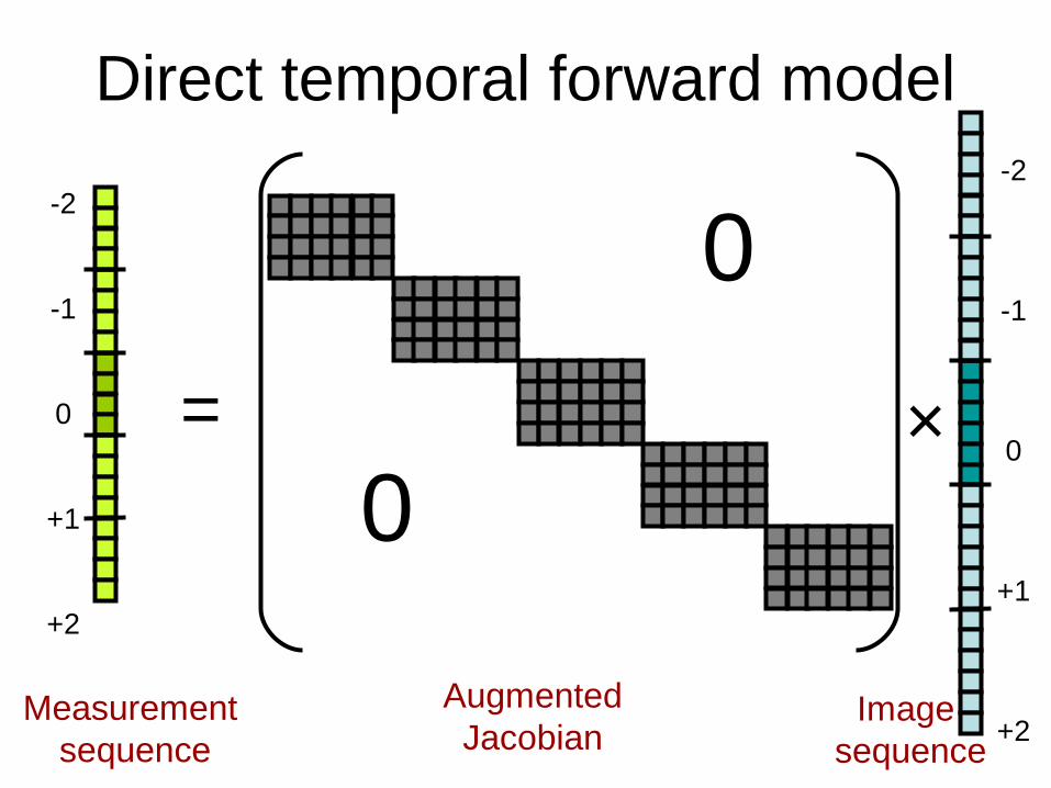
Direct temporal forward model
=
AugmentedJacobian
Image sequence
Measurement sequence
×0
0-2
-1
0
+1
+2
-2
-1
0
+1
+2

Direct temporal inverse model
×0
0– Exp.
image–+
TimePrior
2 2

Temporal Priors
Exp.image–
SpatialPrior
SpatialPrior
SpatialPrior
SpatialPrior
SpatialPrior
TimePrior∆t = 1
TimePrior∆t = 2
TimePrior∆t = 3
TimePrior∆t = 1
TimePrior∆t = 2
TimePrior∆t = 3
TimePrior∆t = 1
TimePrior∆t = 2
TimePrior∆t = 4
TimePrior∆t = 1
TimePrior∆t = 1
TimePrior∆t = 1
TimePrior∆t = 1
TimePrior∆t = 1
TimePrior∆t = 2
TimePrior∆t = 2
TimePrior∆t = 2
TimePrior∆t = 3
TimePrior∆t = 3
TimePrior∆t = 4

EIDORS: community-based extensible software for EIT
Andy Adler1, William R.B. Lionheart2
1Systems and Computer Engineering, Carleton University, Ottawa, Canada
2School of Mathematics, University of Manchester, U.K.
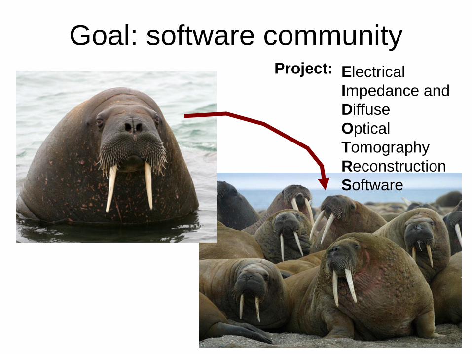
Goal: software communityElectrical Impedance and Diffuse Optical Tomography Reconstruction Software
Project:

Blobby the Walrus?
1. EIT images blobby objects in aqueous media; Blobby the Walrus is a fat animal that lives in water.
2. Walrus is EIDORS logo
3. Walruses are much funnier than a talk about software architecture.
Images: www.biobcc.net© Genny Anderson

EIDORS Features
Open-source: • License: GNU General Public License. • Free to use, modify, and distribute modifications. • May be used in a commercial product
Hosted on Sourceforge.net• Software is available for download (version 2.0)• CVS access to latest developer versions• Group members can modify• Anyone can read and download

Web Site
ReleaseVersion
DeveloperVersion
Walrus
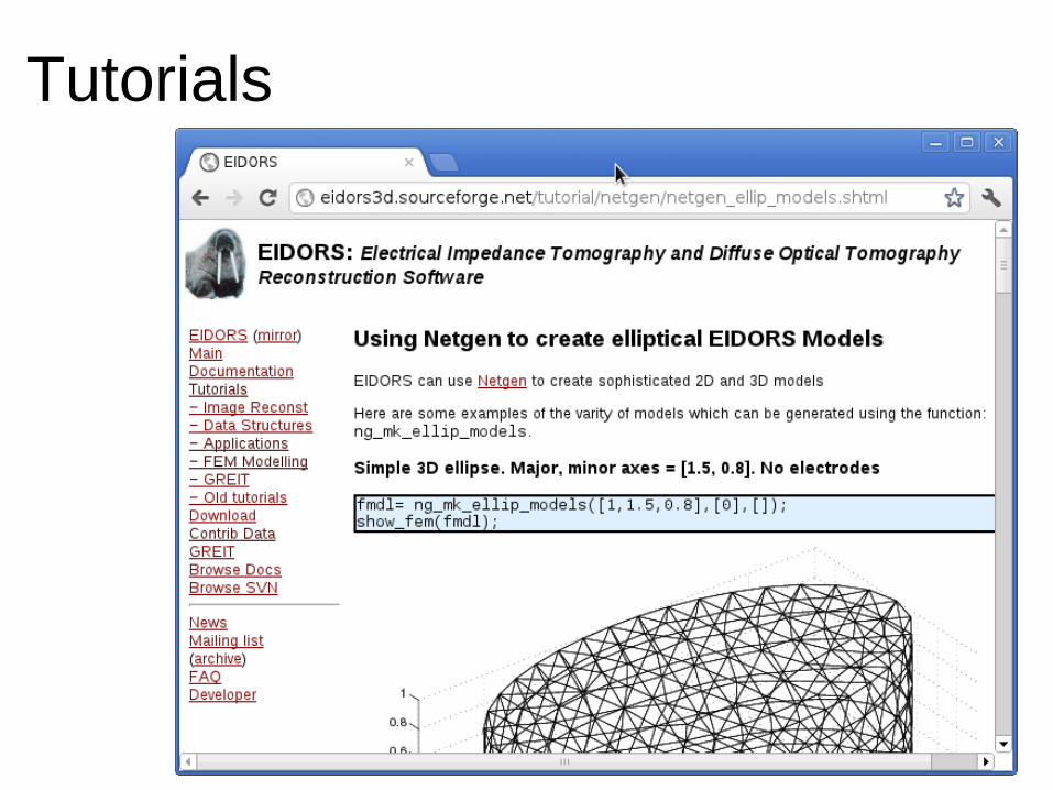
Tutorials
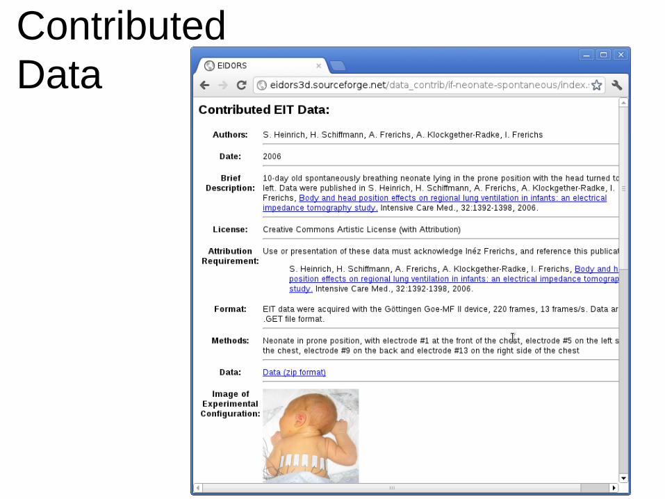
Contributed Data

Source: http://scm-l3.technorati.com/11/12/28/59203/Ottawa-Rideau-Canal-courtesy-city-of-ottawa.jpg
Thank you
Imaging with Impedance: Can We Guide Lung Ventilation? Lecture
Abstract: Electrical Impedance Tomography (EIT) uses a set of electrodes placed around the patient's body to apply current simulation and measure the resulting potentials, from which an image of the internal conductivity distribution is calculated. EIT was invented 100 years ago by the brother's Schlumberger to prospect for conductive minerals. Since EIT is sensitive to physiological phenomena which affect the conductivity, it has been used to image the brain (to view perfusion changes due to epilepsy and stroke), the breast (to screen for cancerous regions), the abdomen (for gastric emptying) and thorax (to image the movement of blood and gas in the heart and lungs).
Patients in respiratory failure require positive pressure ventilation to ensure adequate gas exchange. While ventilation is life-saving, it imposes significant risks. To address these risks, lung EIT has the potential to be a monitoring tool to help guide and optimize lung protective ventilation individually for each patient.
EIT image reconstruction is difficult because of the way current propagates through all paths in the body; EIT image reconstruction is non-linear, spatially variant, and mathematically ill-conditioned. To solve these problems, regularized image reconstruction techniques are used, which use prior models to penalise low probability solutions. Recently, the increase in computer power has facilitated much more powerful algorithms.
This talk will review recent work in EIT image reconstruction, and its application for lung imaging.


